 Daniel Garrigos1,2
 Marta Martínez-Morga1,2
 Angel Toval1,2
 Yevheniy Kutsenko1,2
 Alberto Barreda1,2
 Bruno Ribeiro Do Couto1,2,3
 Fernando Navarro-Mateu2,4,5,6
 José Luis Ferran1,2*
- 1Department of Human Anatomy and Psychobiology, Faculty of Medicine, University of Murcia, Murcia, Spain
- 2Institute of Biomedical Research of Murcia—IMIB, Virgen de la Arrixaca University Hospital, University of Murcia, Murcia, Spain
- 3Faculty of Psychology, University of Murcia, Murcia, Spain
- 4Unidad de Docencia, Investigación y Formación en Salud Mental (UDIF-SM), Servicio Murciano de Salud, Murcia, Spain
- 5CIBER de Epidemiología y Salud Pública (CIBERESP), Madrid, Spain
- 6Departamento de Psicología Básica y Metodología, Universidad de Murcia, Murcia, Spain
A well-documented method and experimental design are essential to ensure the reproducibility and reliability in animal research. Experimental studies using exercise programs in animal models have experienced an exponential increase in the last decades. Complete reporting of forced wheel and treadmill exercise protocols would help to ensure the reproducibility of training programs. However, forced exercise programs are characterized by a poorly detailed methodology. Also, current guidelines do not cover the minimum data that must be included in published works to reproduce training programs. For this reason, we have carried out a systematic review to determine the reproducibility of training programs and experimental designs of published research in rodents using a forced wheel system. Having determined that most of the studies were not detailed enough to be reproducible, we have suggested guidelines for animal research using FORCED exercise wheels, which could also be applicable to any form of forced exercise.
Introduction
Improving the way of reporting animal research is an uphill task that involves the joint effort of authors, editors, reviewers, funding agencies and the painstaking work of ethics committees (1–3). However, the lack of transparency in reporting research methods remains a concern (1, 4–6). Detailed descriptions of methods and experimental designs might be decisive in minimizing unnecessary experimental duplication and ensuring the reproducibility of research results (3, 7, 8). In the long term, inappropriate descriptions of methods might contribute to diminish the credibility and sustainability of the scientific system and damage the reputation of the researchers involved in these studies (3, 9, 10). Increased evidence indicates that there is an appreciable room for improving the ‘reproducibility crisis’ in science (10–12). In this sense, the Animal Research Reporting of In Vivo Experiments (ARRIVE) guidelines were developed for reporting animal research rigorously, providing checklists with the essential information that must be included in research with animal experimentation (3, 6). However, to reproduce an exercise program in rodents, several additional specifications that are not included in any of the current guidelines are required.
Animal research with voluntary or forced exercise reporting the positive impact of physical activity in health is skyrocketing (13–17). Forced treadmill and wheel exercise, but not voluntary, are the preferred modalities to develop the same training in all animals. Specifically, forced wheel is an emergent modality that should ensure the exercise load reproducibility and avoid non-specific stress-related responses (18, 19), but methods provided in most of these works do not seem to be properly detailed. In this sense, current guidelines do not guarantee the reproducibility of the training programs in forced wheel or any other forced modality (6). Therefore, to allow the repeatability of the same experimental design, giving longevity to the research works using forced wheel, reporting accurate information about the ethic committee, housing, husbandry, animals and particularly exercise programs becomes essential (20–27). Furthermore, physical capacity or exercise adaptations promoted by training programs might be affected by housing or husbandry variations in the environmental conditions, food composition, the number of animals per cage or the light/dark cycle period (21, 28, 29). Moreover, the biological and behavioral research output might change if the animals exposed to the same exercise load present differences in terms of age, sex, weight or handling procedures (20, 23, 27, 30).
Particularly in exercise, variables such as habituation, duration of the session, speed, frequency and duration of the exercise program are determinant to guarantee the reproducibility of the forced wheel training (18, 19, 31). Finally, reporting the time elapsed between the last session of exercise and the test or biological analysis should avoid circadian and metabolic misinterpretations (32–34). A large number of studies have proposed the health benefits of exercise (17, 35–37). However, these conclusions could be called into question if the variables affecting the response to exercise have not been adequately controlled. Rodent research in the exercise field, would be benefited if most of these demands were included in the published works.
Our systematic review aimed to evaluate the reproducibility of the training programs and experimental designs of studies using a forced wheel system. Data ensuring the reproducibility of a training program and those that might potentially affect the physical response were selected and arranged in four sections: ethic committee, housing, animals, and exercise. Our results have led us to suggest the FORCED exercise wheel guidelines to improve the reliability of scientific works based on forced training programs. Our FORCED guidelines complement current guidelines by focusing in forced exercise (3, 6). As a novelty, the number of items reached by research works dealing with forced exercise was analyzed.
Methods
Literature Search Strategy
This systematic review was reported following the Preferred Reporting Items for Systematic Reviews and Meta-analysis (PRISMA, see Supplementary File 1) (38). The search was based in two groups of keywords: (i) forced wheel (“forced running wheel*” OR “forced wheel” OR “motorized wheel*”) and (ii) rodents (rodent* OR rat OR rats OR mice OR mouse OR mus OR murine). No additional filters were applied since one of the main objectives of the review was to scope all the available literature. The search was conducted using four electronic bibliographic databases: PubMed, Web of Science, Scopus and Science Direct. The last search was performed on March 23rd, 2020. Detailed examples of the search strategy can be found in supplementary materials (see Supplementary File 4). Reference lists of all the included papers and relevant reviews were scanned to identify any additional studies manually (Identification, Figure 1).
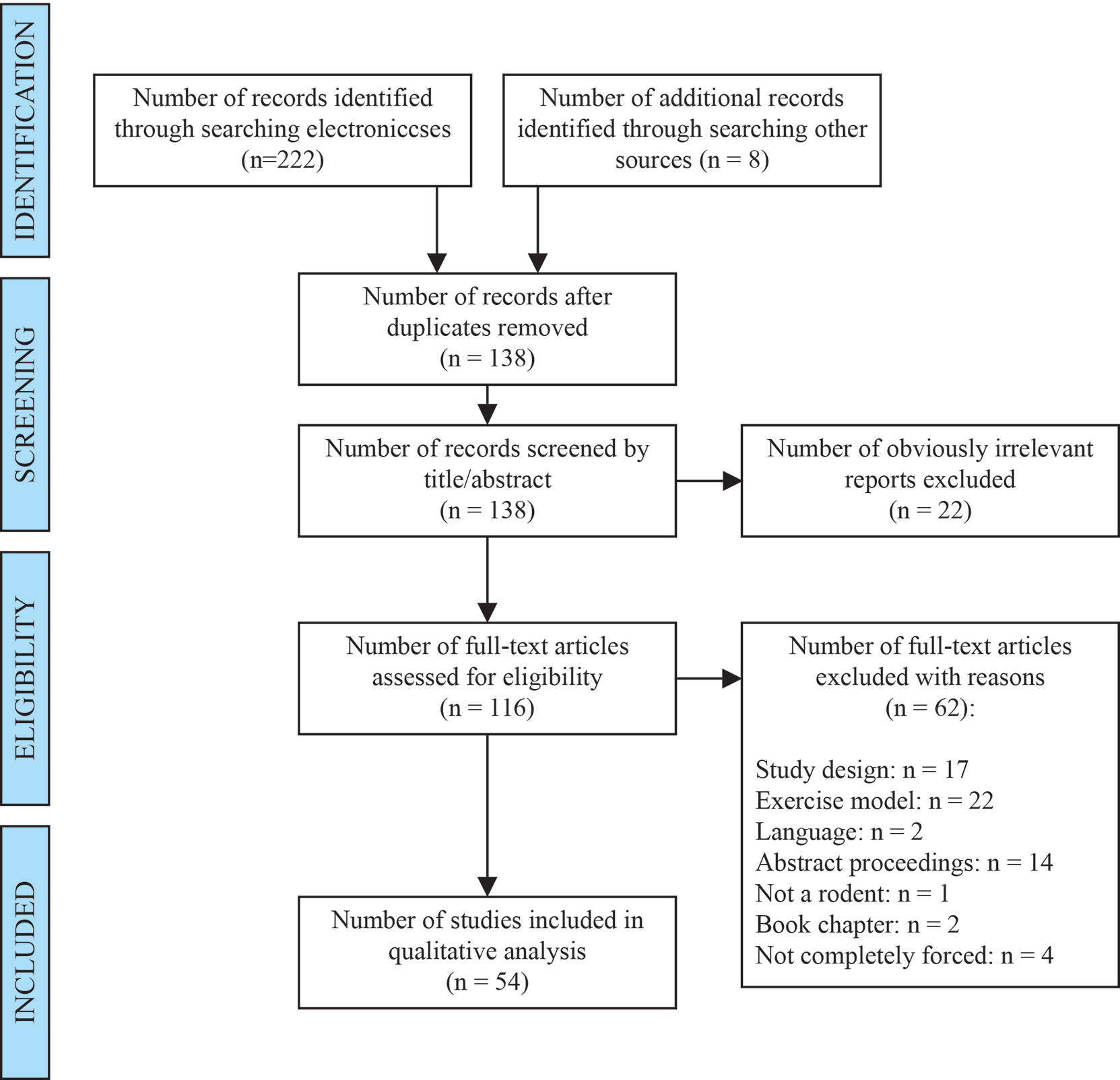
Figure 1 PRISMA flow diagram of study selection.
Study Eligibility Criteria
First screening of obviously irrelevant studies was conducted by two reviewers independently. Resultant full-text studies were then independently assessed using the inclusion and exclusion criteria, which were defined before the screening phase. Any disagreements between the reviewers were resolved by consensus after discussion with a third author. Articles were included in this review if they (1): were published in English (2), intervened on rodents (3), performed forced exercise in a motorized wheel and (4) the study design was a Randomized Controlled Trial or quasi-experimental. Articles were excluded if (1): Only voluntary exercise was performed (2), the forced motorized wheel system implies some additional stimulus to maintain the race. (Screening and eligibility, Figure 1).
For the purpose of this review, it was considered forced exercise when the three main parameters of the load (i.e. volume, intensity and density) were forced. In addition, interventions in which the rodent was manually forced to run on a wheel were not considered forced exercise, since the aim of the review focuses on forced exercise from the movement in a motorized wheel. These studies were discarded in the full-text review phase (Figure 1).
Data Extraction and Management
A data extraction form was elaborated before the fieldwork started (See Supplementary File 5 for detailed extraction protocol). The data extracted from the included studies were categorized in four sections: (i) Animal ethic committee (ii) Housing (iii) Animals and (iv) Exercise. Data extraction was conducted by two authors independently and disagreements were resolved by consensus with a third author.
Risk of Bias Assessment
Bias was assessed using SYRCLE’s risk of bias tool (39). According to the SYRCLE’s guidelines, the symbol ‘+’ was used when the criteria was reported by the authors. The symbol ‘-’ was used when the item was not reported and ‘?’ was used to indicate that the criteria was unclear. These procedures were conducted by two reviewers independently and any discrepancies were resolved by consensus with a third reviewer. The risk of bias establishment was done in an informative way. The data obtained were not used to establish any measure of treatment effect as it was not the aim of this review.
Analysis and Statistics
After data extraction, the number of articles reporting or not each item was calculated as a percentage. To determine the distribution of the studies according to the items described, a frequency histogram was used. The Pearson’s correlation coefficient (r) was used to examine the relationship between the quality of the study and i) the year of publication and ii) current impact factor of the journal in which it was published. The r value was rated as trivial (< 0.10), small (0.10–0.29), moderate (0.30–0.49), large (0.50–0.69), very large (0.70–0.89), or nearly perfect (0.90–0.99) (40). P-value was considered statistically significant at p<0.05. Due to the heterogeneity of topics among the studies that met our inclusion criteria, it was not considered appropriate to perform a meta-analysis of the trials.
Results and Discussion
Study Eligibility
The flow chart of selected studies is shown in Figure 1. A total of 222 studies were found trough database search and 8 studies from manual search. After eliminating duplicates and screening by title and abstracts, 116 studies were assessed for eligibility. Finally, 54 studies met our inclusion criteria.
Characteristics of the Studies
Supplementary File 2 describes a summary of the rodent model and the exercise program used in each study included in this review. Rats were the rodents used in 75.9% (41/54) of the studies (18, 41–80), and mice in 24.1% (13/54) (81–93). In rats, the most frequent strain was Sprague-Dawley (22/54) (18, 43, 46–49, 51–55, 61, 64–67, 70, 71, 76, 77, 79, 80), followed by Wistar (10/54) (44, 45, 57–60, 63, 68, 69, 73). In mice, the most frequent strain was C57BL/6J (10/54) (81, 82, 84–89, 92, 93) and BALB/c (2/54) (83, 90) in second place. Most of the studies used healthy animals (22/54) (18, 41, 44, 48, 49, 53, 56, 57, 59, 60, 68, 69, 72–75, 78, 82, 84, 85, 92), followed by stroke models (5/54) (45, 64, 79, 88, 89). Relative to exercise frequency, 5 days per week was the most common weekly frequency and 1 session per day was the most common daily frequency.
Risk of Bias Assessment
A summary of the risk of bias of the studies can be found in Figure 2. Since the review covered a wide variety of topics, a conservative and general analysis was decided when assessing the baseline characteristics (item 2, Figure 2). Low risk of bias was established if (i) the genetic modification of the animal, (ii) the sex of the sample, and (iii) the age or weight of the sample were described. In case there was no genetic modification: sex, age and weight should be described. When some of these items were described, but not all, it was considered as an unclear risk of bias. High risk was considered when none of the items, or only the sex was mentioned. In none of the studies was it possible to find a high or low risk in the allocation concealment or random housing (items 3 and 4, Figure 2). It was not possible to find other potential risks besides those included in the SYRCLE’s tool.
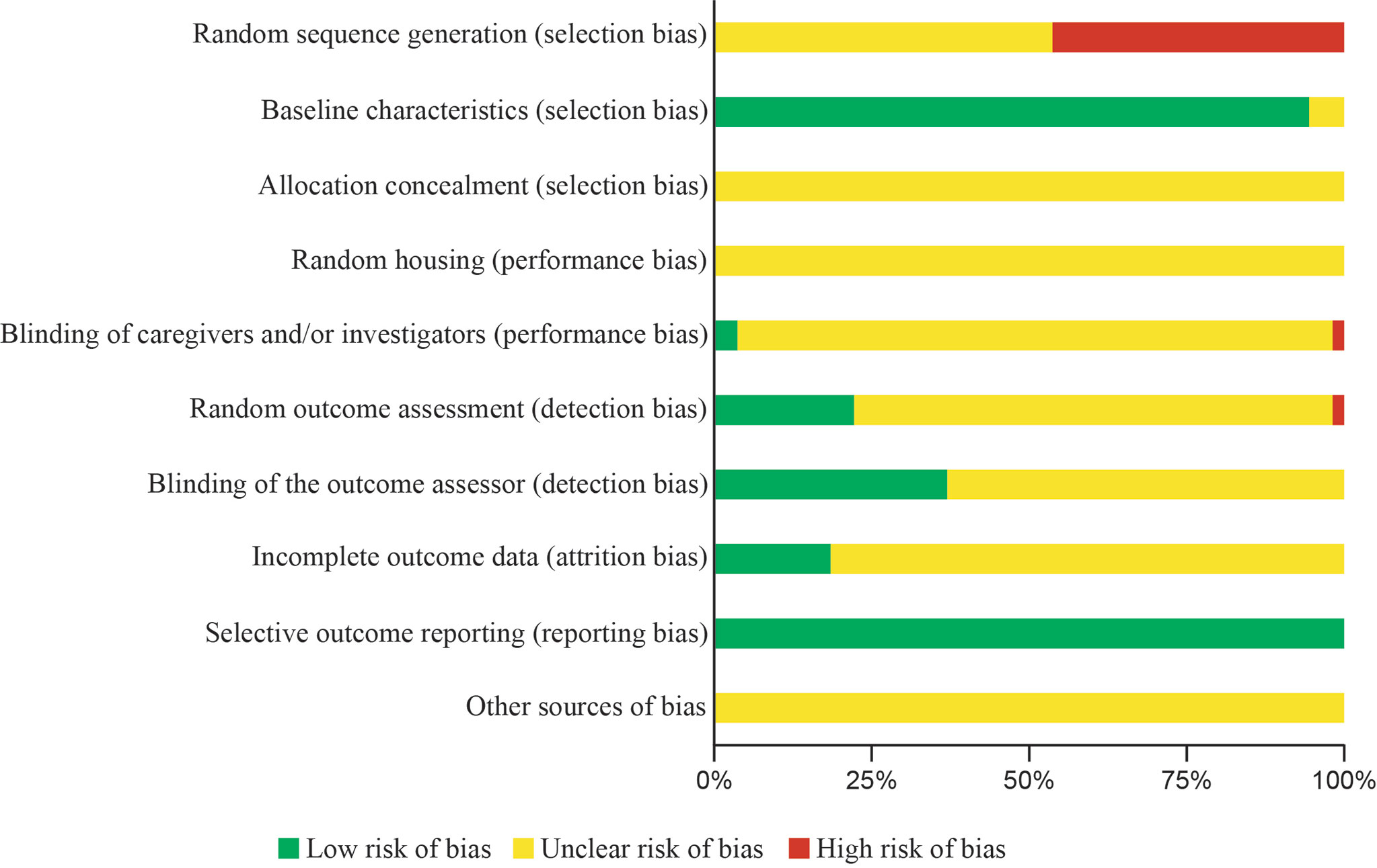
Figure 2 Risk of bias assessment using SYRCLE’s tool. Author’s judgment presented as percentage on each item. (-) High risk of bias, eminent risk of bias for this item; (?) Unclear risk of bias, carefully check the article for this item interpretation; (+) Low risk of bias, free of risk of bias in this item.
Animal Ethics Committees
Almost 76% (41/54) of the studies analyzed described that the experimental procedures were evaluated and approved by an ethics committee (Figure 3). The first animal ethics committees were established during the 70s to review the acceptability of animal research during the experimental procedures, deriving in the 3Rs principles (Replacement, Reduction and Refinement) (2, 94). However, an international consensus of ethical review began 20 years ago, being established as a mandatory requirement for the last 10 years (94, 95). Currently, these committees decide about the ethical acceptability of a research proposal and the researchers’ behavior during the procedures. Singularly, 46.1% (6/13) of the studies that did not describe any ethical committee approval were developed before the international consensus; but 53.8% (7/13) of these works were performed in a period of high consensus.
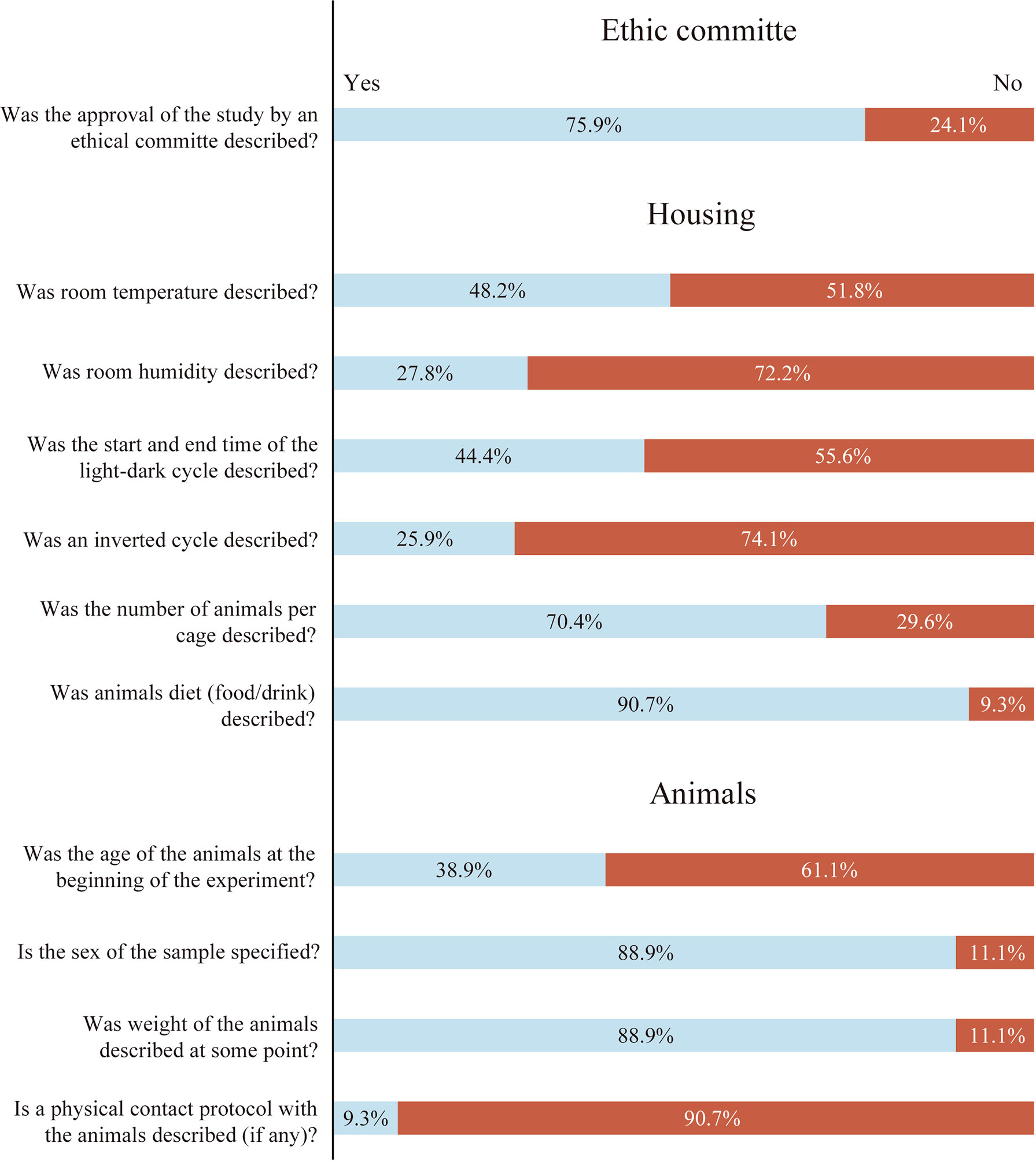
Figure 3 Percentage of studies that reported each item in relation to Ethics Committee, Housing and Animals sections. Light blue box: Item reported percentage. Red box: Item not reported percentage.
Housing
Changes in environmental temperature or humidity, light/dark cycle, number of animals per cage or food and drink availability in the rodents might modify the output results.
Room Temperature
In our analysis, the room temperature was described only in 48.2% (26/54) of the papers (Figure 3). The thermoneutral zone in mice is usually registered at 30°C, within a range of 1-3°C (96). Thus, mice housed at conventional environmental temperatures (~22°C) are below thermoneutrality; and the maintenance of the core body temperature at these conditions requires about half of the total energy expenditure (97). Only a few degrees of variation in the environmental temperature can change the thermogenesis response, as well as metabolic variations related to lipogenesis, adipogenesis or insulin sensitivity (26). Furthermore, temperature fluctuations can affect the cardiovascular response of the animal to carry out the programmed exercise, thus altering the response and the experimental results (21, 98, 99). Given all this, environmental temperature details result mandatory in all published works involving rodent models (100–102).
Environmental Relative Humidity
Unexpectedly, only 27.8% (15/54) of the studies included in our analysis described the room humidity conditions (Figure 3). Fluctuations of relative humidity may contribute to the development of dermal diseases or facilitate the transmission of certain viruses (103, 104). Also, developing an exercise program in a hot and high humidity environment can cause an inflammatory response as well as tissue damage (e.g. liver injury) affecting the internal load carried by the animal during the exercise period (25).
Light/Dark Cycle
In this review, only 44.5% (24/54) described the starting time of the light/dark cycles and a few studies gave precise data about the beginning time of the experiment (See exercise section). Interestingly, it was observed that 74% (18/24) of the reports describing the light-dark cycle developed the experimental phase during the passive period (light phase), but in most of them no justification was found for the chosen period (Figure 3). Most of the rodent models used in experimental research are active during the dark phase (nocturnal) and few of them are active during the light phase (diurnal) (105). Light is the major synchronizer of the circadian rhythms by the action of suprachiasmatic nucleus and peripheral clocks, deriving in different metabolic responses throughout the day (106–108). Also, the disruption of these circadian rhythms can lead to altered cardiovascular, metabolic and neurological responses (32–34, 93). Given this, for an adequate data comparison and reproducibility of the experiment, it is crucial to know the parameters of the light/dark cycle (active or passive) under which the interventions were carried out (28). However, for most of the papers analyzed, it was difficult -or even not possible- to know the period in which the experiment was developed; a situation that can be surpassed by including the start time and duration of the light/dark cycle and the start-end time of the experiments.
Animals per Cage
The number of animals per cage was indicated in 70.4% (38/54) of the studies analyzed (Figure 3). Rodents are social animals, and it is well known that their isolation can modify the experimental results. As a chronic stressor, individual housing can have strong effects on behavior, leading to stereotyped behavior or provoking depression- and anxiety-like symptoms (109, 110). This chronic stress involves structural and molecular alterations in several areas of the brain, particularly the prefrontal cortex and limbic brain structures, deriving in altered psychotic and emotional behavior (111–113). Executing a physical exercise program may mitigate, but not replace the beneficial effects of social interaction (114). It is crucial to report the number of animals per cage, and the sex of the animal (See below), since it can condition other social aspects such as the establishment of hierarchies or dominance behaviors that may affect the results.
Food/Drink
The 90.7% (49/54) of the studies analyzed described the type of food/drink and its availability (Figure 3). Interestingly, 36 of them consisted of a standard ad libitum food/drink consumption, while 13 studies introduced some type of modification of these parameters. Experimental designs with differences in food and water macronutrients composition (fat, protein and carbohydrate percentages) and in its access by the rodent, may strongly affect the experimental outputs (29, 115, 116). Some macronutrient proportions may lead to white adipose tissue inflammation, insulin resistance or obesity (117, 118). On the other hand, changes in intake patterns will derive in a variation of circadian hormones (e.g. insulin, glucagon or corticosterone) (119, 120); or may modify the expression of intestinal enzymes such as maltose and sucrose in rats (121–123). Some experiments are usually finished with a period of fasting before the sacrifice; but this condition changes the expression of PGC-1α, AMPK or PPAR, particularly in rodent muscle (124–126).
Animals
Variations in age, sex, weight, or handling procedures might change both biological and behavioral results.
Age
In our analysis, only 38.9% (21/54) of the studies described the age of the animals at the beginning of the training protocol (Figure 3). Humans and rodents have adolescence, adulthood, or old age periods of life with their own physiological responses. However, these stages of life are significantly shorter in mice and rats and, therefore, reporting the age of the rodents becomes crucial (27). From birth to adulthood, the rodent’s brain increases in size, myelinization (limbic structures are fully developed at six weeks) and remodels its neural networks (127–129). An age-related mitochondrial decline, hearing loss or changes in liver gene expression with high impact in pharmacological or behavioral responses can be observed in mice (130–132). Also, in stroke models (one of the most common models in forced wheel studies), younger animals may respond differently from older animals (133, 134). Due to the mentioned impact of age on the different physiological responses, describing the age of all animals throughout the experiment should be mandatory.
Sex
The 88.9% (48/54) of the studies included in this review described the sex of the rodents (Figure 3). The last decades of experimental research have underlined the need of taking into account the differences between male and female rodents (23, 135). Sex differences in hormone levels may affect decision-making mechanisms (136–138). Furthermore, drug effects and therapies, such as physical exercise, show variations between males and females (139–141).
Weight
The 88.9% (48/54) of the studies described the weight at some point of the research work, but only 63% (34/54) of them measured it regularly (Figure 3). The body weight of the animal may be related to health status and is strongly influenced by numerous nutritional, environmental, husbandry and genetic factors (30, 142–144). Some studies use body weight to deduce the age. However, both age and weight need to be reported together since body weight cannot predict age with precision (145, 146).
Handling
Although all the analyzed studies involved some form of manipulation and contact with the rodents, only 9.3% (5/54) described a handling protocol (Figure 3). The levels of anxiety and stress of the rodents can be reduced with handling procedures (20). In addition, it can help to manipulate the rodents more calmly, reducing possible bites, and with strong repercussions in behavioral tests (e.g. open field) (147, 148).
Exercise
To ensure reproducibility of exercise protocols, it is essential to include details of at least the light/dark cycle, habituation protocol and training parameters.
Exercise and Light/Dark Cycle
In this review, in 55.5% (30/54) of the published works it was possible to know whether the exercise took place in the active or passive phase (Figure 4). Of these studies, 14 developed the exercise program during the active phase and 13 during the passive phase, without justifying the reasons for the choice. Surprisingly, only 3 studies carried out the training during the inactive or active/inactive period under justified experimental reasons. The beginning of the light phase (passive) is considered the zeitgeber time 0 (ZT0). The use of this nomenclature would allow to avoid confusing comparisons of research works using the time of the day as a reference (149). Because almost all experimental research is developed in nocturnal rodents, it is a priority to point out if the experiments were developed during their active (night) or passive phase (day) (150). Exercise may also re-entrain circadian rhythms, producing a misalignment that can lead to low cognitive performance, deterioration of alertness, weight variations and sleep disruption (33, 151–157). Furthermore, forced activity during the inactive phase may disrupt gene expression patterns and hormonal regulations (e.g. insulin, testosterone or cortisol) (158, 159). Knowing the precise light/dark cycle parameters in which the exercise was developed, major misinterpretations of the research output could be avoided.
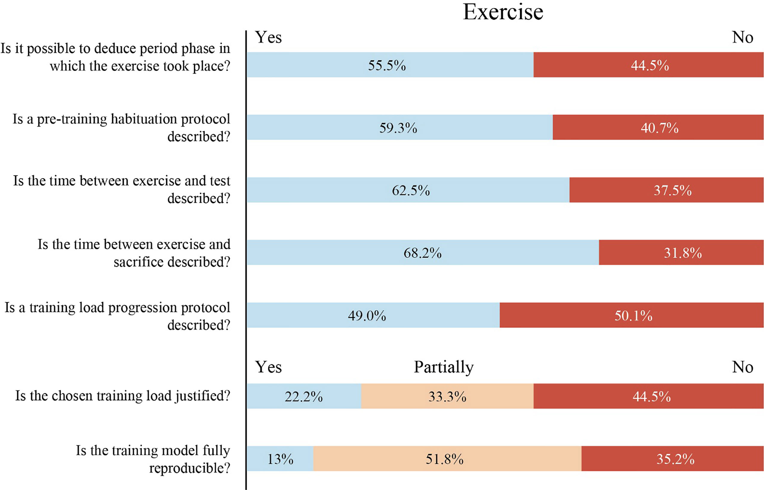
Figure 4 Percentage of studies that reported each item in relation to Exercise. Light blue box: Item reported. Orange box: Item partially reported. Red box: Item not reported.
Habituation Protocol
In this review, 59.3% (32/54) of the analyzed papers described some habituation period before the exercise program (Figure 4). Around 10% of rodents refuse to run in forced running paradigms (160). However, that lack of response can be solved by applying a habituation phase prior to an exercise program (18). This period improves the capacity of rodents to maintain a higher volume and intensity of running and establishes a homogeneous starting point for all the animals to perform a training program (18, 161). In addition, the habituation protocol reduces non-specific stress responses in rats that can modify the physiological and behavioral results in forced models (19).
Training Parameters
Duration, starting time, speed and frequency of the exercise sessions are key parameters to be reported in order to ensure the reproducibility of exercise protocols. Surprisingly, only 13% (7/54) of the analyzed exercise protocols are completely reproducible (Figure 4). Reproducibility was assessed attending to the following factors: (i) duration of each exercise session: specification of series and interspersed rest time (if applies) (min) (ii) speed (m/min), (iii) start time of each exercise session (ZT time), (iv) daily and weekly frequency and (v) total duration of the exercise program (days/weeks). Supplementary File 3, Figure 1 from Toval et al. (18) and Table 1 in Toval et al. (19) are examples where all the mentioned training parameters are described. Reporting these parameters is especially relevant since intensity, volume, and density are the three main factors of the training load and changes in all or some of them can produce different adaptations in the organism (31, 162, 163). We encourage authors to use this table (Supplementary File 3), implementing the required modifications according to the needs of each study.
Load Progression vs Non Progression Protocol
Of the papers that developed more than one training session (51/54), only 49% (25/51) applied a load progression protocol (Figure 4). The implementation of a load progression (intensity, volume, and density of the exercise) during the training program is one of the main training principles (164). A load progression protocol ensures that new adaptations are occurring in the body throughout the weeks of training (165). Therefore, a failure to implement a load progression protocol might produce misinterpretations of the exercise effects.
Training Load Justification
In this review, 44.5% (24/54) of the studies did not justify any load parameter (Figure 4). However, 33.3% (18/54) of them reported some parameters while only 22.2% (12/54) reported some justification for all parameters of the exercise load. The training load (exercise intensity, volume and density) to be developed throughout the experimental research needs to be determined by physiological references (e.g. %VO2Max and lactate thresholds) or be based on well-founded work. Intensity, volume or density can strongly modify the research output, and should be reported and taken into consideration carefully (162, 166). In this sense, we observed a lack of justification among the training protocols.
Time Between Exercise and Test/Sacrifice
In this review, only 62.5% (15/24) of the studies reported the time between exercise and tests, and 68.2% (30/44) reported the time between exercise and sacrifice (Figure 4). Adjustments and adaptations to exercise are produced during an exercise program. Adjustment refers to the short-term changes that occur as a result of increased metabolic demand during the physical exercise (e.g. increased heart rate), while adaptation refers to residual changes in the organism after several exercise sessions (167). Furthermore, these adjustments and adaptations to exercise have an effect on early and late gene expression responses in a time-of-day-dependent way (150, 168). After 1 hour of exercise, the transcriptomic and metabolomic patterns analyzed every 4 hours showed different molecular profiles in a range of 20 hours (168). Thus, establishing and reporting the time between the last session of exercise and any test or analysis (sacrifice) is crucial.
Study Distribution of the Reported Items and Its Correlations With the Impact Factor and Year of Publication
A histogram was used to determine how the papers were distributed according to the number of the items reported (Figure 5A). Items that described weight, training parameters, training load justification and time between exercise and test/sacrifice include an extra point if the items were fully reported, thus being 21 the highest number that could be reached only in entirely detailed works. In our study, a range between 4 and 18 items were reported for the articles analyzed (Figure 5A). However, an average of 11.42 ± 3.46 items reported indicates that most of the articles are not detailed enough to be reproducible. Next, we found that the year of publication and the current impact factor are not correlated with the number of items correctly reported in these research works (Figures 5B, C; p>0.05), being the correlation defined as small (r=0.15) and trivial (r=-0.05) respectively.
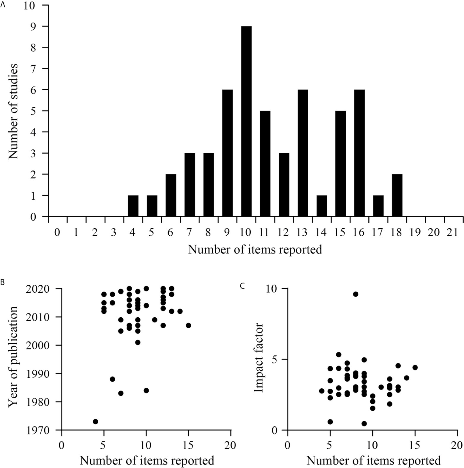
Figure 5 Studies distribution and correlation of the reported items. (A) Histogram representing the distribution of studies according to the items reported by them. (B) Correlation between the number of items reported by each study and the year of publication of the study in the journal (p=0.28, r=0.15, Pearson’s correlation coefficient r). (C) Correlation between the journal’s impact factor (2019) and the number of items reported by each study (p=0.69, r=-0.05, Pearson’s correlation coefficient r).
A strong tendency to claim the reporting of critical data to ensure experimental reproducibility began twenty years ago and led to guidelines to overcome these difficulties (1, 3, 4). Our results raise concerns about the lack of transparency in research reports once again (5, 6). Also, the absence of key information, sometimes related with lack of requirement, could not be linked to the year of publication of the research work (1, 2, 95). Furthermore, the imprecise details of the methods did not depend on the impact factor of the journals, the latter being a measure of the quality of the journal questioned by current criticism (169–171).
FORCED Exercise Wheel Guidelines
After analyzing the degree to which each item mentioned above was reported, we suggested guidelines to facilitate reproducibility, effectiveness and greater transparency of forced-wheel intervention studies (Table 1). The resulting checklist consists of 13 items grouped by (i) Ethic committee, (ii) Housing, (iii) Animals, and (iv) Exercise. Additionally, FORCED guidelines may be used to evaluate other works, in which case, each item will be evaluated as follows: (i) Complete, if the item and sub-items (if any) are described; (ii) Partial, when half or more, but not all of the sub-items have been described; (iii) Absent, when less than half of sub-items have been described. Some parameters analyzed in ethic committee, housing or animal sections are included in ARRIVE guidelines. However, these indications may not be enough to ensure the reproducibility of studies using forced exercise in rodents. We hope that our suggested guidelines will serve as a starting point to open processes with an international and consensus-based approach, in the interest of reproducibility in this type of research.
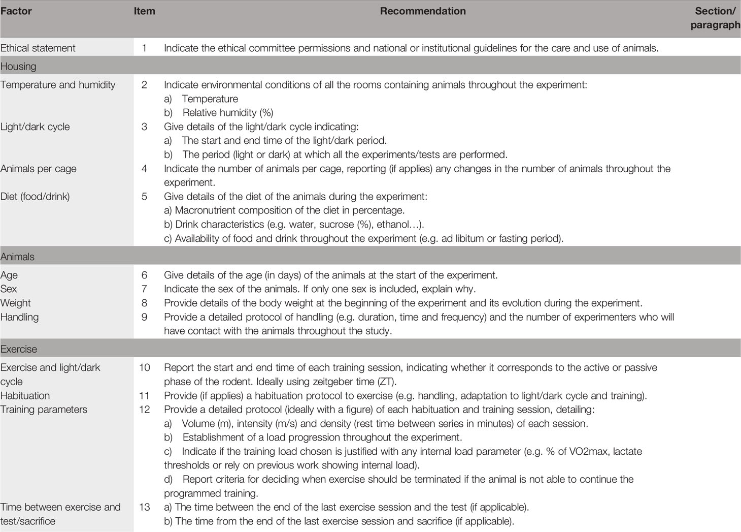
Table 1 FORCED guidelines for authors.
Strengths and Limitations
Strengths
The systematic review was developed according to PRISMA guidelines. The data extraction manual was developed before field work. The risk of bias of the studies was assessed using SYRCLE’s tool. Two independent reviewers applied the exclusion/inclusion criteria, data extraction and the risk of bias assessment, looking for consensus with a third author in case of disagreements.
Limitations
The protocol of the review was not registered in databases such as PROSPERO, since it is required to register it prospectively and the field work had already been initiated. A meta-analysis of the included studies was not performed as the aim of the review was to analyze the characteristics that can affect the reproducibility of forced wheel training protocols, instead of the measurement of a treatment effect.
Conclusion
This systematic review was developed in order to know the reproducibility and reliability of the studies using forced wheel systems in rodents. We concluded that most of the analyzed works do not provide enough data to guarantee the experimental reproducibility and research output comparisons. Our suggested FORCED guidelines are expected to a) be considered to promote a consensus in the field of exercise, b) to be used for in vivo experiments with rodents in forced wheel exercise, and c) extended to other modalities such as treadmill exercise. If the variables mentioned by these guidelines are not accurately described, the reported effects of the exercise could be questioned. Our study reaffirms the need for improved reporting in animal research using forced wheel exercise programs. This task can strongly contribute to the experimental reproducibility in this field, and should be carefully considered by authors, editorial boards, and ethics committees.
Data Availability Statement
The original contributions presented in the study are included in the article/Supplementary Material. Further inquiries can be directed to the corresponding author.
Author Contributions
All authors contributed to the study conception and design. YK, AT, DG, and JF elaborated the extraction manual. DG and MM-M carried out the phases of database searching, screening, and data extraction. Disagreements were revised with AT and AB. DG and AT conducted the risk of bias assessment. Disagreements were revised with AB. DG and JF drafted the initial manuscript. FN-M and BR critically reviewed the manuscript. All authors revised the manuscript and approved the final text as submitted.
Funding
Granted by the Spanish Ministry of Science, Innovation and Universities (MCIU), State Research Agency (AEI) and European Regional Development Fund (FEDER; PGC2018-098229-B-100 to JF), and by Seneca Foundation (19904/GERM/15).
Conflict of Interest
The authors declare that the research was conducted in the absence of any commercial or financial relationships that could be construed as a potential conflict of interest.
Supplementary Material
The Supplementary Material for this article can be found online at: https://www.frontiersin.org/articles/10.3389/fendo.2021.638261/full#supplementary-material
References
1. Smith JA, Birke L, Sadler D. Reporting Animal Use in Scientific Papers. Lab Anim (1997) 31(4):312–7. doi: 10.1258/002367797780596176
2. Rose M, Everitt J, Hedrich H, Schofield J, Dennis M, Scott E, et al. ICLAS Working Group on Harmonization: International Guidance Concerning the Production Care and Use of Genetically-Altered Animals. Lab Anim (2013) 47(3):146–52. doi: 10.1177/0023677213479338
3. Kilkenny C, Browne W, Cuthill IC, Emerson M, Altman DG. Animal Research: Reporting in Vivo Experiments: The ARRIVE Guidelines. Br J Pharmacol (2010) 160(7):1577. doi: 10.1111/j.1476-5381.2010.00872.x
4. Rice AS, Cimino-Brown D, Eisenach JC, Kontinen VK, Lacroix-Fralish ML, Machin I, et al. Animal Models and the Prediction of Efficacy in Clinical Trials of Analgesic Drugs: A Critical Appraisal and Call for Uniform Reporting Standards. Pain (2008) 139(2):243–7. doi: 10.1016/j.pain.2008.08.017
5. Sena ES, Van Der Worp HB, Bath PM, Howells DW, Macleod MR. Publication Bias in Reports of Animal Stroke Studies Leads to Major Overstatement of Efficacy. PloS Biol (2010) 8(3):e1000344. doi: 10.1371/journal.pbio.1000344
6. Percie du Sert N, Hurst V, Ahluwalia A, Alam S, Avey MT, Baker M, et al. The ARRIVE Guidelines 2.0: Updated guidelines reporting animal research. J Cereb Blood Flow Metab (2020) 40(9):1769–77. doi: 10.1177/0271678X20943823
7. Altman DG, Simera I. Responsible Reporting of Health Research Studies: Transparent, Complete, Accurate and Timely. J antimicrobial chemotherapy (2010) 65(1):1–3. doi: 10.1093/jac/dkp410
8. Avey MT, Moher D, Sullivan KJ, Fergusson D, Griffin G, Grimshaw JM, et al. The Devil is in the Details: Incomplete Reporting in Preclinical Animal Research. PloS One (2016) 11(11):e0166733. doi: 10.1371/journal.pone.0166733
9. Kilkenny C, Parsons N, Kadyszewski E, Festing MF, Cuthill IC, Fry D, et al. Survey of the Quality of Experimental Design, Statistical Analysis and Reporting of Research Using Animals. PloS One (2009) 4(11):e7824. doi: 10.1371/journal.pone.0007824
10. Munafò MR, Nosek BA, Bishop DV, Button KS, Chambers CD, Du Sert NP, et al. A Manifesto for Reproducible Science. Nat Hum Behav (2017) 1(1):1–9. doi: 10.1038/s41562-016-0021
11. Weissgerber TL, Garcia-Valencia O, Garovic VD, Milic NM, Winham SJ. Why We Need to Report More Than’data Were Analyzed by T-Tests or ANOVA’. eLife (2018) 7. doi: 10.7554/eLife.36163
12. Makin TR, de Xivry J-JO. Science Forum: Ten Common Statistical Mistakes to Watch Out for When Writing or Reviewing a Manuscript. Elife (2019) 8:e48175. doi: 10.7554/eLife.48175
13. Smith AD, Zigmond MJ. Can the Brain Be Protected Through Exercise? Lessons an Anim Model parkinsonism☆. Exp Neurol (2003) 184(1):31–9. doi: 10.1016/j.expneurol.2003.08.017
14. Cooney G, Dwan K, Mead G. Exercise for Depression. Jama (2014) 311(23):2432–3. doi: 10.1001/jama.2014.4930
15. Marlatt MW, Potter MC, Lucassen PJ, van Praag H. Running Throughout Middle-Age Improves Memory Function, Hippocampal Neurogenesis, and BDNF Levels in Female C57BL/6J Mice. Dev Neurobiol (2012) 72(6):943–52. doi: 10.1002/dneu.22009
16. McGreevy KR, Tezanos P, Ferreiro-Villar I, Pallé A, Moreno-Serrano M, Esteve-Codina A, et al. Intergenerational Transmission of the Positive Effects of Physical Exercise on Brain and Cognition. Proc Natl Acad Sci (2019) 116(20):10103–12. doi: 10.1073/pnas.1816781116
17. Wang R, Tian H, Guo D, Tian Q, Yao T, Kong X. Impacts of Exercise Intervention on Various Diseases in Rats. J Sport Health Sci (2019) 9(3):211–27. doi: 10.1016/j.jshs.2019.09.008
18. Toval A, Banos R, De la Cruz E, Morales-Delgado N, Pallares JG, Ayad A, et al. Habituation Training Improves Locomotor Performance in a Forced Running Wheel System in Rats. Front Behav Neurosci (2017) 11:42. doi: 10.3389/fnbeh.2017.00042
19. Toval A, Vicente-Conesa F, Martínez-Ortega P, Kutsenko Y, Morales-Delgado N, Garrigos D, et al. Hypothalamic Crh/Avp, Plasmatic Glucose and Lactate Remain Unchanged During Habituation to Forced Exercise. Front Physiol (2020) 11:410. doi: 10.3389/fphys.2020.00410
20. Schmitt U, Hiemke C. Strain Differences in Open-Field and Elevated Plus-Maze Behavior of Rats Without and With Pretest Handling. Pharmacol Biochem Behav (1998) 59(4):807–11. doi: 10.1016/S0091-3057(97)00502-9
21. González-Alonso J, Crandall CG, Johnson JM. The Cardiovascular Challenge of Exercising in the Heat. J Physiol (2008) 586(1):45–53. doi: 10.1113/jphysiol.2007.142158
22. Turner PV, Pekow C, Clark JM, Vergara P, Bayne K, White WJ, et al. Roles of the International Council for Laboratory Animal Science (ICLAS) and International Association of Colleges of Laboratory Animal Medicine (IACLAM) in the Global Organization and Support of 3Rs Advances in Laboratory Animal Science. J Am Assoc Lab Anim Sci (2015) 54(2):174–80.
23. Beery AK, Zucker I. Sex Bias in Neuroscience and Biomedical Research. Neurosci Biobehav Rev (2011) 35(3):565–72. doi: 10.1016/j.neubiorev.2010.07.002
24. Chang Y-K, Chi L, Etnier JL, Wang C-C, Chu C-H, Zhou C. Effect of Acute Aerobic Exercise on Cognitive Performance: Role of Cardiovascular Fitness. Psychol Sport Exercise (2014) 15(5):464–70. doi: 10.1016/j.psychsport.2014.04.007
25. Li D, Wang X, Liu B, Liu Y, Zeng Z, Lu L, et al. Exercises in Hot and Humid Environment Caused Liver Injury in a Rat Model. PloS One (2014) 9(12):e111741. doi: 10.1371/journal.pone.0111741
26. Ravussin Y. Temperature Matters With Rodent Metabolic Studies. Obesity (2015) 23(7):1330. doi: 10.1002/oby.21149
27. Jackson SJ, Andrews N, Ball D, Bellantuono I, Gray J, Hachoumi L, et al. Does Age Matter? The impact rodent age study outcomes. Lab Anim (2017) 51(2):160–9. doi: 10.1177/0023677216653984
28. Castelhano-Carlos M, Baumans V. The Impact of Light, Noise, Cage Cleaning and in-House Transport on Welfare and Stress of Laboratory Rats. Lab Anim (2009) 43(4):311–27. doi: 10.1258/la.2009.0080098
29. Pellizzon MA, Ricci MR. Choice of Laboratory Rodent Diet May Confound Data Interpretation and Reproducibility. Curr Developments Nutr (2020) 4(4):nzaa031. doi: 10.1093/cdn/nzaa031
30. Chambers T, Morgan M, Heger A, Sharpe R, Drake A. High-Fat Diet Disrupts Metabolism in Two Generations of Rats in a Parent-of-Origin Specific Manner. Sci Rep (2016) 6(1):1–11. doi: 10.1038/srep31857
31. Brown MB, Neves E, Long G, Graber J, Gladish B, Wiseman A, et al. High-Intensity Interval Training, But Not Continuous Training, Reverses Right Ventricular Hypertrophy and Dysfunction in a Rat Model of Pulmonary Hypertension. Am J Physiology-Regulatory Integr Comp Physiol (2017) 312(2):R197–210. doi: 10.1152/ajpregu.00358.2016
32. Scheer FA, Hilton MF, Mantzoros CS, Shea SA. Adverse Metabolic and Cardiovascular Consequences of Circadian Misalignment. Proc Natl Acad Sci (2009) 106(11):4453–8. doi: 10.1073/pnas.0808180106
33. Garaulet M, Madrid JA. Chronobiology, Genetics and Metabolic Syndrome. Curr Opin lipidology (2009) 20(2):127–34. doi: 10.1097/MOL.0b013e3283292399
34. Barnard AR, Nolan PM. When Clocks Go Bad: Neurobehavioural Consequences of Disrupted Circadian Timing. PloS Genet (2008) 4(5):e1000040. doi: 10.1371/journal.pgen.1000040
35. Duzel E, van Praag H, Sendtner M. Can Physical Exercise in Old Age Improve Memory and Hippocampal Function? Brain (2016) 139(3):662–73. doi: 10.1093/brain/awv407
36. Alessio HM, Hagerman AE, Nagy S, Philip B, Byrnes RN, Woodward JL, et al. Exercise Improves Biomarkers of Health and Stress in Animals Fed Ad Libitum. Physiol Behav (2005) 84(1):65–72. doi: 10.1016/j.physbeh.2004.10.010
37. Ahlskog JE, Geda YE, Graff-Radford NR, Petersen RC.Physical Exercise as a Preventive or Disease-Modifying Treatment of Dementia and Brain Aging. Mayo Clinic Proc (2011) 86(9):876–84. doi: 10.4065/mcp.2011.0252
38. Moher D, Liberati A, Tetzlaff J, Altman DG, Group P. Preferred Reporting Items for Systematic Reviews and Meta-Analyses: The PRISMA Statement. PloS Med (2009) 6(7):e1000097. doi: 10.1371/journal.pmed.1000097
39. Hooijmans CR, Rovers MM, De Vries RB, Leenaars M, Ritskes-Hoitinga M, Langendam MW. SYRCLE’s Risk of Bias Tool for Animal Studies. BMC Med Res Method (2014) 14(1):43. doi: 10.1186/1471-2288-14-43
40. Cohen J. Statistical Power Analysis for the Behavioral Sciences. Cambridge, Massachusetts, United States: Academic press (2013).
41. Arnold MR, Greenwood BN, McArthur JA, Clark PJ, Fleshner M, Lowry CA. Effects of Repeated Voluntary or Forced Exercise on Brainstem Serotonergic Systems in Rats. Behav Brain Res (2020) 378:112237. doi: 10.1016/j.bbr.2019.112237
42. Boersma GJ, Barf RP, Benthem L, van Dijk G, Scheurink AJW. Forced and Voluntary Exercise Counteract Insulin Resistance in Rats: The Role of Coping Style. Hormones Behav (2012) 62(1):93–8. doi: 10.1016/j.yhbeh.2012.05.006
43. Cao JJ, Picklo MJ. Involuntary Wheel Running Improves But Does Not Fully Reverse the Deterioration of Bone Structure of Obese Rats Despite Decreasing Adiposity. Calcified Tissue Int (2015) 97(2):145–55. doi: 10.1007/s00223-015-9992-6
44. Caton SJ, Bielohuby M, Bai Y, Spangler LJ, Burget L, Pfluger P, et al. Low-Carbohydrate High-Fat Diets in Combination With Daily Exercise in Rats: Effects on Body Weight Regulation, Body Composition and Exercise Capacity. Physiol Behav (2012) 106(2):185–92. doi: 10.1016/j.physbeh.2012.02.003
45. Chen CC, Chang MW, Chang CP, Chan SC, Chang WY, Yang CL, et al. A Forced Running Wheel System With a Microcontroller That Provides High-Intensity Exercise Training in an Animal Ischemic Stroke Model. Braz J Med Biol Res (2014) 47(10):858–68. doi: 10.1590/1414-431x20143754
46. Chhaya SJ, Quiros-Molina D, Tamashiro-Orrego AD, Houle JD, Detloff MR. Exercise-Induced Changes to the Macrophage Response in the Dorsal Root Ganglia Prevent Neuropathic Pain After Spinal Cord Injury. J Neurotrauma (2019) 36(6):877–90. doi: 10.1089/neu.2018.5819
47. Detloff MR, Smith EJ, Molina DQ, Ganzer PD, Houlé JD. Acute Exercise Prevents the Development of Neuropathic Pain and the Sprouting of Non-Peptidergic (GDNF-and Artemin-Responsive) C-Fibers After Spinal Cord Injury. Exp Neurol (2014) 255:38–48. doi: 10.1016/j.expneurol.2014.02.013
48. Eccles S, Kim EM, O’Hare E. Granisetron Attenuates Exercise-Induced Conditioned Taste Aversion in the Rat. Appetite (2005) 44(3):325–8. doi: 10.1016/j.appet.2005.02.001
49. Forristall JR, Hookey BL, Grant VL. Conditioned Taste Avoidance Induced by Forced and Voluntary Wheel Running in Rats. Behav Processes (2007) 74(3):326–33. doi: 10.1016/j.beproc.2006.12.002
50. Greenwood BN, Spence KG, Crevling DM, Clark PJ, Craig WC, Fleshner M. Exercise-Induced Stress Resistance is Independent of Exercise Controllability and the Medial Prefrontal Cortex. Eur J Neurosci (2013) 37(3):469–78. doi: 10.1111/ejn.12044
51. Griesbach GS, Tio DL, Vincelli J, McArthur DL, Taylor AN. Differential Effects of Voluntary and Forced Exercise on Stress Responses After Traumatic Brain Injury. J Neurotrauma (2012) 29(7):1426–33. doi: 10.1089/neu.2011.2229
52. Ilback NG, Friman G, Squibb RL, Johnson AJ, Balentine DA, Beisel WR. The Effect of Exercise and Fasting on the Myocardial Protein and Lipid-Metabolism in Experimental Bacterial Myocarditis. Acta Pathologica Microbiologica Et Immunologica Scandinavica Section a-Pathology (1984) 92(4):195–204. doi: 10.1111/j.1699-0463.1984.tb04396.x
53. Jian-Feng J, Sheng-Jun J, Sun R, Li K, Zhang Y, Zhang L-y, et al. Forced Running Exercise Attenuates Hippocampal Neurogenesis Impairment and the Neurocognitive Deficits Induced by Whole-Brain Irradiation Via the BDNF-Mediated Pathway. Biochem Biophys Res Commun (2014) 443(2):646–51. doi: 10.1016/j.bbrc.2013.12.031
54. Kant GJ, Bunnell BN, Mougey EH, Pennington LL, Meyerhoff JL. Effects of Repeated Stress on Pituitary Cyclic AMP, and Plasma Prolactin, Corticosterone and Growth Hormone in Male Rats. Pharmacol Biochem Behav (1983) 18(6):967–71. doi: 10.1016/S0091-3057(83)80022-7
55. Kant GJ, Lenox RH, Bunnell BN, Mougey EH, Pennington LL, Meyerhoff JL. Comparison of Stress Response in Male and Female Rats: Pituitary Cyclic AMP and Plasma Prolactin, Growth Hormone and Corticosterone. Psychoneuroendocrinology (1983) 8(4):421–8. doi: 10.1016/0306-4530(83)90021-5
56. Lloyd BA, Hake HS, Ishiwata T, Farmer CE, Loetz EC, Fleshner M, et al. Exercise Increases Mtor Signaling in Brain Regions Involved in Cognition and Emotional Behavior. Behav Brain Res (2017) 323:56–67. doi: 10.1016/j.bbr.2017.01.033
57. Mancardi D, Tullio F, Crisafulli A, Rastaldo R, Folino A, Penna C, et al. Omega 3 Has a Beneficial Effect on Ischemia/Reperfusion Injury, But Cannot Reverse the Effect of Stressful Forced Exercise. Nutr Metab Cardiovasc Dis (2009) 19(1):20–6. doi: 10.1016/j.numecd.2008.01.004
58. Martinez-Salazar C, Villanueva I, Pacheco-Rosado J, Alva-Sánchez C. Moderate Exercise Prevents the Cell Atrophy Caused by Hypothyroidism in Rats. Acta Neurobiologiae Experimentalis (2020) 80(1):47–56. doi: 10.21307/ane-2020-005
59. Masaki T, Nakajima S. Taste Aversion in Rats Induced by Forced Swimming, Voluntary Running, Forced Running, and Lithium Chloride Injection Treatments. Physiol Behav (2006) 88(4-5):411–6. doi: 10.1016/j.physbeh.2006.04.013
60. Nakajima S. Running Induces Nausea in Rats: Kaolin Intake Generated by Voluntary and Forced Wheel Running. Appetite (2016) 105:85–94. doi: 10.1016/j.appet.2016.05.009
61. O’Dell SJ, Gross NB, Fricks AN, Casiano BD, Nguyen TB, Marshall JF. Running Wheel Exercise Enhances Recovery From Nigrostriatal Dopamine Injury Without Inducing Neuroprotection. Neuroscience (2007) 144(3):1141–51. doi: 10.1016/j.neuroscience.2006.10.042
62. Patel DI, White LJ. Effect of 10-Day Forced Treadmill Training on Neurotrophic Factors in Experimental Autoimmune Encephalomyelitis. Appl Physiol Nutr Metabolism-Physiologie Appliquee Nutr Et Metabolisme (2013) 38(2):194–9. doi: 10.1139/apnm-2012-0303
63. Peng K-T, Tsai M-H, Lee C-W, Chiang Y-C, Chen P-C, Chen C-C, et al. Dysregulated Expression of Antioxidant Enzymes in Polyethylene Particle-Induced Periprosthetic Inflammation and Osteolysis. PloS One (2018) 13(8):e0202501. doi: 10.1371/journal.pone.0202501
64. Pianta S, Lee JY, Tuazon JP, Castelli V, Mantohac LM, Tajiri N, et al. A Short Bout of Exercise Prior to Stroke Improves Functional Outcomes by Enhancing Angiogenesis. Neuromolecular Med (2019) 21(4):517–28. doi: 10.1007/s12017-019-08533-x
65. Picklo MJ, Thyfault JP. Vitamin E and Vitamin C Do Not Reduce Insulin Sensitivity But Inhibit Mitochondrial Protein Expression in Exercising Obese Rats. Appl Physiol Nutr Metab (2015) 40(4):343–52. doi: 10.1139/apnm-2014-0302
66. Ploughman M, Granter-Button S, Chernenko G, Tucker B, Mearow K, Corbett D. Endurance Exercise Regimens Induce Differential Effects on Brain-Derived Neurotrophic Factor, Synapsin-I and Insulin-Like Growth Factor I After Focal Ischemia. Neuroscience (2005) 136(4):991–1001. doi: 10.1016/j.neuroscience.2005.08.037
67. Ploughman M, Granter-Button S, Chernenko G, Attwood Z, Tucker BA, Mearow KM, et al. Exercise Intensity Influences the Temporal Profile of Growth Factors Involved in Neuronal Plasticity Following Focal Ischemia. Brain Res (2007) 1150:207–16. doi: 10.1016/j.brainres.2007.02.065
68. Rezaei S, Agha-Alinejad H, Molanouri Shamsi M, Jafari M, Azevedo Voltarelli F, Naderi A, et al. Evaluation of Efforts in Untrained Wistar Rats Following Exercise on Forced Running Wheel At Maximal Lactate Steady State. J Exerc Nutr Biochem (2017) 21(1):26–32. doi: 10.20463/jenb.2017.0040
69. Saito TR, Hokao R, Terada M, Takahashi KW, Tsubone H, Sugano S. The Telemetric Monitoring of Heart Rate During Copulatory Behavior in the Male Rat. Scandinavian J Lab Anim Sci (2001) 28(2):108–13. doi: 10.23675/sjlas.v28i2.854
70. Sandrow-Feinberg HR, Izzi J, Shumsky JS, Zhukareva V, Houle JD. Forced Exercise as a Rehabilitation Strategy After Unilateral Cervical Spinal Cord Contusion Injury. J neurotrauma (2009) 26(5):721–31. doi: 10.1089/neu.2008.0750
71. Smith GC, Willis GL, Copolow AL, Recher H, Roller L. Cingulotomy in the Rat Fails to Block Opiate Withdrawal Effects But Elevates Stress-Induced Plasma Beta-Endorphin. Prog Neuropsychopharmacol Biol Psychiatry (1988) 12(5):683–8. doi: 10.1016/0278-5846(88)90012-7
72. Smith MA, Fronk GE, Zhang H, Magee CP, Robinson AM. Acute Bouts of Wheel Running Decrease Cocaine Self-Administration: Influence of Exercise Output. Pharmacol Biochem Behav (2016) 150-151:94–9. doi: 10.1016/j.pbb.2016.10.001
73. Spurgeon HA, Steinbach MF, Lakatta EG. Chronic Exercise Prevents Characteristic Age-Related Changes in Rat Cardiac Contraction. Am J Physiol Heart Circ Physiol (1983) 13(4):H513–H8. doi: 10.1152/ajpheart.1983.244.4.H513
74. Stevenson ME, Behnke VK, Swain RA. Exercise Pattern and Distance Differentially Affect Hippocampal and Cerebellar Expression of FLK-1 and FLT-1 Receptors in Astrocytes and Blood Vessels. Behav Brain Res (2018) 337:8–16. doi: 10.1016/j.bbr.2017.09.037
75. Tsai LL, Tsai YC. The Effect of Scheduled Forced Wheel Activity on Body Weight in Male F344 Rats Undergoing Chronic Circadian Desynchronization. Int J Obes (2007) 31(9):1368–77. doi: 10.1038/sj.ijo.0803607
76. Wang Z, Myers KG, Guo Y, Ocampo MA, Pang RD, Jakowec MW, et al. Functional Reorganization of Motor and Limbic Circuits After Exercise Training in a Rat Model of Bilateral Parkinsonism. PloS One (2013) 8(11):e80058. doi: 10.1371/journal.pone.0080058
77. Wang Z, Guo Y, Myers KG, Heintz R, Peng Y-H, Maarek J-MI, et al. Exercise Alters Resting-State Functional Connectivity of Motor Circuits in Parkinsonian Rats. Neurobiol Aging (2015) 36(1):536–44. doi: 10.1016/j.neurobiolaging.2014.08.016
78. Whishaw I, Vanderwolf CH. Hippocampal EEG and Behavior: Change in Amplitude and Frequency of RSA (Theta Rhythm) Associated With Spontaneous and Learned Movement Patterns in Rats and Cats. Behav Biol (1973) 8(4):461–84. doi: 10.1016/S0091-6773(73)80041-0
79. Zhang C, Zou Y, Li K, Li C, Jiang Y, Sun J, et al. Different Effects of Running Wheel Exercise and Skilled Reaching Training on Corticofugal Tract Plasticity in Hypertensive Rats With Cortical Infarctions. Behav Brain Res (2018) 336:166–72. doi: 10.1016/j.bbr.2017.09.002
80. Zhang L, Yang X, Yin M, Yang H, Li L, Parashos A, et al. An Animal Trial on the Optimal Time and Intensity of Exercise After Stroke. Med Sci sports Exercise (2020) 52(8):1699–709. doi: 10.1249/mss.0000000000002318
81. Biondi O, Villemeur M, Marchand A, Chretien F, Bourg N, Gherardi RK, et al. Dual Effects of Exercise in Dysferlinopathy. Am J Pathol (2013) 182(6):2298–309. doi: 10.1016/j.ajpath.2013.02.045
82. Garcia CK, Mattingly AJ, Robinson GP, Laitano O, King MA, Dineen SM, et al. Sex-Dependent Responses to Exertional Heat Stroke in Mice. J Appl Physiol (1985) (2018) 125(3):841–9. doi: 10.1152/japplphysiol.00220.2018
83. Hagar A, Wang Z, Koyama S, Serrano JA, Melo L, Vargas S, et al. Endurance Training Slows Breast Tumor Growth in Mice by Suppressing Treg Cells Recruitment to Tumors. BMC Cancer (2019) 19(1):1–10. doi: 10.1186/s12885-019-5745-7
84. Kang SS, Jeraldo PR, Kurti A, Miller MEB, Cook MD, Whitlock K, et al. Diet and Exercise Orthogonally Alter the Gut Microbiome and Reveal Independent Associations With Anxiety and Cognition. Mol neurodegeneration (2014) 9(1):1–12. doi: 10.1186/1750-1326-9-36
85. Kennard JA, Woodruff-Pak DS. A Comparison of Low- and High-Impact Forced Exercise: Effects of Training Paradigm on Learning and Memory. Physiol Behav (2012) 106(4):423–7. doi: 10.1016/j.physbeh.2012.02.023
86. Kim T-K, Kim J-E, Park J-Y, Lee J-E, Choi J, Kim H, et al. Antidepressant Effects of Exercise are Produced Via Suppression of Hypocretin/Orexin and Melanin-Concentrating Hormone in the Basolateral Amygdala. Neurobiol Dis (2015) 79:59–69. doi: 10.1016/j.nbd.2015.04.004
87. Kim T-K, Han P-L. Chronic Stress and Moderate Physical Exercise Prompt Widespread Common Activation and Limited Differential Activation in Specific Brain Regions. Neurochemistry Int (2016) 99:252–61. doi: 10.1016/j.neuint.2016.08.007
88. King MA, Leon LR, Mustico DL, Haines JM, Clanton TL. Biomarkers of Multiorgan Injury in a Preclinical Model of Exertional Heat Stroke. J Appl Physiol (1985) (2015) 118(10):1207–20. doi: 10.1152/japplphysiol.01051.2014
89. Laitano O, Garcia CK, Mattingly AJ, Robinson GP, Murray KO, King MA, et al. Delayed Metabolic Dysfunction in Myocardium Following Exertional Heat Stroke in Mice. J Physiology-London (2020) 598(5):967–85. doi: 10.1113/jp279310
90. Ranjbar KIA, Ballarò R, Bover Q, Pin F, Beltrà M, Penna F, et al. Combined Exercise Training Positively Affects Muscle Wasting in Tumor-Bearing Mice. Med Sci Sports Exercise (2019) 51(7):1387–95. doi: 10.1249/MSS.0000000000001916
91. Sasaki H, Hattori Y, Ikeda Y, Kamagata M, Iwami S, Yasuda S, et al. Forced Rather Than Voluntary Exercise Entrains Peripheral Clocks Via a Corticosterone/Noradrenaline Increase in PER2::LUC Mice. Sci Rep (2016) 6(1):1–15. doi: 10.1038/srep27607
92. Stoyell-Conti F, Santos F, Machi J, Hernandez D, Barboza C, Irigoyen MC, et al. Measurement of Mouse Heart Rate Variability Using Echocardiographic System. J Cardiovasc Echography (2018) 28(2):90–4. doi: 10.4103/jcecho.jcecho_51_17
93. Tang Y, Preuss F, Turek FW, Jakate S, Keshavarzian A. Sleep Deprivation Worsens Inflammation and Delays Recovery in a Mouse Model of Colitis. Sleep Med (2009) 10(6):597–603. doi: 10.1016/j.sleep.2008.12.009
94. Rose M. Ethical Review of the Use of Animals in Research: A Reflection on the Journey. ALTEX Proc (2012) 1(12):281–8.
95. Smith J, Van Den Broek F, Martorell JC, Hackbarth H, Ruksenas O, Zeller W. Principles and Practice in Ethical Review of Animal Experiments Across Europe: Summary of the Report of a FELASA Working Group on Ethical Evaluation of Animal Experiments. Lab Anim (2007) 41(2):143–60. doi: 10.1258/002367707780378212
96. Hankenson FC, Marx JO, Gordon CJ, David JM. Effects of Rodent Thermoregulation on Animal Models in the Research Environment. Comp Med (2018) 68(6):425–38. doi: 10.30802/AALAS-CM-18-000049
97. Škop V, Guo J, Liu N, Xiao C, Hall KD, Gavrilova O, et al. Mouse Thermoregulation: Introducing the Concept of the Thermoneutral Point. Cell Rep (2020) 31(2):107501. doi: 10.1016/j.celrep.2020.03.065
98. Fuller A, Carter RN, Mitchell D. Brain and Abdominal Temperatures At Fatigue in Rats Exercising in the Heat. J Appl Physiol (1998) 84(3):877–83. doi: 10.1152/jappl.1998.84.3.877
99. Caputa M, Kamari A. Exercise Performance of Normothermic and Hyperthermic Rats: Effect of Warm Rearing. J Thermal Biol (1991) 16(6):363–6. doi: 10.1016/0306-4565(91)90064-9
100. Ganeshan K, Chawla A. Warming the Mouse to Model Human Diseases. Nat Rev Endocrinol (2017) 13(8):458. doi: 10.1038/nrendo.2017.48
101. Hylander BL, Repasky EA. Thermoneutrality, Mice, and Cancer: A Heated Opinion. Trends Cancer (2016) 2(4):166–75. doi: 10.1016/j.trecan.2016.03.005
102. Karp CL. Unstressing Intemperate Models: How Cold Stress Undermines Mouse Modeling. J Exp Med (2012) 209(6):1069–74. doi: 10.1084/jem.20120988
103. Ashida Y, Ogo M, Denda M. Epidermal Interleukin-1α Generation is Amplified At Low Humidity: Implications for the Pathogenesis of Inflammatory Dermatoses. Br J Dermatol (2001) 144(2):238–43. doi: 10.1046/j.1365-2133.2001.04007.x
104. Lowen AC, Mubareka S, Steel J, Palese P. Influenza Virus Transmission is Dependent on Relative Humidity and Temperature. PloS Pathog (2007) 3(10):e151. doi: 10.1371/journal.ppat.0030151
105. Verra DM, Sajdak BS, Merriman DK, Hicks D. Diurnal Rodents as Pertinent Animal Models of Human Retinal Physiology and Pathology. Prog Retinal Eye Res (2020) 74:100776. doi: 10.1016/j.preteyeres.2019.100776
106. Hawkins P, Golledge HD. The 9 to 5 Rodent– Time for Change? Scientific and Animal Welfare Implications of Circadian and Light Effects on Laboratory Mice and Rats. J Neurosci Methods (2018) 300:20–5. doi: 10.1016/j.jneumeth.2017.05.014
107. Peirson SN, Brown LA, Pothecary CA, Benson LA, Fisk AS. Light and the Laboratory Mouse. J Neurosci Methods (2018) 300:26–36. doi: 10.1016/j.jneumeth.2017.04.007
108. Valentinuzzi VS, Menna-Barreto L, Xavier GF. Effect of Circadian Phase on Performance of Rats in the Morris Water Maze Task. J Biol Rhythms (2004) 19(4):312–24. doi: 10.1177/0748730404265688
109. Chang CH, Hsiao YH, Chen YW, Yu YJ, Gean PW. Social Isolation-Induced Increase in NMDA Receptors in the Hippocampus Exacerbates Emotional Dysregulation in Mice. Hippocampus (2015) 25(4):474–85. doi: 10.1002/hipo.22384
110. Djordjevic J, Djordjevic A, Adzic M, Radojcic MB. Effects of Chronic Social Isolation on Wistar Rat Behavior and Brain Plasticity Markers. Neuropsychobiology (2012) 66(2):112–9. doi: 10.1159/000338605
111. Joels M, Karst H, Krugers HJ, Lucassen PJ. Chronic Stress: Implications for Neuronal Morphology, Function and Neurogenesis. Front Neuroendocrinol (2007) 28(2-3):72–96. doi: 10.1016/j.yfrne.2007.04.001
112. Lucassen PJ, Pruessner J, Sousa N, Almeida OF, Van Dam AM, Rajkowska G, et al. Neuropathology of Stress. Acta neuropathologica (2014) 127(1):109–35. doi: 10.1007/s00401-013-1223-5
113. Zlatković J, Todorović N, Bošković M, Pajović SB, Demajo M, Filipović D. Different Susceptibility of Prefrontal Cortex and Hippocampus to Oxidative Stress Following Chronic Social Isolation Stress. Mol Cell Biochem (2014) 393(1-2):43–57. doi: 10.1007/s11010-014-2045-z
114. Gómez-Galán M, Femenía T, Åberg E, Graae L, Van Eeckhaut A, Smolders I, et al. Running Opposes the Effects of Social Isolation on Synaptic Plasticity and Transmission in a Rat Model of Depression. PloS One (2016) 11(10):e0165071. doi: 10.1371/journal.pone.0165071
115. Prado VC, Quines CB, Rosa SG, Cechella JL, Nogueira CW. Oxidative Stress and Metabolic Parameters are Differently Affected by Fructose When Rats Were Kept Sedentary or Underwent Swimming Exercise. Can J Physiol Pharmacol (2019) 97(8):721–8. doi: 10.1139/cjpp-2018-0620
116. Tominaga T, Ma S, Saitou K, Suzuki K. Glucose Ingestion Inhibits Endurance Exercise-Induced IL-6 Producing Macrophage Infiltration in Mice Muscle. Nutrients (2019) 11(7):1496. doi: 10.3390/nu11071496
117. Vieira VJ, Valentine RJ, Wilund KR, Woods JA. Effects of Diet and Exercise on Metabolic Disturbances in High-Fat Diet-Fed Mice. Cytokine (2009) 46(3):339–45. doi: 10.1016/j.cyto.2009.03.006
118. Tanaka S, Hayashi T, Toyoda T, Hamada T, Shimizu Y, Hirata M, et al. High-Fat Diet Impairs the Effects of a Single Bout of Endurance Exercise on Glucose Transport and Insulin Sensitivity in Rat Skeletal Muscle. Metabolism (2007) 56(12):1719–28. doi: 10.1016/j.metabol.2007.07.017
119. Crosby P, Hamnett R, Putker M, Hoyle NP, Reed M, Karam CJ, et al. Insulin/IGF-1 Drives PERIOD Synthesis to Entrain Circadian Rhythms With Feeding Time. Cell (2019) 177(4):896–909. doi: 10.1016/j.cell.2019.02.017
120. Garaulet M, Gómez-Abellán P. Timing of Food Intake and Obesity: A Novel Association. Physiol Behav (2014) 134:44–50. doi: 10.1016/j.physbeh.2014.01.001
121. Allaman-Pillet N, Roduit R, Oberson A, Abdelli S, Ruiz J, Beckmann J, et al. Circadian Regulation of Islet Genes Involved in Insulin Production and Secretion. Mol Cell Endocrinol (2004) 226(1-2):59–66. doi: 10.1016/j.mce.2004.06.001
122. Saito M, Murakami E, Suda M. Circadian Rhythms in Disaccharidases of Rat Small Intestine and Its Relation to Food Intake. Biochim Biophys Acta (BBA)-General Subj (1976) 421(1):177–9. doi: 10.1016/0304-4165(76)90181-1
123. Saito M, Kato H, Suda M. Circadian Rhythm of Intestinal Disaccharidases of Rats Fed With Adiurnal Periodicity. Am J Physiology-Gastrointestinal Liver Physiol (1980) 238(2):G97–G101. doi: 10.1152/ajpgi.1980.238.2.G97
124. de Lange P, Farina P, Moreno M, Ragni M, Lombardi A, Silvestri E, et al. Sequential Changes in the Signal Transduction Responses of Skeletal Muscle Following Food Deprivation. FASEB J (2006) 20(14):2579–81. doi: 10.1096/fj.06-6025fje
125. Zheng D-M, Bian Z, Furuya N, Trejo JAO, Takeda-Ezaki M, Takahashi K, et al. A Treadmill Exercise Reactivates the Signaling of the Mammalian Target of Rapamycin (Mtor) in the Skeletal Muscles of Starved Mice. Biochem Biophys Res Commun (2015) 456(1):519–26. doi: 10.1016/j.bbrc.2014.11.118
126. Jaspers RT, Zillikens MC, Friesema EC, delli Paoli G, Bloch W, Uitterlinden AG, et al. Exercise, Fasting, and Mimetics: Toward Beneficial Combinations? FASEB J (2017) 31(1):14–28. doi: 10.1096/fj.201600652r
127. Bandeira F, Lent R, Herculano-Houzel S. Changing Numbers of Neuronal and Non-Neuronal Cells Underlie Postnatal Brain Growth in the Rat. Proc Natl Acad Sci (2009) 106(33):14108–13. doi: 10.1073/pnas.0804650106
128. Caballero A, Tseng KY. Gabaergic Function as a Limiting Factor for Prefrontal Maturation During Adolescence. Trends Neurosci (2016) 39(7):441–8. doi: 10.1016/j.tins.2016.04.010
129. Downes N, Mullins P. The Development of Myelin in the Brain of the Juvenile Rat. Toxicologic Pathol (2014) 42(5):913–22. doi: 10.1177/0192623313503518
130. Ghosh D, Levault KR, Brewer GJ. Relative Importance of Redox Buffers GSH and NAD (P) H in Age-Related Neurodegeneration and Alzheimer Disease-Like Mouse Neurons. Aging Cell (2014) 13(4):631–40. doi: 10.1111/acel.12216
131. Pibiri M, Sulas P, Leoni VP, Perra A, Kowalik MA, Cordella A, et al. Global Gene Expression Profile of Normal and Regenerating Liver in Young and Old Mice. Age (2015) 37(3):59. doi: 10.1007/s11357-015-9796-7
132. Johnson KR, Zheng QY, Noben-Trauth K. Strain Background Effects and Genetic Modifiers of Hearing in Mice. Brain Res (2006) 1091(1):79–88. doi: 10.1016/j.brainres.2006.02.021
133. Brown AW, Marlowe KJ, Bjelke B. Age Effect on Motor Recovery in a Post-Acute Animal Stroke Model. Neurobiol Aging (2003) 24(4):607–14. doi: 10.1016/S0197-4580(02)00129-X
134. Yager JY, Thornhill JA. The Effect of Age on Susceptibility to Hypoxic-Ischemic Brain Damage. Neurosci Biobehav Rev (1997) 21(2):167–74. doi: 10.1016/S0149-7634(96)00006-1
135. Zucker I, Beery AK. Males Still Dominate Animal Studies. Nature (2010) 465(7299):690–. doi: 10.1038/465690a
136. Eubig PA, Noe TE, Floresco SB, Sable JJ, Schantz SL. Sex Differences in Response to Amphetamine in Adult Long–Evans Rats Performing a Delay-Discounting Task. Pharmacol Biochem Behav (2014) 118:1–9. doi: 10.1016/j.pbb.2013.12.021
137. Hamilton KR, Mitchell MR, Wing VC, Balodis IM, Bickel WK, Fillmore M, et al. Choice Impulsivity: Definitions, Measurement Issues, and Clinical Implications. Pers Disorders: Theory Research Treat (2015) 6(2):182. doi: 10.1037/per0000099
138. Lukkes JL, Thompson BS, Freund N, Andersen SL. The Developmental Inter-Relationships Between Activity, Novelty Preferences, and Delay Discounting in Male and Female Rats. Dev Psychobiology (2016) 58(2):231–42. doi: 10.1002/dev.21368
139. Peterson AB, Hivick DP, Lynch WJ. Dose-Dependent Effectiveness of Wheel Running to Attenuate Cocaine-Seeking: Impact of Sex and Estrous Cycle in Rats. Psychopharmacology (2014) 231(13):2661–70. doi: 10.1007/s00213-014-3437-1
140. Sanchez V, Moore CF, Brunzell DH, Lynch WJ. Sex Differences in the Effect of Wheel Running on Subsequent Nicotine-Seeking in a Rat Adolescent-Onset Self-Administration Model. Psychopharmacology (2014) 231(8):1753–62. doi: 10.1007/s00213-013-3359-3
141. Zhou Y, Zhou C, Li R. Sex Differences in Exercise and Drug Addiction: A Mini Review of Animal Studies. J Sport Health Sci (2014) 3(3):163–9. doi: 10.1016/j.jshs.2014.04.005
142. Abbott C, Small C, Sajedi A, Smith K, Parkinson J, Broadhead L, et al. The Importance of Acclimatisation and Habituation to Experimental Conditions When Investigating the Anorectic Effects of Gastrointestinal Hormones in the Rat. Int J Obes (2006) 30(2):288–92. doi: 10.1038/sj.ijo.0803137
143. Brennan MP, Sinusas AJ, Horvath TL, Collins J, Harding MJ. Correlation Between Body Weight Changes and Postoperative Pain in Rats Treated With Meloxicam or Buprenorphine. Lab Anim (2009) 38(3):87–93. doi: 10.1038/laban0309-87
144. Ellacott KL, Morton GJ, Woods SC, Tso P, Schwartz MW. Assessment of Feeding Behavior in Laboratory Mice. Cell Metab (2010) 12(1):10–7. doi: 10.1016/j.cmet.2010.06.001
145. Keenan KP, Ballam GC, Soper KA, Laroque P, Coleman JB, Dixit R. Diet, Caloric Restriction, and the Rodent Bioassay. Toxicol Sci (1999) 52(suppl_1):24–34. doi: 10.1093/toxsci/52.2.24
146. Laroque P, Keenan K, Soper K, Dorian C, Gerin G, Hoe C, et al. Effect of Early Body Weight and Moderate Dietary Restriction on the Survival of the Sprague-Dawley Rat. Exp Toxicologic Pathol (1997) 49(6):459–65. doi: 10.1016/S0940-2993(97)80135-2
147. Chapillon P, Patin V, Roy V, Vincent A, Caston J. Effects of Pre-and Postnatal Stimulation on Developmental, Emotional, and Cognitive Aspects in Rodents: A Review. Dev Psychobiology: J Int Soc Dev Psychobiology (2002) 41(4):373–87. doi: 10.1002/dev.10066
148. Deacon RM. Housing, Husbandry and Handling of Rodents for Behavioral Experiments. Nat Protoc (2006) 1(2):936. doi: 10.1038/nprot.2006.120
149. Hughes AT, Piggins HD. Feedback Actions of Locomotor Activity to the Circadian Clock. Prog Brain Res 199. Elsevier (2012). p. 305–36. doi: 10.1016/B978-0-444-59427-3.00018-6
150. Sato S, Basse AL, Schönke M, Chen S, Samad M, Altıntaş A, et al. Time of Exercise Specifies the Impact on Muscle Metabolic Pathways and Systemic Energy Homeostasis. Cell Metab (2019) 30(1):92–110. doi: 10.1016/j.cmet.2019.03.013
151. Machado FS, Fóscolo DR, Poletini MO, Coimbra CC. Influence of Time-of-Day on Maximal Exercise Capacity is Related to Daily Thermal Balance But Not to Induced Neuronal Activity in Rats. Front Physiol (2016) 7:464. doi: 10.3389/fphys.2016.00464
152. Marchant EG, Mistlberger RE. Entrainment and Phase Shifting of Circadian Rhythms in Mice by Forced Treadmill Running. Physiol Behav (1996) 60(2):657–63. doi: 10.1016/S0031-9384(96)80045-X
153. Pendergast JS, Branecky KL, Huang R, Niswender KD, Yamazaki S. Wheel-Running Activity Modulates Circadian Organization and the Daily Rhythm of Eating Behavior. Front Psychol (2014) 5:177. doi: 10.3389/fpsyg.2014.00177
154. Wolff G, Esser KA. Scheduled Exercise Phase Shifts the Circadian Clock in Skeletal Muscle. Med Sci sports Exercise (2012) 44(9):1663. doi: 10.1249/MSS.0b013e318255cf4c
155. Yamanaka Y, Honma S, Honma K. Scheduled Exposures to a Novel Environment With a Running-Wheel Differentially Accelerate Re-Entrainment of Mice Peripheral Clocks to New Light–Dark Cycles. Genes to Cells (2008) 13(5):497–507. doi: 10.1111/j.1365-2443.2008.01183.x
156. Dalbram E, Basse AL, Zierath JR, Treebak JT. Voluntary Wheel Running in the Late Dark Phase Ameliorates Diet-Induced Obesity in Mice Without Altering Insulin Action. J Appl Physiol (2019) 126(4):993–1005. doi: 10.1152/japplphysiol.00737.2018
157. Sasaki H, Ohtsu T, Ikeda Y, Tsubosaka M, Shibata S. Combination of Meal and Exercise Timing With a High-Fat Diet Influences Energy Expenditure and Obesity in Mice. Chronobiology Int (2014) 31(9):959–75. doi: 10.3109/07420528.2014.935785
158. Saracino PG, Rossetti ML, Steiner JL, Gordon BS. Hormonal Regulation of Core Clock Gene Expression in Skeletal Muscle Following Acute Aerobic Exercise. Biochem Biophys Res Commun (2019) 508(3):871–6. doi: 10.1016/j.bbrc.2018.12.034
159. Schroeder AM, Truong D, Loh DH, Jordan MC, Roos KP, Colwell CS. Voluntary Scheduled Exercise Alters Diurnal Rhythms of Behaviour, Physiology and Gene Expression in Wild-Type and Vasoactive Intestinal Peptide-Deficient Mice. J Physiol (2012) 590(23):6213–26. doi: 10.1113/jphysiol.2012.233676
160. Kregel KC, Allen DL, Booth FW, Fleshner MR, Henriksen EJ, Musch T, et al. Resource Book for the Design of Animal Exercise Protocols. Am Physiol Soc (2006) 152:583–583. doi: 10.2460/ajvr.68.6.583
161. Toval AGD, Yevheniy K, Miroljub P, Do-Couto R, Bruno, Morales-Delgado N, et al. Dopaminergic Modulation of Forced-Running Performance in Adolescent Rats: Role of Striatal D1 and Extrastriatal D2 Dopamine Receptors. Mol Neurobiol (2021) 58(4):1782–91. doi: 10.1007/s12035-020-02252-2
162. Tine Kartinah N, Rosalyn Sianipar I. The Effects of Exercise Regimens on Irisin Levels in Obese Rats Model: Comparing High-Intensity Intermittent With Continuous Moderate-Intensity Training. BioMed Res Int (2018) 2018. doi: 10.1155/2018/4708287
163. Wang N, Liu Y, Ma Y, Wen D. High-Intensity Interval Versus Moderate-Intensity Continuous Training: Superior Metabolic Benefits in Diet-Induced Obesity Mice. Life Sci (2017) 191:122–31. doi: 10.1016/j.lfs.2017.08.023
164. Bosquet L, Berryman N, Dupuy O, Mekary S, Arvisais D, Bherer L, et al. Effect of Training Cessation on Muscular Performance: A Meta-Analysis. Scandinavian J Med Sci Sports (2013) 23(3):e140–e9. doi: 10.1111/sms.12047
165. Bao C, Yang Z, Cai Q, Li Q, Li H, Shu B. Incremental Load Training Improves Renal Fibrosis by Regulating the TGF− Beta 1/TAK1/MKK3/P38mapk Signaling Pathway and Inducing the Activation of Autophagy in Aged Mice. Int J Mol Med (2019) 44(5):1677–86. doi: 10.3892/ijmm.2019.4344
166. Gradari S, Pallé A, McGreevy KR, Fontán-Lozano Á, Trejo JL. Can Exercise Make You Smarter, Happier, and Have More Neurons? A hormetic perspective. Front Neurosci (2016) 10:93. doi: 10.3389/fnins.2016.00093
167. Rivera-Brown AM, Frontera WR. Principles of Exercise Physiology: Responses to Acute Exercise and Long-Term Adaptations to Training. Pm&r (2012) 4(11):797–804. doi: 10.1016/j.pmrj.2012.10.007
168. Ezagouri S, Zwighaft Z, Sobel J, Baillieul S, Doutreleau S, Ladeuix B, et al. Physiological and Molecular Dissection of Daily Variance in Exercise Capacity. Cell Metab (2019) 30(1):78–91.e4. doi: 10.1016/j.cmet.2019.03.012
169. Falagas ME, Alexiou VG. The Top-Ten in Journal Impact Factor Manipulation. Archivum immunologiae therapiae experimentalis (2008) 56(4):223. doi: 10.1007/s00005-008-0024-5
170. Tort AB, Targino ZH, Amaral OB. Rising Publication Delays Inflate Journal Impact Factors. PloS One (2012) 7(12):e53374. doi: 10.1371/journal.pone.0053374
Keywords: animal research, forced exercise, forced wheel, research guidelines, rodent exercise
Citation: Garrigos D, Martínez-Morga M, Toval A, Kutsenko Y, Barreda A, Do Couto BR, Navarro-Mateu F and Ferran JL (2021) A Handful of Details to Ensure the Experimental Reproducibility on the FORCED Running Wheel in Rodents: A Systematic Review. Front. Endocrinol. 12:638261. doi: 10.3389/fendo.2021.638261
Received: 05 December 2020; Accepted: 16 April 2021;
Published: 10 May 2021.
Edited by:
Jared Rutter, The University of Utah, United StatesReviewed by:
Jonas Thue Treebak, University of Copenhagen, DenmarkMercè Correa, University of Jaume I, Spain
Copyright © 2021 Garrigos, Martínez-Morga, Toval, Kutsenko, Barreda, Do Couto, Navarro-Mateu and Ferran. This is an open-access article distributed under the terms of the Creative Commons Attribution License (CC BY). The use, distribution or reproduction in other forums is permitted, provided the original author(s) and the copyright owner(s) are credited and that the original publication in this journal is cited, in accordance with accepted academic practice. No use, distribution or reproduction is permitted which does not comply with these terms.
*Correspondence: José Luis Ferran, jlferran@um.es