- 1Medical Genetic Center, Changzhi Maternal and Child Health Care Hospital, Changzhi, Shanxi, China
- 2Precision Medicine Research Division, Changzhi Maternal and Child Health Care Hospital, Changzhi, Shanxi, China
- 3Department of Child rehabilitation, Changzhi Maternal and Child Health Care Hospital, Changzhi, Shanxi, China
- 4Department of Pediatrics, Changzhi Maternal and Child Health Care Hospital, Changzhi, Shanxi, China
- 5Science and Education Division, Changzhi Maternal and Child Health Care Hospital, Changzhi, Shanxi, China
Background: Developmental delay (DD) and intellectual disability (ID) are prevalent in children and often have genetic causes, particularly copy number variations (CNVs). Chromosomal microarray analysis (CMA) and whole-exome sequencing (WES) are key diagnostic tools for identifying genetic contributions to these disorders. This study assesses the prevalence and clinical impact of CNVs in pediatric DD and ID patients.
Methods: Ninety-nine pediatric patients with DD or ID underwent CMA or WES. Of these, 82 received SNP array analysis, while 17 had WES. CNV pathogenicity was assessed using established databases and ACMG guidelines, with inheritance patterns determined where possible.
Results: Across the 99 patients, 43 CNVs were identified in 40 individuals, with 32 classified as clinically significant, resulting in a diagnostic rate of 30.3%. These findings included 24 deletions (75%), 7 duplications (22%), and 1 instance of loss of heterozygosity (3%). Of the CNVs with known inheritance, 65.2% were de novo. Recurrent CNVs made up 36.4% of the total, especially in regions 15q11.2-q13.1, 16p11.2, and 22q11.2. Additionally, 11 CNVs were categorized as variants of uncertain significance (VOUS).
Conclusion: This study supports CMA as an effective diagnostic tool for DD and ID, highlighting the importance of family-based CNV testing for genetic counseling. The findings emphasize the need for comprehensive genetic testing to improve diagnostic accuracy, with future multi-omics approaches potentially clarifying VOUS mechanisms and CNV variability in neurodevelopmental disorders.
1 Introduction
Developmental delay (DD) and intellectual disability (ID) rank among the most common neurodevelopmental disorders in children, affecting approximately 1%–3% of the global pediatric population (Marrus and Hall, 2017). These conditions often manifest through delays in language acquisition, motor skills, social interactions, and cognitive development, posing significant challenges to affected individuals and their families. Identifying the underlying etiology is essential, not only for accurate diagnosis and prognosis but also for genetic counseling and targeted interventions (Xiang et al., 2021).
Genetic factors are now recognized as significant contributors to DD and ID, with studies indicating a genetic basis in up to 50% of cases (Jansen et al., 2023; Shchubelka et al., 2024). Copy number variations (CNVs)—duplications or deletions of DNA segments—are key abnormalities linked to these disorders. Chromosomal microarray analysis (CMA), a high-resolution diagnostic tool, has become the first-tier method for detecting CNVs in patients with DD, ID, autism spectrum disorders (ASD), and congenital anomalies, achieving a diagnostic yield of 15%–25%, markedly higher than traditional karyotyping, especially for small microdeletions and duplications (Jang et al., 2019; Kamath et al., 2022; Perovic et al., 2022). Studies have shown that pathogenic CNVs are often associated with complex phenotypes, including DD, ID, epilepsy, and dysmorphisms, and that inheritance patterns—whether inherited or de novo—can offer insights into the severity and recurrence risk, with de novo CNVs typically indicating more severe clinical presentations. CMA thus serves as a critical tool for identifying causative genomic variations, aiding in clinical management and personalized treatment, especially in unexplained cases of DD and ID (Miclea et al., 2022; João et al., 2024).
This study aims to analyze the prevalence and clinical significance of CNVs in a cohort of pediatric patients with DD and ID using CMA. By examining CNV types, inheritance patterns, and associated phenotypes, this research seeks to expand the understanding of the genetic landscape of DD and ID and reinforce the utility of CMA as a diagnostic tool in neurodevelopmental disorder diagnosis. Furthermore, this study aims to contribute to improved genotype-phenotype correlations, ultimately supporting more informed clinical decision-making and genetic counseling for affected families.
2 Materials and methods
2.1 Subject
This retrospective descriptive study was conducted at the Changzhi Maternal and Child Health Care Hospital, targeting pediatric patients diagnosed with dd or ID between August 2021 and December 2022. A total of 99 pediatric patients, each evaluated by clinical geneticists, were included. The study protocol received approval from the Institutional Ethics Committee (CZSFYLL 2021-015), and informed consent was obtained from each participant’s parent or guardian for molecular diagnostic testing and research use. Of these patients, 82 underwent single nucleotide polymorphism (SNP) array testing, while 17 were assessed with whole-exome sequencing (WES) due to complex or specific clinical features.
2.2 DNA extraction
Peripheral blood samples were collected from each patient. DNA extraction was conducted using the microsample genomic DNA extraction kit (DP316, Tiangen BioTech Co., Ltd., Beijing, China) according to the manufacturer’s protocol. The concentration and purity of the extracted genomic DNA were measured using a NanoDrop 2000 spectrophotometer (Thermo Scientific), and samples were stored at −20°C until further processing.
2.3 SNP array analysis
SNP array analysis was performed on 82 patients using the Affymetrix CytoScan® 750K array kit (Affymetrix, Inc., Santa Clara, CA, United States) following the manufacturer’s instructions. Resulting data were processed to detect CNVs using Affymetrix Chromosome Analysis Suite (ChAS) Software version 3.3.
2.4 WES analysis
For the 17 patients selected for WES, the analysis utilized a whole-exome capture kit from MyGenostics Inc. (Beijing, China). SNP and InDel detection was conducted following previously established protocols (Tao et al., 2023). CNVs were identified through relative read depth analysis of NGS data at targeted positions, employing CNVkit (https://cnvkit.readthedocs.io/en/stable/), as outlined in prior studies (Nord et al., 2011; Quenez et al., 2021).
2.5 Pathogenicity assessment
The pathogenicity of CNVs was evaluated using published literature and several public databases, including DGV (http://dgv.tcag.ca/dgv/app/home), ClinGen (https://www.clinicalgenome.org/), DECIPHER (https://decipher.sanger.ac.uk/), ClinVar (https://www.ncbi.nlm.nih.gov/clinvar/), gnomAD (https://gnomad.broadinstitute.org/) and OMIM (https://www.omim.org/). This analysis followed the guidelines of the American College of Medical Genetics and Genomics (ACMG) and the Clinical Genome Resource (ClinGen), using the 2020 classification standards (Riggs et al., 2020).
3 Results
3.1 Patient demographics and clinical characteristics
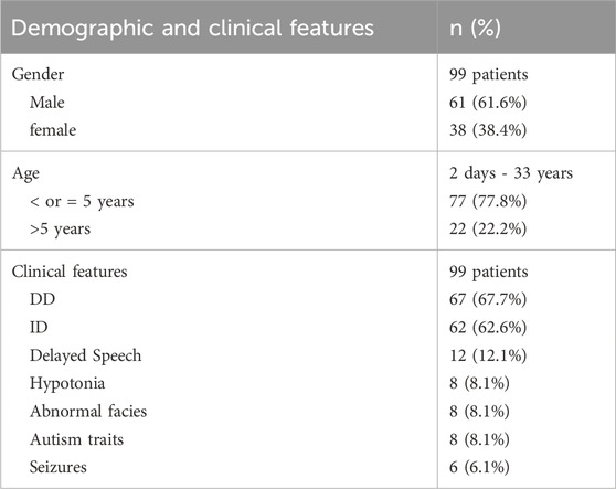
In this study, 99 pediatric patients with a diagnosis of DD or ID were analyzed (Table 1). The cohort included 61 males and 38 females, ranging in age from 2 days to 33 years. The primary clinical features observed were DD in 67 patients and ID in 62 patients. In addition to these primary diagnoses, other frequently observed symptoms included hypotonia (8 cases, 8.08%), delayed or absent speech (12 cases, 12.1%), and abnormal facies (8 cases, 8.08%), which often correlate with genetic syndromic presentations. Autism spectrum-related symptoms were present in 8 cases (8.1%), while seizures were reported in 6 cases (6.1%).
3.2 CNV detection and classification
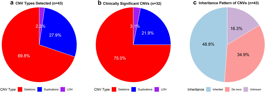
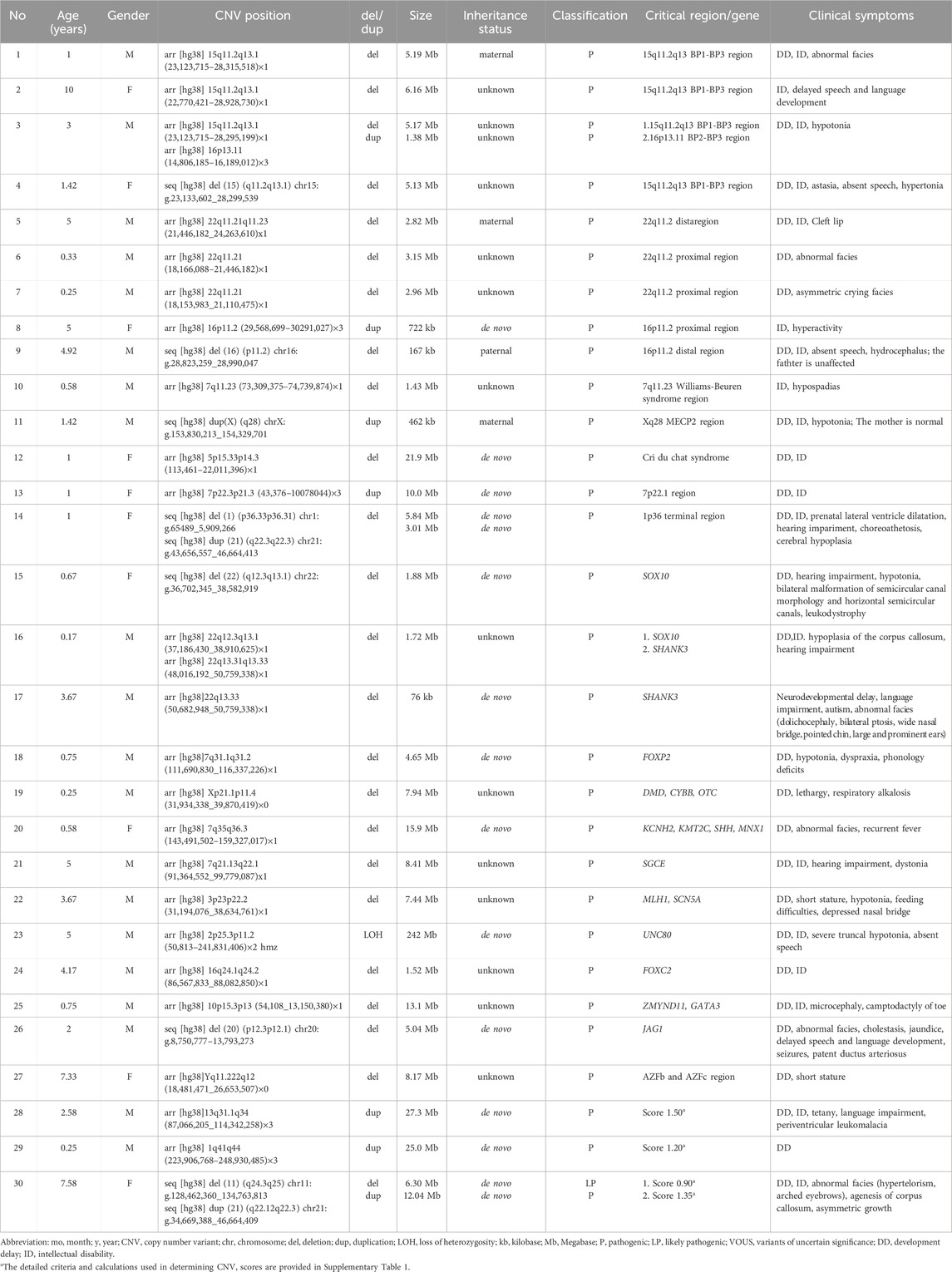
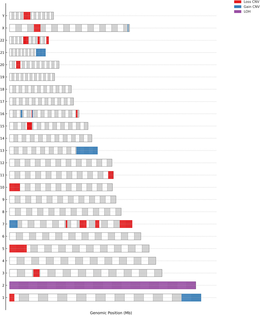
In this cohort of 99 pediatric patients, CMA or WES identified 43 CNVs in 40 patients (Figure 1). Among these, 32 CNVs were deemed clinically significant, impacting 30 patients (30.3%); 31 (96.9%) were classified as pathogenic, while 1 (3.1%) was considered likely pathogenic (Table 2; Figure 2). Additionally, 11 CNVs were identified in 11 patients and were classified as variants of uncertain significance (VOUS) (Supplementary Table 1).

Figure 1. Distribution of CNV types. (a) The number and proportion of deletions, duplications, and loss of heterozygosity (LOH) among all detected CNVs (n = 43). (b) The number and proportion of deletions, duplications, and LOH among clinically significant CNVs (n = 32). (c) Inheritance pattern of all detected CNVs (n = 43).
The 32 clinically significant CNVs included 24 deletions (75.0%), 7 duplications (21.9%), and 1 instance of loss of heterozygosity (LOH) (3.1%).
Of the 43 CNVs identified, inheritance data was available for 21 cases. Fifteen CNVs (65.2%) were de novo. Four CNVs were maternally inherited, including two duplications in regions associated with X-linked syndromes. Two CNVs were paternally inherited. The inheritance patterns of the remaining 22 CNVs were unknown, likely due to limited family information or the high cost of parental testing, which some families may not have been able to afford.
3.3 High-risk CNV regions and associated phenotypes
Recurrent CNVs are defined as copy number variations that frequently occur at specific genomic regions predisposed to deletions or duplications across individuals (Smajlagić et al., 2021). Among the 33 clinically significant CNVs identified in this cohort, 12 (36.4%) were located in recurrent CNV regions, including 15q11.2-q13.1 (4 cases), 16p11.2 (2 cases), 16p13.11 (1 case), 22q11.2 (3 cases), Xq28 (1 case), and 7q11.23 (1 case). Additionally, SOX10-related CNVs were identified in two cases, and SHANK3-related CNVs were also found in two cases, with one case presenting deletions in both SHANK3 and SOX10.
3.4 Normal CMA results and clinical phenotypes
In this cohort, 59 out of the 99 pediatric patients (59.6%) had normal results, with no clinically significant CNVs detected. Despite the absence of detectable CNVs, these patients exhibited a range of clinical phenotypes, including DD in 36 cases, ID in 25 cases, hypotonia in 9 cases, and delayed speech in 8 cases. Additional clinical features observed included ASD in 5 cases and seizures in 3 cases.
4 Discussion
This study investigates the genetic underpinnings of neurodevelopmental disorders through a comprehensive analysis of CNVs in pediatric patients with DD and/or ID. It provides valuable insights into recurrent genomic regions, novel pathogenic variants, and the complexities of inheritance patterns. Recurrent CNVs were identified in 12 cases (36.4%), with 15q11.2-q13.1 being the most frequently observed region (4 cases). This region was predominantly associated with DD (3/4 cases) and ID (3/4 cases), along with variable features such as hypotonia, abnormal facies, and speech delay—phenotypes commonly linked to syndromic conditions like Prader-Willi and Angelman syndromes (Ma et al., 2023). 16p11.2 CNVs, identified in two cases, were associated with ID and behavioral symptoms like hyperactivity and absent speech, aligning with known associations to ASD and ADHD. The observed incomplete penetrance in this region, evidenced by an unaffected parent, underscores the need for family testing, especially with penetrance rates of 33% for distal and 47% for proximal deletions (Goh et al., 2024). This variability is crucial for genetic counseling and reproductive planning, as family testing offers insights into inheritance patterns, allowing for accurate recurrence risk assessment and informed reproductive decisions. 22q11.2 CNVs, observed in three cases, were consistently linked to craniofacial anomalies (e.g., cleft lip, abnormal facies) and DD, findings consistent with DiGeorge syndrome (Szczawińska-Popłonyk et al., 2023). Additional high-risk CNV regions, each identified in one case, included 7q11.23, 16p13.11 and Xq28, each contributing unique but well-documented phenotypic associations in neurodevelopmental presentations. The high recurrence of these CNVs supports the diagnostic value of CMA in neurodevelopmental disorders and further underscores the importance of genetic screening and family testing to clarify inheritance patterns and the variable expressivity of these high-risk regions.
Beyond recurrent CNVs, this study identified unique pathogenic variants affecting genes associated with neurodevelopmental outcomesNotably, deletions related to SOX10 and SHANK3 CNVs were each observed in two patients, with one patient exhibiting concurrent deletions of both SOX10 and SHANK3. SOX10, which is typically associated with neural crest development and conditions such as Waardenburg syndrome and peripheral neuropathies (Pingault et al., 2022), was found in this study to be linked with DD and ID. This finding suggests broader phenotypic implications that may involve additional genes within the deleted region. In one patient with an isolated SOX10 deletion, symptoms included hearing impairment, hypotonia, and bilateral semicircular canal malformations, aligning with known SOX10-related craniofacial and auditory features. Another patient with an isolated SHANK3 deletion presented with neurodevelopmental delay, language impairment, autism, and craniofacial features typical of SHANK3-associated Phelan-McDermid syndrome (Mitz et al., 2024). In contrast, the patient with combined SOX10 and SHANK3 deletions exhibited more complex symptoms, including DD, ID, corpus callosum hypoplasia, and hearing impairment, highlighting potential cumulative effects. This combined deletion profile suggests further investigation into gene interactions and their influence on neurodevelopmental outcomes. Additionally, uniparental disomy (UPD) of chromosome 2 was identified in one patient, attributed to a UNC80 gene homozygous mutation c.5609-4G>A causing Infantile hypotonia with psychomotor retardation and characteristic facies-2 (Tao et al., 2021). The UPD-associated UNC80 mutation underscores the critical importance of employing multiple technologies to uncover the genetic basis of “CNV-negative” cases. These findings emphasize the need for a paradigm shift in diagnostic strategies, advocating for the integration of CNV analysis with complementary genomic approaches to better understand the complexities of neurodevelopmental disorders.
The inheritance analysis conducted in this study revealed that 65.2% of CNVs with established inheritance patterns were de novo. This finding aligns with prior research that associates de novo CNVs with complex phenotypes and heightened risks for developmental disorders (Marrus and Hall, 2017; Brunet et al., 2021). Two CNVs were inherited from the father: one was a distal deletion at 16p11.2 from an unaffected father, and a duplication at 2q12.1q12.3, co-segregating in a father-proband pair exhibiting ID and strabismus. Three additional cases of 2q12.1q12.3 duplications (Kocaay et al., 2022) and two ClinVar/Decipher database entries (Variation ID:152,946; Patient 481,619) consistently report speech delays, cognitive deficits, and motor developmental abnormalities. The 2q12.1q12.3 region of our patients encompasses 30 protein-coding genes, with POU3F3 emerging as the strongest candidate due to its association with Snijders Blok-Fisher syndrome, characterized by DD, ID, and neurological anomalies. Although POU3F3 duplications remain unclassified as pathogenic and their molecular mechanisms are undefined, the concordance of neurodevelopmental deficits—such as speech delays, cognitive impairment, and motor dysfunction—across our cases and five previously reported cases strongly suggests a contributory role. However, no established association exists between this duplication and strabismus, highlighting the necessity for further investigation into this relationship. Four CNVs were inherited from the mother, including a case of Xq28 MECP2 duplication in which the child exhibited DD, ID, and hypotonia, while the mother remained asymptomatic. This finding is consistent with previous studies suggesting that female carriers of MECP2 duplications or mutations frequently remain unaffected, potentially due to skewed X-chromosome inactivation (XCI), which may diminish phenotypic expression (Pascual-Alonso et al., 2020; Sun et al., 2021). The findings highlight the significance of family-based CNV testing for effective genetic counseling and reproductive planning. The 22 cases of CNVs with unknown inheritance highlight a limitation of this study, likely attributable to the high cost of parental testing, which constrained thorough family analysis for certain patients.
This study identified 11 CNVs classified as VOUS, highlighting the ongoing challenges in interpreting these variants in clinical genetics. The VOUS entries ranged from 288 kb to 6.25 Mb and involved genes associated with autosomal or X-linked recessive disorders, such as PCDH15, GPC3, GPC4, CSF2RA, and CASK, although the observed clinical symptoms may not fully align with these gene associations. Assessing the pathogenic significance of these variants typically necessitates further research and supplementary testing (Jansen et al., 2023). Furthermore, 59.6% of patients presented with normal CMA results while displaying DD and ID, indicating that additional genetic factors—such as single nucleotide variants, epigenetic modifications, or environmental influences—could play a role in the observed phenotypes. Integrating these additional factors in further research is essential for improving diagnostic accuracy and advancing the understanding of the genetic landscape of neurodevelopmental disorders.
Several limitations of our study should be considered when interpreting the results. First, this was a single-center study with a relatively small sample size, which may not fully capture the genetic heterogeneity present in broader populations. Second, the resolution of chromosomal microarray analysis (CMA) employed in this study may not have been sufficient to detect certain classes of small or balanced genomic alterations, including low-level mosaicism or balanced translocations. Third, the interpretation of variants, particularly variants of uncertain significance (VOUS), is constrained by current knowledge and may evolve with advances in genomic databases and annotation tools. Finally, although inheritance was analyzed when parental samples were available, not all CNVs could be definitively classified due to incomplete parental testing.
5 Conclusion
This study underscores the utility of CMA and WES as diagnostic tools in pediatric patients with DD and ID, offering insights into the genetic landscape of neurodevelopmental disorders. The findings support CMA as a first-tier diagnostic method and highlight the role of CNV detection for informed clinical decisions and genetic counseling. Limitations include the small cohort size and lack of long-term follow-up, which restrict understanding of the full clinical impact of specific CNVs. Larger, diverse studies with longitudinal tracking are needed to confirm these findings. The interpretation of VOUS remains challenging, and future research using multi-omics approaches, including transcriptomics and proteomics, may help clarify the mechanisms underlying VOUS and CNV variability.
Data availability statement
The dataset associated with our manuscript has been deposited in the China National GeneBank Database (CNGBdb) under the accession numbers CNP0007454 and CVAR0000372. Interested researchers may request access by contacting CNGBdb and citing the accession numbers.
Ethics statement
The studies involving humans were approved by Clinical Research Ethics Committee of Changzhi Maternal and Child Health Care Hospital. The studies were conducted in accordance with the local legislation and institutional requirements. Written informed consent for participation in this study was provided by the participants’ legal guardians/next of kin.
Author contributions
YT: Conceptualization, Formal Analysis, Funding acquisition, Methodology, Project administration, Software, Writing – original draft, Writing – review and editing. HG: Investigation, Methodology, Validation, Writing – original draft. DH: Formal Analysis, Validation, Writing – original draft, Writing – review and editing. MY: Investigation, Validation, Writing – original draft. TL: Investigation, Validation, Writing – original draft. LW: Investigation, Supervision, Validation, Writing – review and editing. WS: Conceptualization, Validation, Writing – original draft. Haiwei Wang: Data curation, Writing – original draft. XL: Supervision, Writing – review and editing.
Funding
The author(s) declare that financial support was received for the research and/or publication of this article. This work was supported by Scientific Research Foundation of the Health Commission of Shanxi Province (No. 2021XM57) and the Natural Science Foundation from the Science and Technology Department of Shanxi Province (No. 202303021222374).
Acknowledgments
We are grateful to the patient and his families in our research. We express our gratitude to all the pediatricians who helped with this study.
Conflict of interest
The authors declare that the research was conducted in the absence of any commercial or financial relationships that could be construed as a potential conflict of interest.
Generative AI statement
The author(s) declare that no Generative AI was used in the creation of this manuscript.
Publisher’s note
All claims expressed in this article are solely those of the authors and do not necessarily represent those of their affiliated organizations, or those of the publisher, the editors and the reviewers. Any product that may be evaluated in this article, or claim that may be made by its manufacturer, is not guaranteed or endorsed by the publisher.
Supplementary material
The Supplementary Material for this article can be found online at: https://www.frontiersin.org/articles/10.3389/fgene.2025.1539902/full#supplementary-material
References
Brunet, T., Jech, R., Brugger, M., Kovacs, R., Alhaddad, B., Leszinski, G., et al. (2021). De novo variants in neurodevelopmental disorders-experiences from a tertiary care center. Clin. Genet. 100, 14–28. doi:10.1111/cge.13946
Goh, S., Thiyagarajan, L., Dudding-Byth, T., Mark, P., and Kirk, E. P. (2024). A systematic review and pooled analysis of penetrance estimates of copy number variants associated with neurodevelopment. Genet. Med. Off. J. Am. Coll. Med. Genet. 27, 101227. doi:10.1016/j.gim.2024.101227
Jang, W., Kim, Y., Han, E., Park, J., Chae, H., Kwon, A., et al. (2019). Chromosomal microarray analysis as a first-tier clinical diagnostic test in patients with developmental delay/intellectual disability, autism spectrum disorders, and multiple congenital anomalies: a prospective multicenter study in korea. Ann. Lab. Med. 39, 299–310. doi:10.3343/alm.2019.39.3.299
Jansen, S., Vissers, L. E. L. M., and de Vries, B. B. A. (2023). The genetics of intellectual disability. Brain Sci. 13, 231. doi:10.3390/brainsci13020231
João, S., Quental, R., Pinto, J., Almeida, C., Santos, H., and Dória, S. (2024). Impact of copy number variants in epilepsy plus neurodevelopment disorders. Seizure 117, 6–12. doi:10.1016/j.seizure.2024.01.009
Kamath, V., Yoganathan, S., Thomas, M. M., Gowri, M., and Chacko, M. P. (2022). Utility of chromosomal microarray in children with unexplained developmental delay/intellectual disability. Fetal Pediatr. Pathol. 41, 208–218. doi:10.1080/15513815.2020.1791292
Kocaay, P., Ceylan, A. C., and Tepe, D. (2022). The case with short stature and intellectual disability caused by a novel 2q12 duplication. J. Coll. Physicians Surg. Pak. 32, S113–S114. doi:10.29271/jcpsp.2022.Supp2.S113
Ma, V. K., Mao, R., Toth, J. N., Fulmer, M. L., Egense, A. S., and Shankar, S. P. (2023). Prader-willi and angelman syndromes: mechanisms and management. Appl. Clin. Genet. 16, 41–52. doi:10.2147/TACG.S372708
Marrus, N., and Hall, L. (2017). Intellectual disability and language disorder. Child. Adolesc. Psychiatr. Clin. N. Am. 26, 539–554. doi:10.1016/j.chc.2017.03.001
Miclea, D., Osan, S., Bucerzan, S., Stefan, D., Popp, R., Mager, M., et al. (2022). Copy number variation analysis in 189 Romanian patients with global developmental delay/intellectual disability. Ital. J. Pediatr. 48, 207. doi:10.1186/s13052-022-01397-1
Mitz, A. R., Boccuto, L., and Thurm, A. (2024). Evidence for common mechanisms of pathology between SHANK3 and other genes of Phelan-McDermid syndrome. Clin. Genet. 105, 459–469. doi:10.1111/cge.14503
Nord, A. S., Lee, M., King, M.-C., and Walsh, T. (2011). Accurate and exact CNV identification from targeted high-throughput sequence data. BMC Genomics 12, 184. doi:10.1186/1471-2164-12-184
Pascual-Alonso, A., Blasco, L., Vidal, S., Gean, E., Rubio, P., O’Callaghan, M., et al. (2020). Molecular characterization of Spanish patients with MECP2 duplication syndrome. Clin. Genet. 97, 610–620. doi:10.1111/cge.13718
Perovic, D., Damnjanovic, T., Jekic, B., Dusanovic-Pjevic, M., Grk, M., Djuranovic, A., et al. (2022). Chromosomal microarray in postnatal diagnosis of congenital anomalies and neurodevelopmental disorders in Serbian patients. J. Clin. Lab. Anal. 36, e24441. doi:10.1002/jcla.24441
Pingault, V., Zerad, L., Bertani-Torres, W., and Bondurand, N. (2022). SOX10: 20 years of phenotypic plurality and current understanding of its developmental function. J. Med. Genet. 59, 105–114. doi:10.1136/jmedgenet-2021-108105
Quenez, O., Cassinari, K., Coutant, S., Lecoquierre, F., Le Guennec, K., Rousseau, S., et al. (2021). Detection of copy-number variations from NGS data using read depth information: a diagnostic performance evaluation. Eur. J. Hum. Genet. 29, 99–109. doi:10.1038/s41431-020-0672-2
Riggs, E. R., Andersen, E. F., Cherry, A. M., Kantarci, S., Kearney, H., Patel, A., et al. (2020). Technical standards for the interpretation and reporting of constitutional copy-number variants: a joint consensus recommendation of the American College of Medical Genetics and Genomics (ACMG) and the Clinical Genome Resource (ClinGen). Genet. Med. 22, 245–257. doi:10.1038/s41436-019-0686-8
Shchubelka, K., Turova, L., Wolfsberger, W., Kalanquin, K., Williston, K., Kurutsa, O., et al. (2024). Genetic determinants of global developmental delay and intellectual disability in Ukrainian children. J. Neurodev. Disord. 16, 13. doi:10.1186/s11689-024-09528-x
Smajlagić, D., Lavrichenko, K., Berland, S., Helgeland, Ø., Knudsen, G. P., Vaudel, M., et al. (2021). Population prevalence and inheritance pattern of recurrent CNVs associated with neurodevelopmental disorders in 12,252 newborns and their parents. Eur. J. Hum. Genet. 29, 205–215. doi:10.1038/s41431-020-00707-7
Sun, Y., Yang, Y., Luo, Y., Chen, M., Wang, L., Huang, Y., et al. (2021). Lack of MECP2 gene transcription on the duplicated alleles of two related asymptomatic females with Xq28 duplications and opposite X-chromosome inactivation skewing. Hum. Mutat. 42, 1429–1442. doi:10.1002/humu.24262
Szczawińska-Popłonyk, A., Schwartzmann, E., Chmara, Z., Głukowska, A., Krysa, T., Majchrzycki, M., et al. (2023). Chromosome 22q11.2 deletion syndrome: a comprehensive review of molecular genetics in the context of multidisciplinary clinical approach. Int. J. Mol. Sci. 24, 8317. doi:10.3390/ijms24098317
Tao, Y., Han, D., Wei, Y., Wang, L., Song, W., and Li, X. (2021). Case report: complete maternal uniparental disomy of chromosome 2 with a novel UNC80 splicing variant c.5609-4g> A in a Chinese patient with infantile hypotonia with psychomotor retardation and characteristic facies 2. Front. Genet. 12, 747422. doi:10.3389/fgene.2021.747422
Tao, Y., Yang, L., Han, D., Zhao, C., Song, W., Wang, H., et al. (2023). A GATA3 gene mutation that causes incorrect splicing and HDR syndrome: a case study and literature review. Front. Genet. 14, 1254556. doi:10.3389/fgene.2023.1254556
Keywords: copy number variations, SNP array, WES, developmental delay, intellectual disability
Citation: Tao Y, Guo H, Han D, Yang M, Lun T, Wang L, Song W, Wang H and Li X (2025) Uncovering genetic contributors to developmental delay and intellectual disability: a focus on CNVs in pediatric patients. Front. Genet. 16:1539902. doi: 10.3389/fgene.2025.1539902
Received: 05 December 2024; Accepted: 21 May 2025;
Published: 23 June 2025.
Edited by:
Kornsorn Srikulnath, Kasetsart University, ThailandReviewed by:
Kuanjun He, Inner Mongolia University for Nationalities, ChinaLuis Alberto Méndez- Rosado, Centro Nacional de Genética Médica, Cuba
Copyright © 2025 Tao, Guo, Han, Yang, Lun, Wang, Song, Wang and Li. This is an open-access article distributed under the terms of the Creative Commons Attribution License (CC BY). The use, distribution or reproduction in other forums is permitted, provided the original author(s) and the copyright owner(s) are credited and that the original publication in this journal is cited, in accordance with accepted academic practice. No use, distribution or reproduction is permitted which does not comply with these terms.
*Correspondence: Yilun Tao, eWx0YW8yMUAxNjMuY29t; Xiaoze Li, bGl4aWFvemU1MjBAMTI2LmNvbQ==
†These authors have contributed equally to this work and share first authorship
 Yilun Tao
 Hongzhi Guo3†

