 Nicolas Henry1†
 Chloé Mellaza1†
 Nicolas Fage1François Beloncle2Franck Genevieve3Guillaume Legendre4Corentin Orvain5Anne-Sophie Garnier1Maud Cousin1Virginie Besson1
 Jean-François Subra1Agnès Duveau1
 Jean-François Augusto1*
 Benoit Brilland1
- 1Service de Néphrologie-Dialyse-Transplantation, Université d'Angers, Centre Hospitalier Universitaire (CHU) Angers, Angers, France
- 2Service de Médecine Intensive et Réanimation, Médecine Hyperbare, Université d'Angers, Centre Hospitalier Universitaire (CHU) Angers, Angers, France
- 3Laboratoire d'Hématologie, Université d'Angers, Centre Hospitalier Universitaire (CHU) Angers, Angers, France
- 4Département de Gynécologie et Obstétrique, Université d'Angers, Centre Hospitalier Universitaire (CHU) Angers, Angers, France
- 5Service d'Hématologie, Université d'Angers, Centre Hospitalier Universitaire (CHU) Angers, Angers, France
Background: Thrombotic microangiopathies (TMAs) are highly suspected in patients showing mechanical hemolytic anemia, thrombocytopenia, and haptoglobin consumption. Primary [thrombotic thrombocytopenic purpura (TTP) and atypical hemolytic uremic syndrome] and secondary TMA are considered. Even if ADAMTS13 measurements and alternative complement pathway explorations have greatly improved the ability to identify primary TMA, their diagnosis remains difficult, and their frequency relative to that of secondary TMA is undetermined. The objectives of the present study were, therefore, to describe the etiologies, management, and the outcomes of patients presenting with TMA in real-life clinical practice.
Methods: We conducted a retrospective study between 01/01/2008 and 31/12/2018 that included all consecutive patients presenting with biological TMA syndrome at admission or developing during hospitalization. Patients were identified from the laboratory databases, and their medical files were reviewed to confirm TMA diagnosis, to determine etiology, and to analyze their therapeutic management and outcomes.
Results: During this period, 239 patients with a full TMA biological syndrome were identified, and the TMA diagnosis was finally confirmed in 216 (90.4%) after the cases were reviewed. Primary TMAs (thrombotic thrombocytopenic purpura or atypical hemolytic uremic syndrome) were diagnosed in 20 of 216 patients (9.3%). Typical HUS was diagnosed in eight patients (3.7%), and the most frequent secondary TMAs were HELLP syndrome (79/216, 36.6%) and active malignancies (30/219, 13.9%). ADAMTS13 measurements and alternative complement pathway analyses were performed in a minority of patients. Multiple factors identified as TMA triggers were present in most patients, in 55% of patients with primary TMA, vs. 44.7% of patients with secondary TMA (p = 0.377). Death occurred in 57 patients (23.4%) during follow-up, and dialysis was required in 51 patients (23.6%). Active malignancies [odds ratio (OR) 13.7], transplantation (OR 4.43), male sex (OR 2.89), and older age (OR 1.07) were significantly associated with death.
Conclusion: Secondary TMAs represent many TMA causes in patients presenting a full TMA biological syndrome during routine clinical practice. Multiple factors favoring TMA are present in about half of primary or secondary TMA. ADAMTS13 and complement pathway were poorly explored in our cohort. The risk of death is particularly high in patients with malignancies as compared with patients with other TMA.
Introduction
Thrombotic microangiopathies (TMAs) are defined by the presence of thrombi in small arterioles and capillaries (1). Thrombosis affects microcirculation and leads to tissue ischemia and organ failure (2). Although the confirmation of TMA diagnosis relies on histological features, a biopsy of an affected organ is rarely performed. Not only do TMAs have, in most cases, a characteristic biological presentation, but also biopsy is often contraindicated, given the bleeding risk. Mechanical hemolytic anemia, schistocytosis, and thrombocytopenia are strongly suggestive of TMA. As in other hemolytic anemia syndromes, haptoglobin consumption, elevated LDH, and elevated free bilirubin levels are also detected.
The understanding of biological mechanisms implicated in TMA development has greatly improved over the past three decades, allowing for the individualization of entities with specific pathophysiology (3, 4). In parallel with the pathophysiological understanding of TMA, the classification of TMA has been enriched with new entities and has further enabled the identification of several favoring or precipitating factors (1, 5). The latter can act as triggers of TMA development in patients with conditions that make them pre-disposed to primary TMA, i.e., genetic defects, or can induce TMA by themselves. TMAs are usually classified into two subsets, primary and secondary. Primary TMAs include thrombotic thrombocytopenic purpura (TTP) and atypical hemolytic uremic syndrome (aHUS). TTP is associated with low or undetectable ADAMTS13 activity (usually <10%), which may be related to a rare genetic defect in the ADAMTS13 gene (Upshaw–Schulman syndrome) or is, in most cases, an autoimmune disease associated with auto-antibodies neutralizing the enzymatic functions of ADAMTS13 (6). aHUS is related to several inherited or acquired abnormalities affecting complement alternative pathway (cAP) (7) that result in its permanent activation. Primary TMA can manifest at all ages and frequently develops after the occurrence of triggers that induce endothelial injury. Secondary TMAs, however, include numerous conditions or diseases that have been associated with TMA development. In secondary TMA, genetic defects and autoimmune abnormalities are rarely detected, and the microvascular endothelial injuries are driven by other factors altogether. The pathophysiology of secondary forms is, therefore, less well-defined, and as a result, diagnosis is rendered much more difficult. In the light of this, several classifications have been proposed to guide clinicians to the right diagnosis. Among secondary TMAs, typical HUS (tHUS) induced by the endothelial toxicity of Shiga toxins from Escherichia coli is the most well-defined secondary TMA. However, numerous other conditions such as a solid organ or bone marrow transplantation, drugs, pregnancy (HELLP syndrome), malignant hypertension, and malignancies can be associated with TMA development (1, 8–11). In such cases, the mechanisms of TMA are largely unknown and frequently multifactorial, and the therapeutic management of these patients is not well-codified.
The treatment of primary TMA has greatly evolved in the last few years using specific drugs such as rituximab in TTP and eculizumab in aHUS (12–15). In these conditions, mortality and organ lesions (mainly brain and kidney injuries) occur early, within the first days or weeks. A patient's prognosis is very dependent upon any delays to commencing with possible specific treatment. It is, therefore, crucial to identify these patients among the flood of those with a secondary cause.
The relative frequency of primary to secondary TMA has been poorly analyzed in the literature (16). Moreover, the management of patients with secondary TMA and their prognosis in comparison with patients with primary TMA are not well-known.
In the present study, we identified all consecutive patients with biological features of TMA (full biological TMA syndrome) in a period spanning 11 years. The objective of the study was to analyze the relative frequency of primary and secondary TMA, their presentation, and their therapeutic management. We also analyzed the prognosis of patients according to the causes of their TMA.
Methods
Selection of Patients
Patients admitted to the University Hospital of Angers between 01/01/2008 and 31/12/2018 and presenting with a full biological TMA syndrome were included in the study. The concomitant association of anemia defined full biological TMA syndrome (<12 g/dl in females and 13 g/dl in males), with thrombopenia ≤ 150 G/L, schistocytosis ≥0.5%, and a decreased haptoglobin level ≤0.4 g/L. These patients were identified from the database of our hematological laboratory. Two datasets listing patients with (1) thrombocytopenia and schistocytosis screening and (2) haptoglobin measurements during the study were extracted and merged. Adult patients (>18 years old) with concomitant thrombocytopenia, schistocytosis, and a decreased haptoglobin level were included in a systematic review of medical records after anemia had been confirmed. The study protocol complied with the standards of the Ethics Committee of the Angers University Hospital (no. 2019/12).
Identification and Classification of Thrombotic Microangiopathic Patients
Medical records of patients identified with a full TMA biological syndrome were analyzed by four physicians (NH, CM, BB, and JFA) trained in nephrology, hematology, and critical care medicine, to confirm or rule out the diagnosis. In patients with a TMA diagnosis, its cause was determined after a hierarchical analysis according to current classifications. In a first step, patients with ADAMTS13 <10% were classified as having TTP. Next, patients with Shiga toxin-positive bacteria (detected in stool culture or polymerase chain reaction) were classified as having tHUS. The other following TMA causes were systematically considered: patients with HELLP syndrome, drugs known to induce TMA, graft vs. host disease (GvHD)-associated TMA, cancers, autoimmune diseases, malignant hypertension, and infections (excluding those related to Shiga toxin-producing bacteria). Patients with TMA and acute renal failure but without evidence of other secondary TMA causes and/or with cAP abnormalities known to be associated with TMA were classified as having aHUS. Patients without evidence of any of the causes of secondary TMA listed earlier, with rare secondary causes of TMA and without evidence of cAP abnormalities, were classified in “other secondary TMA causes.” Patients with multiple TMA potential causes, for whom clinical file review could not determine a predominant mechanism, were also classified in “other secondary TMA causes” (Supplementary Table 1).
In some patients with primary or secondary TMA, several TMA causes could be present. In patients with primary TMA, other possible causes were considered as favoring factors. Patients with secondary TMA were classified within the most probable diagnosis after medical file review, and other associated conditions were considered as favoring factors. Factors considered to favor TMA were concomitant infection, pregnancy, past or present history of malignancy, drugs known to induce TMA, transplantation, autoimmune diseases, and B12 deficiency.
Therapeutic Management and Outcomes
Plasma exchange and/or plasma infusion, eculizumab and rituximab use, and the requirement for transfusion or dialysis were identified in medical charts and collected. Outcomes, including death or need for renal replacement therapy, were also collected.
Statistical Analysis
Continuous variables are presented as median (minimum–maximum). Categorical variables are presented as the absolute value and percentage. Differences between groups were analyzed using the χ2 test (or Fisher exact test if necessary) for categorical variables and the Mann–Whitney U test for continuous variables. Logistic univariate analysis was used to examine factors associated with the outcomes. P-value < 0.05 were considered significant. Statistical analysis was performed using SPSS software® 23.0 and Graphpad Prism®.
Results
Baseline Characteristics
During the period mentioned earlier, we identified 427 patients with thrombocytopenia and schistocytosis ≥0.5% and 4,664 patients with haptoglobin ≤0.4 g/L. After cross-referencing the datasets, we could identify 239 patients with a full biological TMA syndrome. After the review of medical charts, 216 patients were finally diagnosed with TMA, whereas 23 had no evidence of TMA (Figure 1). The diagnosis retained in these 23 patients after reviewing their medical files are given in Supplementary Table 2. Patients with TMA were initially admitted to 17 different medical and surgical departments (Supplementary Figure 1). Among the 216 patients, 81 (37.5%) were admitted to a critical care unit at least partially during their hospitalization.
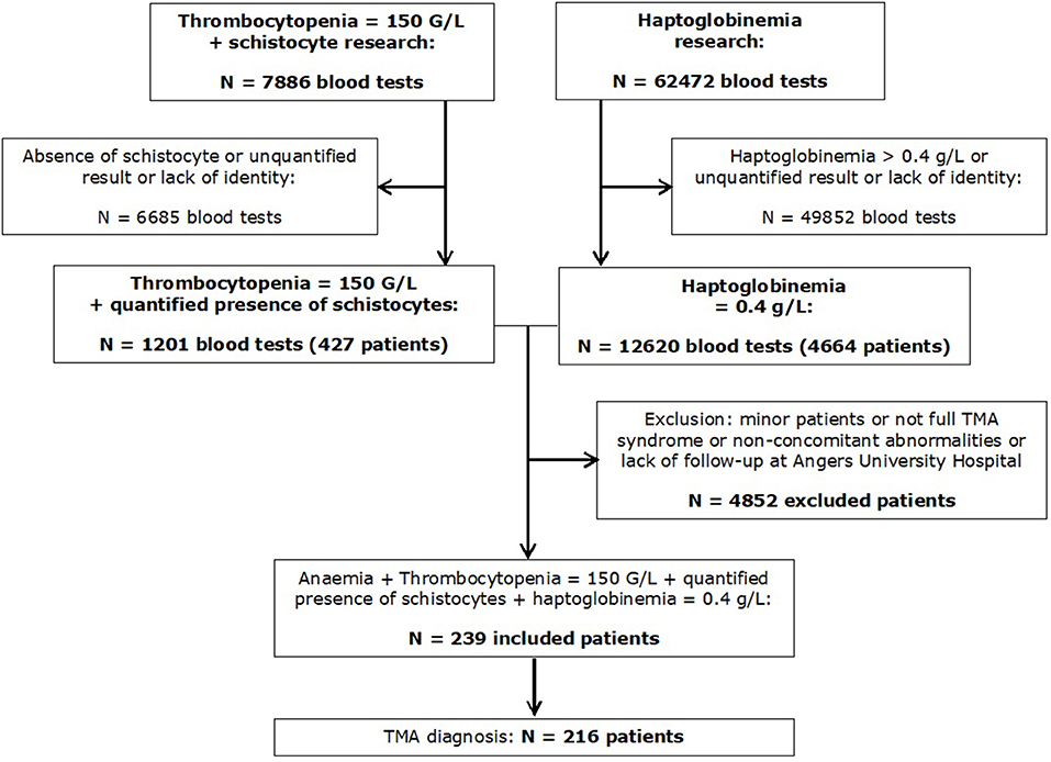
Figure 1. Flowchart of the study.
TMA patients were predominantly females (74.1%), with a median age of 45.3 ± 18.5 years. Hypertension and chronic kidney disease (estimated glomerular filtration rate <60 ml/min/1.73 m2) were found in 54 (25%) and 21 (9.7%) patients, respectively. Primary TMA (TTP or aHUS) was diagnosed in 20 patients (9.3%), with mostly acquired TTP (11 patients, 5.1%), whereas secondary TMA represented 90.7% of all TMA cases. Among secondary TMA, HELLP syndrome and active cancer-associated TMA were the most frequent diagnosis, in 36.6 and 13.9%, respectively. In the case of the latter, solid cancer featured in 19 patients and acute haemopathy in 11 patients. Other secondary TMA causes were less frequently observed, with tHUS representing 3.7% (Table 1 and Supplementary Figure 2). A subgroup of 30 patients was classified as “other TMA.” Some of these patients had a rare TMA cause or had no identified cause of TMA or multiple causes of TMA, not allowing their classification within other groups. The characteristics of patients classified in this subgroup of TMA are given in Supplementary Table 3. Surprisingly, TMA diagnosis was not mentioned within the medical file at the end of the initial admission for 53 of 216 (24.4%) patients. These patients were classified as having mainly secondary TMA after file review (cancer, n = 18; aGVHD, n = 12; undetermined TMA cause, n = 11). ADAMTS13 determination was performed in only 25% of patients, and, therefore, the diagnosis of TTP may have been missed in some patients. C3 and C4 complement fraction concentration and alternative complement pathway were studied in 33.8 and 19.4% of the patients of the cohort, respectively (Supplementary Table 4).
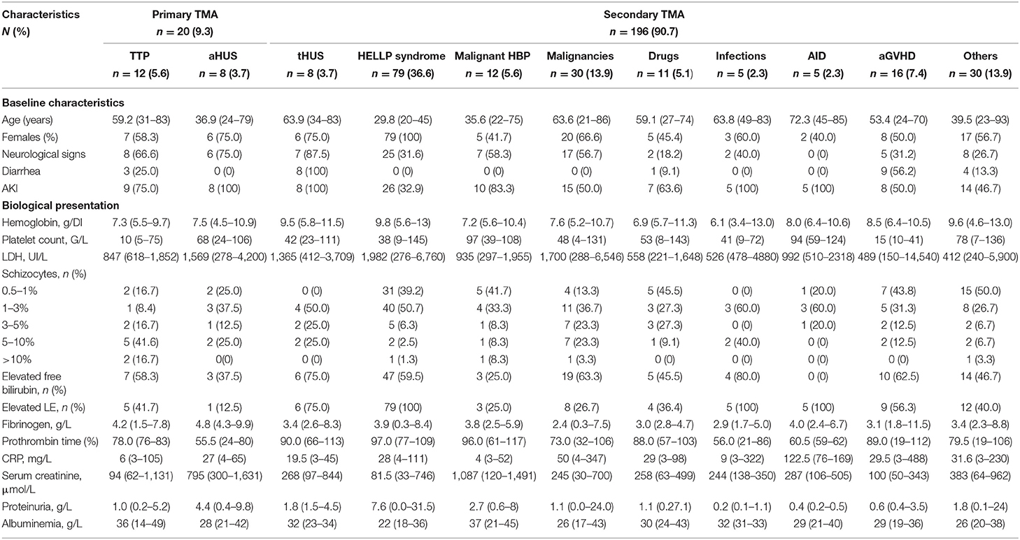
Table 1. Clinical and biological presentation of TMA according to etiology.
Clinical and Biological Presentation According to Thrombotic Microangiopathy Cause
Table 1 reports the clinical and biological presentation according to the TMA etiology. Patients with aHUS tended to be younger compared with patients with TTP. In secondary TMA, patients with HELLP syndrome were younger than those with malignancies. Neurological signs were present at admission with variable frequencies according to TMA causes. They were present in most patients with primary TMA, either TTP or aHUS. Neurological symptoms exhibited greater variety in secondary TMA, most frequently observed in patients with malignant hypertension and malignancies. Acute kidney injury was present in all patients with aHUS and tHUS, in most patients with TTP, and with a variable frequency in other secondary TMA causes. Serum creatinine was greatly higher in patients with aHUS and malignant hypertension, as compared with other causes of TMA. The level of proteinuria showed a similar trend. However, although 75% of TTP patients developed acute kidney injury, their median serum creatinine level at admission was 94 μmol/L and therefore in the same range as HELLP syndrome patients. Diarrhea was present in all patients with tHUS and in most patients with aGvHD but was very inconstantly observed in other conditions. Anemia was present in all patients but with variable severity. Thrombocytopenia was particularly profound in patients with TTP.
Analysis of Thrombotic Microangiopathy-Favoring Factors
Except for patients with HELLP syndrome and active autoimmune diseases, the presence of an additional factor known to favor TMA was frequent (Table 2). There was no difference in the frequency of patients with at least one additional factor between patients with primary TMA and patients with secondary TMA (55.0 vs. 44.7%, p = 0.377). The most frequent additional factor was a history of malignancy and concomitant administration of a drug known to favor TMA, mostly gemcitabine or calcineurin inhibitor. Patients with aGvHD and drug-associated TMA tended to have more associated factors than patients with primary TMA and with other secondary causes (Table 2).
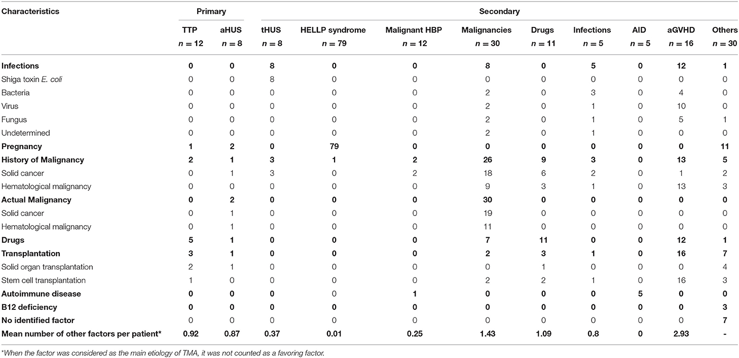
Table 2. TMA-favoring factors according to TMA cause.
Management of Thrombotic Microangiopathy
Red blood cell transfusion was used in 43.5% of patients and platelet transfusion in 22.7% of patients. Plasma exchange or plasma infusion was initiated in 28.2% of patients. Rituximab and eculizumab were administered in 7.9 and 3.7% of patients, respectively. As expected, plasma exchange or plasma infusion was used in most patients with primary TMA. Rituximab was administered in 50% of patients with TTP and at a lower frequency in other TMA groups. Eculizumab was used in eight patients, five of them with aHUS or tHUS (Table 3).
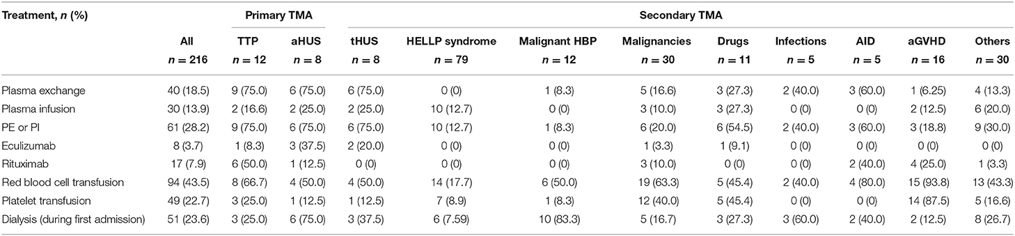
Table 3. Therapeutic management of TMA patients.
Outcomes and Factors Associated With Prognosis
Fifty-one (23.6%) patients required dialysis during initial admission, and 57 (23.4%) died during follow-up at a median time of 42 days (1–1,796) from initial admission. Using univariate analysis, older age [odds ratio (OR) 1.07], male sex (OR 2.89), active malignancies (OR 13.7), and transplantation (OR 4.43) were associated with an increased risk of death. Primary TMA (OR 3.00), aHUS, and tHUS (OR 4.84) were associated with dialysis initiation during the first admission, whereas male sex (OR 0.49) and HELLP syndrome (OR 0.17) were associated with a decreased risk of requiring dialysis (Table 4).
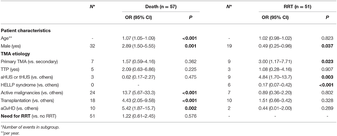
Table 4. Univariate analysis of factors associated with outcomes.
Discussion
The present study gives an overview of TMA etiology and management in routine clinical practice. The major finding of this work is that secondary TMAs are largely much more frequent than primary TMAs (TTP and aHUS). Importantly, using our biologically based selection and systematic medical file review, we were able to conclude TMA diagnosis in ~25% of patients for whom the diagnosis was not clearly mentioned within the medical file. These patients had secondary TMA related to various causes. This observation probably reflects the complexity of TMA diagnosis, especially when multiple factors are involved. Data from TMA registries show a higher proportion of primary TMA. As an example, in the Oklahoma TTP-HUS registry, TTP represented 15% of TMA between 1999 and 2007 (17) and 43% in the UK TTP registry between 2009 and 2013 (18). These discrepancies are probably explained because entries into these registers are linked to plasma exchange requirement and by ADAMTS13 measurement, respectively. Thus, they include selected populations and do not, in fact, reflect the frequency of TMA, nor their distribution in “real-life” conditions. Rather, in the present study, we undertook an unbiased analysis of all consecutive patients with full TMA biological syndrome. Interestingly, a very recent study with a methodology like that used in the present study showed similar frequencies of primary and secondary TMA (16): TTP and aHUS represented 5.9% of TMA, slightly below the 9.3% observed in our study.
It is important to note that ADAMTS13 measurement and cAP analysis were performed in only a minority of patients in our cohort. Therefore, some patients may have been misclassified as having a secondary TMA. This would be in line with recent research showing abnormalities in alternative complement pathway proteins, notably patients with clinical features of malignant hypertension (19, 20) or with TMA associated with drugs, such as calcineurin inhibitors used in transplant patients (21). More recently, patients with HELLP syndrome have also been shown to present a higher incidence of germline alternative complement pathway gene mutations (22). The explanation for such a low rate of patients with specific investigations in our study is likely to be multifaceted, reflecting an unawareness of these relatively new data, the rarity of TMA, and the variable TMA experience of the clinician.
We observed the presence of favorable/precipitating factors that can act as “triggers” in ~50% of patients. Interestingly, their frequency showed no significant difference between patients with primary and secondary TMAs. Therefore, this observation supports the concept of the “multi-hit” theory of TMA (23). TMA has been shown to occur in ~10% of patients with aGvHD (24). It is interesting to note that it was the condition with the higher number of concomitant triggers in our study.
Finally, primary TMAs, for which treatment has been better codified in past years, represent a minority of TMA cases. HELLP syndrome and active malignancies were the most highly represented causes of secondary TMA in our cohort. The repartition between secondary causes in our study is closely aligned with those observed in the study of Bayer et al. (16). However, we observed far fewer patients with infection-associated TMA (2.3%). These discrepancies are probably linked to different methods for selecting patients and for classifying them between the two studies. First, in Bayer et al.'s study, screening constituted a combination of automatized medical file analysis and the inclusion of patients with partial biological TMA syndrome. The differences in patient selection probably explain why Bayer et al. included many more patients. Second, in our study, we considered infectious events as triggers rather than as causative agents per se.
In line with previous data, the mortality rate was high in our study, and death occurred early after TMA diagnosis. In the univariate analysis, active malignancies, transplantation, and aGvHD were significantly associated with a higher risk of death. These observations are in line with the clinical experience we have of TMA and with the few available reports on these topics (24–26). Notably, the differentiation between chemotherapy-induced TMA and cancer-associated TMA represents a challenge, especially when medical files are reviewed retrospectively. Moreover, these patients do not usually undergo extensive biological explorations given their very limited prognosis; this in turn also limits what can be achieved through reviewing files. We may, therefore, have misclassified some of them.
In conclusion, our study highlights that more than 90% of patients with full TMA biological syndrome have secondary TMAs, which are associated with very high mortality. We also point out that it is not uncommon for the diagnosis of TMA not to be mentioned, especially in patients diagnosed with secondary TMA. Therefore, these results suggest that more attention and clinical research should focus on secondary TMA to understand better the mechanisms implicated in it, especially with systematic cAP and ADAMTS13 exploration to rule out primary TMA, improve their classification and codify their treatment.
Data Availability Statement
The datasets presented in this article are not readily available because The datasets generated for this study are available on request to the corresponding author. Requests to access the datasets should be directed to amZhdWd1c3RvQGNodS1hbmdlcnMuZnI=.
Ethics Statement
The studies involving human participants were reviewed and approved by Ethics Committee of the Angers University Hospital (n°2019/12). Written informed consent for participation was not required for this study in accordance with the national legislation and the institutional requirements.
Author Contributions
NH, CM, and J-FA contributed to the conception and design of the study. J-FA supervised the project. NH and CM organized the database and wrote the first draft of the manuscript. NH, CM, J-FA and BB performed the statistical analysis. J-FA and BB provided critical revision of the manuscript. All authors participated in patients care. All authors read and approved the submitted version.
Conflict of Interest
The authors declare that the research was conducted in the absence of any commercial or financial relationships that could be construed as a potential conflict of interest.
Supplementary Material
The Supplementary Material for this article can be found online at: https://www.frontiersin.org/articles/10.3389/fmed.2021.566678/full#supplementary-material
Supplementary Figure 1. Site of initial hospital admission.
Supplementary Figure 2. Number of patients according to TMA etiology.
Supplementary Table 1. Diagnosis criteria for classifying the causes of TMA.
Supplementary Table 2. Patients with full TMA biological syndrome without TMA.
Supplementary Table 3. Characteristics of patients with “other” TMA causes.
Supplementary Table 4. ADAMTS13, complement and autoimmune evaluation.
References
1. George JN, Nester CM. Syndromes of thrombotic microangiopathy. N Engl J Med. (2014) 371:654–66. doi: 10.1056/NEJMra1312353
2. Hofer J, Rosales A, Fischer C, Giner T. Extra-renal manifestations of complement-mediated thrombotic microangiopathies. Front Pediatr. (2014) 2:97. doi: 10.3389/fped.2014.00097
3. Fakhouri F, Fremeaux-Bacchi V. Does hemolytic uremic syndrome differ from thrombotic thrombocytopenic purpura? Nat Clin Pract Nephrol. (2007) 3:679–87. doi: 10.1038/ncpneph0670
4. Zipfel PF, Skerka C. Complement regulators and inhibitory proteins. Nat Rev Immunol. (2009) 9:729–40. doi: 10.1038/nri2620
5. Masias C, Vasu S, Cataland SR. None of the above: thrombotic microangiopathy beyond TTP and HUS. Blood. (2017) 129:2857–63. doi: 10.1182/blood-2016-11-743104
6. Kremer Hovinga JA, Coppo P, Lammle B, Moake JL, Miyata T, Vanhoorelbeke K. Thrombotic thrombocytopenic purpura. Nat Rev Dis Primers. (2017) 3:17020. doi: 10.1038/nrdp.2017.20
8. Gupta M, Feinberg BB, Burwick RM. Thrombotic microangiopathies of pregnancy: differential diagnosis. Pregnancy Hypertens. (2018) 12:29–34. doi: 10.1016/j.preghy.2018.02.007
9. Al-Nouri ZL, Reese JA, Terrell DR, Vesely SK, George JN. Drug-induced thrombotic microangiopathy: a systematic review of published reports. Blood. (2015) 125:616–8. doi: 10.1182/blood-2014-11-611335
10. Noris M, Remuzzi G. Thrombotic microangiopathy after kidney transplantation. Am J Transplant. (2010) 10:1517–23. doi: 10.1111/j.1600-6143.2010.03156.x
11. Obut F, Kasinath V, Abdi R. Post-bone marrow transplant thrombotic microangiopathy. Bone Marrow Transplant. (2016) 51:891–7. doi: 10.1038/bmt.2016.61
12. Scully M, Goodship T. How I treat thrombotic thrombocytopenic purpura and atypical haemolytic uraemic syndrome. Br J Haematol. (2014) 164:759–66. doi: 10.1111/bjh.12718
13. Chander DP, Loch MM, Cataland SR, George JN. Caplacizumab Therapy without plasma exchange for acquired thrombotic thrombocytopenic purpura. N Engl J Med. (2019) 381:92–4. doi: 10.1056/NEJMc1905426
14. Schmidtko J, Peine S, El-Housseini Y, Pascual M, Meier P. Treatment of atypical hemolytic uremic syndrome and thrombotic microangiopathies: a focus on eculizumab. Am J Kidney Dis. (2013) 61:289–99. doi: 10.1053/j.ajkd.2012.07.028
15. Legendre CM, Licht C, Muus P, Greenbaum LA, Babu S, Bedrosian C, et al. Terminal complement inhibitor eculizumab in atypical hemolytic-uremic syndrome. N Engl J Med. (2013) 368:2169–81. doi: 10.1056/NEJMoa1208981
16. Bayer G, von Tokarski F, Thoreau B, Bauvois A, Barbet C, Cloarec S, et al. Etiology and outcomes of thrombotic microangiopathies. Clin J Am Soc Nephrol. (2019) 14:557–66. doi: 10.2215/CJN.11470918
17. George JN. The thrombotic thrombocytopenic purpura and hemolytic uremic syndromes: overview of pathogenesis (experience of the Oklahoma TTP-HUS registry, 1989-2007). Kidney Int Suppl. (2009) 112:S8–10. doi: 10.1038/ki.2008.609
18. Hassan S, Westwood JP, Ellis D, Laing C, Mc Guckin S, Benjamin S, et al. The utility of ADAMTS13 in differentiating TTP from other acute thrombotic microangiopathies: results from the UK TTP Registry. Br J Haematol. (2015) 171:830–5. doi: 10.1111/bjh.13654
19. Cavero T, Arjona E, Soto K, Caravaca-Fontan F, Rabasco C, Bravo L, et al. Severe and malignant hypertension are common in primary atypical hemolytic uremic syndrome. Kidney Int. (2019) 96:995–1004. doi: 10.1016/j.kint.2019.05.014
20. Timmermans S, Abdul-Hamid MA, Vanderlocht J, Damoiseaux J, Reutelingsperger CP, van Paassen P, et al. Patients with hypertension-associated thrombotic microangiopathy may present with complement abnormalities. Kidney Int. (2017) 91:1420–5. doi: 10.1016/j.kint.2016.12.009
21. Le Quintrec M, Lionet A, Kamar N, Karras A, Barbier S, Buchler M, et al. Complement mutation-associated de novo thrombotic microangiopathy following kidney transplantation. Am J Transplant. (2008) 8:1694–701. doi: 10.1111/j.1600-6143.2008.02297.x
22. Vaught AJ, Braunstein EM, Jasem J, Yuan X, Makhlin I, Eloundou S, et al. Germline mutations in the alternative pathway of complement predispose to HELLP syndrome. JCI Insight. (2018) 3:e99128. doi: 10.1172/jci.insight.99128
23. Miyata T, Fan X. A second hit for TMA. Blood. (2012) 120:1152–4. doi: 10.1182/blood-2012-06-433235
24. Kraft S, Bollinger N, Bodenmann B, Heim D, Bucher C, Lengerke C, et al. High mortality in hematopoietic stem cell transplant-associated thrombotic microangiopathy with and without concomitant acute graft-vs.-host disease. Bone Marrow Transplant. (2019) 54:540–8. doi: 10.1038/s41409-018-0293-3
25. Izzedine H, Perazella MA. Thrombotic microangiopathy, cancer, and cancer drugs. Am J Kidney Dis. (2015) 66:857–68. doi: 10.1053/j.ajkd.2015.02.340
Keywords: Thrombotic microangiopathies, etiology, primary, secondary, thrombotic thrombocytopenic purpura, hemolytic uremic syndrome, real-life
Citation: Henry N, Mellaza C, Fage N, Beloncle F, Genevieve F, Legendre G, Orvain C, Garnier A-S, Cousin M, Besson V, Subra J-F, Duveau A, Augusto J-F and Brilland B (2021) Retrospective and Systematic Analysis of Causes and Outcomes of Thrombotic Microangiopathies in Routine Clinical Practice: An 11-Year Study. Front. Med. 8:566678. doi: 10.3389/fmed.2021.566678
Received: 29 May 2020; Accepted: 28 January 2021;
Published: 26 February 2021.
Edited by:
Hollie Marie Reeves, University Hospitals Cleveland Medical Center, United StatesReviewed by:
Mutlu Arat, Istanbul Florence Nightingale Hospital, TurkeyHong Hong, Northwell Health, United States
Copyright © 2021 Henry, Mellaza, Fage, Beloncle, Genevieve, Legendre, Orvain, Garnier, Cousin, Besson, Subra, Duveau, Augusto and Brilland. This is an open-access article distributed under the terms of the Creative Commons Attribution License (CC BY). The use, distribution or reproduction in other forums is permitted, provided the original author(s) and the copyright owner(s) are credited and that the original publication in this journal is cited, in accordance with accepted academic practice. No use, distribution or reproduction is permitted which does not comply with these terms.
*Correspondence: Jean-François Augusto, amZhdWd1c3RvQGNodS1hbmdlcnMuZnI=
†These authors have contributed equally to this work