- 1Department of Nuclear Medicine, Affiliated Hospital of Zunyi Medical University, Zunyi, China
- 2Department of Pathology, Affiliated Hospital of Zunyi Medical University, Zunyi, China
Splenosis occurring in adrenal glands is relatively rare and is easily misdiagnosed as neoplastic lesions. Herein, we present a case of a 39-year-old woman who underwent a pancreatic tail resection and splenectomy 8 years ago due to caudal pancreatic neuroendocrine tumor and splenic invasion. She underwent abdominal ultrasound examination in an external hospital a month ago due to abdominal discomfort and found a lump in the left adrenal gland. She was admitted to our hospital for further diagnosis and treatment. Abdominal computed tomography (CT) examination revealed a nodule of equal soft tissue density on her left adrenal gland, which presented obvious uniform enhancement on contra-enhanced CT. Subsequently, she underwent fluorine-18 fluorodeoxyglucose (18F-FDG) and gallium-68 labeld 1, 4, 7, 10-tetraazacyclododecane-1, 4, 7, 10-tetraaceticacid -D-Phel-Tyr3-Thr8-OC (68Ga-DOTATATE) positron emission tomography (PET)/CT imagings, and showed slightly increased 18F-FDG uptake and obviously increased 68Ga-DOTATATE uptake in the lesion, suggesting the possibility of neuroendocrine tumor metastasis. However, postoperative pathology confirmed that the lesion was splenosis. Our case suggests that adrenal gland splenosis should be considered as a differential diagnosis of adrenal tumors, understanding the clinical and imaging features of splenosis can reduce misdiagnosis and avoid unnecessary surgical intervention.
Introduction
Splenosis is an autologous implantation of spleen tissue caused by trauma or splenectomy. It is a completely isolated splenic tissue that exists outside the normal spleen and has no anatomical relationship such as blood supply and innervation with the normal spleen, and its blood supply comes from the small blood vessels that penetrate the capsule at the implantation site (1). There are three methods in which the spleen can be implanted into other organ tissues, including direct implantation of spleen tissue fragments separated after spleen injury, usually implanted into omentum, parietal peritoneum, intestinal wall serous layer and pelvic cavity, which are the main methods of spleen implantation; secondly, part of the debris of spleen tissue can be planted on the surface of organs far from the spleen area by washing the abdominal cavity with normal saline during the operation; and splenic myeloid cells can be disseminated through the splenic vein and are often implanted in the liver, while this type is rare (2). Splenosis can occur in any part of the abdominal cavity, mainly in the serous membrane of small intestine, omentum, parietal peritoneum, mesentery, pelvis, pancreas, stomach, etc., which is related to the normal anatomical location of the spleen and its implantation route, while it is rare in the adrenal gland or other distant diaphragmatic organs (3). Most splenosis have no clinical symptoms and are usually found by chance during physical examination. Depending on the location and size of the splenosis, some patients may develop gastrointestinal symptoms, such as abdominal pain, abdominal distension, intestinal obstruction, and anemia caused by gastrointestinal bleeding (4). Herein, we present a case of a 39-year-old woman who underwent a pancreatic tail resection and splenectomy 8 years ago due to caudal pancreatic neuroendocrine tumor and splenic invasion. A month ago, she underwent abdominal ultrasound examination due to abdominal discomfort and found a mass in the left adrenal gland, suspected to be a neuroendocrine tumor metastasis. To further evaluate the nature of the lesion, she underwent fluorine-18 fluorodeoxyglucose (18F-FDG) and gallium-68 labeld 1, 4, 7, 10-tetraazacyclododecane-1, 4, 7, 10-tetraaceticacid -D-Phel-Tyr3-Thr8-OC (68Ga-DOTATATE) positron emission tomography (PET)/computed tomography (CT) imagings, and showed slightly increased 18F-FDG uptake and obviously increased 68Ga-DOTATATE uptake in the lesion, suggesting the possibility of neuroendocrine tumor metastasis. However, postoperative pathology confirmed that the lesion was splenosis.
Case report
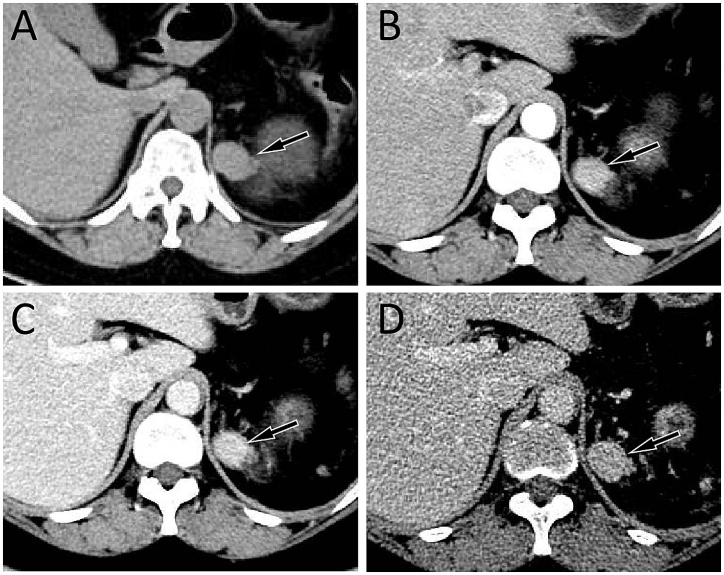
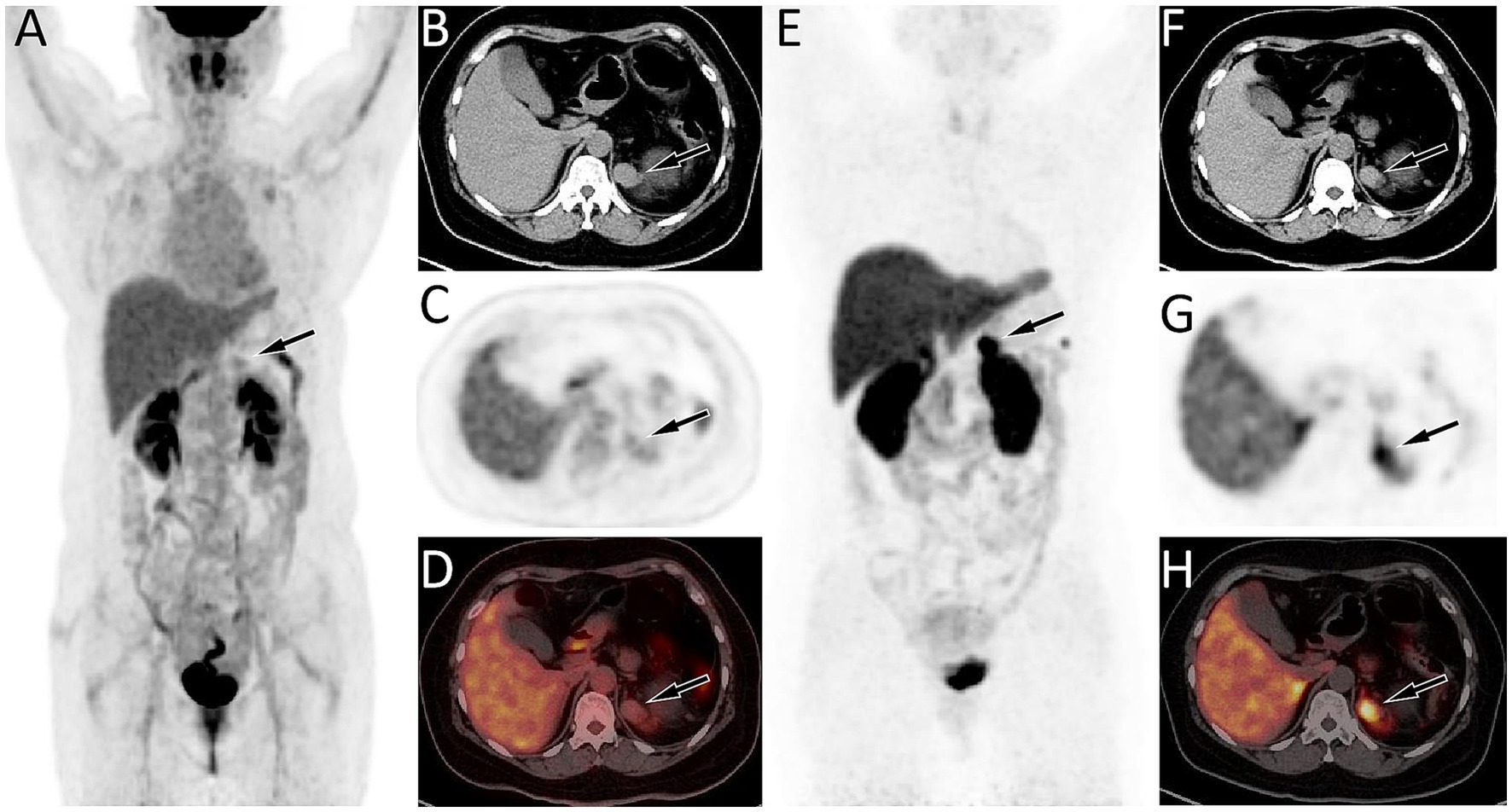
A 39 year old woman was found to have a left adrenal gland mass during abdominal ultrasound examination at an external hospital 1 month ago due to abdominal discomfort. For further diagnosis and treatment, she visited the Department of Hepatobiliary and Pancreatic Surgery at our hospital on September 21, 2023. The patient had a medical history of pancreatic tail resection and splenectomy 8 years ago due to neuroendocrine tumor in the tail of the pancreas and invasion of the spleen, whose condition has remained stable since the surgery. Physical examination showed slight tenderness in the left upper abdomen, and no significant positive signs in the rest. On September 22, 2023, the patient’s fasting blood routine and digestive system tumor markers and other laboratory test results were all within the normal reference value range. On the same day, CT examination revealed a soft tissue density nodule about 2.8 cm × 2.0 cm in size in the patient’s left adrenal gland, which showed significant enhancement on contrast-enhanced CT scan (as shown in Figure 1). To further assess the nature of this lesion, the patient underwent a dual tracer (i.e., 18F-FDG and 68Ga-DOTATATE) PET/CT examination over the following 2 days, and the results showed slightly increased 18F-FDG uptake and obviously increased 68Ga-DOTATATE uptake in the lesion (Figure 2), which was suspected to be a neuroendocrine tumor metastasis. Because the lesion was localized, the patient underwent a left adrenalectomy on September 25, 2023. Postoperatively, the excised adrenal mass was sent for pathological examination. Microscopically, the resected tissue contained red pulp and white pulp (Figure 3), suggesting splenic tissue, which was consistent with the diagnosis of splenosis. Subsequently, the patient was discharged after receiving 3 days of anti-inflammatory treatment. Follow-up up to now, the patient has not complained of any discomfort.

Figure 1. (A) Abdominal computed tomography (CT) revealed a soft tissue density nodule about 2.8 cm × 2.0 cm in size on the left adrenal gland (arrow); In the arterial phase (B), venous phase (C), and delayed phase (D) of contrast-enhanced CT, the lesion showed obvious and continuous uniform enhancement (arrows).

Figure 2. (A–D) Fluorine-18 fluorodeoxyglucose (18F-FDG) positron emission tomography (PET)/CT imaging of the patient; The maximum intensity projection (MIP, A) showed a slightly increased 18F-FDG uptake in the left upper abdomen (arrow). Axial CT (B) showed an isodense nodule in the left adrenal gland (arrow). The corresponding lesion had mildly increased 18F-FDG uptake on axial PET (C, arrow) and PET/CT fusion (D, arrow), with a maximum standardized uptake value (SUVmax) of 2.7. (E–H) 68Ga-DOTATATE PET/CT imaging; The MIP (E) showed a significantly increased 68Ga-DOTATATE uptake in the left upper abdomen (arrow). Axial CT (F), PET (G) and PET/CT fusion (H) showed this increased focal uptake in the left adrenal gland (arrow), in the same location as the lesion shown on 18F-FDG PET/CT, with a SUVmax of 24.3.

Figure 3. Hematoxylin–eosin staining (magnification, ×100) showing red pulp (black arrow) and white pulp (red arrow) within the tissue, suggesting splenic tissue.
Discussion
The most common parenchymal organs for splenosis include pancreas, stomach, small intestine and kidney, but are relatively rare in adrenal glands (5). The possible reason for adrenal gland splenosis is that after splenectomy, the splenic myeloid cells were seeded to the adrenal gland by blood due to splenic vein embolism (1). Splenosis often presents as nodules with a diameter of about 1–3 cm, which are locally dark red or blue black, and can be single or multiple, which can occur within 5 months to 32 years after trauma or splenectomy, with an average of 10 years (6). Our patient was found to have adrenal gland splenosis 8 years after splenectomy, and the length of the nodule was about 2.8 cm, which was consistent with the characteristics of splenosis reported in the literature.
The imaging findings of ectopic splenosis in literature mostly focus on splenic implantation in the pancreas, stomach, intestines, liver, etc., the density of which on CT plain scan is similar to that of normal splenic tissue, exhibiting uniform or slightly higher soft tissue density (7–10). On contrast-enhanced CT scan, the enhancement mode of splenosis was related to the size of the lesion. Lesions smaller than 3 cm showed uniform enhancement in the arterial phase due to small red and white pulp content, short blood flow stroke and insignificant blood flow difference; However, lesions larger than 3 cm showed large red and white pulp content, long blood flow stroke and large difference in blood flow velocity, so presenting as uneven enhancement in the arterial phase. In the portal phase and the delayed phase, it showed continuous uniform or uneven strengthening (9). The MRI of splenosis was also similar to the signal of normal spleen tissue, that is, low signal on T1WI and high signal on T2WI. When the splenosis nodules contained hemosidin or mineral deposits, both T1WI and T2WI showed low signals, and when steatosis was present, the in-phase of T1WI showed high signals and the out-phase of T1WI signal decreased (4, 9, 11). The patient we reported presented with uniform soft tissue density nodules on CT plain and significant and sustained uniform enhancement on three-phase contrast-enhanced CT scan, consistent with literature reports.
Usually, when splenosis can be diagnosed through routine imaging examinations, further examination such as PET/CT is not necessary, and there are also rare descriptions of its PET/CT findings in the literature. However, our patient had a history of pancreatic neuroendocrine neoplasms, and as splenosis in the adrenal gland was rare, little was known about it, which led us to initially consider the possibility of metastatic tumor and eventually to perform PET/CT examination on the patient. Normal spleen tissue will have mild physiological uptake of 18F-FDG and significant uptake of 68Ga-DOTATATE. The mechanism of increased 68Ga-DOTATATE uptake in splenosis is related to the fact that somatostatin receptor type 2 (SSTR2) can be expressed in some immune cells such as macrophages and splenic sinus endothelial cells in normal spleen tissue, resulting in increased 68Ga-DOTATATE uptake. The function of splenosis is the same as that of normal spleen tissue, so its PET/CT findings are also similar, both showing increased 68Ga-DOTATATE uptake (12). As is well known, 18F-FDG and 68Ga-DOTATATE dual nuclear tracer PET/CT have been widely used in the diagnosis, staging, and post-treatment evaluation of neuroendocrine tumors, and G1-G2 grade neuroendocrine tumors can show mildly to moderately increased 18F-FDG uptake and significantly increased 68Ga-DOTATATE uptake (13–15). These imaging features are similar to the splenosis, combined with the patient’s history of neuroendocrine tumors, which ultimately led to our misdiagnosis of the disease.
The imaging findings of splenosis in the adrenal gland should be distinguished from the neoplastic lesions of the adrenal gland, mainly including neuroendocrine tumors such as pheochromocytoma, neuroblastoma, and ganglioneuroma, adrenal adenoma and metastatic tumor. Neuroendocrine tumors may present varying levels of increased 18F-FDG and 68Ga-DOTATATE uptake on PET/CT imaging (16). However, it varies in size and often shows bleeding, cystic changes, and necrosis within the tumor, resulting in uneven density on CT, and contrast enhanced CT scans usually show uneven enhancement (17, 18). For neuroblastoma, calcified foci are frequently observed within the tumor tissue (19). Adrenal adenoma is usually a solitary nodule with small volume and uniform density, which appears as mild and uniform enhancement on contrast-enhanced CT scans (20). The incidence of adrenal metastasis in malignant tumors is 8.6–27.0%, when the lesion size is small, it shows uniform or slightly low density on CT. When the lesion size is large, it is easy to become cystic necrosis and show uneven density, and it can show different degrees of uniform enhancement or ring enhancement on contrast- enhanced CT (20). These variable CT signs make it difficult to distinguish it from the splenosis. For patients suspected of having ectopic splenosis nodules, preoperative technetium-99 m (99mTc) labeled sulfur colloid (99mTc-SC) scanning or 99mTc labeled heat-damaged red blood cell (99mTc-DRBC) imaging is feasible (21). There are a large number of macrophages in the spleen, which can phagocytose colloid and denatured red blood cells. 99mTc-SC or 99mTc-DRBC can be phagocytosed by mononuclear macrophages in the spleen after entering the body intravenously, and then remain in the spleen for imaging diagnosis (22).
Splenosis nodules have compensatory and proliferative functions, which plays a wide range of immune functions in hematopoiesis and red blood cell clearance, which can reduce the incidence of explosive infections (23, 24). Therefore, it is important to obtain an accurate diagnosis of splenosis before surgery, as most ectopic splenic implantation nodules can avoid surgical removal (25). Surgical intervention is required only if the implanted spleen compresses adjacent tissues causing symptoms such as pain, vomiting, etc. (26, 27). Like many cases reported in the literature, our patient also underwent unnecessary surgical resection treatment due to being misdiagnosed before surgery. As splenosis is a phenomenon of autoimplantation of splenic tissue fragments caused by splenic trauma or splenectomy, the prognosis of patients is good.
Conclusion
Splenosis occurring in adrenal glands is relatively rare and is easily misdiagnosed as neoplastic lesions. Our case study suggests that adrenal gland splenosis should be considered as a differential diagnosis of adrenal tumors, especially in patients with a history of splenic trauma or splenectomy. Understanding the clinical and imaging features of splenosis can reduce misdiagnosis and avoid unnecessary surgical intervention.
Data availability statement
The original contributions presented in the study are included in the article/Supplementary material, further inquiries can be directed to the corresponding author.
Ethics statement
The present study was approved by the Ethics Committee of Affiliated Hospital of Zunyi Medical University (approval no. KLL-2024-066). Written informed consent was obtained from the patient for the publication of any potentially identifiable images or data included in this article.
Author contributions
XH: Conceptualization, Data curation, Formal analysis, Funding acquisition, Writing – original draft. WZ: Investigation, Methodology, Project administration, Writing – original draft. PW: Investigation, Supervision, Validation, Writing – review & editing.
Funding
The author(s) declare that financial support was received for the research and/or publication of this article. This study was funded by the Guizhou Province Science and Technology Plan Project (grant numbers: Qiankehe-ZK[2024]-329) and Zunyi Science and Technology Joint Fund (grant number: HZ-2023-284).
Conflict of interest
The authors declare that the research was conducted in the absence of any commercial or financial relationships that could be construed as a potential conflict of interest.
Generative AI statement
The authors declare that no Gen AI was used in the creation of this manuscript.
Publisher’s note
All claims expressed in this article are solely those of the authors and do not necessarily represent those of their affiliated organizations, or those of the publisher, the editors and the reviewers. Any product that may be evaluated in this article, or claim that may be made by its manufacturer, is not guaranteed or endorsed by the publisher.
Supplementary material
The Supplementary material for this article can be found online at: https://www.frontiersin.org/articles/10.3389/fmed.2025.1578613/full#supplementary-material
References
1. Kwok, CM, Chen, YT, Lin, HT, Su, CH, Liu, YS, and Chiu, YC. Portal vein entrance of splenic erythrocytic progenitor cells and local hypoxia of liver, two events cause intrahepatic splenosis. Med Hypotheses. (2006) 67:1330–2. doi: 10.1016/j.mehy.2006.04.064
2. Kanagalingam, G, Vyas, V, Sostre, V, and Mo, A. Gastric splenosis mimicking gastrointestinal stromal tumor. Cureus. (2021) 13:E12816. doi: 10.7759/cureus.12816
3. Li, X, Hu, X, Wang, P, Hu, G, Zhou, B, and Cai, J. A large gastric splenosis mimicking gastrointestinal stromal tumor: a case report and literature review. Exp Ther Med. (2024) 27:186. doi: 10.3892/etm.2024.12474
4. Smoot, T, Revels, J, Soliman, M, Liu, P, Menias, CO, Hussain, HH, et al. Abdominal and pelvic Splenosis: atypical findings, pitfalls, and mimics. Abdom Radiol (NY). (2022) 47:923–47. doi: 10.1007/s00261-021-03402-3
5. Xiao, SM, Xu, R, Tang, XL, Ding, Z, Li, JM, and Zhou, X. Splenosis with lower gastrointestinal bleeding mimicking Colonical gastrointestinal stromal tumour. World J Surg Oncol. (2017) 15:78. doi: 10.1186/s12957-017-1153-0
6. Berman, AJ, Zahalsky, MP, Okon, SA, and Wagner, JR. Distinguishing splenosis from renal masses using Ferumoxide-enhanced magnetic resonance imaging. Urology. (2003) 62:748. doi: 10.1016/S0090-4295(03)00509-0
7. Li, B, Huang, Y, Chao, B, Zhao, Q, Hao, J, Qin, C, et al. Splenosis in gastric fundus mimicking gastrointestinal stromal tumor: a report of two cases and review of the literature. Int J Clin Exp Pathol. (2015) 8:6566–70.
8. Deng, Y, Jin, Y, Li, F, and Zhou, Y. Splenosis mimicking an extramural duodenal mass: a case report. Oncol Lett. (2014) 8:2811–3. doi: 10.3892/ol.2014.2609
9. Vernuccio, F, Dimarco, M, Porrello, G, Cannella, R, Cusmà, S, Midiri, M, et al. Abdominal splenosis and its differential diagnoses: what the radiologist needs to know. Curr Probl Diagn Radiol. (2021) 50:229–35. doi: 10.1067/j.cpradiol.2020.04.012
11. Nalbant, MO. Intrahepatic Splenosis: a rare case. Curr Med Imaging. (2023) 19:640–3. doi: 10.2174/1573405619666221212153639
12. Siebinga, H, De Wit-Van Der Veen, BJ, Beijnen, JH, Stokkel, M, Dorlo, T, Huitema, A, et al. A physiologically based pharmacokinetic (Pbpk) model to describe organ distribution of (68)Ga-DOTATATE in patients without neuroendocrine tumors. EJNMMI Res. (2021) 11:73. doi: 10.1186/S13550-021-00821-7
13. Chan, DL, Pavlakis, N, Schembri, GP, Bernard, EJ, Hsiao, E, Hayes, A, et al. Dual somatostatin receptor/FDG PET/CT imaging in metastatic neuroendocrine Tumours: proposal for a novel grading scheme with prognostic significance. Theranostics. (2017) 7:1149–58. doi: 10.7150/thno.18068
14. Fortunati, E, Argalia, G, Zanoni, L, Fanti, S, and Ambrosini, V. New pet radiotracers for the imaging of neuroendocrine neoplasms. Curr Treat Options in Oncol. (2022) 23:703–20. doi: 10.1007/s11864-022-00967-z
15. Hu, X, Li, D, Wang, R, Wang, P, and Cai, J. Comparison of the application of 18F-FDG and 68Ga-DOTATATE PET/CT in neuroendocrine tumors: a retrospective study. Medicine (Baltimore). (2023) 102:E33726. doi: 10.1097/MD.0000000000033726
16. Jha, A, Patel, M, Ling, A, Shah, R, Chen, CC, Millo, C, et al. Diagnostic performance of [(68)Ga]DOTATATE PET/CT, [(18)F]FDG PET/CT, Mri of the spine, and whole-body diagnostic Ct and Mri in the detection of spinal bone metastases associated with Pheochromocytoma and Paraganglioma. Eur Radiol. (2024) 34:6488–98. doi: 10.1007/s00330-024-10652-4
17. Farrugia, FA, Martikos, G, Tzanetis, P, Charalampopoulos, A, Misiakos, E, Zavras, N, et al. Pheochromocytoma, diagnosis and treatment: review of the literature. Endocr Regul. (2017) 51:168–81. doi: 10.1515/enr-2017-0018
18. Lafont, M, Fagour, C, Haissaguerre, M, Darancette, G, Wagner, T, Corcuff, JB, et al. Per-operative hemodynamic instability in normotensive patients with incidentally discovered Pheochromocytomas. J Clin Endocrinol Metab. (2015) 100:417–21. doi: 10.1210/jc.2014-2998
19. Hao, J, Sang, J, Xu, X, and Bao, A. Diagnostic value of Ct and Mri combined with serum Ldh, Nse, Cea, and Mycn in pediatric neuroblastoma. World J Surg Oncol. (2023) 21:251. doi: 10.1186/S12957-023-03131-5
20. Schieda, N, and Siegelman, ES. Update on Ct and Mri of adrenal nodules. AJR Am J Roentgenol. (2017) 208:1206–17. doi: 10.2214/AJR.16.17758
21. Grande, M, Lapecorella, M, Ianora, AA, Longo, S, and Rubini, G. Intrahepatic and widely distributed intraabdominal Splenosis: multidetector CT, us and Scintigraphic findings. Intern Emerg Med. (2008) 3:265–7. doi: 10.1007/s11739-008-0112-8
22. Singh, P, Munn, NJ, and Patel, HK. Thoracic Splenosis. N Engl J Med. (1995) 333:882. doi: 10.1056/NEJM199509283331318
23. Patil, MS, Goodin, SZ, and Findeiss, LK. Update: splenic artery embolization in blunt abdominal trauma. Semin Intervent Radiol. (2020) 37:097–102. doi: 10.1055/s-0039-3401845
24. Dehli, T, Bågenholm, A, Trasti, NC, Monsen, SA, and Bartnes, K. The treatment of spleen injuries: a retrospective study. Scand J Trauma Resusc Emerg Med. (2015) 23:85. doi: 10.1186/s13049-015-0163-6
25. Harfouche, MN, Dhillon, NK, and Feliciano, DV. Update on nonoperative management of the injured spleen. Am Surg. (2022) 88:2649–55. doi: 10.1177/00031348221114025
26. Braga, J, Pereira, F, Fernandes, C, Silva, M, Boncoraglio, T, and Oliveira, C. Abdominal splenosis mimicking a colon tumour. Eur J Case Rep Intern Med. (2021) 8:002219. doi: 10.12890/2021_002219
Keywords: splenosis, adrenal gland, neuroendocrine tumor, PET/CT, CT
Citation: Hu X, Zhao W and Wang P (2025) Case Report: Adrenal gland splenosis mimicking a neuroendocrine tumor on 68Ga-DOTATATE and 18F-FDG PET/CT imaging. Front. Med. 12:1578613. doi: 10.3389/fmed.2025.1578613
Edited by:
Romina Grazia Giancipoli, Agostino Gemelli University Polyclinic (IRCCS), ItalyReviewed by:
Chunyin Zhang, Southwest Medical University, ChinaSelin Kesim, Istanbul Kartal Dr. Lutfi Kirdar Education and Research Hospital, Türkiye
Copyright © 2025 Hu, Zhao and Wang. This is an open-access article distributed under the terms of the Creative Commons Attribution License (CC BY). The use, distribution or reproduction in other forums is permitted, provided the original author(s) and the copyright owner(s) are credited and that the original publication in this journal is cited, in accordance with accepted academic practice. No use, distribution or reproduction is permitted which does not comply with these terms.
*Correspondence: Pan Wang, MTI5ODE3ODgyOEBxcS5jb20=
 Xianwen Hu
 Wei Zhao
 Pan Wang1*