 Letícia Ferreira Lima1,2
 Mauricio Gonçalves Da Costa Sousa3*
 Gisele Regina Rodrigues1,2,4
 Kamila Botelho Sampaio de Oliveira1,2
 Ana Margarida Pereira5,6André da Costa5,6
 Raul Machado5,6
 Octavio Luiz Franco1,2,4
 Simoni Campos Dias1,2,7
- 1Centro de Análises Proteômicas e Bioquímicas, Universidade Católica de Brasília (UCB), Brasília, Brazil
- 2Departamento de Pós-Graduação em Ciências Genômicas e Biotecnologia, Universidade Católica de Brasília (UCB), Brasília, Brazil
- 3School of Dentistry, Oregon Health and Science University OHSU, Portland, OR, United States
- 4S-Inova Biotech, Pós-Graduação em Biotecnologia, Universidade Católica Dom Bosco, Campo Grande, Brazil
- 5CBMA (Centre of Molecular and Environmental Biology), Department of Biology, University of Minho, Braga, Portugal
- 6IB-S (Institute of Science and Innovation for Bio-Sustainability), University of Minho, Braga, Portugal
- 7Pós-Graduação em Biologia Animal, Campus Universitário Darcy Ribeiro, Universidade de Brasília (UnB), Brasília, Brazil
Elastin-like polypeptides (ELPs) are biopolymers formed by amino acid sequences derived from tropoelastin. These biomolecules can be soluble below critical temperatures, forming aggregates at higher temperatures, which makes them an interesting source for the design of different nanobiomaterials. These nanobiomaterials can be obtained from heterologous expression in several organisms such as bacteria, fungi, and plants. Thanks to the many advantages of ELPs, they have been used in the biomedical field to develop nanoparticles, nanofibers, and nanocomposites. These nanostructures can be used in multiple applications such as drug delivery systems, treatments of type 2 diabetes, cardiovascular diseases, tissue repair, and cancer therapy. Thus, this review aims to shed some light on the main advances in elastin-like-based nanomaterials, their possible expression forms, and importance to the medical field.
Introduction
Nanotechnology is promising because it allows new applications to be used in a wide range of fields, especially medical ones. To develop new bionanotechnologies, several natural-based components, such as silk (Chouhan and Mandal, 2020), chitosan (Qi et al., 2004), elastin-like polypeptides (ELPs) (Rodríguez-Cabello et al., 2016), and others (Ding et al., 2014) have been studied to design nanomaterials. Elastin-like polypeptides (ELPs), for instance, are biosynthetic structures based on natural elastin, which occurs naturally in mammals.
ELPs present elastomeric characteristics acquired from the monomeric precursor tropoelastin, formed from its alignment, and monomer binding (Kaur and Reinhardt, 2015; Coenen et al., 2018; Varanko et al., 2020). The most frequently used ELPs consist of n tandem repeats of the VPGXG pentapeptide, where X is a residue which can be any amino acid except for proline (Kim and Chaikof, 2010). The presence of a proline amino acid residue in the X position causes transition temperature (Tt) loss due to its rigid conformational characteristic (Urry, 1992; Trabbic-Carlson et al., 2004; Quintanilla-Sierra et al., 2019). ELPs exhibit a reversible phase-transitional behavior at a certain temperature threshold, referred to as the transition temperature. In aqueous solution and below the Tt, ELPs are highly soluble, but they undergo phase-separate aggregation into coacervates that may coalesce into supra-hierarchical structures at temperatures above Tt, which is mostly 37°C (Chow et al., 2008; Floss et al., 2010; Saxena and Nanjan, 2015; Fletcher et al., 2019; Quintanilla-Sierra et al., 2019). The insoluble phase occurs because ELP bonds are disrupted by water molecules (Urry, 1992). Besides being affected by polymer concentration and molecular weight, pH and ionic strength, Tt is also dependent on the guest residue composition (Urry et al., 1991; Meyer and Chilkoti, 2004; Li et al., 2014; Zhao et al., 2016). Of remarkable interest, the fusion of peptides or small molecules to ELPs was not seen to significantly compromise thermally responsive behavior (Trabbic-Carlson et al., 2004; Christensen et al., 2013; da Costa et al., 2015a; da Costa et al., 2018; da Costa et al., 2021). In addition, ELPs have been demonstrated to be biodegradable, biocompatible, and non-cytotoxic (Nair and Laurencin., 2007; Rodriguez-Cabello et al., 2017), thus becoming increasingly attractive for the development of materials suitable for biomedical applications (Despanie et al., 2016; Varanko et al., 2020; Mbundi et al., 2021).
ELPs can be chemically synthetized (McGrath et al., 1990; Urry et al., 1990; Urry and Pattanaik, 2006; Aladini et al., 2016), but this process presents considerable limitations involving several complex steps, resulting in low yields of a polydisperse mixture of polypeptides with low molecular weight (McGrath et al., 1990). In contrast, the production of recombinant ELPs represents an efficient and high-yield approach, producing monodisperse polymers with high molecular weight (McGrath et al., 1990; Nettles et al., 2010; Mbundi et al., 2021; Rodriguez-Cabello et al., 2021). Owing to the reversible phase transition behavior of ELPs, purification can be achieved by employing simple heating/cooling cycles, avoiding the need for complex chromatographic methods, assisting cost reduction and avoiding the use of cumbersome steps, because there is no need for chromatography purification (Figure 1) (Meyer and Chilkoti, 1999; Banki et al., 2005; Trabbic-Carlson et al., 2009; Hu et al., 2010; Yang et al., 2012; da Costa et al., 2018; Paiva dos Santos et al., 2019; Pereira et al., 2021a).
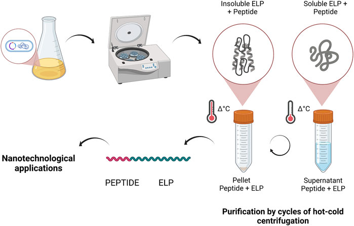
FIGURE 1. Heterologous expression of ELP fused with proteins or peptides and the purification process for hysteresis, due to their characteristic Tt, which is separated based on insoluble or soluble ELPs by centrifugation of the other proteins. Created with BioRender.com.
Besides chemical production, elastin-like polypeptides can also be obtained by heterologous expression (da Costa et al., 2015b), which is less costly than chemical synthesis (Basu et al., 2014). Heterologous expression is an affordable and effective way of using yeasts, bacteria, and plants to produce these polypeptides (Chow et al., 2006; Lin et al., 2006; Schipperus et al., 2009). ELPs can be fused with peptides of interest and obtained on a large scale, by means such as fermentation (Lindbo, 2007; Schipperus et al., 2009; Martínez-Alarcón et al., 2018). Therefore, the choice of the microorganism depends on what the focus is, and generally prokaryotes, in particular Escherichia coli, are used (Kuthning et al., 2015). The heterologous expression of ELPs can be scaled, favoring their adoption in the industrial sector (Fong et al., 2009; Cardoso et al., 2020).
Moreover, the use of DNA recombinant technology allows fine-tuning of the structure of ELRs to incorporate bioactive domains with precise control over size and composition (Meyer and Chilkoti, 2002; Mbundi et al., 2021), leading to customized functional materials (Richman et al., 2005; da Costa et al., 2015a; Lee et al., 2019; Salinas-Fernández et al., 2020; Pereira et al., 2021b; da Costa et al., 2021). These materials in nanoscale are used in a wide range of studies, mainly in the medical field, for applications such as biosensors, wound dressing, fighting infections acquired by acute or chronic injuries, or skin burns (Li et al., 2020; Sarangthem et al., 2021).
The elastin-like polypeptides have intrinsic and versatile characteristics that allow them to form different nanostructures, such as nanoparticles (Machado et al., 2009; Peddi et al., 2020), nanofibers (Mahara et al., 2017; Sarangthem et al., 2021), and nanocomposites (Lin et al., 2019). These nanotechnologies, when used in applications like drug delivery, improve the efficacy of treatment since the nanostructure can direct the molecule, thus enhancing the stability and boosting the contact surface, which can enhance the activity of these structures, important for their thermodynamic properties (Javili et al., 2013; Butcher et al., 2016; Schöttler et al., 2016). Studies that have already passed through phase 2 of clinical trials demonstrated that, compared to free medication, nanoparticle-assembled ELPs enhanced the drug’s half-life and its focus on the tumor, which is an interesting and promising construction, but yet to be approved as a drug delivery system (Macewan and Chilkoti, 2014).
Therefore, due to the benefits of the biotechnological applications of ELPs, this review presents the significant advances in using elastin-like polypeptides in the biomedical field, their production in different systems, and how they can be used to develop different nanomaterials.
Elastin Biosynthesis and Derivatives
Elastin and Tropoelastin
Elastin is a structural protein which is the main component of elastic fibers present in the extracellular matrix (ECM), imparting structural support to numerous organs and tissues, for example, large blood vessels (e.g., aorta artery), skin, cartilage, ligaments, vocal cords, bladder, and lungs (Mithieux and Weiss, 2005; Nettles et al., 2010; Roberts et al., 2017). This proteinaceous material comprises approximately 90% of the elastic fibers (Mithieux and Weiss, 2005; Wang et al., 2019; Ozsvar et al., 2021), conferring resilience and elasticity to organs and tissues that require the ability to undergo a lifetime of repetitive cycles of deformation and relaxation without rupture (Vrhovski and Weiss, 1998; Floss et al., 2010). In its natural form, elastin is heavily cross-linked and therefore insoluble (O’Neill Moore et al., 2020; Rodriguez-Cabello et al., 2021), providing great stability and an estimated half-life of 70 years (Mithieux and Weiss, 2005; Rodriguez-Cabello et al., 2021). Its soluble precursor, tropoelastin, presents 60–72 kDa; it is an alternatively spliced protein, rich in non-polar residues including glycine, alanine, valine and proline, and composed of alternated hydrophobic and hydrophilic domains (Wise et al., 2014; Wang et al., 2019; Ozsvar et al., 2021). While the hydrophilic domains comprise a high content of alanine (A) and lysine (K) residues that promote intra- and intermolecular crosslinking, the hydrophobic component is mainly composed of the non-polar amino acids valine (V), glycine (G), alanine (A), and proline (P) (Wise and Weiss, 2009; Wise et al., 2014; Rodriguez-Cabello et al., 2021). The hydrophobic moieties often occur as repeats of the tetra-, penta-, and hexa-peptides VPGG, VPGVG and APGVGV, with the latter being the most common (Floss et al., 2010; Casal et al., 2013; Wise et al., 2014). Multiple and periodic PG motifs promote the formation of repeated fluctuating β-type turns, imparting to tropoelastin a highly hydrated structure with conformational flexibility (Wise et al., 2014; Wang et al., 2019). Under physiological conditions, tropoelastin monomers reversibly self-aggregate into spherical globules in an intrinsic process known as coacervation, which plays a pivotal role in elastin biosynthesis (Yeo et al., 2011).
Elastogenesis
The formation of elastic fibers (Figure 2) is a hierarchical and complex process that occurs during prenatal and childhood development, involving tropoelastin synthesis, coacervation, cross-linking, and microfibrillar deposition (Yeo et al., 2011; Ozsvar et al., 2021). Initially, tropoelastin is expressed in elastogenic cells (smooth muscle, mesothelial and endothelial cells, chondrocytes, and fibroblasts) (Vrhovski and Weiss, 1998; Mithieux and Weiss, 2005) in response to biological signals such as developmental stage, mechanical stress, cytokines, and growth factors (Yeo et al., 2011; Wang et al., 2019). After the transcription and translation steps, tropoelastin binds to elastin binding protein (EBP), a 67 kDa chaperone that prevents self-aggregation and proteolysis of tropoelastin (Vrhovski and Weiss, 1998; Mithieux and Weiss, 2005; O’Neill Moore et al., 2020; Ozsvar et al., 2021). The tropoelastin-EBP complex is then transported to the extracellular space and the complex dissociates. EBP is recycled intracellularly by endocytosis, and the cross-linking of the secreted tropoelastin is initiated, catalyzed by lysyl oxidase (LOX) present at the cell surface (Vrhovski and Weiss, 1998; Sato et al., 2017; O’Neill Moore et al., 2020; Ozsvar et al., 2021). Finally, the cross-linked tropoelastin spherules (elastin) coacervate and deposit onto microfibril scaffolds of the extracellular space (Vrhovski and Weiss, 1998; Yeo et al., 2011; O’Neill Moore et al., 2020; Ozsvar et al., 2021), initiating the assembly of elastic fibers. As the biosynthesis into the ECM proceeds, the final products are insoluble mature elastin fibers.
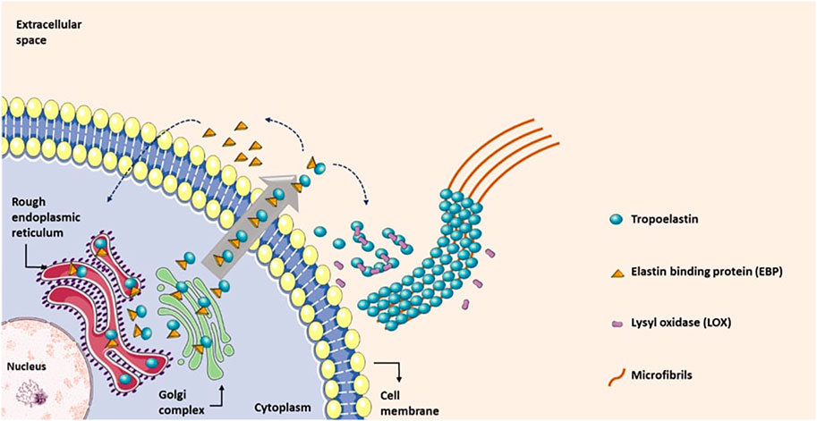
FIGURE 2. Elastin biosynthesis: after synthesis in the rough endoplasmic reticulum (rER), the tropoelastin-EBP complex is transported through the Golgi complex to the extracellular space, aggregating on the cell surface. EBP then unbinds from tropoelastin, and tropoelastin molecules are cross-linked to each other via lysine residues that are oxidized by lysyl oxidase (LOX) generating cross-linked elastin. The cross-linked molecules deposit onto microfibrils which direct elastin deposition for the formation of the elastic fibers. EBP is then recycled back into the cell and binds newly synthesized tropoelastin molecules. This process continues, producing insoluble elastin fibres.
Applications of Elastin and Elastin-Derived Peptides
The structural role of elastin is well recognized, imparting elastic recoil and resilience to tissues and organs. Nevertheless, elastin is also directly or indirectly involved in physiological processes such as cell adhesion (Senior et al., 1984; Mithieux and Weiss, 2005; Rodgers and Weiss, 2005; Mithieux et al., 2013; Lee et al., 2017). Due to its diverse biological properties, elastin has gained special interest in biomedical applications, namely tissue engineering and regenerative medicine (Daamen et al., 2007; Wang et al., 2021). Some examples include the use of elastin from decellularized tissues combined with endothelial cells for the development of vascular grafts (Amiel et al., 2006) and heart valves (Bader et al., 1998), the use of purified elastin for the development of gastrointestinal patches to repair duodenal injuries (Kajitani et al., 2000), or as a coating material to promote cell adhesion and proliferation in tissue scaffolds (Sales et al., 2007) or metallic surfaces (Yin et al., 2009). Despite its potential and attractiveness, the use of elastin as a biomaterial is limited due to the highly dense cross-linked network that is insoluble and extremely stable. The insoluble nature of elastin makes its purification highly challenging and ineffective (Wise et al., 2014), often leading to contamination of elastin with other proteins that can elicit immunological responses (Daamen et al., 2001). Isolation and purification of tropoelastin are also troublesome processes, due to extensive soluble precursor degradation during and even after purification (Vrhovski and Weiss, 1998). Moreover, isolation and purification of tropoelastin from natural sources present ethical issues. The isolation from animals is typically a low-yield process, demanding the use of many animals, and/or fetal and neonatal animal tissues, since the biosynthesis of tropoelastin occurs mainly during early development stages (Vrhovski and Weiss, 1998; Wen et al., 2020).
While mature elastin fibers have an insoluble nature, initial studies on elastin’s characteristics and remodeling have shown the possibility of obtaining soluble elastin variants, α- and k-elastin, depending on the extraction methods (Adair et al., 1951; Partridge, 1963). The extraction processes involve the scission of the covalent bonds by hydrolysis under harsh acidic (α-elastin) or alkaline (k-elastin) chemical conditions. Usually, α-elastin is obtained by hydrolysis in hot oxalic acid, whereas κ-elastin is obtained by hydrolysis with 1 M potassium hydroxide in 80% ethanol (Adair et al., 1951; Partridge, 1963). The corresponding degradation products are termed elastin-derived peptides (EDPs) or elastokines (le Page et al., 2019). Table 1 enumerates some examples of EDPs obtained by hydrolysis or proteolytic digestion. EDPs also occur naturally, as a result of ageing due to the many insults and increased protease activity, and have many implications in health and disease. They are not only a hallmark of ageing, but also influence T-cell modulation, tumor progression, or diabetes (Robert and Labat-Robert, 2014; Meghraoui-Kheddar et al., 2017; Boraldi et al., 2018; Salesse et al., 2018; le Page et al., 2019). κ-EDPs have a bioactive sequence based on the xGxxPG motif that binds to the elastin receptor complex and produces biological effects, while α-EDPs have a molecular weight and sequence much more similar to tropoelastin (Qin, 2015). The solubility of EDPs is advantageous, allowing their use for several applications in biomedicine (Gigante et al., 2003; Pocza et al., 2008; Szychowski and Gmiński, 2019; Amakye et al., 2021) and the formation of self-assembled supramolecular architectures of fibrils (Bochicchio et al., 2015). In addition to biomedical applications, EDPs are frequently used in the cosmetic industry in creams, shampoos, or even as nutraceuticals (Arany et al., 2006). In such cases, they act as antistatic, film forming, skin and hair conditioner, and emollient (Hunter et al., 1991) and are usually obtained from bovine tendons or from the skin of fish such as salmon or tuna (Hyun et al., 2004).
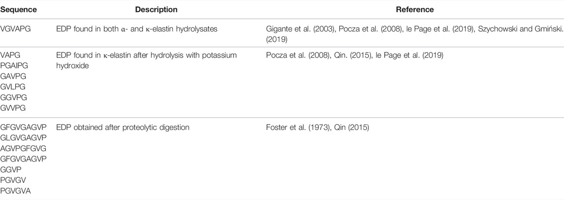
TABLE 1. Examples of some elastin-derived peptide (EDP) sequences obtained by hydrolysis or proteolytic digestion.
To overcome the limitations of natural insoluble elastin, great attention has been paid to the synthesis of artificial elastin-mimetic polypeptides, termed elastin-like polypeptides (ELPs) (Roberts et al., 2017), or elastin-like recombinamers (ELRs) (Rodríguez-Cabello et al., 2009). These biomimetic sequence-repetitive molecules are based on the repeating motifs present in the hydrophobic domain of tropoelastin and can undergo coacervation in a similar way to elastin (Le and Sugawara-Narutaki, 2019). Nevertheless, these artificial biomolecules are inspired by and do not adequately represent the diversity of natural tropoelastin sequences per se. The canonical sequence for ELPs is based on repetitions of the pentapeptide VPGVG, but the most common sequence found in tropoelastin is the hexapeptide APGVGV (Conticello and Carpenter Desai, 2012). Still, early studies with the hexapeptide demonstrated the absence of a reversible coacervation process (Conticello and Carpenter Desai, 2012). On the other hand, ELPs based on the canonical pentapeptide VPGVG sequence can undergo a fully reversible temperature-dependent coacervation process (van Eldijk et al., 2012).
Heterologous Expression of Elastin-Like Polypeptides
As described above, elastin is structural protein present in all vertebrates (Liu et al., 2018), and the synthetic product is denominated elastin-like polypeptides (ELPs) (Fletcher et al., 2019). In addition to synthetic production, they can be produced by recombinant DNA technology (Girotti et al., 2011; Fletcher et al., 2019; Saha et al., 2020). ELPs can be produced alone or used in fusion with protein or peptides (Trabbic-Carlson et al., 2004; Walker et al., 2014). This technology can provide scalable, sustainable and cheaper production, and ELPs can be produced in bacteria, yeast and plants (Girotti et al., 2011; Sampaio de Oliveira et al., 2020).
Escherichia coli
E. coli has been extensively applied as a host to produce recombinant proteins for therapeutic use. Moreover, its use has demonstrated advantages like rapid growth rate, easier genetic manipulations, high yield of product, and scalability (Kaur et al., 2018; Sampaio de Oliveira et al., 2020). Additionally, E. coli presents different strains and expression vectors and a relatively simple mechanism of protein folding, and it has been used in several applications in the biotechnology industry (Sampaio de Oliveira et al., 2020). Some studies used the capacity of E. coli to produce ELPs via heterologous expression (Figure 1).
In a study to produce the ABP-CM4 peptide with broad antimicrobial activity, the researchers used the elastin-like recombinant consisting of 200 repetitions of the VPAVG pentamer fused to ABP-CM4 in the terminal N portion, denominated CM4-A200. The plasmid [pET25b (+)] containing the sequence of CM4-A200 was transformed into E. coli BL21 (DE3). CM4-A200 was purified using the thermo-responsive behavior of the A200 polymer. This polymer was processed into free-standing films and displayed significant antimicrobial activity against yeasts, Gram-positive and Gram-negative bacteria, and filamentous fungi. Authors also reported that CM4-A200 did not have a cytotoxic effect on human skin fibroblasts (da Costa et al., 2015b).
An ELP sequence was used in fusion with human interferon-γ (hIFN-γ). This construction was cloned into the pET-28a (+) expression vector with 50 repeats of ELP (VPGVG), and then transferred into competent E. coli strain BL21 (DE3). Authors described the ELP construction raising the accumulation of hIFN tenfold, and the average expression of total soluble protein (TSP) rose by 46.85%. In addition, using inverse transition cycling (ITC), they obtained hIFN-γ-ELP with 98 ± 5% of purity. Another result described by the same authors is related to the bioactivity of recombinant hIFN-γ-ELP, which was comparable to commercial hIFN-γ, (7.55 × 10⁶ IU/ml) (Heidari-Japelaghi et al., 2019).
Another study used ELP fused to a glucagon-like peptide, using a type-2 diabetes drug to produce a novel peptide delivery system. According to the authors, this system undergoes a transition phase between room temperature and body temperature, and the system was tested as an injection. Researchers tested the proteolytic stability and activity in vitro. Tests with mice identified that an injection of GLP-1-ELP fusions decreased blood glucose levels for up to 5 days, which is 120 times longer than an injection of the native peptide. Results illustrated the benefit of working with ELPs to release peptide-ELP fusions (Amiram et al., 2013).
Yeast
The use of yeast cells as an expression system which, compared to other systems, demonstrated some benefits, including simple genetic manipulation, rapid growth, and the ability to perform adequate post-translational modifications (PTM) and achieve high cell density. It also demonstrated the ability to produce and secrete biologically active proteins, and easily adapted to industrial-scale conditions (Gomes et al., 2018). The most common of post-translational modifications, such as methylation, can modified the structure or hydrophobicity of the protein (Owen and Shewmaker, 2019), and N-myristoylation can aid the molecular assembly of ELPs and influence the Tt. (Scheibel et al., 2020).
In this study, the authors tested the 90 repetitions of ELP-VPGXG (with any amino acid except proline at the X position). They demonstrated ELP production using the method of methanol-induced fed-batch cultures of Pichia pastoris (Çelik and Çalik, 2012; Wang et al., 2017). This study also evaluated the influence of pH (pH 3–7) on culture growth. Their results showed that pH 6 was optimum for production of ELP with a yield of 255 mg L−1 of purified ELP of cell-free medium (Schipperus et al., 2009). Another study produced an ELP with 21 repeats of the amino acid sequence (VPGVG)2VPGEG (VPGVG)2 in P. pastoris. This construction presents ∼47 kDa and in the C-terminal includes c-Myc and His-tag, which were used for purification. The ELP produced was purified, and the yield after metal ion affinity chromatography was 2.5 mg L−1 in shake flask cultures (Sallach et al., 2009). The difference in yield observed in these studies can be explained by the optimal pH evaluated for enhaced production of ELP in fed-batch fermentation in Schipperus et al. (2009) study, while the Sallach et al. (2009) study was produced in baffled flask and no optimization yield was evaluated yet.
Plants
Plants have been widely used as hosts and are able to produce biologically active recombinant products (da Cunha et al., 2017; Margolin et al., 2018). They present advantageous heterologous expression systems, due to high production and low cost, do not generate endotoxins, and do not present pathogens mutual to humans (Twyman et al., 2003; Basaran and Rodriguez-Cerezo, 2008).
In this context, Conley et al. (2009) tested different sizes of ELP (VGVPG)n to identify the optimal construction for the accumulation of recombinant proteins in Nicotiana benthamiana. The results demonstrated that ELP tag (n = 5–40) repeats provided the best results when evaluating recombinant protein accumulation, and the larger ELP tags (n = 80–160) showed high efficiency during the purification by inverse transition cycling (ITC). Results showed that the use of ELP fusion tags contributed to raising the production of recombinant proteins in plants (Conley et al., 2009).
A different strategy was used in another study, expressing single-chain variable fragments (scFvs) in transgenic tobacco seeds, with fusions on the C-terminal based on ELPs composed of Val-Pro-Gly-Xaa-Gly, where Xaa is valine, glycine or alanine with 100 repetitions. This strategy allowed a 40-fold increase in scFv accumulation, with levels nearing 25% of total soluble seed protein. In addition, ELPylated scFv continued stable and functional in mature seeds that were stored for a long period at room temperature (Scheller et al., 2006).
In another study, the authors used 2-cell suspension to produce human interleukin-10 (IL-10) in tobacco, and they evaluated the effect of an ELP with 28 repetitions and a green fluorescent protein (GFP) tag on IL-10 accumulation. The IL-10 obtained via expression demonstrated high accumulation levels. IL-10-ELP demonstrated cytokine activity, but this activity was reduced compared to unfused IL-10 (Kaldis et al., 2013).
Nanomaterials Based on Elastin-Like Polypeptides and Their Medical Applications
Nanoparticles
The ELP self-assembly process is directly associated with the polymer architecture, which can result in coacervate and subsequently different organizations according to the transition temperature (Tt) (Figure 3). Some ELPs have alternating hydrophobic and hydrophilic domains along the backbone of the polypeptide. This complex structure results in the formation of nanoparticles (Smits et al., 2015; Rodríguez-Cabello et al., 2018). The typical hydrophobic block transition occurs when the temperature of the solution is raised above the lower Tt, causing the hydrophobic portion to fold and segregate from the aqueous solution. The hydrophilic block (top Tt) remains soluble and hydrated in contact with the surrounding water, forming the crown of a micellar structure. This feature allows the hydrophobic core to store nonpolar drugs, while polar molecules can be kept on the hydrophilic surface (Rodríguez-Cabello et al., 2016).
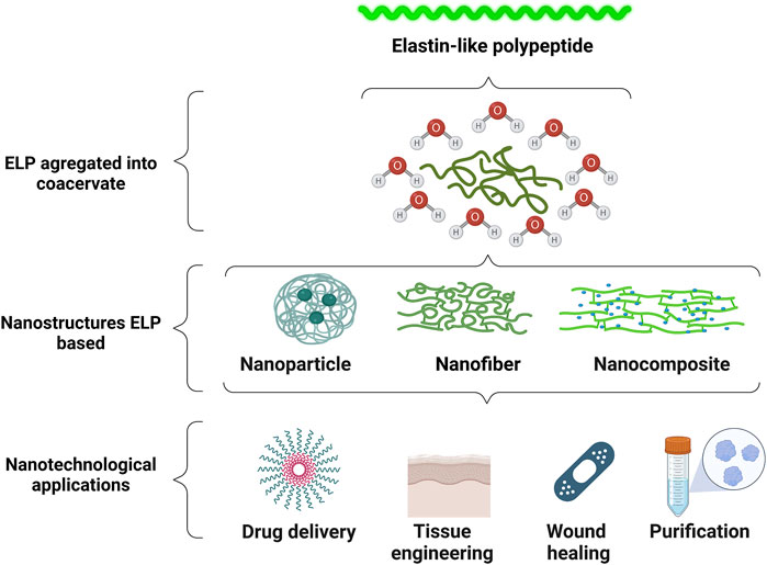
FIGURE 3. ELPs aggregated coacervate involved in water molecules in a conformation in different types of nanostructures that can be used in various nanotechnological applications. Created with BioRender.com.
The adoption of ELP nanoparticles as nanodrugs is of great interest in different biomedical areas, as they have a broad-spectrum therapeutic potential (Smits et al., 2015). The biological activity of protein motifs can be maintained when fused to ELPs, and the phase transition property of ELPs as well, enabling the formation of nanoparticles above their transition temperature (Monfort and Koria, 2017). These features make these nanostructures attractive as delivery vehicles. The loading, targeting, and delivery of drugs can be optimized using ELP nanoparticles.
As described above, ELPs can be fused to a wide variety of bioactive peptides. Nanoparticles formed from fusion proteins based on ELPs showed potential as drug carriers, enabling the supply of the active principle for a long time and protection against proteolytic degradation (Yeboah et al., 2016). The self-assembled nanoparticles from a recombinant fusion protein composed of cell-derived growth factor-1 (SDF1) and an elastin-like peptide have shown promise for the treatment of chronic skin wounds. SDF1-ELP nanoparticles were used in the treatment of full-thickness skin wounds in diabetic mice and demonstrated significantly higher healing activity than free SDF1. By 28 days, the wounds were fully closed, while wounds treated with free SDF1 or ELP alone took 42 days to close fully (Yeboah et al., 2016).
ELPs are also used as purification tags and solubility enhancers. The human granulocyte-macrophage colony stimulating factor (hGMCSF), an essential molecule in the immune system, was fused to an ELP, enabling its direct purification from the soluble fraction of the E. coli lysate. Furthermore, this fusion provided the formation of small and stable spherical nanoparticles that can maintain the pro-mitotic activity of hGMCSF. Fusion of ELPs to different proteins can stabilize bioactive nanoparticles based on these proteins of interest, providing their wide application in medicine and biology. The hGMCSF-A192 nanoparticles were able to stimulate TF-1 cell proliferation [EC50 of 0.29 ± 0.07 nM (mean ± SD, n = 3)], demonstrating that they are biologically active (Park et al., 2020).
In another study, ELPs were fused to nerve growth factor (NGF) and brain-derived neurotrophic factor (BDNF) to improve their biological activity in neural injuries, providing a robust delivery system that increases the bioavailability and half-life of these proteins. The fused proteins NGF-ELP and BDNF-ELP were able to self-assemble into nanoparticles at their respective transition temperatures. According to the results, NGF-ELP nanoparticles induced neurite outgrowth in PC12 cells, while BDNF-ELP nanoparticles induced TrkB receptor phosphorylation in transfected cells. Data indicate that these nanoparticle fusion proteins can be applied in neural regeneration, as they retain the biological activity of nerotrophins and increase their bioavailability (Johnson and Koria, 2016).
ELP nanoparticles also play an important role as a tumor-targeting drug (Mie et al., 2019). In a study described by Matsumoto et al., epidermal growth factor (EGF) fused to genetically engineered ELPs with a fused polyaspartic acid (ELPD) tail was loaded with the anticancer drug paclitaxel. The nanoparticles formed were able to induce the death of the human lung adenocarcinoma epithelial cell line, A549 cells, known to express large amounts of the EGF receptor (EGFR). According to the data, cell proliferation was at least 10 times lower in the presence of the generated nanoparticles, when compared to the presence of EGF alone. This result suggests that the EGF contained in nanoparticles retained its ability to bind to EGFR and induce cell proliferation (Matsumoto et al., 2014).
Nanofibers
Nanofibers are nanomaterials that present characteristics such as biocompatibility and biodegradability of elastin-like polypeptides, which is an interesting strategy for the development of bionanomaterials focused on the medical field (Figure 3) (Shah et al., 2018; Sarangthem et al., 2021; Sugioka et al., 2021). Furthermore, nanofibers can be associated with antimicrobial peptides for the development of smart wound dressings (Pfalzgraff et al., 2018). These constructions are attractive in treatments such as drug delivery (Aluri et al., 2012), wound healing (Kang et al., 2021), and tissue engineering (Chen et al., 2021). They can be produced by distinct methods, especially electrospinning and self-assembly (Benitez et al., 2013; Machado et al., 2013; Le et al., 2017; Iscen and Schatz, 2019), a low-cost technique that provides mass production (Valizadeh and Farkhani, 2014).
In this regard, Le et al. (2017) developed nanofibers by self-assembly based on a double-hydrophobic sequence of elastin-like polypeptides, called GPG1, GPG2, and GPG3. The proliferative potential of these fibers was tested on NIH-3T3 fibroblasts. The GPG3 nanofibers were able to stimulate more proliferation after 3 days than negative and positive control, which were non-coating with polystyrene and human fibronectin, respectively. They also presented higher proliferation than GPG 1 and 2, making them an interesting development for tissue engineering (Le et al., 2017). These nanofiber constructions treated with trifluoroethanol can also help in self-assembly (Le et al., 2015).
The study carried out by Sugioka et al. (2021) used GPG constructions of ELP hydrophobic hydrogel formed into a self-assembly nanofiber. Characteristics that are important for nanofibers for use as tissue engineering were evaluated, such as thixotropicity, and the results demonstrated enhanced thixotropicity of the GPG1 nanofiber with genipin, an agent for cross-linking (Sugioka et al., 2021).
Nanofibers formed by electrospinning have also been studied. Constructions of ELP with a sequence of amino acid residues (VPGIG) and motif (RGD) were tested ex vivo in abdominal aortae from rats, and ELP was used as control. The evaluation of regeneration was observed in the nanofiber ELP/RGD and not in the control, indicating that the construction can regenerate small-caliber vessel tissues (Mahara et al., 2017). Nanofibers developed for drug delivery can improve limitations of traditional drug carriers, such as non-targeted delivery, low stability and others, which can make treatment more effective (Fan and Moon, 2015; Isaacson et al., 2018; Shah et al., 2018). Polymers constructed from silk-elastin-like polypeptide nanogels self-assembled into nanofibers were evaluated, based on the stability from dilution in PBS, and sodium dodecyl sulfate as control (Isaacson et al., 2018). Their stability is interesting because it can provide a biomaterial for drug delivery (Isaacson et al., 2018).
For the wound healing process, some recent smart biomaterials have been developed to contribute to wound healing (Sousa et al., 2021). In this regard, antimicrobial peptides that inhibit infections can be used topically for burn wounds (Mofazzal Jahromi et al., 2018). Antimicrobial peptides can be used to treat wound infections, mainly acting against different pathogens (Pfalzgraff et al., 2018). The antimicrobial peptide ABP-CM4, associated with 200 repetitions of VPAVG pentamer of elastin-like polypeptides, demonstrated antimicrobial activity against strains of Gram-negative and Gram-positive bacteria (da Costa et al., 2017). More than 70% of Staphilococcus aureus was killed and almost 100% of Pseudomonas aeruginosa, when the peptide was immobilized in electrospun nanofiber (da Costa et al., 2017). This study demonstrated hydrolytic degradation of nanofibers, which is an important characteristic of nanofibers for wound healing (da Costa et al., 2017).
Nanocomposites
Composites are materials that present at least two components or phases, with different chemical and physical properties (Figure 3) (Roy et al., 1986). These materials are classified as matrix and reinforcement (Alshabib et al., 2019). They can be found in biological systems such as tissues and bones (Gaharwar et al., 2014). When at least one of the components of these composites presents a nanometric scale they are denominated nanocomposites (Motealleh and Kehr, 2017). They are subdivided into categories based on their compositions or connections (Gaharwar et al., 2014). Currently, nanocomposites are being studied by many scientists because of their properties such as the greater matrix/reinforcement surface when at the nanoscale (Motealleh and Kehr, 2017). These are promising characteristics for industrial uses, such as biosensors, and mostly in the medical field (Sahoo et al., 2013; Cheikh et al., 2019; Hatami et al., 2020).
In the medical field, the use of nanocomposites for controlled release of drugs from stimulus in drug delivery is possible due to characteristics present in the matrix and reinforcement (Li et al., 2018; Cheikh et al., 2019). The addition of an inorganic phase can help to increase controlled drug release, assisting treatments (Liu et al., 2008).
The use of nanocomposites composed of nanoparticles has an important role because their characteristics influence bioactivity, biodegradability, biocompatibility, and other properties (Sahoo et al., 2013; Asadi et al., 2018). ELP nanocomposites are attractive strategies that can improve mechanical properties such as film-silk-ELP-carbon nanotubes that then enhance characteristics such as elongation and tensile strength (Correia et al., 2019). To enhance mechanical properties, an inorganic matrix can be used, such as hydroxyapatite (Wang et al., 2011). Hydroxyapatite is a component present in bones and teeth that can be obtained after precipitation in a mixture of calcium phosphate. It can be used in tissue engineering to reconstruct parts of lost tissues (Kikuchi, 1666; Chang et al., 2003). Wang et al. (2011), for instance, conducted studies with hydroxyapatite bonds with ELP segments. They observed that the connection of ELP and hydroxyapatite was sequence-dependent from binding assays (Wang et al., 2011). The result of the ELP-hydroxyapatite nanocomposite linked with calcium phosphate cement demonstrated enhanced strength and washout resistance properties (Wang et al., 2011).
One problem in the application of new materials is their potential cytotoxicity ((Patlolla et al. 2010). These biomaterials might also simulate an inflammatory process (Yuan et al., 2019). The nanocomposites produced nine repetitions in tandem of elastin-like polypeptides and silk with different percentages of multiwall carbon nanotubes. They demonstrated no cytotoxicity in C2C12 myoblast mice cells, and viability was analyzed in the MTT test. Previous studies also demonstrated no cytotoxicity to Bj-5ta cells from human skin fibroblast, revealing the importance of nanotechnologies for use in treatments (Pereira et al., 2017; Correia et al., 2019).
Conclusion and Prospects
Nanotechnology is an important science that is still emerging. The nanoscale of structures can improve therapies that are currently difficult to treat. It also presents benefits such as a larger contact surface for materials in nanoscale, mostly in the medical field, providing several alternatives to conventional treatment (Xin et al., 2016).
When formulated into nanostructures, ELPs can be influenced by several properties such as pH, temperature, sequence of amino acid residues and post-translational modifications. Studies of ELPs with in vitro and in vivo tests with characteristics such as non-cytotoxicity, potential antimicrobial activity, biocompatibility, biodegradability and other advantages demonstrate that ELP nanostructures are a promising medical tool for the near future, and they will likely improve the treatment of difficult-to-treat diseases. They can also be a cheaper medical option, since they can be produced by heterologous expression.
In this review, we examined nanostructures based on ELP biomaterials and their advantages. They can be produced from heterologous expression in several organisms, which provides the possibility of fusion with antimicrobial peptides or proteins. These ELP-based bionanomaterials can be an alternative in producing novel treatments in the medical field.
Several nanomedicines were approved by the FDA between 1990 and 2015, with an emphasis on some that were indicated for more than one treatment, and mostly providing enhanced stability (Bobo et al., 2016). Clinical trials in phase 1, 2, or 3, from 2001 to 2015, showed a considerable increase in 2014–2015 (Bobo et al., 2016). ELPs have also been studied in clinical trials. The conjugated construction of cell penetration peptide, ELPs and doxorubicin (SynB1-ELP-Dox) was developed to deliver doxorubicin in glioblastoma tumor chemotherapy based on the Tt of ELPs which inhibit tumor cell proliferation. It was efficiently demonstrated to be a potential drug delivery system, due to ELP’s characteristic which directs the drug to the tumor (Dragojevic et al., 2019). Furthermore, lacritin, a prosecretory protein present in human tears, was associated with ELPs expressed in E. coli BLR (DE3) and purified with Tt; then, size exclusion chromatography was evaluated in vivo. The fused ELPs retained the prosecretory activity of lacritin, based on increasing secretion of tears in mice, maintaining and enhancing the retention time, demonstrating that this could be an interesting means of drug delivery for dry eye disease (Wang et al., 2015).
ELP-based nanostructures present a number of advantages and advances in their application in the medical field. However, before being adopted in clinical practice, they must undergo phase 4 of clinical trials, which have not yet approved ELP purification commercially or for therapeutic use (Yeboah et al., 2016; Peddi et al., 2020). However, nanotechnology is a novel science, and many aspects still need to be further explored.
Author Contributions
All authors listed have made a substantial, direct, and intellectual contribution to the work and approved it for publication.
Conflict of Interest
The authors declare that the research was conducted in the absence of any commercial or financial relationships that could be construed as a potential conflict of interest.
Publisher’s Note
All claims expressed in this article are solely those of the authors and do not necessarily represent those of their affiliated organizations, or those of the publisher, the editors and the reviewers. Any product that may be evaluated in this article, or claim that may be made by its manufacturer, is not guaranteed or endorsed by the publisher.
Acknowledgments
The authors are grateful to the Fundação de Apoio à Pesquisa do Distrito Federal (FAPDF), the Coordenação de Aperfeiçoamento de Pessoal de Nível Superior (CAPES), and the Conselho Nacional de Desenvolvimento Científico e Tecnológico (CNPq) and the Fundação de Apoio ao Desenvolvimento do Ensino, Ciência e Tecnologia do Estado de Mato Grosso do Sul (FUNDECT). MR acknowledges FCT I.P. in the scope of the Scientific Employment Stimulus instrument (CEECIND/00526/2018). The authors also acknowledge Fundação para a Ciência e a Tecnologia (FCT I.P., Portugal) under the scope of “Contrato-Programa” UIDB/04050/2020.
References
Adair, G. S., Davis, H. F., and Partridge, S. M. (1951). A Soluble Protein Derived from Elastin. Nature 167, 605. doi:10.1038/167605a0
Aladini, F., Araman, C., and Becker, C. F. W. (2016). Chemical Synthesis and Characterization of Elastin-like Polypeptides (ELPs) with Variable Guest Residues. J. Pept. Sci. 22, 334–342. doi:10.1002/psc.2871
Alshabib, A., Silikas, N., and Watts, D. C. (2019). Hardness and Fracture Toughness of Resin-Composite Materials with and without Fibers. Dental Mater. 35, 1194–1203. doi:10.1016/j.dental.2019.05.017
Aluri, S., Pastuszka, M. K., Moses, A. S., and MacKay, J. A. (2012). Elastin-like Peptide Amphiphiles Form Nanofibers with Tunable Length. Biomacromolecules 13, 2645–2654. doi:10.1021/bm300472y
Amakye, W. K., Yang, L., Yao, M., Yuan, E., Ren, R., and Ren, J. (2021). Skipjack (Katsuwonus pelamis) Elastin Hydrolysate‐derived Peptides Attenuate UVA Irradiation‐induced Cell Damage in Human HaCaT Keratinocytes. Food Front. 2, 184–194. doi:10.1002/fft2.74
Amiel, G. E., Komura, M., Shapira, O., Yoo, J. J., Yazdani, S., Berry, J., et al. (2006). Engineering of Blood Vessels from Acellular Collagen Matrices Coated with Human Endothelial Cells. Tissue Eng. 12, 2355–2365. doi:10.1089/ten.2006.12.2355
Amiram, M., Luginbuhl, K. M., Li, X., Feinglos, M. N., and Chilkoti, A. (2013). A Depot-Forming Glucagon-like Peptide-1 Fusion Protein Reduces Blood Glucose for Five Days with a Single Injection. J. Controlled Release 172, 144–151. doi:10.1016/j.jconrel.2013.07.021
Arany, P. R., Flanders, K. C., Kobayashi, T., Kuo, C. K., Stuelten, C., Desai, K. V., et al. (2006). Smad3 Deficiency Alters Key Structural Elements of the Extracellular Matrix and Mechanotransduction of Wound Closure. Proc. Natl. Acad. Sci. U.S.A. 103, 9250–9255. doi:10.1073/pnas.0602473103
Asadi, N., Alizadeh, E., Salehi, R., Khalandi, B., Davaran, S., and Akbarzadeh, A. (2018). Nanocomposite Hydrogels for Cartilage Tissue Engineering: a Review. Artif. Cell Nanomedicine, Biotechnol. 46, 465–471. doi:10.1080/21691401.2017.1345924
Bader, A., Schilling, T., Teebken, O. E., Brandes, G., Herden, T., Steinhoff, G., et al. (1998). Tissue Engineering of Heart Valves - Human Endothelial Cell Seeding of Detergent Acellularized Porcine Valves1. Eur. J. Cardio-Thoracic Surg. 14, 279–284. doi:10.1016/S1010-7940(98)00171-7
Banki, M. R., Feng, L., and Wood, D. W. (2005). Simple Bioseparations Using Self-Cleaving Elastin-like Polypeptide Tags. Nat. Methods 2, 659–662. doi:10.1038/nmeth787
Basaran, P., and Rodríguez-Cerezo, E. (2008). Plant Molecular Farming: Opportunities and Challenges. Crit. Rev. Biotechnol. 28, 153–172. doi:10.1080/07388550802046624
Basu, A., Mishra, B., Dey, S., and Leong, S. S. J. (2014). Intein Based Bioprocess for Production of a Synthetic Antimicrobial Peptide: an Alternative Route to Solid Phase Peptide Synthesis. RSC Adv. 4, 31564–31572. doi:10.1039/C4RA04056B
Benitez, P. L., Sweet, J. A., Fink, H., Chennazhi, K. P., Nair, S. v., Enejder, A., et al. (2013). Sequence-Specific Crosslinking of Electrospun, Elastin-like Protein Preserves Bioactivity and Native-like Mechanics. Adv. Healthc. Mater. 2, 114–118. doi:10.1002/adhm.201200115
Bobo, D., Robinson, K. J., Islam, J., Thurecht, K. J., and Corrie, S. R. (2016). Nanoparticle-Based Medicines: A Review of FDA-Approved Materials and Clinical Trials to Date. Pharm. Res. 33, 2373–2387. doi:10.1007/s11095-016-1958-5
Bochicchio, B., Pepe, A., Crudele, M., Belloy, N., Baud, S., and Dauchez, M. (2015). Tuning Self-Assembly in Elastin-Derived Peptides. Soft Matter 11, 3385–3395. doi:10.1039/c5sm00072f
Boraldi, F., Moscarelli, P., Bochicchio, B., Pepe, A., Salvi, A. M., and Quaglino, D. (2018). Heparan Sulfates Facilitate Harmless Amyloidogenic Fibril Formation Interacting with Elastin-like Peptides. Sci. Rep. 8, 21472. doi:10.1038/s41598-018-21472-0
Butcher, N. J., Mortimer, G. M., and Minchin, R. F. (2016). Unravelling the Stealth Effect. Nat. Nanotech 11, 310–311. doi:10.1038/nnano.2016.6
Cardoso, V. M., Campani, G., Santos, M. P., Silva, G. G., Pires, M. C., Gonçalves, V. M., et al. (2020). Cost Analysis Based on Bioreactor Cultivation Conditions: Production of a Soluble Recombinant Protein Using Escherichia coli BL21(DE3). Biotechnol. Rep. 26, e00441. doi:10.1016/j.btre.2020.e00441
Casal, M., Cunha, A. M., and Machado, R. (2013). “Future Trends for Recombinant Protein-Based Polymers: The Case Study of Development and Application of Silk-elastin-Like Polymers,” in Bio-Based Plastics (Chichester, United Kingdom: John Wiley & Sons), 311–329. doi:10.1002/9781118676646.ch12
Çelik, E., and Çalık, P. (2012). Production of Recombinant Proteins by Yeast Cells. Biotechnol. Adv. 30, 1108–1118. doi:10.1016/j.biotechadv.2011.09.011
Chang, M. C., Ko, C.-C., and Douglas, W. H. (2003). Preparation of Hydroxyapatite-Gelatin Nanocomposite. Biomaterials 24, 2853–2862. doi:10.1016/S0142-9612(03)00115-7
Cheikh, D., García-Villén, F., Majdoub, H., Viseras, C., and Zayani, M. B. (2019). Chitosan/beidellite Nanocomposite as Diclofenac Carrier. Int. J. Biol. Macromolecules 126, 44–53. doi:10.1016/j.ijbiomac.2018.12.205
Chen, Z., Zhang, Q., Li, H., Wei, Q., Zhao, X., and Chen, F. (2021). Elastin-like Polypeptide Modified Silk Fibroin Porous Scaffold Promotes Osteochondral Repair. Bioactive Mater. 6, 589–601. doi:10.1016/j.bioactmat.2020.09.003
Chouhan, D., and Mandal, B. B. (2020). Silk Biomaterials in Wound Healing and Skin Regeneration Therapeutics: From Bench to Bedside. Acta Biomater. 103, 24–51. doi:10.1016/j.actbio.2019.11.050
Chow, D. C., Dreher, M. R., Trabbic-Carlson, K., and Chilkoti, A. (2006). Ultra-high Expression of a Thermally Responsive Recombinant Fusion Protein in E. coli. Biotechnol. Prog. 22, 638–646. doi:10.1021/bp0503742
Chow, D., Nunalee, M. L., Lim, D. W., Simnick, A. J., and Chilkoti, A. (2008). Peptide-based Biopolymers in Biomedicine and Biotechnology. Mater. Sci. Eng. R: Rep. 62, 125–155. doi:10.1016/j.mser.2008.04.004
Christensen, T., Hassouneh, W., Trabbic-Carlson, K., and Chilkoti, A. (2013). Predicting Transition Temperatures of Elastin-like Polypeptide Fusion Proteins. Biomacromolecules 14, 1514–1519. doi:10.1021/bm400167h
Coenen, A. M. J., Bernaerts, K. v., Harings, J. A. W., Jockenhoevel, S., and Ghazanfari, S. (2018). Elastic Materials for Tissue Engineering Applications: Natural, Synthetic, and Hybrid Polymers. Acta Biomater. 79, 60–82. doi:10.1016/j.actbio.2018.08.027
Conley, A. J., Joensuu, J. J., Jevnikar, A. M., Menassa, R., and Brandle, J. E. (2009). Optimization of Elastin-like Polypeptide Fusions for Expression and Purification of Recombinant Proteins in Plants. Biotechnol. Bioeng. 103, 562–573. doi:10.1002/bit.22278
Conticello, V. P., and Carpenter Desai, H. E. (2012). Elastins. Polym. Sci. A Compr, 10, 71–103. doi:10.1016/B978-0-444-53349-4.00248-X
Correia, D. M., Ribeiro, S., da Costa, A., Ribeiro, C., Casal, M., Lanceros-Mendez, S., et al. (2019). Development of Bio-Hybrid Piezoresistive Nanocomposites Using Silk-Elastin Protein Copolymers. Composites Sci. Technol. 172, 134–142. doi:10.1016/j.compscitech.2019.01.017
da Costa, A., Machado, R., Ribeiro, A., Collins, T., Thiagarajan, V., Neves-Petersen, M. T., et al. (2015a). Development of Elastin-like Recombinamer Films with Antimicrobial Activity. Biomacromolecules 16, 625–635. doi:10.1021/bm5016706
da Costa, A., Pereira, A. M., Gomes, A. C., Rodriguez-Cabello, J. C., Casal, M., and Machado, R. (2018). Production of Bioactive Hepcidin by Recombinant DNA Tagging with an Elastin-like Recombinamer. New Biotechnol. 46, 45–53. doi:10.1016/j.nbt.2018.07.001
da Costa, A., Pereira, A. M., Gomes, A. C., Rodriguez-Cabello, J. C., Sencadas, V., Casal, M., et al. (2017). Single Step Fabrication of Antimicrobial Fibre Mats from a Bioengineered Protein-Based Polymer. Biomed. Mater. 12, 045011. doi:10.1088/1748-605X/aa7104
da Costa, A., Pereira, A. M., Sampaio, P., Rodríguez-Cabello, J. C., Gomes, A. C., Casal, M., et al. (2021). Protein-Based Films Functionalized with a Truncated Antimicrobial Peptide Sequence Display Broad Antimicrobial Activity. ACS Biomater. Sci. Eng. 7, 451–461. doi:10.1021/acsbiomaterials.0c01262
da Costa, J. P., Cova, M., Ferreira, R., and Vitorino, R. (2015b). Antimicrobial Peptides: an Alternative for Innovative Medicines? Appl. Microbiol. Biotechnol. 99, 2023–2040. doi:10.1007/s00253-015-6375-x
da Cunha, N. B., Cobacho, N. B., Viana, J. F. C., Lima, L. A., Sampaio, K. B. O., Dohms, S. S. M., et al. (2017). The Next Generation of Antimicrobial Peptides (AMPs) as Molecular Therapeutic Tools for the Treatment of Diseases with Social and Economic Impacts. Drug Discov. Today 22, 234–248. doi:10.1016/j.drudis.2016.10.017
Daamen, W. F., Hafmans, T., Veerkamp, J. H., and van Kuppevelt, T. H. (20012005). Comparison of Five Procedures for the Purification of Insoluble Elastin. Biomaterials 22, 1997–2005. doi:10.1016/S0142-9612(00)00383-5
Daamen, W., Veerkamp, J., Vanhest, J., and Vankuppevelt, T. (2007). Elastin as a Biomaterial for Tissue Engineering. Biomaterials 28, 4378–4398. doi:10.1016/j.biomaterials.2007.06.025
Despanie, J., Dhandhukia, J. P., Hamm-Alvarez, S. F., and MacKay, J. A. (2016). Elastin-like Polypeptides: Therapeutic Applications for an Emerging Class of Nanomedicines. J. Controlled Release 240, 93–108. doi:10.1016/j.jconrel.2015.11.010
Ding, F., Deng, H., Du, Y., Shi, X., and Wang, Q. (2014). Emerging Chitin and Chitosan Nanofibrous Materials for Biomedical Applications. Nanoscale 6, 9477–9493. doi:10.1039/c4nr02814g
Dragojevic, S., Mackey, R., and Raucher, D. (2019). Evaluation of Elastin-like Polypeptides for Tumor Targeted Delivery of Doxorubicin to Glioblastoma. Molecules 24, 3242. doi:10.3390/molecules24183242
Fan, Y., and Moon, J. (2015). Nanoparticle Drug Delivery Systems Designed to Improve Cancer Vaccines and Immunotherapy. Vaccines 3, 662–685. doi:10.3390/vaccines3030662
Fletcher, E. E., Yan, D., Kosiba, A. A., Zhou, Y., and Shi, H. (2019). Biotechnological Applications of Elastin-like Polypeptides and the Inverse Transition Cycle in the Pharmaceutical Industry. Protein Expr. Purif. 153, 114–120. doi:10.1016/j.pep.2018.09.006
Floss, D. M., Schallau, K., Rose-John, S., Conrad, U., and Scheller, J. (2010). Elastin-like Polypeptides Revolutionize Recombinant Protein Expression and Their Biomedical Application. Trends Biotechnology 28, 37–45. doi:10.1016/j.tibtech.2009.10.004
Fong, B. A., Wu, W.-Y., and Wood, D. W. (2009). Optimization of ELP-Intein Mediated Protein Purification by Salt Substitution. Protein Expr. Purif. 66, 198–202. doi:10.1016/j.pep.2009.03.009
Foster, J. A., Bruenger, E., Gray, W. R., and Sandberg, L. B. (1973). Isolation and Amino Acid Sequences of Tropoelastin Peptides. J. Biol. Chem. 248, 2876–2879. doi:10.1016/S0021-9258(19)44088-X
Gaharwar, A. K., Peppas, N. A., and Khademhosseini, A. (2014). Nanocomposite Hydrogels for Biomedical Applications. Biotechnol. Bioeng. 111, 441–453. doi:10.1002/bit.25160
Gigante, A., Chillemi, C., Bevilacqua, C., Greco, F., Bisaccia, F., and Tamburro, A. M. (2003). Effects of Elastin-Derived Peptide on Achilles’ Tendon Healing: An Experimental Study. J. Mater. Sci. Mater. Med. 14, 717–720. doi:10.1023/a:1024967801131
Girotti, A., Fernández-Colino, A., López, I. M., Rodríguez-Cabello, J. C., and Arias, F. J. (2011). Elastin-like Recombinamers: Biosynthetic Strategies and Biotechnological Applications. Biotechnol. J. 6, 1174–1186. doi:10.1002/biot.201100116
Hatami, Z., Ragheb, E., Jalali, F., Tabrizi, M. A., and Shamsipur, M. (2020). Zinc Oxide-Gold Nanocomposite as a Proper Platform for Label-free DNA Biosensor. Bioelectrochemistry 133, 107458. doi:10.1016/j.bioelechem.2020.107458
Heidari-Japelaghi, R., Haddad, R., Valizadeh, M., Dorani-Uliaie, E., and Jalali-Javaran, M. (2019). Elastin-like Polypeptide Fusions for High-Level Expression and Purification of Human IFN-γ in Escherichia coli. Anal. Biochem. 585, 113401. doi:10.1016/j.ab.2019.113401
Hu, F., Ke, T., Li, X., Mao, P. H., Jin, X., Hui, F. L., et al. (2010). Expression and Purification of an Antimicrobial Peptide by Fusion with Elastin-like Polypeptides in Escherichia coli. Appl. Biochem. Biotechnol. 160, 2377–2387. doi:10.1007/s12010-009-8850-2
Hunter, G. C., Dubick, M. A., Keen, C. L., and Eskelson, C. D. (1991). Effects of Hypertension on Aortic Antioxidant Status in Human Abdominal Aneurysmal and Occlusive Disease. Exp. Biol. Med. 196, 273–279. doi:10.3181/00379727-196-43188
Hyun, J., Lee, W.-K., Nath, N., Chilkoti, A., and Zauscher, S. (2004). Capture and Release of Proteins on the Nanoscale by Stimuli-Responsive Elastin-like Polypeptide "Switches". J. Am. Chem. Soc. 126, 7330–7335. doi:10.1021/ja049721e
Isaacson, K. J., Jensen, M. M., Watanabe, A. H., Green, B. E., Correa, M. A., Cappello, J., et al. (2018). Self‐Assembly of Thermoresponsive Recombinant Silk‐Elastinlike Nanogels. Macromol. Biosci. 18, 1700192. doi:10.1002/mabi.201700192
Iscen, A., and Schatz, G. C. (2019). Hofmeister Effects on Peptide Amphiphile Nanofiber Self-Assembly. J. Phys. Chem. B 123, 7006–7013. doi:10.1021/acs.jpcb.9b05532
Javili, A., McBride, A., and Steinmann, P. (2013). Thermomechanics of Solids with Lower-Dimensional Energetics: On the Importance of Surface, Interface, and Curve Structures at the Nanoscale. A Unifying Review. Appl. Mech. Rev. 65, 4023012. doi:10.1115/1.4023012
Johnson, T., and Koria, P. (2016). Expression and Purification of Neurotrophin-elastin-like Peptide Fusion Proteins for Neural Regeneration. BioDrugs 30, 117–127. doi:10.1007/s40259-016-0159-4
Kajitani, M., Wadia, Y., Xie, H., Hinds, M. T., Shalaby, S. W., Swartz, K. R., et al. (2000). Use of a New Elastin Patch and Glue for Repair of a Major Duodenal Injury. ASAIO J. 46, 409–414. doi:10.1097/00002480-200007000-00007
Kaldis, A., Ahmad, A., Reid, A., Mcgarvey, B., Brandle, J., Ma, S., et al. (2013). High‐level Production of Human Interleukin‐10 Fusions in Tobacco Cell Suspension Cultures. Plant Biotechnol. J. 11, 535–545. doi:10.1111/pbi.12041
Kang, H. J., Chen, N., Dash, B. C., Hsia, H. C., and Berthiaume, F. (2021). Self-Assembled Nanomaterials for Chronic Skin Wound Healing. Adv. Wound Care 10, 221–233. doi:10.1089/wound.2019.1077
Kaur, J., Kumar, A., and Kaur, J. (2018). Strategies for Optimization of Heterologous Protein Expression in E. coli: Roadblocks and Reinforcements. Int. J. Biol. Macromolecules 106, 803–822. doi:10.1016/j.ijbiomac.2017.08.080
Kaur, J., and Reinhardt, D. P. (2015). “Extracellular Matrix (ECM) Molecules,” in Stem Cell Biology and Tissue Engineering in Dental Sciences (Elsevier), 25–45. doi:10.1016/B978-0-12-397157-9.00003-5
Kikuchi, M. (2013). Hydroxyapatite/Collagen Bone-like Nanocomposite. Biol. Pharm. Bull. 36, 1666–1669. doi:10.1248/bpb.b13-00460
Kim, W., and Chaikof, E. L. (2010). Recombinant Elastin-Mimetic Biomaterials: Emerging Applications in Medicine. Adv. Drug Deliv. Rev. 62, 1468–1478. doi:10.1016/j.addr.2010.04.007
Kuthning, A., Mösker, E., and Süssmuth, R. D. (2015). Engineering the Heterologous Expression of Lanthipeptides in Escherichia coli by Multigene Assembly. Appl. Microbiol. Biotechnol. 99, 6351–6361. doi:10.1007/s00253-015-6557-6
Le, D. H. T., Okubo, T., and Sugawara-Narutaki, A. (2015). Beaded Nanofibers Assembled from Double-Hydrophobic Elastin-like Block Polypeptides: Effects of Trifluoroethanol. Biopolymers 103, 175–185. doi:10.1002/bip.22582
Le, D. H. T., and Sugawara-Narutaki, A. (2019). Elastin-like Polypeptides as Building Motifs toward Designing Functional Nanobiomaterials. Mol. Syst. Des. Eng. 4, 545–565. doi:10.1039/c9me00002j
Le, D. H. T., Tsutsui, Y., Sugawara-Narutaki, A., Yukawa, H., Baba, Y., and Ohtsuki, C. (2017). Double-hydrophobic Elastin-like Polypeptides with Added Functional Motifs: Self-Assembly and Cytocompatibility. J. Biomed. Mater. Res. 105, 2475–2484. doi:10.1002/jbm.a.36105
le Page, A., Khalil, A., Vermette, P., Frost, E. H., Larbi, A., Witkowski, J. M., et al. (2019). The Role of Elastin-Derived Peptides in Human Physiology and Diseases. Matrix Biol. 84, 81–96. doi:10.1016/j.matbio.2019.07.004
Lee, K.-M., Kim, J.-H., Choi, E.-S., Kim, E., Choi, S.-K., and Jeon, W. B. (2019). RGD-containing Elastin-like Polypeptide Improves Islet Transplantation Outcomes in Diabetic Mice. Acta Biomater. 94, 351–360. doi:10.1016/j.actbio.2019.06.011
Lee, P., Yeo, G. C., and Weiss, A. S. (2017). A Cell Adhesive Peptide from Tropoelastin Promotes Sequential Cell Attachment and Spreading via Distinct Receptors. Febs J. 284, 2216–2230. doi:10.1111/febs.14114
Li, J., Tian, S., Tao, Q., Zhao, Y., Gui, R., Yang, F., et al. (2018). Montmorillonite/chitosan Nanoparticles as a Novel Controlled-Release Topical Ophthalmic Delivery System for the Treatment of Glaucoma. Ijn Vol. 13, 3975–3987. doi:10.2147/IJN.S162306
Li, L., Shields, C. W., Huang, J., Zhang, Y., Ohiri, K. A., Yellen, B. B., et al. (2020). Rapid Capture of Biomolecules from Blood via Stimuli-Responsive Elastomeric Particles for Acoustofluidic Separation. Analyst 145, 8087–8096. doi:10.1039/d0an01164a
Li, N. K., Quiroz, F. G., Hall, C. K., Chilkoti, A., and Yingling, Y. G. (2014). Molecular Description of the LCST Behavior of an Elastin-like Polypeptide. Biomacromolecules 15, 3522–3530. doi:10.1021/bm500658w
Lin, M., Rose-John, S., Grötzinger, J., Conrad, U., and Scheller, J. (2006). Functional Expression of a Biologically Active Fragment of Soluble Gp130 as an ELP-Fusion Protein in Transgenic Plants: Purification via Inverse Transition Cycling. Biochem. J. 398, 577–583. doi:10.1042/BJ20060544
Lin, Y., Jin, W., Qiu, Y., and Zhang, G. (2019). Programmable Stimuli-Responsive Polypeptides for Biomimetic Synthesis of Silica Nanocomposites and Enzyme Self-Immobilization. Int. J. Biol. Macromolecules 134, 1156–1169. doi:10.1016/j.ijbiomac.2019.05.159
Lindbo, J. A. (2007). High-efficiency Protein Expression in Plants from Agroinfection-Compatible Tobacco Mosaic Virus Expression Vectors. BMC Biotechnol. 7, 52. doi:10.1186/1472-6750-7-52
Liu, D., Du, K., and Feng, W. (2018). Immobilization of Enzymes Using a Multifunctional Fusion Polypeptide. Biotechnol. Lett. 40, 181–187. doi:10.1007/s10529-017-2458-3
Liu, K.-H., Liu, T.-Y., Chen, S.-Y., and Liu, D.-M. (2008). Drug Release Behavior of Chitosan-Montmorillonite Nanocomposite Hydrogels Following Electrostimulation. Acta Biomater. 4, 1038–1045. doi:10.1016/j.actbio.2008.01.012
Macewan, S. R., and Chilkoti, A. (2014). Applications of Elastin-like Polypeptides in Drug Delivery. J. Controlled Release 190, 314–330. doi:10.1016/j.jconrel.2014.06.028
Machado, R., da Costa, A., Sencadas, V., Garcia-Arévalo, C., Costa, C. M., Padrão, J., et al. (2013). Electrospun Silk-elastin-like Fibre Mats for Tissue Engineering Applications. Biomed. Mater. 8, 065009. doi:10.1088/1748-6041/8/6/065009
Machado, R., Ribeiro, A. J., Padrão, J., Silva, D., Nobre, A., Teixeira, J. A., et al. (2009). Exploiting the Sequence of Naturally Occurring Elastin: Construction, Production and Characterization of a Recombinant Thermoplastic Protein-Based Polymer. JNanoR 6, 133–145. doi:10.4028/www.scientific.net/jnanor.6.133
Mahara, A., Kiick, K. L., and Yamaoka, T. (2017). In Vivo guided Vascular Regeneration with a Non‐porous Elastin‐like Polypeptide Hydrogel Tubular Scaffold. J. Biomed. Mater. Res.A. 105, 1746–1755. doi:10.1002/jbm.a.36018
Margolin, E., Chapman, R., Williamson, A.-L., Rybicki, E. P., and Meyers, A. E. (2018). Production of Complex Viral Glycoproteins in Plants as Vaccine Immunogens. Plant Biotechnol. J. 16, 1531–1545. doi:10.1111/pbi.12963
Martínez-Alarcón, D., Blanco-Labra, A., and García-Gasca, T. (2018). Expression of Lectins in Heterologous Systems. Int. J. Mol. Sci. 19, 616. doi:10.3390/ijms19020616
Matsumoto, R., Hara, R., Andou, T., Mie, M., and Kobatake, E. (2014). Targeting of EGF-Displayed Protein Nanoparticles with Anticancer Drugs. J. Biomed. Mater. Res. 102, 1792–1798. doi:10.1002/jbm.b.33162
Mbundi, L., González-Pérez, M., González-Pérez, F., Juanes-Gusano, D., and Rodríguez-Cabello, J. C. (2021). Trends in the Development of Tailored Elastin-like Recombinamer-Based Porous Biomaterials for Soft and Hard Tissue Applications. Front. Mater. 7, 601795. doi:10.3389/fmats.2020.601795
McGrath, K. P., Tirrell, D. A., Kawai, M., Mason, T. L., and Fournier, M. J. (1990). Chemical and Biosynthetic Approaches to the Production of Novel Polypeptide Materials. Biotechnol. Prog. 6, 188–192. doi:10.1021/bp00003a004
Meghraoui-Kheddar, A., Pierre, A., Sellami, M., Audonnet, S., Lemaire, F., and le Naour, R. (2017). Elastin Receptor (S-Gal) Occupancy by Elastin Peptides Modulates T-Cell Response during Murine Emphysema. Am. J. Physiol. Lung Cel Mol Physiol 313, L534–L547. doi:10.1152/ajplung.00465.2016.-Chronic
Meyer, D. E., and Chilkoti, A. (2002). Genetically Encoded Synthesis of Protein-Based Polymers with Precisely Specified Molecular Weight and Sequence by Recursive Directional Ligation: Examples from the Elastin-like Polypeptide System. Biomacromolecules 3, 357–367. doi:10.1021/bm015630n
Meyer, D. E., and Chilkoti, A. (1999). Purification of Recombinant Proteins by Fusion with Thermally-Responsive Polypeptides. Nat. Biotechnol. 17, 1112–1115. doi:10.1038/15100
Meyer, D. E., and Chilkoti, A. (2004). Quantification of the Effects of Chain Length and Concentration on the thermal Behavior of Elastin-like Polypeptides. Biomacromolecules 5, 846–851. doi:10.1021/bm034215n
Mie, M., Matsumoto, R., Mashimo, Y., Cass, A. E. G., and Kobatake, E. (2019). Development of Drug-Loaded Protein Nanoparticles Displaying Enzymatically-Conjugated DNA Aptamers for Cancer Cell Targeting. Mol. Biol. Rep. 46, 261–269. doi:10.1007/s11033-018-4467-2
Mithieux, S. M., and Weiss, A. S. (2005). Elastin. Advantages Protein Chem. 70, 437–461. doi:10.1016/S0065-3233(04)70013-310.1016/s0065-3233(05)70013-9
Mithieux, S. M., Wise, S. G., and Weiss, A. S. (2013). Tropoelastin - A Multifaceted Naturally Smart Material. Adv. Drug Deliv. Rev. 65, 421–428. doi:10.1016/j.addr.2012.06.009
Mofazzal Jahromi, M. A., Sahandi Zangabad, P., Moosavi Basri, S. M., Sahandi Zangabad, K., Ghamarypour, A., Aref, A. R., et al. (2018). Nanomedicine and Advanced Technologies for burns: Preventing Infection and Facilitating Wound Healing. Adv. Drug Deliv. Rev. 123, 33–64. doi:10.1016/j.addr.2017.08.001
Monfort, D. A., and Koria, P. (2017). Recombinant Elastin-Based Nanoparticles for Targeted Gene Therapy. Gene Ther. 24, 610–620. doi:10.1038/gt.2017.54
Motealleh, A., and Kehr, N. S. (2017). Nanocomposite Hydrogels and Their Applications in Tissue Engineering. Adv. Healthc. Mater. 6, 1600938. doi:10.1002/adhm.201600938
Nair, L. S., and Laurencin, C. T. (2007). Biodegradable Polymers as Biomaterials. Prog. Polym. Sci. 32, 762–798. doi:10.1016/j.progpolymsci.2007.05.017
Nettles, D. L., Chilkoti, A., and Setton, L. A. (2010). Applications of Elastin-like Polypeptides in Tissue Engineering. Adv. Drug Deliv. Rev. 62, 1479–1485. doi:10.1016/j.addr.2010.04.002
O’Neill Moore, S., Grubb, T. J., and Kothapalli, C. R. (2020). Insights into the Biophysical Forces between Proteins Involved in Elastic Fiber Assembly. J. Mater. Chem. B 8, 9239–9250. doi:10.1039/d0tb01591a
Owen, I., and Shewmaker, F. (2019). The Role of Post-Translational Modifications in the Phase Transitions of Intrinsically Disordered Proteins. Int. J. Mol. Sci. 20, 5501. doi:10.3390/ijms20215501
Ozsvar, J., Yang, C., Cain, S. A., Baldock, C., Tarakanova, A., and Weiss, A. S. (2021). Tropoelastin and Elastin Assembly. Front. Bioeng. Biotechnol. 9. doi:10.3389/fbioe.2021.643110
Paiva dos Santos, B., Garbay, B., Pasqua, M., Chevron, E., Chinoy, Z. S., Cullin, C., et al. (2019). Production, Purification and Characterization of an Elastin-like Polypeptide Containing the Ile-Lys-Val-Ala-Val (IKVAV) Peptide for Tissue Engineering Applications. J. Biotechnol. 298, 35–44. doi:10.1016/j.jbiotec.2019.04.010
Park, M., Vaikari, V. P., Dhandhukia, J. P., Alachkar, H., and MacKay, J. A. (2020). Human Granulocyte-Macrophage Colony-Stimulating Factor Fused to Elastin-like Polypeptides Assembles Biologically-Active Nanoparticles. Bioconjug. Chem. 31, 1551–1561. doi:10.1021/acs.bioconjchem.0c00204
Patlolla, A., Knighten, B., and Tchounwou, P. (2010). Multi-Walled Carbon Nanotubes Induce Cytotoxicity, Genotoxicity and Apoptosis in Normal Human Dermal Fibroblast Cells. Ethn. Dis. 20, S1–S65.
Peddi, S., Roberts, S. K., and Mackay, J. A. (2020). Nanotoxicology of an Elastin-like Polypeptide Rapamycin Formulation for Breast Cancer. Biomacromolecules 21, 1091–1102. doi:10.1021/acs.biomac.9b01431
Pereira, A. M., Costa, A. d., Dias, S. C., Casal, M., and Machado, R. (2021a). Production and Purification of Two Bioactive Antimicrobial Peptides Using a Two-step Approach Involving an Elastin-like Fusion Tag. Pharmaceuticals 14, 956. doi:10.3390/ph14100956
Pereira, A. M., Gomes, D., da Costa, A., Dias, S. C., Casal, M., and Machado, R. (2021b). Protein-engineered Polymers Functionalized with Antimicrobial Peptides for the Development of Active Surfaces. Appl. Sci. 11, 5352. doi:10.3390/app11125352
Pereira, A. M., Machado, R., da Costa, A., Ribeiro, A., Collins, T., Gomes, A. C., et al. (2017). Silk-based Biomaterials Functionalized with Fibronectin Type II Promotes Cell Adhesion. Acta Biomater. 47, 50–59. doi:10.1016/j.actbio.2016.10.002
Pfalzgraff, A., Brandenburg, K., and Weindl, G. (2018). Antimicrobial Peptides and Their Therapeutic Potential for Bacterial Skin Infections and Wounds. Front. Pharmacol. 9, 281. doi:10.3389/fphar.2018.00281
Pocza, P., Süli-Vargha, H., Darvas, Z., and Falus, A. (2008). Locally Generated VGVAPG and VAPG Elastin-Derived Peptides Amplify Melanoma Invasion via the Galectin-3 Receptor. Int. J. Cancer 122, 1972–1980. doi:10.1002/ijc.23296
Qi, L., Xu, Z., Jiang, X., Hu, C., and Zou, X. (2004). Preparation and Antibacterial Activity of Chitosan Nanoparticles. Carbohydr. Res. 339, 2693–2700. doi:10.1016/j.carres.2004.09.007
Qin, Z. (2015). Soluble Elastin Peptides in Cardiovascular Homeostasis: Foe or Ally. Peptides 67, 64–73. doi:10.1016/j.peptides.2015.03.006
Quintanilla-Sierra, L., García-Arévalo, C., and Rodriguez-Cabello, J. C. (2019). Self-assembly in Elastin-like Recombinamers: a Mechanism to Mimic Natural Complexity. Mater. Today Bio 2, 100007. doi:10.1016/j.mtbio.2019.100007
Robert, L., and Labat-Robert, J. (2014). Circulating Elastin Peptides, Role in Vascular Pathology. Pathologie Biologie 62, 337–341. doi:10.1016/j.patbio.2014.05.020
Roberts, S., Costa, S., Schaal, J., Simon, J. R., Dzuricky, M., Quiroz, F. G., et al. (2017). “2.5 Elastin-Like Polypeptides ☆,” in Comprehensive Biomaterials II (Elsevier), 90–108. doi:10.1016/B978-0-12-803581-8.09308-5
Rodgers, U. R., and Weiss, A. S. (2005). Cellular Interactions with Elastin. Pathologie Biologie 53, 390–398. doi:10.1016/j.patbio.2004.12.022
Rodríguez-Cabello, J. C., Arias, F. J., Rodrigo, M. A., and Girotti, A. (2016). Elastin-like Polypeptides in Drug Delivery. Adv. Drug Deliv. Rev. 97, 85–100. doi:10.1016/j.addr.2015.12.007
Rodriguez-Cabello, J. C., Gonzalez De Torre, I., González-Pérez, M., González-Pérez, F., and Montequi, I. (2021). Fibrous Scaffolds from Elastin-Based Materials. Front. Bioeng. Biotechnol. 9, 652384. doi:10.3389/fbioe.2021.652384
Rodríguez-Cabello, J. C., González de Torre, I., Ibañez-Fonseca, A., and Alonso, M. (2018). Bioactive Scaffolds Based on Elastin-like Materials for Wound Healing. Adv. Drug Deliv. Rev. 129, 118–133. doi:10.1016/j.addr.2018.03.003
Rodriguez-Cabello, J. C., Ibáñez Fonseca, A., Alonso, M., Poocza, L., Cipriani, F., and González de Torre, I. (2017). “Elastin-Like Polymers: Properties, Synthesis, and Applications,” in Encyclopedia of Polymer Science and Technology (John Wiley & Sons, Inc), 1–36. doi:10.1002/0471440264.pst656
Rodríguez-Cabello, J. C., Martín, L., Alonso, M., Arias, F. J., and Testera, A. M. (2009). "Recombinamers" as Advanced Materials for the post-oil Age. Polymer 50, 5159–5169. doi:10.1016/j.polymer.2009.08.032
Roy, R., Roy, R. A., and Roy, D. M. (1986). Alternative Perspectives on “‘quasicrystallinity’”: Non-uniformity and Nanocomposites. Nanoscience. Technol Open Access 326–328. doi:10.1016/0167-577X(86)90063-7
Saha, S., Banskota, S., Roberts, S., Kirmani, N., and Chilkoti, A. (2020). Engineering the Architecture of Elastin‐Like Polypeptides: From Unimers to Hierarchical Self‐Assembly. Adv. Therap. 3, 1900164. doi:10.1002/adtp.201900164
Sahoo, N. G., Pan, Y. Z., Li, L., and He, C. B. (2013). Nanocomposites for Bone Tissue Regeneration. Nanomedicine 8, 639–653. doi:10.2217/nnm.13.44
Sales, V. L., Engelmayr, G. C., Johnson, J. A., Gao, J., Wang, Y., Sacks, M. S., et al. (2007). Protein Precoating of Elastomeric Tissue-Engineering Scaffolds Increased Cellularity, Enhanced Extracellular Matrix Protein Production, and Differentially Regulated the Phenotypes of Circulating Endothelial Progenitor Cells. Circulation 116. doi:10.1161/CIRCULATIONAHA.106.6806637
Salesse, S., Odoul, L., Chazée, L., Garbar, C., Duca, L., Martiny, L., et al. (2018). Elastin Molecular Aging Promotes MDA ‐ MB ‐231 Breast Cancer Cell Invasiveness. FEBS Open Bio 8, 1395–1404. doi:10.1002/2211-5463.12455
Salinas-Fernández, S., Santos, M., Alonso, M., Quintanilla, L., and Rodríguez-Cabello, J. C. (2020). Genetically Engineered Elastin-like Recombinamers with Sequence-Based Molecular Stabilization as Advanced Bioinks for 3D Bioprinting. Appl. Mater. Today 18, 100500. doi:10.1016/j.apmt.2019.100500
Sallach, R. E., Conticello, V. P., and Chaikof, E. L. (2009). Expression of a Recombinant Elastin-like Protein Inpichia Pastoris. Biotechnol. Prog. 25, NA. doi:10.1002/btpr.208
Sampaio de Oliveira, K. B., Leite, M. L., Rodrigues, G. R., Duque, H. M., da Costa, R. A., Cunha, V. A., et al. (2020). Strategies for Recombinant Production of Antimicrobial Peptides with Pharmacological Potential. Expert Rev. Clin. Pharmacol. 13, 367–390. doi:10.1080/17512433.2020.1764347
Sarangthem, V., Singh, T. D., and Dinda, A. K. (2021). Emerging Role of Elastin-like Polypeptides in Regenerative Medicine. Adv. Wound Care 10, 257–269. doi:10.1089/wound.2019.1085
Sato, F., Seino-Sudo, R., Okada, M., Sakai, H., Yumoto, T., and Wachi, H. (2017). Lysyl Oxidase Enhances the Deposition of Tropoelastin through the Catalysis of Tropoelastin Molecules on the Cell Surface. Biol. Pharm. Bull. 40, 1646–1653. doi:10.1248/bpb.b17-00027
Saxena, R., and Nanjan, M. J. (2015). Elastin-like Polypeptides and Their Applications in Anticancer Drug Delivery Systems: A Review. Drug Deliv. 22, 156–167. doi:10.3109/10717544.2013.853210
Scheibel, D. M., Hossain, M. S., Smith, A. L., Lynch, C. J., and Mozhdehi, D. (2020). Post-translational Modification Mimicry for Programmable Assembly of Elastin-Based Protein Polymers. ACS Macro Lett. 9, 371–376. doi:10.1021/acsmacrolett.0c00041
Scheller, J., Leps, M., and Conrad, U. (2006). Forcing Single-Chain Variable Fragment Production in Tobacco Seeds by Fusion to Elastin-like Polypeptides. Plant Biotechnol. J. 4, 243–249. doi:10.1111/j.1467-7652.2005.00176.x
Schipperus, R., Teeuwen, R. L. M., Werten, M. W. T., Eggink, G., and de Wolf, F. A. (2009). Secreted Production of an Elastin-like Polypeptide by Pichia pastoris. Appl. Microbiol. Biotechnol. 85, 293–301. doi:10.1007/s00253-009-2082-9
Schöttler, S., Becker, G., Winzen, S., Steinbach, T., Mohr, K., Landfester, K., et al. (2016). Protein Adsorption Is Required for Stealth Effect of Poly(ethylene Glycol)- and Poly(phosphoester)-Coated Nanocarriers. Nat. Nanotech 11, 372–377. doi:10.1038/nnano.2015.330
Senior, R. M., Griffin, G. L., Mecham, R. P., Wrenn, D. S., Prasad, K. U., and Urry, D. W. (1984). Val-Gly-Val-Ala-Pro-Gly, a Repeating Peptide in Elastin, Is Chemotactic for Fibroblasts and Monocytes. J. Cel Biol. 99, 870–874. doi:10.1083/jcb.99.3.870
Shah, M. D., Yates, D., Hunt, J., and Murrell, J. C. (2018). A Comparison between Methadone and Buprenorphine for Perioperative Analgesia in Dogs Undergoing Ovariohysterectomy. J. Small Anim. Pract. 59, 539–546. doi:10.1111/jsap.12859
Smits, F. C. M., Buddingh, B. C., van Eldijk, M. B., and van Hest, J. C. M. (2015). Elastin-Like Polypeptide Based Nanoparticles: Design Rationale toward Nanomedicine. Macromol. Biosci. 15, 36–51. doi:10.1002/mabi.201400419
Sousa, M. G. C., Rezende, T. M. B., and Franco, O. L. (2021). Nanofibers as Drug-Delivery Systems for Antimicrobial Peptides. Drug Discov. Today 26, 2064–2074. doi:10.1016/j.drudis.2021.03.008
Sugioka, Y., Nakamura, J., Ohtsuki, C., and Sugawara-Narutaki, A. (2021). Thixotropic Hydrogels Composed of Self-Assembled Nanofibers of Double-Hydrophobic Elastin-like Block Polypeptides. Ijms 22, 4104. doi:10.3390/ijms22084104
Szychowski, K. A., and Gmiński, J. (2019). Impact of Elastin-Derived VGVAPG Peptide on Bidirectional Interaction between Peroxisome Proliferator-Activated Receptor Gamma (Pparγ) and Beta-Galactosidase (β-Gal) Expression in Mouse Cortical Astrocytes In Vitro. Naunyn-schmiedeberg's Arch. Pharmacol. 392, 405–413. doi:10.1007/s00210-018-1591-4
Trabbic-Carlson, K., Liu, L., Kim, B., and Chilkoti, A. (2009). Expression and Purification of Recombinant Proteins from Escherichia coli: Comparison of an Elastin-like Polypeptide Fusion with an Oligohistidine Fusion. Protein Sci. 13, 3274–3284. doi:10.1110/ps.04931604
Trabbic-Carlson, K., Meyer, D. E., Liu, L., Piervincenzi, R., Nath, N., LaBean, T., et al. (2004). Effect of Protein Fusion on the Transition Temperature of an Environmentally Responsive Elastin-like Polypeptide: A Role for Surface Hydrophobicity? Protein Eng. Des. Selection 17, 57–66. doi:10.1093/protein/gzh006
Twyman, R. M., Stoger, E., Schillberg, S., Christou, P., and Fischer, R. (2003). Molecular Farming in Plants: Host Systems and Expression Technology. Trends Biotechnol. 21, 570–578. doi:10.1016/j.tibtech.2003.10.002
Urry, D. W. (1992). Free Energy Transduction in Polypeptides and Proteins Based on Inverse Temperature Transitions. Prog. Biophys. Mol. Biol. 57, 23–57. doi:10.1016/0079-6107(92)90003-O
Urry, D. W., Jaggard, J., Harris, R. D., Chang, D. K., and Prasad, K. U. (1990). “The Poly(nonapeptide) of Elastin: A New Elastomeric Polypeptide Biomaterial,” in Progress in Biomedical Polymers (Boston, MA: Springer US), 171–178. doi:10.1007/978-1-4899-0768-4_18
Urry, D. W., LuanGowda, C. H. D., Parker, T. M., Gowda, D. C., Prasad, K. U., Reid, M. C., et al. (1991). Temperature of Polypeptide Inverse Temperature Transition Depends on Mean Residue Hydrophobicity. J. Am. Chem. Soc. 113, 4346–4348. doi:10.1021/ja00011a057
Urry, D. W., and Pattanaik, A. (2006). Elastic Protein-Based Materials in Tissue Reconstructiona. Ann. N Y Acad. Sci. 831, 32–46. doi:10.1111/j.17496632.1997.tb52182.x
Valizadeh, A., and Mussa Farkhani, S. (2014). Electrospinning and Electrospun Nanofibres. IET nanobiotechnol. 8, 83–92. doi:10.1049/iet-nbt.2012.0040
van Eldijk, M. B., McGann, C. L., Kiick, K. L., and van Hest, J. C. M. (2011). Elastomeric Polypeptides. Top. Curr. Chem. 310, 71–116. doi:10.1007/128_2011_205
Varanko, A. K., SuforApplications., J. C. B., and Chilkoti, A. (2020). Elastin-Like Polypeptides for Biomedical Applications. Annu. Rev. Biomed. Eng. 22, 343–369. doi:10.1146/annurev-bioeng-092419-061127
Vieira Gomes, A., Souza Carmo, T., Silva Carvalho, L., Mendonça Bahia, F., and Parachin, N. (2018). Comparison of Yeasts as Hosts for Recombinant Protein Production. Microorganisms 6, 38. doi:10.3390/microorganisms6020038
Vrhovski, B., and Weiss, A. S. (1998). Biochemistry of Tropoelastin. Eur. J. Biochem. 258, 1–18. doi:10.1046/j.1432-1327.1998.2580001.x
Walker, L., Ryu, J. S., Perkins, E., McNally, L., and Raucher, D. (2014). Fusion of Cell-Penetrating Peptides to Thermally Responsive Biopolymer Improves Tumor Accumulation of P21 Peptide in a Mouse Model of Pancreatic Cancer. Dddt 8, 1649–1658. doi:10.2147/DDDT.S60451
Wang, E., Lee, S.-H., and Lee, S.-W. (2011). Elastin-like Polypeptide Based Hydroxyapatite Bionanocomposites. Biomacromolecules 12, 672–680. doi:10.1021/bm101322m
Wang, G., Huang, M., and Nielsen, J. (2017). Exploring the Potential of Saccharomyces cerevisiae for Biopharmaceutical Protein Production. Curr. Opin. Biotechnol. 48, 77–84. doi:10.1016/j.copbio.2017.03.017
Wang, R., Ozsvar, J., Yeo, G. C., and Weiss, A. S. (2019). Hierarchical Assembly of Elastin Materials. Curr. Opin. Chem. Eng. 24, 54–60. doi:10.1016/j.coche.2019.01.004
Wang, W., Jashnani, A., Aluri, S. R., Gustafson, J. A., Hsueh, P.-Y., Yarber, F., et al. (2015). A Thermo-Responsive Protein Treatment for Dry Eyes. J. Controlled Release 199, 156–167. doi:10.1016/j.jconrel.2014.11.016
Wang, Z., Liu, L., Mithieux, S. M., and Weiss, A. S. (2021). Fabricating Organized Elastin in Vascular Grafts. Trends Biotechnol. 39, 505–518. doi:10.1016/j.tibtech.2020.09.003
Wen, Q., Mithieux, S. M., and Weiss, A. S. (2020). Elastin Biomaterials in Dermal Repair. Trends Biotechnol. 38, 280–291. doi:10.1016/j.tibtech.2019.08.005
Wise, S. G., and Weiss, A. S. (2009). Tropoelastin. Int. J. Biochem. Cel Biol. 41, 494–497. doi:10.1016/j.biocel.2008.03.017
Wise, S. G., Yeo, G. C., Hiob, M. A., Rnjak-Kovacina, J., Kaplan, D. L., Ng, M. K. C., et al. (2014). Tropoelastin: A Versatile, Bioactive Assembly Module. Acta Biomater. 10, 1532–1541. doi:10.1016/j.actbio.2013.08.003
Xin, Y., Huang, Q., Tang, J.-Q., Hou, X.-Y., Zhang, P., Zhang, L. Z., et al. (2016). Nanoscale Drug Delivery for Targeted Chemotherapy. Cancer Lett. 379, 24–31. doi:10.1016/j.canlet.2016.05.023
Yang, K., Su, Y., Li, J., Sun, J., and Yang, Y. (2012). Expression and Purification of the Antimicrobial Peptide Cecropin AD by Fusion with Cationic Elastin-like Polypeptides. Protein Expr. Purif. 85, 200–203. doi:10.1016/j.pep.2012.04.007
Yeboah, A., Cohen, R. I., Faulknor, R., Schloss, R., Yarmush, M. L., and Berthiaume, F. (2016). The Development and Characterization of SDF1α-Elastin-Like-Peptide Nanoparticles for Wound Healing. J. Controlled Release 232, 238–247. doi:10.1016/j.jconrel.2016.04.020
Yeo, G. C., Keeley, F. W., and Weiss, A. S. (2011). Coacervation of Tropoelastin. Adv. Colloid Interf. Sci. 167, 94–103. doi:10.1016/j.cis.2010.10.003
Yin, Y., Wise, S. G., Nosworthy, N. J., Waterhouse, A., Bax, D. v., Youssef, H., et al. (2009). Covalent Immobilisation of Tropoelastin on a Plasma Deposited Interface for Enhancement of Endothelialisation on Metal Surfaces. Biomaterials 30, 1675–1681. doi:10.1016/j.biomaterials.2008.11.009
Yuan, X., Zhang, X., Sun, L., Wei, Y., and Wei, X. (2019). Cellular Toxicity and Immunological Effects of Carbon-Based Nanomaterials. Part. Fibre Toxicol. 16, 18. doi:10.1186/s12989-019-0299-z
Keywords: elastin-like polypeptides, biosynthesis, heterologous expression, nanomaterials, medical field
Citation: Lima LF, Sousa MGDC, Rodrigues GR, de Oliveira KBS, Pereira AM, da Costa A, Machado R, Franco OL and Dias SC (2022) Elastin-like Polypeptides in Development of Nanomaterials for Application in the Medical Field. Front. Nanotechnol. 4:874790. doi: 10.3389/fnano.2022.874790
Received: 13 February 2022; Accepted: 05 April 2022;
Published: 25 April 2022.
Edited by:
Srinivasan Ramanathan, Prince of Songkla University, ThailandReviewed by:
Carlos Moya, Université Libre de Bruxelles, BelgiumAyae Sugawara-Narutaki, Nagoya University, Japan
Arti Vashist, Florida International University, United States
Copyright © 2022 Lima, Sousa, Rodrigues, de Oliveira, Pereira, da Costa, Machado, Franco and Dias. This is an open-access article distributed under the terms of the Creative Commons Attribution License (CC BY). The use, distribution or reproduction in other forums is permitted, provided the original author(s) and the copyright owner(s) are credited and that the original publication in this journal is cited, in accordance with accepted academic practice. No use, distribution or reproduction is permitted which does not comply with these terms.
*Correspondence: Mauricio Gonçalves Da Costa Sousa, c291c2FtQG9oc3UuZWR1