 Andrew M. Durso1,2*
 Gokula Krishnan Moorthy3*
 Sharada P. Mohanty4
 Isabelle Bolon2
 Marcel Salathé4,5
 Rafael Ruiz de Castañeda2
- 1Department of Biological Sciences, Florida Gulf Coast University, Ft. Myers, FL, United States
- 2Institute of Global Health, Faculty of Medicine, University of Geneva, Geneva, Switzerland
- 3Eloop Mobility Solutions, Chennai, India
- 4AICrowd, Lausanne, Switzerland
- 5Digital Epidemiology Laboratory, École Polytechnique Fédérale de Lausanne, Geneva, Switzerland
We trained a computer vision algorithm to identify 45 species of snakes from photos and compared its performance to that of humans. Both human and algorithm performance is substantially better than randomly guessing (null probability of guessing correctly given 45 classes = 2.2%). Some species (e.g., Boa constrictor) are routinely identified with ease by both algorithm and humans, whereas other groups of species (e.g., uniform green snakes, blotched brown snakes) are routinely confused. A species complex with largely molecular species delimitation (North American ratsnakes) was the most challenging for computer vision. Humans had an edge at identifying images of poor quality or with visual artifacts. With future improvement, computer vision could play a larger role in snakebite epidemiology, particularly when combined with information about geographic location and input from human experts.
Introduction
Snake identification to the species level is challenging for the majority of people (Henke et al., 2019; Wolfe et al., 2020), including healthcare providers who may need to identify snakes (Bolon et al., 2020) involved in the ∼5 million snakebite cases that take place annually worldwide (Williams et al., 2019). The current gold standard in the clinical management of snakebite is identification by an expert (usually a herpetologist; Bolon et al., 2020; Warrell, 2016), but experts are limited in their number, geographic distribution, and availability. Snakes are never identified in nearly 50% of snakebite cases globally (Bolon et al., 2020) and even in developed countries with detailed record keeping, species-level identification of snakes in snakebite cases could be improved. For instance, only 5% of snake bites in the United States from 2001 to 2005 were reported at the species level and 30% of bites were from totally unknown snakes (Langley, 2008); more recently, only 45% of snake bites in the United States from 2013 to 2015 were identified to the species level (Ruha et al., 2017).
Computer vision can make an impact by speeding up the process of suggesting an identification to a healthcare provider or other person in need of snake identification. Once just a dream (Gaston and O’Neill, 2004), AI-based identification exists for other groups of organisms (e.g., plants, fishes, insects, birds; Hernández-Serna and Jiménez-Segura, 2014; Barry 2016; Seeland et al., 2019; Wäldchen et al., 2018; Wäldchen and Mäder, 2018) but all applications for snakes have so far been quite limited in scope (James et al., 2014; Amir et al., 2016; James, 2017; Joshi et al., 2018; Joshi et al., 2019; Rusli et al., 2019; Patel et al., 2020), being focused on only a few species or using only high-quality training images from a limited number of individuals often taken under captive conditions that do not reflect the variation in quality and background that characterize photos taken by amateurs in the wild.
Performance of computer vision algorithms depends on the quality of the training data as well as on the learning mechanism. Generating realistic and unbiased training and testing data is a major challenge for most computer vision applications, especially for species of animals, which have greater intraclass (inter-individual) variation than manufactured objects such as street signs or license plates (Stallkamp et al., 2012). Although models that are pre-trained on generic publicly available image datasets (e.g., ImageNet, Object Net) can meet or exceed state-of-the-art performance on several vision benchmarks after fine-tuning on just a few samples (Barbu et al., 2019), biodiversity is so vast that targeted labeled training datasets where each species represents one class must be used to achieve desired performance benchmarks.
An ideal diagnostic tool for snakebite would support healthcare providers in reporting the taxonomic identity of biting snakes, which would vastly improve articulation of taxonomic names of snake species with medical records of bite symptoms and improve snakebite epidemiology data, responses to specific treatment, and antivenom efficacy [see also Garg et al. (2019) for discussion of this problem with genetic resources]. In certain cases, improved snake identification capacity could also aid in clinical management; for example, asymptomatic patients with bites from non-venomous snakes could be released sooner, and knowing which species of medically important venomous snake (which make up ∼20% of all snake species) is involved could allow healthcare providers to select among a possible diversity of monovalent or polyvalent antivenoms and anticipate the appearance of particular symptoms. Currently, the approach to all these diagnostics is primarily syndromic—in the absence of herpetological expertise, many healthcare providers await the appearance of symptoms and then treat based on a diagnosis made from these symptoms.
Our goal was to develop a computer vision algorithm to identify species of snakes to support healthcare providers and other health professionals and neglected communities affected by snakebite (Ruiz De Castaneda et al., 2019). Our project is a use case of the ITU-WHO Focus Group on “AI for Health” (Wiegand et al., 2019), with the aim of creating “a rigorous, standardised evaluation framework” in order to promote the responsible adoption of algorithmic decision-making tools in health. In our opinion, an ideal benchmark in snake identification would be >99% top-1 identification accuracy to the species level from a single photo of low quality.
Methods
Classes and Training Dataset
Although there are >3,700 species of snakes worldwide (Uetz et al., 2020), 600–800 of which are medically important (Uetz et al., 2020), we chose to initially focus on 45 of the most well-represented species, each with ≥500 photos per species (i.e., per class). We gathered a total of 82,601 images from open online citizen science biodiversity platforms (iNaturalist, HerpMapper) and photo sharing sites (Flickr). The photos were labeled by users of these platforms; either by the user who uploaded the photo (Flickr, HerpMapper) or through a consensus reached by users who viewed the photo and submitted an identification (iNaturalist; see Hochmair et al., 2020 for a synopsis). The data from iNaturalist were collected Oct 24, 2018 using the iNaturalist export tool (https://www.inaturalist.org/observations/export) with the parameters quality_grade=research&identifications=any&captive=false&taxon_id=85553. The HerpMapper data were provided via a partner request on 19 Sept 2018 (see https://www.herpmapper.org/data-request). The Flickr data were collected Nov 5, 2018 using a python script (https://github.com/cam4ani/snakes/blob/master/get_flickr_data.ipynb). The full training dataset can be accessed at (https://datasets.aicrowd.com/aws-eu-central1/aicrowd-static/datasets/snake-species-identification-challenge/train.tar.gz)
In order to improve the accuracy of labels used for image classification, we employed several best practices. For the iNaturalist data, only “research grade” observations, which require a community-supported and agreed-upon identification at the species taxonomic rank, were used. Additionally, we selected a subset (N = 336) of these images for additional label validation by human experts (Durso et al., 2021) and found that just five (1.5%) were misidentified (these have been corrected on iNaturalist). HerpMapper is used primarily by experienced enthusiasts with a lot of experience in snake identification; we found no misidentifications in the subset (N = 200) we examined. Finally, we used only scientific names in our Flickr search to target photographers with a more serious interest in biodiversity. We found no misidentifications in the subset (N = 63) we examined, although one image showed only the habitat without an actual snake. Subset sample sizes were chosen to represent 0.5% of the total dataset from each source. Human experts (N = 250) were recruited via social media, targeting Facebook groups that specialize in snake identification (Durso et al., 2021) as well as by email or private messages over iNaturalist to top identifiers. Because of privacy issues, we were unable to collect demographic information about this community of experts. Their accuracy in aggregate was 43% at the species level, 56% at the genus level, and 75% at the family level, with important variation among individuals, snake species, and global region (Durso et al., 2021).
The distribution of photos among classes was unequal: the class with the most photos had 11,092, while the class with the fewest photos had 517. Some species classes have high intraclass variance (Figure 1), due to geographic and ontogenetic variation (e.g., Manier, 2004), and to color and pattern polymorphism (e.g., Shannon and Humphery, 1963; Cox et al., 2012), and others have very low interclass variance (Figure 1), due to morphological similarity among closely related species as well as inter-species and even inter-family mimicry (e.g., Sweet, 1985; Akcali and Pfennig, 2017). The 45 species we used are found largely in North America (42 species in 19 genera; see Appendix I for a full list), with two Eurasian species (Hierophis viridiflavus and Natrix natrix) and one Central/South American species (Boa constrictor). An interactive version of Figure 1 is available at https://chart-studio.plotly.com/∼amdurso/1/#/.
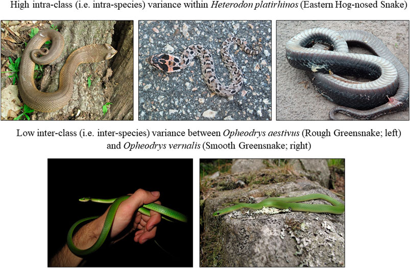
FIGURE 1. Examples of high intra-class (i.e., intra-species) and low inter-class (i.e., inter-species) variance among snake images. All photos by Andrew M. Durso (CC-BY).
Algorithm Development Challenge
We used the platform AICrowd, which takes a collaborative approach to the development of algorithms, by inviting data science experts and enthusiasts to collaboratively solve real-world problems by participating in challenges in which the solutions are automatically evaluated in real-time. On January 21, 2019, we launched a “Snake Species Identification Challenge”. The first round lasted until May 31, 2019, and the second round (during which live code collection was implemented) until July 31, 2019. We offered prizes as incentives for the best algorithms submitted (a travel grant and co-authorship on this manuscript). A total of 24 participants made 356 submissions, resulting in five algorithms with an F1 ≥ 0.75 and a top score of F1 = 0.861 with a log-loss = 0.53.
Test Datasets
We evaluated the identification accuracy of the submitted algorithms using two test datasets (TD2 and TD3), both distinct from the training dataset described above and its subset (TD1) used for pre-submission testing by the challenge participant (Table 1). One (TD2) was made up of 42,688 photos of the same 45 species of snakes submitted to iNaturalist between January 1, 2019 and September 2, 2019 (after the beginning of our challenge; range 23–6,228 photos per class). The other (TD3) was made up of 248 undisclosed images from 27 classes (1–10 images per class) that were collected from private individuals. Many of the images in this dataset were purposefully chosen to be as difficult to identify as possible—e.g., low resolution, out of focus, with the snake filling only a small part of the frame, and/or obscured by vegetation. The identity of species in these images was confirmed by a herpetologist (A. Durso) and they were used in a citizen science challenge where they were presented to participants recruited from online snake identification communities (largely Facebook snake identification groups and iNaturalist) who suggested species identifications, resulting in 68–157 labels of 5–47 classes per image (Durso et al., 2021). We further subdivided TD3 to take all 45 classes into account (TD3a; for fairer comparison with human labeling, because humans were allowed to choose any of the >3,700 snake species classes from a list), and to evaluate only the 27 classes in common (TD3b; for fairer comparison with TD2).
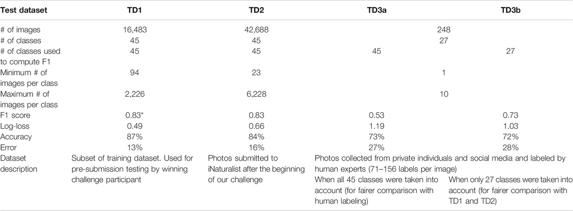
TABLE 1. Summary of three test datasets used to evaluate identification accuracy of top algorithm. Performance of humans on TD3a yielded F1 = 0.76, accuracy = 68%, error = 32%, and on TD3b F1 = 0.79, accuracy = 79%, error = 21%. *The winning F1 high score of 0.861 reported above comes from a different randomly generated subset, not reported here in detail.
Winning Algorithm
Applying large, deep convolutional neural networks for image classification is a well-studied problem (Krizhevsky et al., 2012; Huang et al., 2017; He et al., 2019). The top algorithm made use of incremental learning in neural networks and incorporated elements of EfficientNet from Google Brain (Tan and Le, 2019), a pre-trained network from ILSRVC (Russakovsky et al., 2015), discriminative learning, cyclic learning rates and automated image object detection (Figure 2).
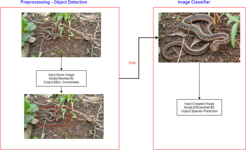
FIGURE 2. Diagram of overall pipeline including both object detection and classification.
As shown by Kornblith et al. (2019), models trained on a standard data distribution generalize better than the models trained from scratch. The ImageNet Large Scale Visual Recognition Competition (ILSVRC) dataset is the most widely used dataset for benchmarking image classifiers, comprising 1.2 million images classified into 1,000 different classes. The winning solution applied incremental learning on a pre-trained EfficientNet network. Specifically, this involved retaining what the model has learned from the ILSVRC dataset and performing incremental learning on the snake species domain. The final layer from the pretrained network was removed and replaced with a domain specific head, a fully connected layer of size 45, each representing the probability of the snakes being in a particular class. Discriminative learning strategy was also used to train the network. Specifically, different layers of the network are responsible for capturing different types of information (Yosinski et al., 2014) and discriminative learning allows us to set the rate at which different components of the network learn. The initial layers are trained at much lower learning rates to inhibit the loss of learned information while the final layers are trained at higher learning rates. A general update to model parameters ⍵ at time step t looks like:
where λ denotes the learning rate and ΔωJ(ω) denotes the gradient with respect to the model’s objective function. In discriminative learning, the model is split into N components, {ω1, ω2,…ωN} where ωn contains the layers of the nth component of the model. Each component can have any number of layers. An update to model parameters then becomes:
where λn denotes the learning rate of the nth component of the model.
In the first attempt, an image classifier was trained using progressive resizing, starting with Densenet121 (Huang et al., 2017) architecture with a modified focal loss function (Lin et al., 2017) for multi-class image classification and resizing image sizes from (256,256) → (384,384) → (512,512) → (768,768) → (1024,1024). Using this as an initial method, the scores plateaued at F1 ∼0.67. As an alternative, a new image classifier was trained from scratch using Resnet152 with modifications as suggested by Bag of tricks (He et al., 2019) called XResnet152 (152 indicating the number of layers). This time, the scores plateaued at ∼0.75.
Adding a preprocessing pipeline to predict the four coordinates of the corners of the box bounding the snake itself (which may be any size and shape and in any orientation within the image, against any background) and crop the images, as well as handling orientation variance, was accomplished by annotating 30–32 images from the training dataset from each species category using an annotation tool called sloth (https://github.com/cvhciKIT/sloth/). Pipelining preprocessing and training with XResnet architecture together increases the accuracy to 0.78–0.79.
Finally, a pre-trained EfficientNet (Tan and Le, 2019), which balances the depth of the architecture, the width of the architecture [Mobile Inverted Bottleneck Convolutional block (Sandler et al., 2018) with Swish activation function (Ramachandran et al., 2017)], image resolution and uses appropriate drop-outs, was fine-tuned using preprocessed images. Our winning algorithm carefully tuned hyper-parameters for EfficientNetB0 (Supplementary Figure S1) and used the exact same parameters for EfficientNetB5 (Supplementary Figure S2), although it is expected that higher accuracy is possible using the more time-intensive checkpoints/pretrained weights for B6 and B7 and by fine-tuning EfficientNetB6/B7 pre-trained on ImageNet. Some of the best hyperparameters required to train the EfficientNet include: 1) taking off the final layer of EfficientNetB5 and adding a single layer of size 45 (fastai adds an additional layer, which was not optimal for this case); 2) using the LabelSmoothingCrossEntropy Loss function; 3) using RMSProp optimizer with centered = True; 4) setting bn_wd = false, no batchnorm weight decay; 5) training with discriminative learning; 6) using resize method as SQUISH and not CROP; 7) no mixup augmentation; 8) use rotation augmentation (rotating the training image from −90 to +90 with a probability of 0.8) as shown in Figure 3; and 9) training on 95% of the dataset instead of the 80–20 split.
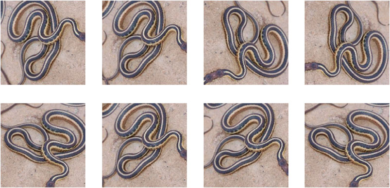
FIGURE 3. Rotation augmentation of a sample image.
The network was trained on a single Tesla V100 GPU with a batch size of 8 for 12 epochs. The learning rate schedule is shown in Figure 4. Each epoch took approximately 55 min for completion. Other approaches that were tried but did not work include building a genus classifier to predict the snake genus and then the snake species within genus, focal loss, a weighted sampler/oversampling and batch accumulation (updates happen every n epochs, where n > 1).
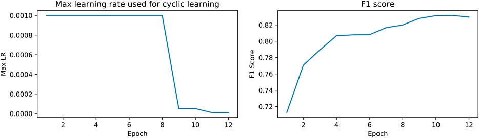
FIGURE 4. Max learning rate schedule and corresponding F1 score.
The algorithm and readme are available at https://github.com/GokulEpiphany/contests-final-code/tree/master/aicrowd-snake-species.
Results and Discussion
First Test Dataset (TD1)
A confusion matrix generated using TD1 (a withheld subset of the training data) is shown in Figure 5. Species classification accuracy ranged from 96% for Rhinocheilus lecontei to 35% for Pantherophis spiloides. Interclass confusion was 0 for 1332/2025 (66%) of class pairs. F1 for this dataset was 0.83 and log-loss was 0.49 (Table 1). Relatively high confusion remains between some similar species pairs. In particular, the three putative species of the Pantherophis obsoletus (North American ratsnake) complex (Burbrink, 2001) were frequently confused (Table 2).
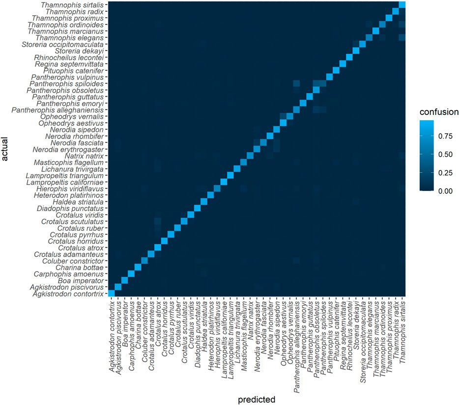
FIGURE 5. Confusion matrix for the top algorithm, using TD1 and an 80-20 split (subset of training data). The final model was trained on a 95-5 split. For an interactive version see https://chart-studio.plotly.com/∼amdurso/1/#/.
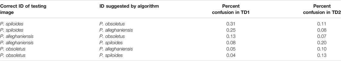
TABLE 2. Confusion among putative species of the North American Ratsnake (Pantherophis obsoletus) complex (Burbrink, 2001) in TD1 and TD2 (the three putative species were combined in TD3). The identity of species in training and testing data was done exclusively from photos, taking into account the geographic location but not information from scale counts or DNA.
Among species that are uncontroversially delimited, there were seven species pairs with confusion >10% in TD1 (max = 26% for Thamnophis elegans as Thamnophis sirtalis; Table 3). The highest confusion between species in different genera was Natrix natrix as Thamnophis sirtalis (7%). Although these genera are found in different hemispheres, confusion among co-occurring genera exceeded 5% for Coluber constrictor as Pantherophis alleghaniensis (6%) and for Lichanura trivirgata as Crotalus atrox (5%). The last is particularly troubling because Lichanura trivirgata is a harmless boid whereas Crotalus atrox is a large, potentially dangerous rattlesnake; these species co-occur in the Sonoran Desert and are probably frequently photographed against similar backgrounds (Rorabaugh, 2008). Other frequently confused intergeneric and intercontinental species pairs were Hierophis viridiflavus (as Thamnophis sirtalis and as Masticophis flagellum; both 5%) and Natrix natrix (as Nerodia rhombifer; 5%).
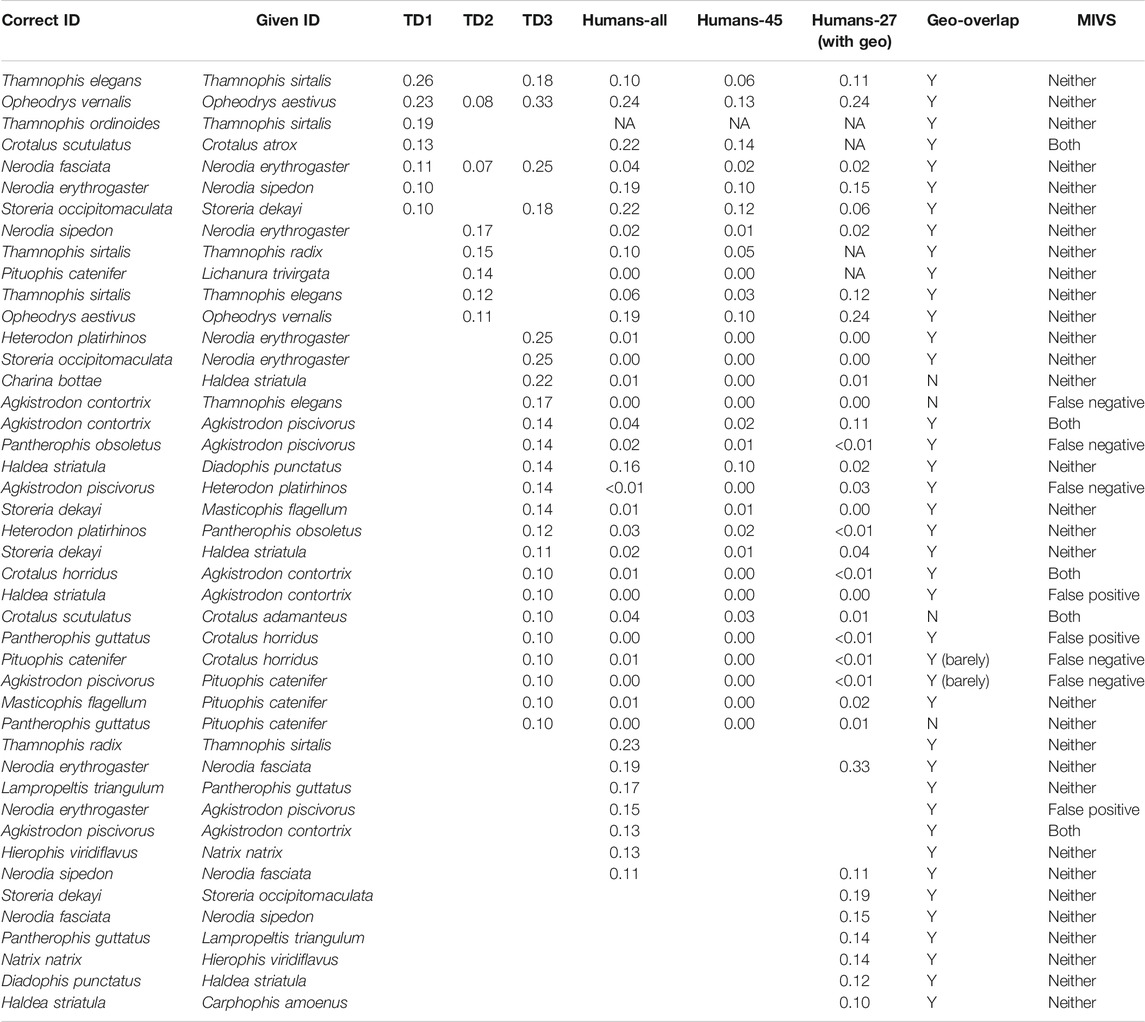
TABLE 3. Confusion among species that are uncontroversially delimited, showing only pairs with confusion >10% in TD1, TD2, or TD3 (by algorithm or humans). MIVS = medically important venomous snakes.
Second Test Dataset (TD2)
In our second test dataset (TD2, containing images for all 45 classes taken from iNaturalist after the challenge had started), F1 = 0.83 and log-loss = 0.66 (Table 1). Species classification accuracy ranged from 97% for Pantherophis guttatus to 61% for Pantherophis alleghaniensis. Interclass confusion was 0 for 1118/2025 (55%) of class pairs. Again, the most frequently confused were members of the Pantherophis obsoletus complex (Table 2).
Among species that are uncontroversially delimited, there were five species pairs with confusion >10% in TD2 (Table 3). The highest confusion among species in different genera was Pituophis catenifer as Lichanura trivirgata (14%). As in TD1, Pituophis catenifer and Lichanura trivirgata co-occur in the Sonoran Desert and are probably frequently photographed against similar backgrounds, although they are dissimilar in appearance. The highest confusion among species/genera found on different continents in TD2 was Coluber constrictor as Hierophis viridiflavus (7%). Both are slender, fast-moving, diurnal snakes, and the subspecies H. v. carbonarius (sometimes considered a full species; Mezzasalma et al., 2015) from Italy is all black, making it very similar in appearance to adult forms of C. c. constrictor and C. c. priapus in the eastern United States.
Species pairs that were commonly confused in both TD1 and TD2 were Coluber constrictor as Pantherophis alleghaniensis (9%; also confused 6% of the time in TD1), Opheodrys vernalis as Opheodrys aestivus (8%; also confused 23% of the time in TD1) and Nerodia fasciata as Nerodia erythrogaster (7%; also confused 11% of the time in TD1).
Third Test Dataset (TD3)
In our third test dataset (TD3, containing images for just 27 of the 45 classes hand-selected from other sources), F1 = 0.53 and log-loss = 1.19 when all 45 classes were considered (TD3a), and F1 = 0.73 and log-loss = 1.03 when only the 27 classes relevant to both datasets were considered (TD3b; Table 1). In TD3, species classification accuracy ranged from 100% for 10 species to 25% for Nerodia erythrogaster. Interclass confusion was 0 for 648/729 (89%) of class pairs.
Among species that are uncontroversially delimited, there were 23 pairs with confusion >10% in TD3 (Table 3), including two that were also commonly confused in both TD1 and TD2 (Opheodrys vernalis as Opheodrys aestivus; 33% in TD3, 8% in TD2, 23% in TD1; Nerodia fasciata as Nerodia erythrogaster; 25% in TD3, 7% in TD2, 11% in TD1) and one that was also commonly confused in TD1 (Storeria occipitomaculata as Storeria dekayi; 18% in TD3 vs 10% in TD1). The highest confusion among species in different genera was 25% for two species pairs (Heterodon platirhinos as Nerodia erythrogaster and Storeria occipitomaculata as Nerodia erythrogaster) and the highest confusion among species that do not occur in sympatry was Charina bottae as Haldea striatula (22%) (see Table 3 for detailed comparison).
When we asked human experts to identify the same images, they performed slightly better overall (species-level F1 = 0.76 for 45 classes, F1 = 0.79 for 27 classes; Table 1), with significant variation among species (Durso et al., 2021). Humans correctly labeled the 248 images in TD3 at the species level 53% of the time (N = 26,672), although this varied by species from 83% for Agkistrodon contortrix to 30% for Nerodia erythrogaster.
Thirteen species pairs were confused >10% of the time by humans, five of which also exceeded 10% in at least one of the three test datasets and all but one of which involved species that were also commonly confused by the algorithm (Table 3). Among frequently confused species from TD1 and TD2, humans only had the option to select “Black Ratsnake complex”, so there were no opportunities for confusion among the three putative species (P. obsoletus, P. alleghaniensis, and P. spiloides). Recent changes to taxonomy resulting in unfamiliar names may also hinder human experts’ ability to identify snakes (Carrasco et al., 2016), which could partially explain why e.g., Haldea striatula (more widely known as Virginia striatula in recent decades; Powell et al., 1994; McVay and Carstens, 2013) was correctly identified to the species level by humans only 31% of the time, or why Lampropeltis californiae was identified as Lampropeltis getula (the species from which it was split; Pyron and Burbrink, 2009) 23% of the time.
Some of the images in this dataset were purposefully chosen to be as difficult to identify as possible—e.g., low resolution, out of focus, with the snake filling only a small part of the frame, and/or obscured by vegetation. Stallkamp et al. (2012) also found that humans were superior to computer vision at identifying images with visual artifacts (e.g., traffic signs with graffiti or a lot of glare).
Patterns Across Test Datasets
Because TD1 is a withheld subset of the training data and TD2 was collected using very similar methods, their similarity to the training data is very high. In contrast, TD3 images were selected to be representative of realistic difficult cases. We suggest that this explains the drop in performance for TD3, because image datasets often have built-in biases that are difficult to pinpoint and tests of cross-dataset generalization are rare (Dollár et al., 2009) but generally show that algorithms perform better on their “native” test sets (here, TD1 and TD2) than on entirely novel testing datasets (here, TD3) (Torralba and Efros, 2011). Of the types of bias discussed by Torralba and Efros (2011), we took pains to eliminate or account for label bias (inconsistent category definitions across datasets) by using the same snake taxonomy consistently and paying particular attention to cases where instability in species definitions might sabotage identification predictions. Our dataset is probably most susceptible to capture bias (photographers tending to take pictures of objects in similar ways) and perhaps to selection bias (source datasets that prefer particular kinds of images). Future studies should address the trade-offs between effort and information gain that may result from attempting to acquire images taken from particular angles or of particular anatomical features (Rzanny et al., 2017; Rzanny et al., 2019).
Species that were consistently identified with high accuracy across the three test datasets include:
• Boa constrictor (Boa Constrictor) had the highest average F1 (Figure 6), the fourth highest average recall (Supplementary Figure S3) and the third highest average precision (Supplementary Figure S4), as well as among the lowest average false positive (6.3 ± 2.7%) and average false negative (4.0 ± 2.3%) rates, across the three test datasets. These large, iconic snakes are easily recognized and there are few similar species with which they could be confused, especially within their range. Potential for confusion with large Python and Eunectes (anaconda) species exists. Several recent studies have suggested that Boa constrictor is likely to be a species complex containing as many as nine species (see Reynolds and Henderson, 2018 for a summary).
• Lichanura trivirgata (Rosy Boa) had the highest average recall (true positive rate) across three test datasets (95.7 ± 3.3%; Supplementary Figure S3). These short, stocky snakes are native to southwestern North America and are quite distinct in shape within their range; they most closely resemble species of Eryx (sand boas) from the Middle East and south Asia, which were not represented in our dataset. However, this species had only moderate average precision; in particular, it was confused with other species that are often photographed against similar backgrounds (e.g., Crotalus atrox in TD1, Pituophis catenifer in TD2).
• Pantherophis guttatus (Red Cornsnake) had the highest average precision (positive predictive value) across three test datasets (98.1 ± 1.6%; Supplementary Figure S4). These snakes are native to the southeastern United States and are common in the pet trade. Our dataset may contain more images of captive P. guttatus than any other species, although we attempted to filter these out. The impact of incorporating images of captive snakes in training data intended to be used for identifying wild snakes is not well understood, but the plethora of designer color morphs that have been produced by captive breeding of cornsnakes probably influences the algorithm’s ability to recognize this species.
• Rhinocheilus lecontei (Long-nosed Snake) had the second-highest average precision (97.2 ± 0.6%; Supplementary Figure S4) and second-highest average F1 (94.3 ± 4.4%; Figure 6) and relatively high average recall (91.8 ± 8.8%; Supplementary Figure S3). This species is placed in its own monotypic genus, but it is part of a mimicry complex including its non-venomous relatives in the genera Lampropeltis (kingsnakes and milksnakes) and Cemophora (Scarletsnake) (as well as many other mimics in Central and South America) and their dangerous models in the genera Micrurus and Micruroides (coralsnakes) (Savage and Slowinski, 1992; Davis Rabosky et al., 2016). The mimicry of Rhinocheilus is not as exact as that of many species, but addition of similar species to the training dataset would certainly increase the computer vision algorithm’s confusion of species that currently have low confusion (e.g., Rhinocheilus lecontei could be confused with several Lampropeltis species on which the algorithm is not currently trained; Shannon and Humphery, 1963; Manier, 2004).
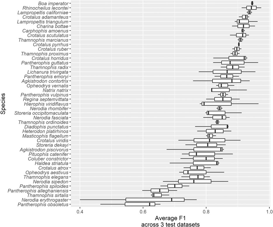
FIGURE 6. Average F1 across three test datasets for all 45 species.
Species groups that were consistently confused across the three test datasets include:
• The two greensnake species in the genus Opheodrys: O. aestivus (Rough Greensnake) and O. vernalis (Smooth Greensnake; formerly placed in the genus Liochlorophis). Average F1 across test datasets and species was 81.4 ± 6.9% (Figure 6). These sister taxa are similar in their bright green color, slender bodies, small size, and gestalt; they differ anatomically mainly in that O. aestivus has keeled dorsal scales in 17 rows, whereas O. vernalis has smooth dorsal scales in 15 rows, subtle characteristics that may not be discernible in photos that are not taken at close range. Habitat and geographic range are also useful in distinguishing them (Ernst and Ernst, 2003). Although telling Opheodrys species apart is not likely to ever be clinically or epidemiologically useful, there are many other solid bright green snakes lacking easy-to-distinguish features in Africa, including non-venomous Philothamnus and Hapsidophrys as well as some highly dangerous Dispholidus (boomslangs) and Dendroaspis (mambas); see e.g., Broadley and Fitzsimons (1983) and Chippaux and Jackson (2019).
• Watersnake species in the genus Nerodia, especially N. fasciata (Banded Watersnake), N. erythrogaster (Plain-bellied Watersnake), and N. sipedon (Northern Watersnake). Average F1 across test datasets and species was 75.9 ± 13.8% (Figure 6). All species in this genus are brown and blotched, but they can be reliably identified using differences in dorsal and ventral pattern, scalation, and range (Gibbons and Dorcas, 2004). However, a photo that does not show the posterior part of the body (where the alignment of blotches is an important characteristic) or the venter (which is commonly concealed in photos) may represent a challenging ID for a human, especially in the absence of geographic information. Again, telling Nerodia species apart is not likely to ever be clinically or epidemiologically useful, but they are often confused with Agkistrodon piscivorus (Cottonmouth) by laypeople, and worldwide there are many brown, blotched snakes that look similar and may mimic one another (e.g., Gans, 1961; Gans and Latifi, 1973; Kroon, 1975) as well as species that can only be reliably identified if a particular part of the body is visible. Although k nearest-neighbor techniques have been used to semi-automatically classify snakes from images based on their anatomical features (James et al., 2014; James, 2017), these involve laborious and detail-oriented curation of feature databases that should include every possible snake species, a gargantuan task that, although useful for snake taxonomy, would require constant updating (see e.g., Meirte, 1992 for an example of a dichotomous key based on scale characters used by humans). Semi-supervised self-learning algorithms such as ours do not require such databases and represent a more efficient, modern approach to image classification, and wisely chosen pipelines of local feature detection, extraction, encoding, fusion and pooling allows for high accuracy in species classification in groups even more biodiverse than snakes (Seeland et al., 2017).
• The Pantherophis obsoletus (North American Ratsnake) species complex (Burbrink, 2001). All three putative species were among the bottom five in terms of F1, never exceeding 74.6% (average across all test datasets and all three species = 63.5 ± 9.0%; Figure 6), as well as the bottom five for recall (Supplementary Figure S3) and the bottom 10 for precision (Supplementary Figure S4). The practice of delimiting species largely on the basis of mitochondrial DNA haplotypes (or combining these with nuclear genetic data sets with little phylogenetic signal) has been criticized for being sensitive to gaps in data caused by limited geographic sampling, which are artifactually detected as species boundaries by clustering algorithms used in the Multispecies Coalescent Model for species delimitation, leading to “over-splitting” of widely distributed species and assignment of species names to “slices” of continuous geographic clines that are not evolving independently (Hillis, 2019; Chambers and Hillis, 2020) and may not be morphologically diagnosable (Guyer et al., 2019; Freitas et al., 2020; Mason et al., 2020). Here we show that computer vision algorithms have low power to distinguish at least some putative species that have been delineated primarily using molecular methods and where training data were identified exclusively from photos, presumably primarily based on geographic location (Table 2). Although Burbrink (2001) provided multivariate analyses of 67 mensural and meristic characters (mostly related to scalation) that corresponded to mitochondrial lineages, the most obvious color and pattern variants intergrade with one another over large areas and correspond to former subspecies designations rather than Burbrink’s species concepts (recently refined to better match phenotypic variation; Burbrink et al., 2020; Hillis and Wüster, 2021), and numerous color patterns can be found within each putative species, especially P. alleghaniensis (not to mention the marked ontogenetic change characteristic of all three, wherein juveniles are most similar to adult P. spiloides from the southern portion of their range). This, combined with the widely overlapping distribution between P. alleghaniensis and P. spiloides, makes the three putative species nearly impossible for both humans and AI to differentiate in the absence of information about geographic location. We also note that training images in this species complex are probably more likely to be mis-labeled due to confusion over how to differentiate the putative species.
• Other taxa used in our dataset that may be susceptible to the same problem as the Pantherophis obsoletus complex include Lampropeltis californiae and Lampropeltis triangulum (both members of widespread species complexes that were formerly treated as a single species, of which only one putative species per species complex is present in our dataset; Pyron and Burbrink, 2009; Ruane et al., 2014) and Agkistrodon contortrix and Agkistrodon piscivorus (both of which have recently been split into two putative species along former subspecies lines but were treated as single species in our dataset; Burbrink and Guiher, 2015) as well as Boa constrictor (the taxonomy of which is even less settled; see Reynolds and Henderson, 2018 for a summary). See Appendix I for a full list of species in our dataset together with notes on recent taxonomic changes that may affect their diagnosability based purely on a photograph.
Of the 44 species pairs that exceeded 10% confusion on any test dataset (including human IDs), only four had no overlap in geographic range (two others had very little geographical overlap). Incorporating information on the geography has the potential to better discriminate these species pairs (Wittich et al., 2018), but in the most biodiverse regions of the world, >125 species of snakes may be sympatric within the same 50 × 50 km2 area (Roll et al., 2017).
Most commonly confused species pairs involved either two non-venomous or two medically important venomous species (hereafter “venomous”). Of the 44 species pairs that exceeded 10% confusion on any test dataset (including human IDs), 31 involved two non-venomous species and five involved two venomous species (Table 3). Only three of the 44 pairs were “false positives” (a non-venomous species identified as a venomous one) and only five were “false negatives” (a venomous species identified as a non-venomous one). Bites from all medically important venomous snakes in our dataset (all North American pit vipers) would be treated using the same antivenom (Dart et al., 2001; Gerardo et al., 2017) but this would not be as straightforward in other regions of the world (e.g., Bitis vs. Echis in sub-Saharan Africa) and indeed is becoming more complex in North America as new products enter the market (Bush et al., 2015; Cocchio et al., 2020).
Finally, we found that among the 163 confused species pairs in TD3, the algorithm suggested the species with more images in the training dataset as the identity of the species with fewer images far more often (78% of pairs) than the other way around. For example, Opheodrys vernalis (1,471 training images) was misidentified as O. aestivus (3,312 training images) 33% of the time in TD3, but O. aestivus was never misidentified as O. vernalis in TD3, even though humans confused these two species nearly equally often in both directions (12 and 10%). Humans were marginally more likely to suggest the species from a given pair with more images even when it was incorrect (65 of 118 confused pairs; 55%), but the effect was much more pronounced for AI than for humans (Figure 7). For the algorithm, this is probably due to the imbalance in the amount of training data among classes; for humans, we suggest that it may be caused by the same processes that lead a species to be represented by fewer images in the training data: smaller geographic ranges or lower probabilities of encounter, leading to less familiarity with the rarer species.
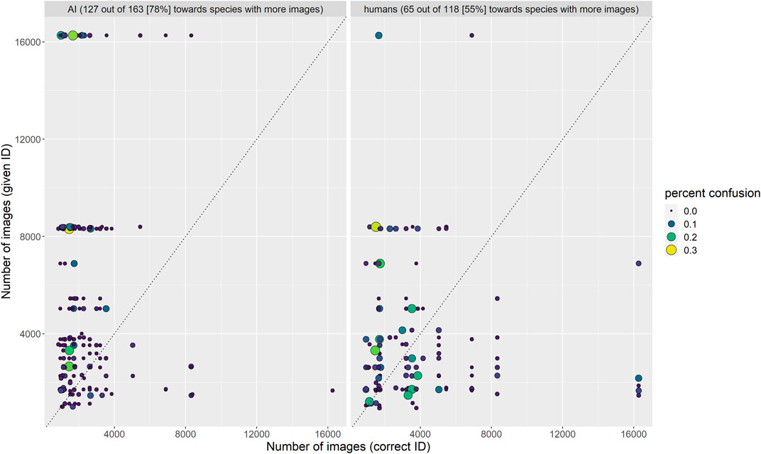
FIGURE 7. Frequency with which confused species pairs were incorrectly identified by algorithm and humans. The algorithm erred on the side of the species with more training images much more often than humans.
Long-tailed distributions are commonplace and have been called “the enemy of machine learning” (Bengio, 2015). As we enlarge our dataset to include more of the snake species with few images per species (i.e., those in the “long-tailed” part of the distribution), we must pay particular attention to rare species. Previous studies on faces (Zhang et al., 2017) and scene parsing (Yang et al., 2014) suggest approaches including rare class enrichment (Yang et al., 2014) and clustering objects into visually similar hierarchical groups (in our case, snake genera or families) (Ouyang et al., 2016).
Conclusion
It is extremely important to keep in mind that the 45 species that the algorithm was trained on were selected solely based on the quantity of photos available, and no effort was made to include all possible similar or closely related species. Additionally, human experts could suggest any of the >3,700 species of snakes as an identification, and species other than the 45 used to train the computer vision algorithm were suggested (always incorrectly) 76% of the time overall (min = 7% for Agkistrodon contortrix, max = 50% for Hierophis viridiflavus). The average number of species that a human expert knows well (analogous to the number of classes that an algorithm is trained on) is hard to estimate. The grand total number of species suggested by all 580 human experts was 457, but the mean ± SD per person was just 35 ± 18 (min = 18, max = 61; Durso et al., 2021). Another important difference was that human experts had access to the geographic location of each image, whereas the computer vision algorithm does not yet incorporate this information. To further explore these issues, additional rounds of our AICrowd challenge, including more photos, additional species classes, and information on geography at the continent and country level have allowed us to assess the generality of patterns discovered here (Bloch et al., 2020; Moorthy, 2020; Picek et al., 2020). Future directions include finer image segmentation (e.g., the head, body, and tail of the snake) and hierarchical (e.g., genus level) classification, which might work better when more species from larger genera are included.
The accuracy of our algorithm at predicting species-level identity ranged from 72 to 87% depending on the test dataset (Table 1), which is comparable to that for other taxa (see Table 2 of Weinstein, 2018). A promising future awaits snake identification as AI begins to compliment static photos, diagrams, and audio-visual media, interactive multiple-access keys, species checklists that can be customized to particular locations, dynamic range maps, and online communities in which people share species observations and identifications in “next-generation field guides” (Farnsworth et al., 2013). In particular, we emphasize the need to keep “humans in the loop” in order to validate labels in training datasets as well as AI predictions, particularly for healthcare applications (Holzinger, 2016; Holzinger et al., 2016).
We suggest that AI could play a larger role in disease surveillance and disease ecology (Hosny and Aerts, 2019; Pandit and Han, 2020), particularly as a widely available rapid diagnostic test for improving detailed epidemiological reporting and mapping of snakebite, as well as clinical management (Ruiz De Castañeda et al., 2019). However, a more complete understanding of how AI and humans perform and interact with one another when identifying species classes from photos with regard to image quality, inter-species similarity, and prior information about geographic location is essential before this technology could aid in the snakebite crisis. Critically, increasing data availability in regions of high snake diversity and snakebite prevalence is a pre-requisite for applying data-hungry algorithms to the regions of the world where they are most needed.
Data Availability Statement
The raw data supporting the conclusions of this article will be made available by the authors, without undue reservation.
Ethics Statement
Ethical review and approval was not required for the study on human participants in accordance with the local legislation and institutional requirements. The patients/participants provided their written informed consent to participate in this study.
Author Contributions
AMD collected the training and testing datasets, made comparisons among testing datasets, and wrote most of the article. GM created the winning algorithm and tested it on TD1, and wrote the remainder of the article. SPM hosted the challenge on AICrowd and tested the winning algorithm on TD2 and TD3. IB supported collection of the training and testing datasets and contributed to writing and revising the article. MS hosted the challenge on AICrowd and contributed to writing and revising the article. RRdC supported collection of the training and testing datasets and contributed to writing and revising the article.
Funding
This research was supported by the Fondation privée des Hôpitaux Universitaires de Genève (award QS04-20). A. Flahault and the Fondation Louis-Jeantet at the Institute of Global Health, and F. Chappuis at the Department of Community Health and Medicine of the University of Geneva supported R. Ruiz de Castañeda.
Conflict of Interest
Authors SM and MS are CEO and co-founders of AICrowd, which was used in the research and sponsors this call for manuscripts. Author GM was employed by the company Eloop Mobility Solutions.
The remaining authors declare that the research was conducted in the absence of any commercial or financial relationships that could be construed as a potential conflict of interest.
The handling editor declared a past co-authorship with one of the authors SH.
Acknowledgments
We thank all the participants in all rounds of the AICrowd Snake ID Challenge, and the users and admins of open citizen science initiatives (iNaturalist, HerpMapper, and Flickr) for their efforts building, curating, and sharing their image libraries. C. Montalcini supported the gathering of images and metadata. S. Khandelwal provided assistance testing and summarizing datasets. P. Uetz provided support for snake taxonomy through the Reptile database. D. Becker, C. Smith, and M. Pingleton provided support for and access to HerpMapper data. We thank the WHO-ITU Focus Group on AI for Health for their guidance and technical support during the development of this benchmark.
Supplementary Material
The Supplementary Material for this article can be found online at: https://www.frontiersin.org/articles/10.3389/frai.2021.582110/full#supplementary-material.
References
Akcali, C. K., and Pfennig, D. W. (2017). Geographic variation in mimetic precision among different species of coral snake mimics. J. Evol. Biol. 30, 1420–1428. doi:10.1111/jeb.13094
Amir, A., Zahri, N. A. H., Yaakob, N., and Ahmad, R. B. (2016). “Image classification for snake species using machine learning techniques,” in CIIS 2016: computational intelligence in information systems, Brunei, November 18–20, 2016. Editors S. Phon-Amnuaisuk, T. W. Au, and S. Omar (Cham, Switzerland: Springer), 52–59. doi:10.1007/978-3-319-48517-1_5
Barbu, A., Mayo, D., Alverio, J., Luo, W., Wang, C., Gutfreund, D., et al. (2019). “Objectnet: a large-scale bias-controlled dataset for pushing the limits of object recognition models,” in Advances in neural information processing systems (NIPS 2019), Vancouver, Canada, December 8–14, 2019. Editors H. Wallach, H. Larochelle, A. Beygelzimer, F. d'Alché-Buc, E. Fox, and R. Garnett (Red Hook, NY: Curran Associates, Inc.), Vol. 32, 9453–9463.
Barry, J. (2016). “Identifying biodiversity using citizen science and computer vision: introducing Visipedia,” in TDWG 2016 annual conference, Santa Clara de San Carlos, Costa Rica, December 5–9, 2016 (St. Louis, MI: Missouri Botanical Garden). Available at: https://mbgocs.mobot.org/index.php/tdwg/tdwg2016/paper/view/1112/.
Bengio, S. (2015). “The battle against the long tail,” in Workshop on big data and statistical machine learning (Toronto, ON: Fields Institute for Research in Mathematical Sciences).
Bloch, L., Boketta, A., Keibel, C., Mense, E., Michailutschenko, A., Pelka, O., et al. (2020). “Combination of image and location information for snake species identification using object detection and efficientnets,” in CLEF: conference and labs of the evaluation forum, Thessaloniki, Greece.
Bolon, I., Durso, A. M., Botero Mesa, S., Ray, N., Alcoba, G., Chappuis, F., et al. (2020). Identifying the snake: first scoping review on practices of communities and healthcare providers confronted with snakebite across the world. PLoS One 15, e0229989. doi:10.1371/journal.pone.0229989
Broadley, D. G., and Fitzsimons, V. F. M. (1983). Fitzsimons’ snakes of southern Africa. Johannesburg: Delta Books.
Burbrink F. T., Gehara M., McKelvy A. D., and Myers E. A. (2020). Resolving spatial complexities of hybridization in the context of the gray zone of speciation in North American ratsnakes (Pantherophis obsoletus complex). Evolution 75, 260–277.
Burbrink, F. T., and Guiher, T. J. (2015). Considering gene flow when using coalescent methods to delimit lineages of North American pitvipers of the genus Agkistrodon. Zool. J. Linn. Soc. 173, 505–526. doi:10.1111/zoj.12211
Burbrink, F. T. (2001). Systematics of the eastern ratsnake complex (Elaphe obsoleta). Herpetol. Monogr. 15, 1–53. doi:10.2307/1467037
Bush, S. P., Ruha, A.-M., Seifert, S. A., Morgan, D. L., Lewis, B. J., Arnold, T. C., et al. (2015). Comparison of F(ab')(2) versus Fab antivenom for pit viper envenomation: a prospective, blinded, multicenter, randomized clinical trial. Clin. Toxicol. 53, 37–45. doi:10.3109/15563650.2014.974263
Carrasco, P. A., Venegas, P. J., Chaparro, J. C., and Scrocchi, G. J. (2016). Nomenclatural instability in the venomous snakes of the Bothrops complex: implications in toxinology and public health. Toxicon 119, 122–128. doi:10.1016/j.toxicon.2016.05.014
Chambers, E. A., and Hillis, D. M. (2020). The multispecies coalescent over-splits species in the case of geographically widespread taxa. Syst. Biol. 69, 184–193. doi:10.1093/sysbio/syz042
Chippaux, J.-P., and Jackson, K. (2019). Snakes of central and western Africa. Baltimore: Johns Hopkins University Press.
Cocchio, C., Johnson, J., and Clifton, S. (2020). Review of North American pit viper antivenoms. Am. J. Health Syst. Pharm. 77, 175–187. doi:10.1093/ajhp/zxz278
Cox, C. L., Davis Rabosky, A. R., Reyes-Velasco, J., Ponce-Campos, P., Smith, E. N., Flores-Villela, O., et al. (2012). Molecular systematics of the genus Sonora (Squamata: Colubridae) in central and western Mexico. Syst. Biodivers. 10, 93–108. doi:10.1080/14772000.2012.666293
Dart, R. C., Seifert, S. A., Boyer, L. V., Clark, R. F., Hall, E., Mckinney, P., et al. (2001). A randomized multicenter trial of crotalinae polyvalent immune Fab (ovine) antivenom for the treatment for crotaline snakebite in the United States. Arch. Intern. Med. 161, 2030–2036. doi:10.1001/archinte.161.16.2030
Davis Rabosky, A. R., Cox, C. L., Rabosky, D. L., Title, P. O., Holmes, I. A., Feldman, A., et al. (2016). Coral snakes predict the evolution of mimicry across New World snakes. Nat. Commun. 7, 1–9. doi:10.1038/ncomms11484
Dollár, P., Wojek, C., Schiele, B., and Perona, P. (2009). “Pedestrian detection: a benchmark,” in Proceedings of the IEEE conference on computer vision and pattern recognition (CVPR), Miami, FL, June 20–25, 2009 (IEEE), 304–311.
Durso, A. M., Bolon, I., Kleinhesselink, A. R., Mondardini, M. R., Fernandez-Marquez, J. L., Gutsche-Jones, F., et al. (2021). Crowdsourcing snake identification with online communities of professionals and avocational enthusiasts. R. Soc. Open Sci. 8, 201273. doi:10.1098/rsos.201273
Ernst, C. H., and Ernst, E. M. (2003). Snakes of the United States and Canada. Washington DC: Smithsonian Institution Press.
Farnsworth, E. J., Chu, M., Kress, W. J., Neill, A. K., Best, J. H., Pickering, J., et al. (2013). Next-generation field guides. BioScience 63, 891–899. doi:10.1525/bio.2013.63.11.8
Freitas, I., Ursenbacher, S., Mebert, K., Zinenko, O., Schweiger, S., Wüster, W., et al. (2020). Evaluating taxonomic inflation: towards evidence-based species delimitation in Eurasian vipers (Serpentes: Viperinae). Amphibia-Reptilia 41, 285–311. doi:10.1163/15685381-bja10007
Gans, C. (1961). Mimicry in procryptically colored snakes of the genus Dasypeltis. Evolution 15, 72–91. doi:10.2307/2405844
Gans, C., and Latifi, M. (1973). Another case of presumptive mimicry in snakes. Copeia 1973, 801–802. doi:10.2307/1443081
Garg, A., Leipe, D., and Uetz, P. (2019). The disconnect between DNA and species names: lessons from reptile species in the NCBI taxonomy database. Zootaxa 4706, 401–407. doi:10.11646/zootaxa.4706.3.1
Gaston, K. J., and O'Neill, M. A. (2004). Automated species identification: why not? Philos. Trans. R. Soc. Lond. Ser. B Biol. Sci. 359, 655–667. doi:10.1098/rstb.2003.1442
Gerardo, C. J., Quackenbush, E., Lewis, B., Rose, S. R., Greene, S., Toschlog, E. A., et al. (2017). The efficacy of crotalidae polyvalent immune fab (ovine) antivenom versus placebo plus optional rescue therapy on recovery from copperhead snake envenomation: a randomized, double-blind, placebo-controlled, clinical trial. Ann. Emerg. Med. 70, 233–244. doi:10.1016/j.annemergmed.2017.04.034
Gibbons, J. W., and Dorcas, M. E. (2004). North American watersnakes: a natural history. Norman, OK: University of Oklahoma Press.
Guyer, C., Folt, B., Hoffman, M., Stevenson, D., Goetz, S. M., Miller, M. A., et al. (2019). Patterns of head shape and scutellation in Drymarchon couperi (squamata: colubridae) reveal a single species. Zootaxa 4695, 168–174. doi:10.11646/zootaxa.4695.2.6
He, T., Zhang, Z., Zhang, H., Zhang, Z., Xie, J., and Li, M. (2019). “Bag of tricks for image classification with convolutional neural networks,” in Proceedings of the IEEE conference on computer vision and pattern recognition (CVPR), Long Beach, CA, June 16–20, 2019, 558–567.
Henke, S. E., Kahl, S. S., Wester, D. B., Perry, G., and Britton, D. (2019). Efficacy of an online native snake identification search engine for public use. Hum. Wildl. Interact. 13, 290–307. doi:10.26077/pg70-1r55
Hernández-Serna, A., and Jiménez-Segura, L. F. (2014). Automatic identification of species with neural networks. PeerJ 2, e563. doi:10.7717/peerj.563
Hillis, D. M. (2019). Species delimitation in herpetology. J. Herpetol. 53, 3–12. doi:10.1670/18-123
Hillis, D. M., and Wüster, W. (2021). Taxonomy and nomenclature of the Pantherophis obsoletus complex. Herpetol. Rev. 52, 51–52.
Hochmair, H. H., Scheffrahn, R. H., Basille, M., and Boone, M. (2020). Evaluating the data quality of iNaturalist termite records. PLoS One 15, e0226534. doi:10.1371/journal.pone.0226534
Holzinger, A. (2016). Interactive machine learning for health informatics: when do we need the human-in-the-loop? Brain Inform. 3, 119–131. doi:10.1007/s40708-016-0042-6
Holzinger, A., Valdez, A. C., and Ziefle, M. (2016). “Towards interactive recommender systems with the doctor-in-the-loop,” in Mensch und Computer 2016–workshopband, Aachen, Germany, September 4–7, 2016 (Leibniz, Austria: Schloss Dagstuhl: Leibniz Center for Informatics). doi:10.18420/muc2016-ws11-0001
Hosny, A., and Aerts, H. J. W. L. (2019). Artificial intelligence for global health. Science 366, 955–956. doi:10.1126/science.aay5189
Huang, G., Liu, Z., Van Der Maaten, L., and Weinberger, K. Q. (2017). “Densely connected convolutional networks,” in Proceedings of the IEEE conference on computer vision and pattern recognition (CVPR), Honolulu, HI, July 21–26, 2017, 4700–4708. doi:10.1109/CVPR.2017.243
James, A. P., Mathews, B., Sugathan, S., and Raveendran, D. K. (2014). Discriminative histogram taxonomy features for snake species identification. Hum. Centric Comput. Inf. Sci. 4, 3. doi:10.1186/s13673-014-0003-0
James, A. (2017). Snake classification from images. PeerJ Preprints 5, e2867v2861. doi:10.7287/peerj.preprints.2867v1
Joshi, P., Sarpale, D., Sapkal, R., and Rajput, A. (2018). A survey on snake species identification using image processing technique. Int. J. Comp. Appl. 181, 22–24. doi:10.5120/ijca2018918144
Joshi, P., Sarpale, D., Sapkal, R., and Rajput, A. (2019). “Snake species recognition using tensor flow machine learning algorithm & effective convey system,” in Proceedings of international conference on communication and information processing (ICCIP) 2019, Chongqing, China, November 15–17, 2019 (New York, NY: Association for Computing Machinery). Available at: https://ssrn.com/abstract=3421658.
Kornblith, S., Shlens, J., and Le, Q. V. (2019). “Do better imagenet models transfer better?,” in Proceedings of the IEEE conference on computer vision and pattern recognition (CVPR), Long Beach, CA, June 16–20, 2019, 2661–2671.
Krizhevsky, A., Sutskever, I., and Hinton, G. E. (2012). ImageNet classification with deep convolutional neural networks. Adv. Neural Inf. Process. Syst. 25, 1097–1105. doi:10.1145/3065386
Kroon, C. (1975). A possible Müllerian mimetic complex among snakes. Copeia 1975, 425–428. doi:10.2307/1443639
Langley, R. L. (2008). Animal bites and stings reported by United States poison control centers, 2001–2005. Wilderness Environ. Med. 19, 7–14. doi:10.1580/07-WEME-OR-111.1
Lin, T. Y., Goyal, P., Girshick, R., He, K., and Dollár, P. (2017). “Focal loss for dense object detection,” in Proceedings of the IEEE conference on computer vision and pattern recognition (CVPR), Honolulu, HI, July 21–26, 2017, 2980–2988.
Manier, M. K. (2004). Geographic variation in the long-nosed snake, Rhinocheilus lecontei (Colubridae): beyond the subspecies debate. Biol. J. Linn. Soc. 83, 65–85. doi:10.1111/j.1095-8312.2004.00373.x
Mason, N. A., Fletcher, N. K., Gill, B. A., Funk, W. C., and Zamudio, K. R. (2020). Coalescent-based species delimitation is sensitive to geographic sampling and isolation by distance. Syst. Biodivers. 18, 269–280. doi:10.1080/14772000.2020.1730475
McVay, J. D., and Carstens, B. (2013). Testing monophyly without well-supported gene trees: evidence from multi-locus nuclear data conflicts with existing taxonomy in the snake tribe Thamnophiini. Mol. Phylogenet. Evol. 68, 425–431. doi:10.1016/j.ympev.2013.04.028
Mezzasalma, M., Dall'asta, A., Loy, A., Cheylan, M., Lymberakis, P., Zuffi, M. A., et al. (2015). A sisters’ story: comparative phylogeography and taxonomy of Hierophis viridiflavus and H. gemonensis (Serpentes, Colubridae). Zool. Scr. 44, 495–508. doi:10.1111/zsc.12115
Moorthy, G. K. (2020). “Impact of pretrained networks for snake species classification,” in CLEF: conference and labs of the evaluation forum, Thessaloniki, Greece, September 25, 2020. Editors L. Cappellato, C. Eickhoff, N. Ferro and A. Névéol.
Ouyang, W., Wang, X., Zhang, C., and Yang, X. (2016). “Factors in finetuning deep model for object detection with long-tail distribution,” in Proceedings of the IEEE conference on computer vision and pattern recognition (CVPR), Las Vegas, NV, June 27–30, 2016, 864–873. doi:10.1109/CVPR.1997.609286
Pandit, P., and Han, B. A. (2020). Rise of machines in disease ecology. Bull. Ecol. Soc. Am. 101, 1–4. doi:10.1002/bes2.1625
Patel, A., Cheung, L., Khatod, N., Matijosaitiene, I., and Arteaga, A. (2020). Revealing the unknown: real-time recognition of Galápagos snake species using deep learning. Animals 10, 806. doi:10.3390/ani10050806
Picek, L., Bolon, I., Ruiz De Castañeda, R., and Durso, A. M. (2020). “Overview of the SnakeCLEF 2020: automatic snake species identification Challenge,” in CLEF: conference and Labs of the evaluation forum, Thessaloniki, Greece, September 25, 2020. Editors L. Cappellato, C. Eickhoff, N. Ferro and A. Névéol.
Powell, R., Collins, J. T., and Fish, L. D. (1994). Virginia striatula (Linnaeus). Rough earth snake. Cat. Am. Amphib. Reptil. 599, 1–6.
Pyron, R. A., and Burbrink, F. T. (2009). Systematics of the common kingsnake (Lampropeltis getula; Serpentes: Colubridae) and the burden of heritage in taxonomy. Zootaxa 2241, 22–32. doi:10.11646/zootaxa.2241.1.2
Ramachandran, P., Zoph, B., and Le, Q. V. (2017). Swish: a self-gated activation function. Available at: https://arxiv.org/abs/1710.05941v1.
Reynolds, R. G., and Henderson, R. W. (2018). Boas of the world (superfamily Booidae): a checklist with systematic, taxonomic, and conservation assessments. Bull. Mus. Comp. Zool. 162, 1–58. doi:10.3099/mcz48.1
Roll, U., Feldman, A., Novosolov, M., Allison, A., Bauer, A. M., Bernard, R., et al. (2017). The global distribution of tetrapods reveals a need for targeted reptile conservation. Nat. Ecol. Evol. 1, 1677–1682. doi:10.1038/s41559-017-0332-2
Rorabaugh, J. C. (2008). An introduction to the herpetofauna of mainland Sonora, México, with comments on conservation and management. J. Arizona-Nevada Acad. Sci. 40, 20–65. doi:10.2181/1533-6085(2008)40[20:aittho]2.0.co;2
Ruane, S., Bryson, R. W., Pyron, R. A., and Burbrink, F. T. (2014). Coalescent species delimitation in milksnakes (genus Lampropeltis) and impacts on phylogenetic comparative analyses. Syst. Biol. 63, 231–250. doi:10.1093/sysbio/syt099
Ruha, A. M., Kleinschmidt, K. C., Greene, S., Spyres, M. B., Brent, J., Wax, P., et al. (2017). The epidemiology, clinical course, and management of snakebites in the North American snakebite registry. J. Med. Toxicol. 13, 309–320. doi:10.1007/s13181-017-0633-5
Ruiz De Castañeda, R., Durso, A. M., Ray, N., Fernández, J. L., Williams, D. J., Alcoba, G., et al. (2019). Snakebite and snake identification: empowering neglected communities and health-care providers with AI. Lancet Digit. Health 1, e202–e203. doi:10.1016/s2589-7500(19)30086-x
Rusli, N. L. I., Amir, A., Zahri, N. A. H., and Ahmad, R. B. (2019). Snake species identification by using natural language processing. Indones. J. Electr. Eng. Comp. Sci. 13, 999–1006. doi:10.11591/ijeecs.v13.i3.pp999-1006
Russakovsky, O., Deng, J., Su, H., Krause, J., Satheesh, S., Ma, S., et al. (2015). ImageNet large scale visual recognition challenge. Int. J. Comp. Vis. 115, 211–252. doi:10.1007/s11263-015-0816-y
Rzanny, M., Mäder, P., Deggelmann, A., Chen, M., and Wäldchen, J. (2019). Flowers, leaves or both? How to obtain suitable images for automated plant identification. Plant Methods 15, 77. doi:10.1186/s13007-019-0462-4
Rzanny, M., Seeland, M., Wäldchen, J., and Mäder, P. (2017). Acquiring and preprocessing leaf images for automated plant identification: understanding the tradeoff between effort and information gain. Plant Methods 13, 97. doi:10.1186/s13007-017-0245-8
Sandler, M., Howard, A., Zhu, M., Zhmoginov, A., and Chen, L. C. (2018). “Mobilenetv2: inverted residuals and linear bottlenecks,” in Proceedings of the IEEE conference on computer vision and pattern recognition (CVPR), Salt Lake City, UT, June 18–22, 2018, 4510–4520.
Savage, J. M., and Slowinski, J. B. (1992). The coloration of the venomous coral snakes (family Elapidae) and their mimics (families Aniliidae and Colubridae). Biol. J. Linn. Soc. 45, 235–254. doi:10.1111/j.1095-8312.1992.tb00642.x
Seeland, M., Rzanny, M., Alaqraa, N., Wäldchen, J., and Mäder, P. (2017). Plant species classification using flower images—a comparative study of local feature representations. PLoS One 12, e0170629. doi:10.1371/journal.pone.0170629
Seeland, M., Rzanny, M., Boho, D., Wäldchen, J., and Mäder, P. (2019). Image-based classification of plant genus and family for trained and untrained plant species. BMC Bioinfom. 20, 4. doi:10.1186/s12859-018-2474-x
Shannon, F. A., and Humphery, F. L. (1963). Analysis of color pattern polymorphism in the snake, Rhinocheilus lecontei. Herpetologica 19, 153–160.
Stallkamp, J., Schlipsing, M., Salmen, J., and Igel, C. (2012). Man vs. computer: benchmarking machine learning algorithms for traffic sign recognition. Neural Netw. 32, 323–332. doi:10.1016/j.neunet.2012.02.016
Sweet, S. (1985). Geographic variation, convergent crypsis, and mimicry in gopher snakes (Pituophis melanoleucus) and western rattlesnakes (Crotalus viridis). J. Herpetol. 19, 55–67. doi:10.2307/1564420
Tan, M., and Le, Q. V. (2019). EfficientNet: rethinking model scaling for convolutional neural networks. Available at: https://arxiv.org/abs/1905.11946
Torralba, A., and Efros, A. A. (2011). “An unbiased look at dataset bias,” in Proceedings of the IEEE conference on computer vision and pattern recognition (CVPR), Colorado Springs, CO, June 20–25, 2011, 1521–1528.
Uetz, P., Hallermann, J., and Hošek, J. (2020). The Reptile Database. Available at: http://reptile-database.reptarium.cz.
Wäldchen, J., and Mäder, P. (2018). Plant species identification using computer vision techniques: a systematic literature review. Arch. Comput. Methods Eng. 25, 507–543. doi:10.1007/s11831-016-9206-z
Wäldchen, J., Rzanny, M., Seeland, M., and Mäder, P. (2018). Automated plant species identification—trends and future directions. PLoS Comput. Biol. 14, e1005993. doi:10.1371/journal.pcbi.1005993
Warrell, D. A. (2016). Guidelines for the management of snakebites. 2nd Edn. Geneva, Switzerland: WHO/Regional Office for South-East AsiaAvailable at: https://www.who.int/snakebites/resources/9789290225300/en/.
Weinstein, B. G. (2018). A computer vision for animal ecology. J. Anim. Ecol. 87, 533–545. doi:10.1111/1365-2656.12780
Wiegand, T., Krishnamurthy, R., Kuglitsch, M., Lee, N., Pujari, S., Salathé, M., et al. (2019). WHO and ITU establish benchmarking process for artificial intelligence in health. Lancet 394, 9–11. doi:10.1016/S0140-6736(19)30762-7
Williams, D. J., Faiz, M. A., Abela-Ridder, B., Ainsworth, S., Bulfone, T. C., Nickerson, A. D., et al. (2019). Strategy for a globally coordinated response to a priority neglected tropical disease: snakebite envenoming. PLoS Negl. Trop. Dis. 13, e0007059. doi:10.1371/journal.pntd.0007059
Wittich, H. C., Seeland, M., Wäldchen, J., Rzanny, M., and Mäder, P. (2018). Recommending plant taxa for supporting on-site species identification. BMC Bioinfom. 19, 190. doi:10.1186/s12859-018-2201-7
Wolfe, A. K., Fleming, P. A., and Bateman, P. W. (2020). What snake is that? common Australian snake species are frequently misidentified or unidentified. Hum. Dimen. Wildl. 125, 517–530. doi:10.1080/10871209.2020.1769778
Yang, J., Price, B., Cohen, S., and Yang, M.-H. (2014). “Context driven scene parsing with attention to rare classes,” in Proceedings of the IEEE conference on computer vision and pattern recognition (CVPR), Honolulu, HI, July 21–26, 2017, 3294–3301.
Yosinski, J., Clune, J., Bengio, Y., and Lipson, H. (2014). How transferable are features in deep neural networks? Adv. Neural Inf. Process. Syst. 27, 3320–3328.
Keywords: fine-grained image classification, crowd-sourcing, reptiles, epidemiology, biodiversity
Citation: Durso AM, Moorthy GK, Mohanty SP, Bolon I, Salathé M and Ruiz de Castañeda R (2021) Supervised Learning Computer Vision Benchmark for Snake Species Identification From Photographs: Implications for Herpetology and Global Health. Front. Artif. Intell. 4:582110. doi: 10.3389/frai.2021.582110
Received: 10 July 2020; Accepted: 09 February 2021;
Published: 20 April 2021.
Edited by:
Sriraam Natarajan, The University of Texas at Dallas, United StatesReviewed by:
Riccardo Zese, University of Ferrara, ItalyMichael Rzanny, Max Planck Institute for Biogeochemistry, Jena, Germany
Benjamin Michael Marshall, Suranaree University of Technology, Thailand
Copyright © 2021 Durso, Moorthy, Mohanty, Bolon, Salathé and Ruiz de Castañeda. This is an open-access article distributed under the terms of the Creative Commons Attribution License (CC BY). The use, distribution or reproduction in other forums is permitted, provided the original author(s) and the copyright owner(s) are credited and that the original publication in this journal is cited, in accordance with accepted academic practice. No use, distribution or reproduction is permitted which does not comply with these terms.
*Correspondence: Andrew M. Durso, YW1kdXJzb0BnbWFpbC5jb20=; Gokula Krishnan Moorthy, Z29rdWxAZWxvb3AuYWk=