 Claire Condon1
 Oriol Careta-Borràs2
 Judit Fodor1
 Amani B. Al Riyami1Xiao Xiao3
 Jianzhang Zhao3
 Carme Nogués2*
 Sylvia M. Draper1*
- 1School of Chemistry, Trinity College Dublin, Dublin, Ireland
- 2Departament de Biologia Cellular, Fisiología i Immunologia, Universitat Autònoma de Barcelona, Barcelona, Spain
- 3State Key Laboratory of Fine Chemicals, Dalian University of Technology, Dalian, China
Two previously investigated Ru(II) triplet photosensitisers (PS) ([Ru-2ENR][(PF6)2] and [Ru-3ENR][(PF6)2]) comprising a bipyridyl frame and acetylene appended Nile red chromophore, were converted to their chloride salts for cytotoxicity and phototoxicity measurements. SKBR3-cell lines were used to evaluate the photodynamic activity of the compounds. Irradiation (
Introduction
Our group previously reported efficient Ru(II) and Ir(III) triplet photosensitisers (PS) when functionalised with pyrene, BODIPY, carbazole, courmarin-6, and Nile red chromophores, in mono- and dinuclear complexes, and in multinuclear porphyrin-based compounds (Conway-Kenny et al., 2021; Cabrera-González et al., 2019; Hallen et al., 2023; Lu et al., 2017; Lu et al., 2018; Lu et al., 2016a; Wang et al., 2018; Wang et al., 2016; Lu et al., 2016b). The purpose of this new work is to evaluate the PS efficiency of the Nile red systems more thoroughly and to explore whether their photophysical properties, which are determined by their structural and electronic features, change when tested in conditions more closely aligned to those used in Photodynamic Therapy (PDT) applications.
Transition metal (TM) polypyridyl compounds benefit from the heavy atom effect. This increases spin orbit coupling and leads to the greater intersystem crossing from the singlet to triplet state, so essential to PDT applications (Josefsen and Boyle, 2008). Metal-polypyridyl frameworks offer diversity and tunability of the photophysical properties; through changing the metal centre or ligand, extending the aromaticity/conjugation of the coordinating ligands and/or attaching chromophores. In particular, Ru(II) complexes often have favourable properties for PDT, such as low dark toxicity and biocompatibility (Mital and Ziora, 2018). TM complexes for PDT are widely explored in the literature (Huang et al., 2019; Mani et al., 2023; Wang et al., 2023; Lazic et al., 2017; Parella et al., 2024), and TLD-1433, a Ru(II) PS, is currently in clinical trials as a photosensitiser for bladder cancer treatment (Chamberlain et al., 2020; Monro et al., 2019). Evaluating the features of the molecules from a modular approach to PS design, has potential benefits to a wide range of applications.
[Ru-2ENR][(PF6)2] and [Ru-3ENR][(PF6)2] (Figure 1) have a Ru(II) bipyridyl frame with an appended Nile red chromophore. The latter is connected to the complex by an acetylene linker, as is characteristic of our group’s molecular design motif. The complexes were formed utilising an ‘on the complex’ synthetic strategy involving Sonogashira coupling reaction (Conway-Kenny et al., 2021; Cabrera-González et al., 2019; Lu et al., 2016a; Condon et al., 2021). Both compounds were shown to absorb strongly in the visible region, with the [Ru-2ENR][(PF6)2] (ε450/557 nm = 11,900/33,500 M−1 cm−1) having reduced molar extinction coefficient compared to [Ru-3ENR][(PF6)2] (ε450/560 nm = 12,600/47,300 M−1 cm−1). Both complexes have two weakly emissive states originating from an intraligand charge transfer, 1ILCT* (557/565 nm), and a metal to ligand charge transfer, 3MLCT* (450 nm) transition. A long lived, ILCT based, non-emissive triplet excited state was also identified (
![Chemical structures of two ruthenium complexes. The top complex is labeled [Ru-2ENR][(PF₆)₂]/[(Cl)₂], and the bottom complex is labeled [Ru-3ENR][(PF₆)₂]/[(Cl)₂]. Both structures feature a central ruthenium atom coordinated with ligands containing nitrogen, oxygen, and aromatic rings. The labels indicate a positive charge of two.](https://www.frontiersin.org/files/Articles/1620562/fchbi-04-1620562-HTML/image_m/fchbi-04-1620562-g001.jpg)
Figure 1. Structure of [Ru-2ENR]2+ and [Ru-3ENR]2+.
Further to our previous work on [Ru-2ENR][(PF6)2] and [Ru-3ENR][(PF6)2] (Condon et al., 2021), recent computational reports by Scolotti and Mazzone et al. have used these two PF6 Ru complexes as benchmarks for proposing, via computational analyses, new and analogous compounds for PDT (Barretta et al., 2024a; Barretta et al., 2024b). Their theoretical calculations were in good agreement with our own previously reported computational and photophysical studies. The lowest energy absorptions, assigned as ILCTs in the calculated spectra of [Ru-2ENR][(PF6)2] and [Ru-3ENR][(PF6)2] were at 561 and 571 nm respectively (557 and 565 nm experimentally). The triplet states reside on the chromophore in both compounds and are ILCT in origin. They are localised on the ethynyl-Nile red moiety for [Ru-2ENR][(PF6)2] but extend to include the pyridyl ring on the appended bpy in [Ru-3ENR][(PF6)2] as this is the more linear and conjugated of the two ligands.
Computationally determined rate constants of the intersystem crossing, kISC, for the most plausible ISC process, indicated how well the complexes might populate their triplet excited states. While concluding that both complexes can generate 1O2, [Ru-2ENR][(PF6)2] with the lower kISC, was computationally predicted to have lower efficiency or lower singlet oxygen quantum yields (Barretta et al., 2024a; Barretta et al., 2024b).
Based on the experimental and computational photophysical results, we were encouraged to undertake a preliminary biological evaluation of these two PSs to experimentally determine their potential in solvents and conditions more aligned to those appropriate to photodynamic therapeutic agents and to compare our findings with those anticipated computationally.
Firstly, to further explore the two emissive excited states, nanosecond time-resolved absorption studies were carried out, exciting into the MLCT (450 nm) and the ILCT bands (560 nm) respectively (Supplementary Figures S1, S2). Significant bleaching was observed at circa 560 nm in both complexes due to the depletion of the ground state in the Nile red moiety when exciting at 450 or 560 nm, with the longer wavelength causing considerably greater bleaching. The triplet excited lifetimes (
The ΦΔ measurements for the two [Ru(II)][(PF6)2] complexes were repeated at a longer excitation wavelength of 610 nm (
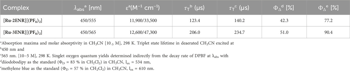
Table 1. Photophysical data of [Ru-2ENR][(PF6)2] and [Ru-3ENR][(PF6)2].
To improve the solubility of the complexes in aqueous media, their counterions were exchanged for chloride ions by passing them through a short silica column (CH3CN, H2O, KCl(aq), 100:10:1 v/v/v). Complete anion exchange was confirmed via 35Cl and 19F NMR spectra (Supplementary Figures S4, S5). It was shown previously that the 1ILCT absorption bands of the complexes were sensitive to the solvent environment and the absorption maxima red-shifted in methanol relative to the spectra in acetonitrile (MeCN) (Condon et al., 2021). The UV-Vis spectra of the chloride salts were therefore measured in MeCN, DMSO and water (Figures 2a,b). As for the PF6 salts, the ILCT absorption maxima for both Cl compounds are considerably affected showing positive solvatochromism, and significant red shifts in the more polar DMSO and water. Broadened red-shifted absorption bands can be indicative of increased molecular/solvent interactions and the substantial effect of the electronic environment confirms the charge transfer character of this transition. The wavelength of the MLCT absorption maxima are unaffected for [Ru-2ENR][(Cl)2] and only slight variation is seen in the extinction coefficient of this band for [Ru-3ENR][(Cl)2].
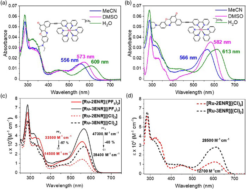
Figure 2. UV-visible absorption spectra of (a) [Ru-2ENR][(Cl)2] (b) [Ru-3ENR][(Cl)2], in MeCN, DMSO and H2O: [10-6 M], 298 K. Overlaid UV-visible absorption spectra of (c) [Ru-2ENR][(Cl)2], [Ru-3ENR][(Cl)2], Ru-2ENR][(PF6−)2], [Ru-3ENR][(PF6−)2] in acetonitrile and (d) [Ru-2ENR][(Cl)2], [Ru-3ENR][(Cl)2] in water at 298 K.
In water, the ILCT
Our preliminary biological evaluation began with an investigation of the dark toxicity of [Ru-2ENR][(Cl)2] and [Ru-3ENR][(Cl)2] using an Alamar Blue assay. This test primarily measures metabolic activity and specifically the redox activity of living cells, which reflects the activity of mitochondrial and cytosolic enzymes. It does not directly measure cell proliferation or growth, but is widely used as a proxy in the literature for cell viability and cytotoxicity due to its sensitivity to changes in cellular metabolic activity.
The dark toxicity of [Ru-2ENR][(Cl)2] and [Ru-3ENR][(Cl)2] was evaluated using the SKBR-3 cell line (a human breast cancer cell line) to find the concentration at which more than 80% of the cells remain active for the phototoxicity studies. The cells were incubated for 24 h and tested with the Alamar Blue assay (24 h). They were then incubated for another 48 h and a second Alamar Blue assay was performed (72 h) (Supplementary Figure S6). The PSs needed to exhibit less than 20% dark toxicity after 72 h incubation to test for phototoxicity. Between 0.1 and 10 μM concentrations, the toxicity of the compounds was too high and exceeded the maximum 20% (Figure 3; Supplementary Figure S7). Over 80% cell viability was achieved at 0.01 and 0.05 μM for [Ru-2ENR][(Cl)2] and 0.01 μM for [Ru-3ENR][(Cl)2] (Figures 3a,b).The non-negligible dark toxicity at concentrations in excess of 0.05 μM might be expected to limit the therapeutic application of the materials. To contextualise this information the phototoxicity selectivity indices based on metabolic read-outs from the Alamar Bue Assay are provided (Supplementary Figure S8). The highest selectivity index (2.31) is seen for [Ru-3ENR][(Cl)2] at 52h and 0.05 μM.
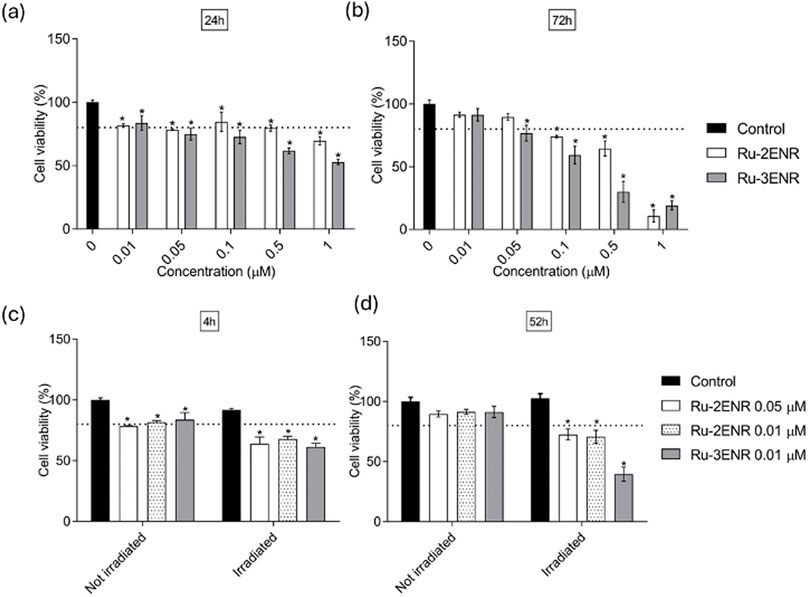
Figure 3. Dark toxicity. (a,b) Cell viability of SKBR-3 cells incubated in dark conditions with different concentrations of [[Ru-2ENR][(Cl)2] and [Ru-3ENR][(Cl)2], (0.01, 0.05, 0.1, 0.5 and 1 µM), and control (0 µM) at (a) 24 h and (b) 72 h. Photodynamic treatment effects after 15 min irradiation (c,d). Cell viability of SKBR-3 cells incubated without (control) or with 0.01 and 0.05 µM of [Ru-2ENR][(Cl)2] and 0.01 µM of [Ru-3ENR][(Cl)2] for 4 h followed by cell wash. Cell viability was determined immediately after incubation (c) (4 h) either in dark conditions (not irradiated) or after 15 min irradiation at
The phototoxicity of the complexes was tested by irradiating the cells after 4 h incubation with the PSs, at λex 630 nm (33 J cm−2) for 15 min. After irradiation, cell viability was tested immediately (4 h, Figure 3c). The cells were then incubated in the dark for 48 h, after which time cell viability was tested again (52 h, Figure 3d). Immediately after irradiation (Figure 3c), a decrease in cell survival was observed for both compounds. After 48 h (Figure 3d), a further decrease in cell viability was also observed. The 3ENR-appended complex performed better than the 2ENR. The 3ENR appended complex showed an increase in ΦΔ compared to its 2ENR analogue and had a significantly higher absorption coefficient (55%) in support of the enhanced photodynamic action seen for [Ru-3ENR][(Cl)2].
The cellular uptake of the Cl complexes was evaluated by confocal microscopy, at a concentration of 10 μM. The results are presented in Figure 4. For both compounds, most of the PS remained attached to the plasma membrane, with a small amount of the product internalised.
![Microscopy images showing cellular structures treated with [Ru-2ENR][Cl]₂ and [Ru-3ENR][Cl]₂. Each row contains four images: plasma membrane in green, photosensitiser in red, a merged image in yellow, and an orthogonal projection combining both colors. The arrangement highlights cellular localization and interaction of these compounds.](https://www.frontiersin.org/files/Articles/1620562/fchbi-04-1620562-HTML/image_m/fchbi-04-1620562-g004.jpg)
Figure 4. Product internalisation. Live SKBR-3 cells incubated with 10 µM of (a) [Ru-2ENR][(Cl)2], (b) [Ru-3ENR][(Cl)2], for 4 h and observed under confocal microscope. To analyse the localisation of the product, fluorescence mode was used. Product fluorescence emission was detected in the range of
Fluorescence intensity profiles extracted from the confocal images (Supplementary Figure S9) revealed peak intensities at the plasma membrane, consistent with dominant membrane accumulation. Intracellular puncta also displayed localized signals, suggesting vesicular internalization via endocytosis.
Conclusion
[Ru-2ENR][(Cl)2] and [Ru-3ENR][(Cl)2], two PSs with long excitation wavelengths and excellent singlet oxygen quantum yields (
Both photophysical measurements and theoretical calculations suggested that [Ru-3ENR][(PF6)2] would be a better performing PS, due to having higher ΦΔ, higher absorption and a higher rate constant of the intersystem crossing, kISC. These predictions carried through into the in vitro cell studies however experimentally determining the influence of the solvent was important particularly due to the IL* charge transfer nature of the excited state. In addition, the photophysical properties such as absorption, emission, and singlet oxygen quantum yields were found to change significantly and in some cases favourably upon anion exchange (from PF6 to Cl). The suitability of PS in biological applications such as PDT is also determined by the dark toxicity and cellular uptake. We will direct further studies with a view to exploring analogue modifications of the above complex salts. These will include detailed analyses involving mitochondrial assays, the mechanism of cell death, and the quantification of ROS generation, as appropriate. The aim will be to target improvements in terms of their cellular uptake while retaining the very advantageous photophysical properties of the materials examined to date.
Data availability statement
The datasets presented in this study can be found in online repositories. The names of the repository/repositories and accession number(s) can be found in the article/Supplementary Material.
Ethics statement
Ethical approval was not required for the studies on humans in accordance with the local legislation and institutional requirements because only commercially available established cell lines were used.
Author contributions
CC: Investigation, Conceptualization, Writing – review and editing, Software, Methodology, Formal Analysis, Visualization, Data curation, Writing – original draft. OC-B: Writing – original draft, Formal Analysis, Visualization, Data curation, Investigation, Methodology, Writing – review and editing. JF: Writing – original draft, Formal Analysis, Visualization, Writing – review and editing, Data curation. AA: Writing – review and editing, Visualization. XX: Data curation, Methodology, Investigation, Software, Formal Analysis, Writing – review and editing. JZ: Supervision, Writing – review and editing, Resources. CN: Writing – review and editing, Resources, Writing – original draft, Visualization, Formal Analysis, Investigation, Supervision, Methodology. SD: Validation, Resources, Data curation, Conceptualization, Project administration, Formal Analysis, Investigation, Funding acquisition, Methodology, Writing – review and editing, Supervision, Writing – original draft.
Funding
The author(s) declare that financial support was received for the research and/or publication of this article. This publication has emanated from research conducted with the financial support of Science Foundation Ireland (Grant Number 15/IA/3046), and Research Ireland (Grant Number 12/RC/2278_2) which is co-funded under the European Regional Development Fund under the AMBER award. A.Al-R. acknowledges funding from a TCD Postgraduate Studentship 1252 Award. C.C., and J.F. acknowledge funding support from the Trinity Research Doctoral Higher Education Authority and the Department of Further and Higher Education, Research, Innovation and Science.
Conflict of interest
The authors declare that the research was conducted in the absence of any commercial or financial relationships that could be construed as a potential conflict of interest.
The author(s) declared that they were an editorial board member of Frontiers, at the time of submission. This had no impact on the peer review process and the final decision.
Generative AI statement
The author(s) declare that no Generative AI was used in the creation of this manuscript.
Publisher’s note
All claims expressed in this article are solely those of the authors and do not necessarily represent those of their affiliated organizations, or those of the publisher, the editors and the reviewers. Any product that may be evaluated in this article, or claim that may be made by its manufacturer, is not guaranteed or endorsed by the publisher.
Supplementary material
The Supplementary Material for this article can be found online at: https://www.frontiersin.org/articles/10.3389/fchbi.2025.1620562/full#supplementary-material
References
Barretta, P., Ponte, F., Scoditti, S., and Mazzone, G. (2024a). Computational assessment of novel ruthenium phenoxazine-based complexes as photosensitizers in photodynamic therapy. Eur. J. Inorg. Chem. 27 (n/a), e202400309. doi:10.1002/ejic.202400309
Barretta, P., Scoditti, S., Belletto, D., Ponte, F., Vigna, V., Mazzone, G., et al. (2024b). Ruthenium complexes bearing nile red chromophore and one of its derivative: theoretical evaluation of PDT-related properties. J. Comput. Chem. 45 (23), 2034–2041. doi:10.1002/jcc.27392
Cabrera-González, J., Soriano, J., Conway-Kenny, R., Wang, J., Lu, Y., Zhao, J., et al. (2019). Multinuclear Ru(ii) and Ir(iii) decorated tetraphenylporphyrins as efficient PDT agents. Biomaterials Sci. 7 (8), 3287–3296. doi:10.1039/C9BM00192A
Chamberlain, S., Cole, H. D., Roque, J., Bellnier, D., McFarland, S. A., and Shafirstein, G. (2020). TLD1433-Mediated photodynamic therapy with an optical surface applicator in the treatment of lung cancer cells in vitro. Pharmaceuticals 13 (7), 137. doi:10.3390/ph13070137
Condon, C., Conway-Kenny, R., Cui, X., Hallen, L. J., Twamley, B., Zhao, J., et al. (2021). Exploring the dark: detecting long-lived nile red 3ILCT states in Ru(ii) polypyridyl photosensitisers. J. Mater. Chem. C 9 (41), 14573–14577. doi:10.1039/D1TC02830H
Conway-Kenny, R., Ferrer-Ugalde, A., Careta, O., Cui, X., Zhao, J., Nogués, C., et al. (2021). Ru(ii) and Ir(iii) phenanthroline-based photosensitisers bearing o-carborane: PDT agents with boron carriers for potential BNCT. Biomaterials Sci. 9 (16), 5691–5702. doi:10.1039/D1BM00730K
Hallen, L., Horan, A. M., Twamley, B., McGarrigle, E. M., and Draper, S. M. (2023). Accessing unsymmetrical ru(ii) bipyridine complexes: a versatile synthetic mechanism for fine tuning photophysical properties. Chem. Commun. 59 (3), 330–333. doi:10.1039/D2CC04910D
Huang, H., Banerjee, S., Qiu, K., Zhang, P., Blacque, O., Malcomson, T., et al. (2019). Targeted photoredox catalysis in cancer cells. Nat. Chem. 11 (11), 1041–1048. doi:10.1038/s41557-019-0328-4
Josefsen, L. B., and Boyle, R. W. (2008). Photodynamic therapy and the development of metal-based photosensitisers. Metal-Based Drugs 2008 (1), 1–23. doi:10.1155/2008/276109
Lazic, S., Kaspler, P., Shi, G., Monro, S., Sainuddin, T., Forward, S., et al. (2017). Novel osmium-based coordination complexes as photosensitizers for panchromatic photodynamic therapy. Photochem. Photobiol. 93 (5), 1248–1258. doi:10.1111/php.12767
Lu, Y., Wang, J., McGoldrick, N., Cui, X., Zhao, J., Caverly, C., et al. (2016a). Iridium(III) complexes bearing pyrene-functionalized 1,10-Phenanthroline ligands as highly efficient sensitizers for triplet-triplet annihilation upconversion. Angew. Chem. Int. Ed. 55 (47), 14688–14692. doi:10.1002/anie.201608442
Lu, Y., McGoldrick, N., Murphy, F., Twamley, B., Cui, X., Delaney, C., et al. (2016b). Highly efficient triplet photosensitizers: a systematic approach to the application of IrIII complexes containing extended phenanthrolines. Chem. – A Eur. J. 22 (32), 11349–11356. doi:10.1002/chem.201601534
Lu, Y., Conway-Kenny, R., Twamley, B., McGoldrick, N., Zhao, J., and Draper, S. M. (2017). 1,10-Phenanthroline Ruthenium(II) complexes as model systems in the search for high-performing triplet photosensitisers: addressing ligand versus metal effects. ChemPhotoChem 1 (12), 544–552. doi:10.1002/cptc.201700158
Lu, Y., Conway-Kenny, R., Wang, J., Cui, X., Zhao, J., and Draper, S. M. (2018). Exploiting coumarin-6 as ancillary ligands in 1,10-phenanthroline Ir(iii) complexes: generating triplet photosensitisers with high upconversion capabilities. Dalton Trans. 47 (26), 8585–8589. doi:10.1039/C8DT00231B
Mani, A., Feng, T., Gandioso, A., Vinck, R., Notaro, A., Gourdon, L., et al. (2023). Structurally simple Osmium(II) polypyridyl complexes as photosensitizers for photodynamic therapy in the near infrared. Angew. Chem. Int. Ed. 62 (20), e202218347. doi:10.1002/anie.202218347
Mital, M., and Ziora, Z. (2018). Biological applications of Ru(II) polypyridyl complexes. Coord. Chem. Rev. 375, 434–458. doi:10.1016/j.ccr.2018.02.013
Monro, S., Colón, K. L., Yin, H., Roque, J., Konda, P., Gujar, S., et al. (2019). Transition metal complexes and photodynamic therapy from a tumor-centered approach: challenges, opportunities, and highlights from the development of TLD1433. Chem. Rev. 119 (2), 797–828. doi:10.1021/acs.chemrev.8b00211
Parella, C., Blanquer, A., Sinha, S., Hümpfner, E., Hernando, J., Mora, E., et al. (2024). Developing photo-activable ruthenium (II) complexes for PDT: synthesis, characterization, photophysical and biological studies. Dyes Pigments 224, 111985. doi:10.1016/j.dyepig.2024.111985
Wang, J., Lu, Y., McGoldrick, N., Zhang, C., Yang, W., Zhao, J., et al. (2016). Dual phosphorescent dinuclear transition metal complexes, and their application as triplet photosensitizers for TTA upconversion and photodynamic therapy. J. Mater. Chem. C 4 (25), 6131–6139. doi:10.1039/C6TC01926A
Wang, J., Lu, Y., McCarthy, W., Conway-Kenny, R., Twamley, B., Zhao, J., et al. (2018). Novel ruthenium and iridium complexes of N-substituted carbazole as triplet photosensitisers. Chem. Commun. 54 (9), 1073–1076. doi:10.1039/C7CC08535D
Keywords: ruthenium, coordination chemistry, photobiology, SKBR-3 breast cancer cells, photodynamic therapy, Nile red
Citation: Condon C, Careta-Borràs O, Fodor J, Al Riyami AB, Xiao X, Zhao J, Nogués C and Draper SM (2025) Preliminary investigation of Ru(II) complexes bearing nile red chromophores for photodynamic therapy. Front. Chem. Biol. 4:1620562. doi: 10.3389/fchbi.2025.1620562
Received: 29 April 2025; Accepted: 29 July 2025;
Published: 14 August 2025.
Edited by:
Roy Planalp, University of New Hampshire, United StatesReviewed by:
Aakriti Garg, University of New Hampshire, United StatesNir Hananya, Technion Israel Institute of Technology, Israel
Copyright © 2025 Condon, Careta-Borràs, Fodor, Al Riyami, Xiao, Zhao, Nogués and Draper. This is an open-access article distributed under the terms of the Creative Commons Attribution License (CC BY). The use, distribution or reproduction in other forums is permitted, provided the original author(s) and the copyright owner(s) are credited and that the original publication in this journal is cited, in accordance with accepted academic practice. No use, distribution or reproduction is permitted which does not comply with these terms.
*Correspondence: Sylvia M. Draper, c21kcmFwZXJAdGNkLmll; Carme Nogués, Y2FybWUubm9ndWVzQHVhYi5jYXQ=