 Marília Ladeira de Araújo1
 Bruno Costa Gomes2
 Paula Pícoli Devóz3
 Nathália de Assis Aguilar Duarte1
 Diego Luis Ribeiro4
 Adriana Ladeira de Araújo5
 Bruno Lemos Batista6
 Lusânia Maria Greggi Antunes3
 Fernando Barbosa Jr.3
 António Sebastião Rodrigues2
 José Rueff2
 Gustavo Rafael Mazzaron Barcelos1*
- 1Department of Biosciences, Institute of Health and Society, Federal University of São Paulo, Santos, Brazil
- 2Center for Toxicogenomics and Human Health, NOVA Medical School (NMS), Universidade Nova de Lisboa, Lisbon, Portugal
- 3Department of Clinical Analyses, Toxicology and Food Sciences, School of Pharmaceutical Sciences of Ribeirão Preto, University of São Paulo, Ribeirão Preto, Brazil
- 4Department of Genetics, Ribeirão Preto Medical School, University of São Paulo, Ribeirão Preto, Brazil
- 5Anhembi Morumbi University, São Paulo, Brazil
- 6Center for Natural and Human Sciences, Federal University of ABC, Santo André, Brazil
Experimental and epidemiologic studies have shown that lead (Pb) is able to induce epigenetic modifications, such as changes in DNA methylation profiles, in chromatin remodeling, as well as the expression of non-coding RNAs (ncRNAs). However, very little is known about the interactions between microRNAs (miRNAs) expression and DNA methylation status in individuals exposed to the metal. The aim of the present study was to investigate the impact of hsa-miR-148a expression on DNA methylation status, in 85 workers exposed to Pb. Blood and plasma lead levels (BLL and PLL, respectively) were determined by ICP-MS; expression of the miRNA-148a was quantified by RT-qPCR (TaqMan assay) and assessment of the global DNA methylation profile (by measurement of 5-methylcytosine; % 5-mC) was performed by ELISA. An inverse association was seen between miR-148a and % 5-mC DNA, as a function of BLL and PLL (β = −3.7; p = 0.071 and β = −4.1; p = 0.049, respectively) adjusted for age, BMI, smoking, and alcohol consumption. Taken together, our study provides further evidence concerning the interactions between DNA methylation profile and miR-148a, in individuals exposed to Pb.
Introduction
Previous studies have shown that exposure to toxic metals, such as lead (Pb), induces alterations in gene expression, by modulation of epigenetic status and, consequently, may influence Pb-induced toxicity (Senut et al., 2012; Kim et al., 2014; Nye et al., 2014). In this context, it is noteworthy that an increasing number of studies is being conducted to understand the impact of disturbances on epigenetic status induced by Pb exposure, with special focus on DNA methylation and on the expression of non-coding RNAs (nc-RNAs), mainly micro-RNAs (miRNAs).
Earlier epidemiological studies showed associations between Pb exposure and changes in DNA global methylation profiles (Pilsner et al., 2009; Nye et al., 2015; Sen et al., 2015; Devóz et al., 2017). However, the molecular mechanisms underlying these events are still unclear. Experimental evidences give support to the fact that Pb-induced alterations of the activity of DNA methyl transferases (DNMTs) may be related to these observations (Schneider et al., 2013; Sanchez et al., 2017; Sobolewski et al., 2018). DNMTs play a key role in the maintenance of genes’ promoter region methylation, especially in CpG islands. It is well known that hypo- and hypermethylation of CpG islands in promoter regions are associated with an increase or decrease of mRNA transcription, respectively (for a review, see Ehrlich and Lacey, 2013; Moore et al., 2013).
The identification of molecular biomarkers that may precede the adverse health effects related to the exposure of toxicants significantly increased in the last decade. This approach is particularly interesting and cost-effective, since it provides evidence about underlying toxic effects, being possible to draw safety decisions prior to acute toxicity responses (for a review, see Califf, 2018; Turesky and Lu, 2020). In this context, the detection of miRNAs seems to be a promising tool for biomonitoring individuals who are exposed to chemical compounds, such as toxic metals and organic pollutants (Kotsyfakis and Patelarou, 2019; Sisto et al., 2019). Several studies showed clear associations between the expression of miRNAs and the increase in cancers (Sohel, 2020). On the other hand, a few studies have been performed aiming to assess the impact of toxic metals, such as Pb, arsenic (As), cadmium (Cd) and mercury (Hg) on miRNAs expression (Wallace et al., 2020); moreover, most of them were carried out using in vitro or in vivo laboratory models (Xu et al., 2015; Bihaqi, 2019; Wallace et al., 2020) and fewer studies have assessed the impact of Pb on miRNA expression profile in individuals exposed to the metal.
For example, Xu et al. (2017) observed an inverse association between expression of miR-520c-3p, miR-211, and miR-148a and high blood lead levels (BLL), while miR-572 levels increased in individuals with high BLL, in workers from China. In another study, Kong et al. (2012) assessed the association between the exposure to toxic metals, including Pb and miRNAs expression, in adolescents from Hong Kong, and an inverse association was observed between miR-21 and miR-221 and urinary Pb and As levels, when compared to the non-exposed ones.
As described above, there are few studies focusing on the disturbances of miRNA expressions induced by Pb exposure, in humans. Therefore, the aim of the present study was to investigate the impact of Pb exposure on the expression of miR-148a and its association with DNA methylation, in individuals exposed to metal, from automotive battery plants.
Materials and Methods
Population and Study Design
A cross-sectional study with 85 male individuals (>18 years old) was carried out from automotive battery plants, in Paraná State, Brazil. An interviewed-administrated questionnaire was applied in order to collect socio-demographic, lifestyle, and health information, such as age, body mass index (BMI), time of exposure (working time), medical history, medication use, smoking, and alcohol intake. Participants who drank alcoholic beverages at least five times per week were considered alcohol users and those who had smoked at least five cigarettes per day for the previous 5 years were classified as smokers (Barcelos et al., 2013, 2015a, 2015b; de Oliveira et al., 2014; Gomes et al., 2018).
The present study was approved by the Ethics Committee of University Federal of São Paulo, Santos, Brazil (approval number: 0292/2018), and the corresponding methods were carried out in accordance with the approved guidelines. All participating workers were advised about the content of the investigation and signed the written informed consents before starting the study.
Quantification of Pb in Blood and in Plasma
Samples were taken on site, in the infirmary station of each plant. Blood samples were collected by a qualified nurse using evacuated tubes: (I) for Pb determination and DNA isolation: Vacutainer Trace-Elements and Vacutainer PST (BD, Franklin Lakes, NJ, United States) and (II) for miRNA isolation: PAXgene Blood RNA Tubes (PreAnalytiX, Hombrechtikon, Switzerland); plasma samples were obtained by centrifugation (10 min at 1,200 g). Transportation of the samples was carried out using Styrofoam boxes with dry ice until they arrived at the laboratory; samples were kept at −80°C till further handling and analyses.
Total BLL and PLL levels were determined by inductively coupled plasma mass spectrometry (ICP-MS; ELAN DRC II, Perkin Elmer, Norwalk, CT, United States) as previously described by Batista et al., 2009a, b). Results are expressed as μg dl–1.
hsa-miR-148a-5p Expression Assessment
Total RNA was extracted from whole blood using MagMax for Stabilized Blood Tubes RNA Isolation Kit (Applied Biosystems, Foster City, CA, United States) according to the manufacturer’s instructions. The quality of the RNA was verified by measuring the 260/280 and 260/230 nm ratio (Nanodrop 2000, Invitrogen, California, CA, United States). Samples were quantified using Qubit RNA BR Assay Kit (Invitrogen, California, CA, United States) on a fluorimeter (Quibit 3.0, Invitrogen, California, CA, United States), according to the manufacturer’s recommendations.
TaqMan Advanced miRNA Assay (Applied Biosystems, Foster City, CA, United States) was used for cDNA synthesis. Monitoring of miR-148a expression was assessed using TaqMan Advanced miRNA Assay (assay #478718_mir; Applied Biosystems, Foster City, CA, United States), according to the manufacturer’s instructions; moreover, quantification of expression of has-miRNA-miR-16-5p was used as an endogenous control (assay #477860_mir; Applied Biosystems, Foster City, CA, United States). miRNAs were quantified using the relative quantification method [2–(Δ Ctx – Δ Ctr) = 2–Δ Ct]. All RT-qPCR reactions were performed in a QuantStudio 3 Real Time PCR System Thermal Cycler (Applied Biosystems, Foster City, CA, United States).
Global Methylation Assays
Genomic DNA (gDNA) was extracted from peripheral blood using the ReliaPrep Blood gDNA Miniprep System (Promega, Wisconsin, WI, EUA) according to the manufacturer’s instructions. The quality of the DNA was verified by measuring the 260/280 and 260/230 nm ratio (Nanodrop 2000, Invitrogen, California, CA, United States). Subsequently, gDNA was quantified by use of the Qubit dsDNA BR Assay Kit (Invitrogen, California, CA, United States) in a fluorimeter (Qubit 3.0, Invitrogen, California, CA, United States).
Quantification of the global DNA methylation status was performed using the 5-mC DNA ELISA Kit (Zymo Research, Irvine, CA, United States), according to the manufacturer’s recommendations; absorbance was read at 405 nm (Biotek Elx800—Winooski, VT, United States). Results are expressed as % DNA global methylation (% 5-mC DNA).
Data Interpretation
Age (years), body mass index (BMI), BLL, PLL, miR-148a expression, and % 5-mC DNA were analyzed as continuous variables; alcohol consumption (yes or no) and smoking (yes or no) were assessed as categorical ones. Due to their skewed distribution, BLL and BMI were sqrt transformed, while PLL data were log10-transformed.
Descriptive statistics were run for reporting the general characteristics of the participants. Non-parametric correlations (Spearman’s rho) were performed in order to examine the associations between age, BMI, alcohol consumption, smoking, exposure period, BLL, PLL, % 5-mC DNA, and miR-148. Then, multivariate linear regression models were performed to assess the associations of Pb biomarkers, miR-148a and % 5-mC DNA, adjusted for age, BMI, alcohol consumption, and smoking.
Analyses were run using SPSS 23 Statistics software (IBM; Armonk, NY, United States), and a p < 0.050 was set as significant.
Results
The general population characteristics, Pb concentrations, and % 5-mC DNA for all participants enrolled in the present study are summarized in Table 1. The age ranged from 19 to 69 years (mean 37 ± 11 years), while the mean exposure period was 3.2 ± 3.4 years (from 2 months to 20 years). Alcohol was consumed by 31% of the participants, and 12% of the individuals were declared as smokers. Mean BLL was 19 ± 11 μg dl–1 (ranging from 2.6 to 48 μg dl–1), and mean PLL was 0.54 ± 0.65 μg dl–1, reaching values up to 4.0 μg dl–1.
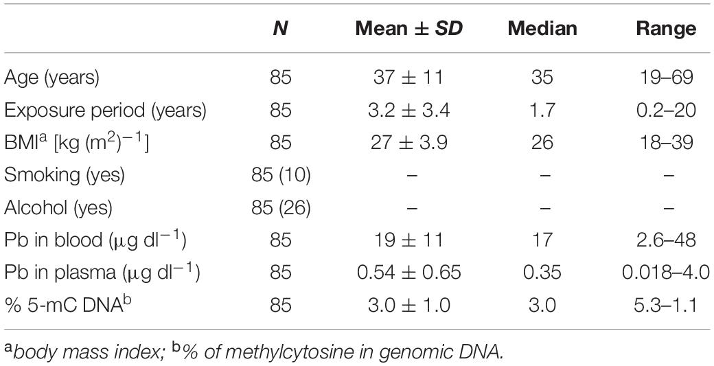
Table 1. General characteristics of study population.
Table 2 presents the Spearman’s correlations between age, BMI, alcohol consumption, smoking, biomarkers of Pb exposure, % 5-mC, and miR-148a expression. As expected, BLL and PLL were strongly correlated to each other (p < 0.0010); also, a significant correlation was seen between age and BMI (p = 0.010). On the other hand, none of the assessed variables were significantly correlated with any of the biomarkers related to metal exposure (p > 0.050).
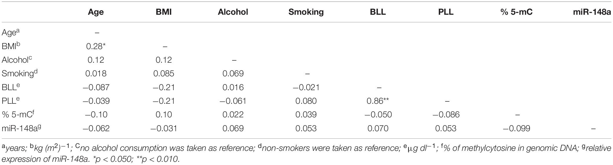
Table 2. Spearman’s correlations between age, body mass index (BMI), alcohol consumption, smoking, working time, blood and plasma lead levels (BLL and PLL, respectively), % DNA global methylation (% 5-mC), and expression of miR-148a.
Tables 3, 4 summarize the impact of miR-148a expression on % 5-mC DNA through data obtained by multivariate linear regression analyses, adjusted by age, BMI, alcohol consumption, smoking, and BLL or PLL. It can be seen that the content of 5-mC DNA tended to decrease as the miR-148a expression increases, as a function of BLL (β = −3.7; p = 0.071); however, this observation did not reach statistical significance. Further, it can be observed that miR-148a expression is able to decrease the % 5-mC DNA, as a function of PLL (β = −4.1; p = 0.049).
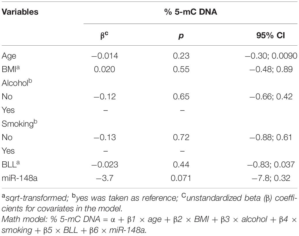
Table 3. Impact of miR-148a expression on % DNA global methylation (% 5-mC DNA) of Pb-exposed workers from automotive battery factories. Adjusted for age, BMI, alcohol consumption, smoking, and blood lead levels (BLL).
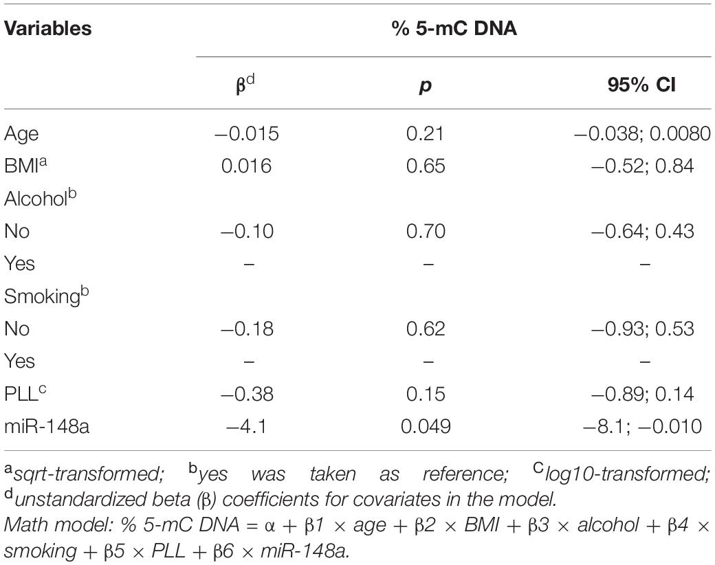
Table 4. Impact of miR-148a expression on % DNA global methylation (% 5-mC DNA) of Pb-exposed workers from automotive battery factories. Adjusted for age, BMI, alcohol consumption, smoking, and plasma lead levels(PLL).
Finally, no effects of other variables (age, BMI, alcohol intake, and smoking) were seen on % 5-mC DNA (Tables 3, 4), and no significant interaction terms (IT) between the biomarkers of Pb exposure and miR-148a were able to induce disturbances of DNA methylation profile (IT for BLL∗miR-148a: β = 0.43; p = 0.45; IT for PLL∗miR-148a: β = 2.3; p = 0.54; not in Tables).
Discussion
It is well stablished that Pb exposure is able to induce several adverse health effects, such as cardiovascular disorders (Lustberg and Silbergeld, 2002), kidney injuries (Weaver et al., 2005), and cognitive dysfunctions (Shih et al., 2007); previous data showed that BLL as low as 10 μg dl–1 can cause hypertension, tremors, and renal dysfunction (National Toxicology Program, 2012), and BLL up to 20 μg dl–1 induces several neurological injuries, in adults (Murata et al., 2009). Moreover, recent studies suggest that exposure to the metal is associated with changes in epigenetic status and, consequently, may modulate the toxic effects related to Pb exposure (Senut et al., 2012; Kim et al., 2014; Nye et al., 2014).
We previously showed that Pb exposure is able to decrease the DNA global methylation profile of leukocytes from peripheral blood, in a subgroup of this study population (Devóz et al., 2017). Moreover, other studies also showed the impact of Pb-induced disturbances on the DNA methylation profile. For example, Zhang et al. (2019) observed alterations in DNA methylation profile of 356 significant CpG sites of blood cells, and these changes were related to the levels of exposure to the metal, in workers from car battery facilities, in China; Li et al. (2013) demonstrated that individuals exposed to Pb levels similar to those found in our study (21 μg dl–1) had lower % of DNA methylation assessed by LINE-1 than the non-exposed ones (3.7 μg dl–1). Besides, associations between Pb exposure and DNA hypomethylation were also seen in animal laboratory models (Dou et al., 2019; Nakayama et al., 2019).
Studies performed with laboratory models showed that Pb is able to inhibit the DNA methyl transferase (Dnmts) activities. For example, Sanchez et al. (2017) observed that zebra fish exposed to the metal throughout embryogenesis (500 ppb, i.e., 500 μg l–1) had lower Dnmt1 activity than non-exposed animals, while Nakayama et al. (2019) showed that Wistar rats exposed to Pb- and Cd-contaminated soil (3,750 and 6.0 mg kg–1 bw, respectively) had higher Dnmt3a and Dnmt3b mRNA expression in testis than the control subjects (Nakayama et al., 2019).
miRNAs are being widely used as epigenetic biomarkers, since they canregulate various cellular and molecular pathways (Gholamin et al., 2018; Rabieian et al., 2018). Currently, these biomarkers are not limited to diagnosis or to therapeutic monitoring response; miRNAs are also used as predictive tools for underlying adverse health effects induced by exposure to toxicants, predicting by molecular signaling the onset of pathologies and prior to manifestation of symptoms and complications. For example, miRNAs are widely used as a prognostic biomarker for myocardial infarction (Wang et al., 2016), and for type 1 and 2 diabetes (Nielsen et al., 2012; Villard et al., 2015; Marchand et al., 2016). The monitoring of miRNAs as predictive tools is particularly advantageous, due their non-invasive collection (mostly plasma and saliva) and good correlation to disorders of several inner organs and systems (Condrat et al., 2020).
Alterations of miRNA expression profile can be a sensitive indicator related to acute and/or chronic exposures to several inorganic and organic toxicants. Chronic Pb poisoning is a complex disease, due to interactions between genetic backgrounds and environmental variables, and also because of the long latency between the beginning of exposure to the metal and the onset of adverse health effects (Mani et al., 2019). In this context, biomonitoring alterations of miRNA expression may be a useful tool for predicting cellular and systemic response against Pb-induced toxicity (for a comprehensive review, see Wallace et al., 2020).
We observed an inverse association between miR-148a expression and % 5-mC DNA. Data from in silico and from in vitro studies showed that miR-148a has the DNMT1 gene as target (Long et al., 2014; Zhan et al., 2015; Sengupta et al., 2018). DNMT1 plays a key role on the maintenance of genes’ promoter region methylation, especially in CpG islands. It is well-known that hypo- and hypermethylation of CpG sites in promoter regions are associated to an increase and decrease in mRNA translation, respectively (for a review, see Ehrlich and Lacey, 2013; Moore et al., 2013). One hypothesis of our findings may be explained to the premature degradation of DNMT1 mRNAs induced by overexpression of miR-148a, which would impact the global DNA methylation status.
Previous studies give further support to this explanation; Lujambio et al. (2008) showed that miR-148a silencing was associated with DNA hypermethylation in different metastatic cell lines, while Sengupta et al. (2018) observed that miR-148a suppressed DNMT1 expression, in prostate cancer cells. Moreover, Wang et al. (2019) found similar results of those previously reported by Lujambio et al. (2008) and Sengupta et al. (2018), in an acute myeloid leukemia cell line, i.e., the increase in miR-148a decreases the expression of DNMT1 (both mRNA and protein), which may impact the DNA methylation profile, suggesting that lower DNMT1 activity is related to a decrease in the methylation levels of promoter region’s CpG islands of miR-148a and, consequently, increases miR-148a expression, creating a negative feedback system. It is important to note that these studies were carried out in cancer cell lines, and the mechanisms may differ in normal cells, as well as in complex organisms, such as mammals and humans, for example.
To the best of our knowledge, we have found a few studies related to Pb exposure and miRNA expression in humans, suggesting that the metal is able to induce disturbances on miRNA expression profiles. For example, in a study conducted with workers from battery car plants in China, it was seen that individuals who had higher BLL (higher exposure: BLL mean 51 ± 6.4 μg dl–1) had lower expression of plasmatic miRNAs miR-520c-3p, miR-211, and miR-148a, when compared to those that were categorized as lower exposure persons (BLL 8.9 ± 1.5 μg dl–1), while opposite effect was seen concerning miR-572 (Xu et al., 2017). In contrast, we did not find a significant association between BLL and PLL, and miR-148a expression. These contradictory results may be partly explained by differences of studied populations, such as variation on genetic background, dietary habits, and lifestyle; moreover, it is important to highlight that the level of Pb exposure in the study carried out by Xu et al. (2017) is much higher than those found in our study (BLL mean 51 ± 6.4 vs. 19 ± 11 μg dl–1, respectively) suggesting that high Pb levels may trigger metal–epigenetic interactions.
Taken together, our study provides further evidence concerning decrease in DNA global methylation induced by miR-148a in workers exposed to Pb. The consequences may result in impairment in the regulation of gene expression and, consequently, modulate the adverse health effects induced by exposure to the metal.
Data Availability Statement
The raw data supporting the conclusions of this article will be made available by the authors, without undue reservation.
Ethics Statement
The studies involving human participants were reviewed and approved by the Ethics Committee of Federal University of São Paulo, São Paulo, Brazil. The patients/participants provided their written informed consent to participate in this study.
Author Contributions
AR, GB, JR, and MA conceptualized the study. BG and MA developed the methodology and performed the miRNA assays. BB and FB performed the ICP-MS analysis. DR, LA, and PD performed the global methylation assays. GB, MA, and ND conducted the investigation. AR, GB, and JR wrote and prepared the original draft. AR, GB, JR, MA, and ND wrote reviewed, and edited the manuscript. All authors have read and agreed to the published version of the manuscript.
Funding
This study was supported by the grants #2013/06033-8, #2016/24520-1 and #2018/24643-1 (São Paulo Research Foundation, FAPESP, Brazil), by the National Council for Scientific and Technological Development (Brazil) and by grant UID-BIM-00009-2020 (Foundation for Science and Technology, FCT, Portugal).
Conflict of Interest
The authors declare that the research was conducted in the absence of any commercial or financial relationships that could be construed as a potential conflict of interest.
References
Barcelos, G. R. M., de Marco, K. C., Rezende, V. B., Braga, G. U. L., Antunes, L. M. G., Tanus-Santos, J. E., et al. (2015a). Genetic effects of eNOS polymorphisms on miomarkers related to cardiovascular status in a population coexposed to methylmercury and lead. Arch. Environ. Contam. Toxicol. 69, 173–180. doi: 10.1007/s00244-015-0137-8
Barcelos, G. R. M., Grotto, D., de Marco, K. C., Valentini, J., Lengert, A.vH, Oliveira, A. A. S., et al. (2013). Polymorphisms in glutathione-related genes modify mercury concentrations and antioxidant status in subjects environmentally exposed to methylmercury. Sci. Total Environ. 463-464, 319–325. doi: 10.1016/j.scitotenv.2013.06.029
Barcelos, G. R. M., Souza, M. F., Oliveira, A. A. S., Lengert, A.v. H, Oliveira, M. T., Camargo, R. B., et al. (2015b). Effects of genetic polymorphisms on antioxidant status and concentrations of the metals in the blood of riverside Amazonian communities co-exposed to Hg and Pb. Environ. Res. 138, 224–232. doi: 10.1016/j.envres.2015.02.017
Batista, B. L., Rodrigues, J. L., Nunes, J. A., Souza, V. C., and Barbosa, F. Jr. (2009a). Exploiting dynamic reaction cell inductively coupled plasma mass spectrometry (DRC-ICP-MS) for sequential determination of trace elements in blood using a dilute-and-shoot procedure. Anal. Chim. Acta 639, 13–18. doi: 10.1016/j.aca.2009.03.016
Batista, B. L., Rodrigues, J. L., Tormen, L., Curtius, A. J., and Barbosa, F. Jr. (2009b). Reference concentrations for trace elements in urine for the Brazilian population based on q-ICP-MS with a simple dilute-and-shoot procedure. J. Braz. Chem. Soc. 20, 1406–1413. doi: 10.1590/S0103-50532009000800004
Bihaqi, S. W. (2019). Early life exposure to lead (Pb) and changes in DNA methylation: relevance to Alzheimer’s disease. Rev. Environ. Health 26, 187–195. doi: 10.1515/reveh-2018-0076
Califf, R. M. (2018). Biomarker definitions and their applications. Exp. Biol. Med. (Maywood) 243, 213–221. doi: 10.1177/1535370217750088
Condrat, C. E., Thompson, D. C., Barbu, M. G., Bugnar, O. L., Boboc, A., Cretoiu, D., et al. (2020). miRNAs as biomarkers in disease: latest findings regarding their role in diagnosis and prognosis. Cells 9:276. doi: 10.3390/cells9020276
de Oliveira, A. A. S., Souza, M. F., Lengert, A.vH, Oliveira, M. T., Camargo, R. B., Braga, G. U. L., et al. (2014). Genetic polymorphisms in glutathione (GSH-) related genes affect the plasmatic Hg/whole blood Hg partitioning and the distribution between inorganic and methylmercury levels in plasma collected from a fish-eating population. Biomed. Res. Int. 2014:940952. doi: 10.1155/2014/940952
Devóz, P. P., Gomes, W. R., Araújo, M. L., Ribeiro, D. L., Pedron, T., Antunes, L. M. G., et al. (2017). Lead (Pb) exposure induces disturbances in epigenetic status in workers exposed to this metal. J. Toxicol. Environ. Health A 80, 1098–1105. doi: 10.1080/15287394.2017.1357364
Dou, J. F., Farooqui, Z., Faulk, C. D., Barks, A. K., Jones, T., Dolinoy, D. C., et al. (2019). Perinatal lead (Pb) exposure and cortical neuron-specific DNA methylation in male mice. Genes (Basel) 10:274. doi: 10.3390/genes10040274
Ehrlich, M., and Lacey, M. (2013). DNA methylation and differentiation: silencing, upregulation and modulation of gene expression. Epigenomics 5, 553–568. doi: 10.2217/epi.13.43
Gholamin, S., Mirzaei, H., Razavi, S. M., Hassanian, S. M., Saadatpour, L., Masoudifar, A., et al. (2018). GD2-targeted immunotherapy and potential value of circulating microRNAs in neuroblastoma. J. Cell. Physiol. 233, 866–879. doi: 10.1002/jcp.25793
Gomes, W. R., Devóz, P. P., Luiz, B. L. C., Grotto, D., Batista, B. L., and Barbosa, F. Jr., et al. (2018). Polymorphisms of genes related to metabolism of lead (Pb) are associated with the metal body burden and with biomarkers of oxidative stress. Mutat. Res. Genet. Toxicol. Environ. Mutagen. 836, 42–46. doi: 10.1016/j.mrgentox.2018.05.016
Kim, J., Lee, Y., and Yang, M. (2014). Environmental exposure to lead (Pb) and variations in its susceptibility. J. Environ. Sci. Health C Environ. Carcinog. Ecotoxicol. Rev. 32, 159–185. doi: 10.1080/10590501.2014.907461
Kong, A. P., Xiao, K., Choi, K. C., Wang, G., Chan, M. H., Ho, C. S., et al. (2012). Associations between microRNA (miR-21, 126, 155 and 221), albuminuria and heavy metals in Hong Kong Chinese adolescents. Clin. Chim. Acta 413, 1053–1057. doi: 10.1016/j.cca.2012.02.014
Kotsyfakis, M., and Patelarou, E. (2019). MicroRNAs as biomarkers of harmful environmental and occupational exposures: a systematic review. Biomarkers 24, 623–630. doi: 10.1080/1354750X.2019.1652348
Li, C., Yang, X., Xu, M., Zhang, J., and Sun, N. (2013). Epigenetic marker (LINE-1 promoter) methylation level was associated with occupational lead exposure. Clin. Toxicol. (Phila) 51, 225–229. doi: 10.3109/15563650.2013.782410
Long, X. R., He, Y., Huang, C., and Li, J. (2014). MicroRNA-148a is silenced by hypermethylation and interacts with DNA methyltransferase 1 in hepatocellular carcinogenesis. Int. J Oncol. 44, 1915–1922. doi: 10.3892/ijo.2014.2373
Lujambio, A., Calin, G. A., Villanueva, A., Ropero, S., Sánchez-Céspedes, M., Blanco, D., et al. (2008). A microRNA DNA methylation signature for human cancer metastasis. Proc. Natl. Acad. Sci. U.S.A. 105, 13556–13561. doi: 10.1073/pnas.0803055105
Lustberg, M., and Silbergeld, E. (2002). Blood lead levels and mortality. Arch. Intern. Med. 162, 2443–2449. doi: 10.1001/archinte.162.21.2443
Mani, M. S., Kabekkodu, S. P., Joshi, M. B., and Dsouza, H. S. (2019). Ecogenetics of lead toxicity and its influence on risk assessment. Hum. Exp. Toxicol. 38, 1031–1059. doi: 10.1177/0960327119851253
Marchand, L., Jalabert, A., Meugnier, E., Van den Hende, K., Fabien, N., Nicolino, M., et al. (2016). miRNA-375 a sensor of glucotoxicity is altered in the serum of children with newly diagnosed type 1 diabetes. J. Diabetes Res. 2016:1869082. doi: 10.1155/2016/1869082
Moore, L. D., Le, T., and Fan, G. (2013). DNA methylation and its basic function. Neuropsychopharmacology 38, 23–38. doi: 10.1038/npp.2012.112
Murata, K., Iwata, T., Dakeishi, M., and Karita, K. (2009). Lead toxicity: does the critical level of lead resulting in adverse effects differ between adults and children? J. Occup. Health 51, 1–12. doi: 10.1539/joh.k8003
Nakayama, S. M. M., Nakata, H., Ikenaka, Y., Yabe, J., Oroszlany, B., Yohannes, Y. B., et al. (2019). One year exposure to Cd- and Pb-contaminated soil causes metal accumulation and alteration of global DNA methylation in rats. Environ. Pollut. 252, 1267–1276. doi: 10.1016/j.envpol.2019.05.038
National Toxicology Program (2012). Monograph on Health Effects of Low-Level Lead. Available online at: https://ntp.niehs.nih.gov/ntp/ohat/lead/final/monographhealtheffectslowlevellead_newissn_508.pdf (accessed August 26, 2020).
Nielsen, L. B., Wang, C., Sørensen, K., Bang-Berthelsen, C. H., Hansen, L., Andersen, M. L., et al. (2012). Circulating levels of microRNA from children with newly diagnosed type 1 diabetes and healthy controls: evidence that miR-25 associates to residual beta-cell function and glycaemic control during disease progression. Exp. Diabetes Res. 2012:896362. doi: 10.1155/2012/896362
Nye, M. D., Fry, R. C., Hoyo, C., and Murphy, S. K. (2014). Investigating epigenetic effects of prenatal exposure to toxic metals in newborns: challenges and cenefits. Med. Epigenet. 2, 53–59. doi: 10.1159/000362336
Nye, M. D., Hoyo, C., and Murphy, S. K. (2015). In vitro lead exposure changes DNA methylation and expression of IGF2 and PEG1/MEST. Toxicol. Vitro 29, 544–550. doi: 10.1016/j.tiv.2015.01.002
Pilsner, J. R., Hu, H., Ettinger, A., Sánchez, B. N., Wright, R. O., Cantonwine, D., et al. (2009). Influence of prenatal lead exposure on genomic methylation of cord blood DNA. Environ. Health Perspect. 117, 1466–1471. doi: 10.1289/ehp.0800497
Rabieian, R., Boshtam, M., Zareei, M., Kouhpayeh, S., Masoudifar, A., and Mirzaei, H. (2018). Plasminogen activator inhibitor type-1 as a regulator of fibrosis. J. Cell. Biochem. 119, 17–27. doi: 10.1002/jcb.26146
Sanchez, O. F., Lee, J., Yu, K. H. N., Kim, S. E., Freeman, J. L., and Yuan, C. (2017). Lead (Pb) exposure reduces global DNA methylation level by non-competitive inhibition and alteration of dnmt expression. Metallomics 9, 149–160. doi: 10.1039/c6mt00198j
Schneider, J. S., Kidd, S. K., and Anderson, D. W. (2013). Influence of developmental lead exposure on expression of DNA methyltransferases and methyl cytosine-binding proteins in hippocampus. Toxicol. Lett. 217, 75–81. doi: 10.1016/j.toxlet.2012.12.004
Sen, A., Cingolani, P., Senut, M. C., Land, S., Mercado-Garcia, A., Tellez-Rojo, M. M., et al. (2015). Lead exposure induces changes in 5-hydroxymethylcytosine clusters in CpG islands in human embryonic stem cells and umbilical cord blood. Epigenetics 10, 607–621. doi: 10.1080/15592294.2015.1050172
Sengupta, D., Deb, M., and Patra, S. K. (2018). Antagonistic activities of miR-148a and DNMT1: ectopic expression of miR-148a impairs DNMT1 mRNA and dwindle cell proliferation and survival. Gene 660, 68–79. doi: 10.1016/j.gene.2018.03.075
Senut, M. C., Cingolani, P., Sen, A., Kruger, A., Shaik, A., Hirsch, H., et al. (2012). Epigenetics of early-life lead exposure and effects on brain development. Epigenomics 4, 665–674. doi: 10.2217/epi.12.58
Shih, R. A., Hu, H., Weisskopf, M. G., and Schwartz, B. S. (2007). Cumulative lead dose and cognitive function in adults: a review of studies that measured both blood lead and bone lead. Environ. Health Perspect. 115, 483–492. doi: 10.1289/ehp.9786
Sisto, R., Capone, P., Cerini, L., Sanjust, F., Paci, E., Pigini, D., et al. (2019). Circulating microRNAs as potential biomarkers of occupational exposure to low dose organic solvents. Toxicol. Rep. 6, 126–135. doi: 10.1016/j.toxrep.2019.01.001
Sobolewski, M., Varma, G., Adams, B., Anderson, D. W., Schneider, J. S., and Cory-Slechta, D. A. (2018). Developmental lead exposure and prenatal stress result in sex-specific reprograming of adult stress physiology and epigenetic profiles in brain. Toxicol. Sci. 163, 478–489. doi: 10.1093/toxsci/kfy046
Sohel, M. M. H. (2020). Circulating microRNAs as biomarkers in cancer diagnosis. Life Sci. 248:117473. doi: 10.1016/j.lfs.2020.117473
Turesky, R. J., and Lu, K. (2020). Biomarkers of environmental toxicants: exposure and biological effects. Toxics 8:37. doi: 10.3390/toxics8020037
Villard, A., Marchand, L., Thivolet, C., and Rome, S. (2015). Diagnostic value of cell-free circulating MicroRNAs for obesity and type 2 diabetes: a meta-analysis. J. Mol. Biomark. Diagn. 6:251. doi: 10.4172/2155-9929.1000251
Wallace, D. R., Taalab, Y. M., Heinze, S., Tariba, L. B., Pizent, A., Renieri, E., et al. (2020). Toxic-metal-induced alteration in miRNA expression profile as a proposed mechanism for disease development. Cells 9:901. doi: 10.3390/cells9040901
Wang, J., Chen, J., and Sem, S. (2016). MicroRNA as biomarkers and diagnostics. J. Cell. Physiol. 231, 25–30. doi: 10.1002/jcp.25056
Wang, X. X., Zhang, H., and Li, Y. (2019). Preliminary study on the role of miR-148a and DNMT1 in the pathogenesis of acute myeloid leukemia. Mol. Med. Rep. 19, 2943–2952. doi: 10.3892/mmr.2019.9913
Weaver, V. M., Jaar, B. G., Schwartz, B. S., Todd, A. C., Ahn, K. D., Lee, S. S., et al. (2005). Associations among lead dose biomarkers, uric acid, and renal function in Korean lead workers. Environ. Health Perspect. 113, 36–42. doi: 10.1289/ehp.7317
Xu, L. H., Mu, F. F., Zhao, J. H., He, Q., Cao, C. L., Yang, H., et al. (2015). Lead induces apoptosis and histone hyperacetylation in rat cardiovascular tissues. PLoS One 10:e0129091. doi: 10.1371/journal.pone.0129091
Xu, M., Yu, Z., Hu, F., Zhang, H., Zhong, L., Han, L., et al. (2017). Identification of differential plasma miRNA profiles in Chinese workers with occupational lead exposure. Biosci. Rep. 37:BSR20171111. doi: 10.1042/BSR20171111
Zhan, Q., Fang, Y., Deng, X., Chen, H., Jin, J., Lum, X., et al. (2015). The interplay between miR-148a and DNMT1 might be exploited for pancreatic cancer therapy. Cancer Invest. 33, 267–275. doi: 10.3109/07357907.2015.1025794
Keywords: epigenetics, miRNA, ncRNA, occupational exposure, toxicity, lead
Citation: Araújo ML, Gomes BC, Devóz PP, Duarte NAA, Ribeiro DL, Araújo AL, Batista BL, Antunes LMG, Barbosa F Jr, Rodrigues AS, Rueff J and Barcelos GRM (2021) Association Between miR-148a and DNA Methylation Profile in Individuals Exposed to Lead (Pb). Front. Genet. 12:620744. doi: 10.3389/fgene.2021.620744
Received: 23 October 2020; Accepted: 07 January 2021;
Published: 17 February 2021.
Edited by:
Siddhartha Kumar Mishra, Chhatrapati Shahu Ji Maharaj University, IndiaReviewed by:
Eunus S. Ali, Northwestern University, United StatesCristina Matzenbacher, Federal University of Rio Grande do Sul, Brazil
Copyright © 2021 Araújo, Gomes, Devóz, Duarte, Ribeiro, Araújo, Batista, Antunes, Barbosa, Rodrigues, Rueff and Barcelos. This is an open-access article distributed under the terms of the Creative Commons Attribution License (CC BY). The use, distribution or reproduction in other forums is permitted, provided the original author(s) and the copyright owner(s) are credited and that the original publication in this journal is cited, in accordance with accepted academic practice. No use, distribution or reproduction is permitted which does not comply with these terms.
*Correspondence: Gustavo Rafael Mazzaron Barcelos, Z3VzdGF2by5iYXJjZWxvc0B1bmlmZXNwLmJy