Abstract
Introduction:
Osteoarthritis (OA) is a common degenerative joint disease resulting from the breakdown of multiple joint tissues, remains a leading cause of disability with limited therapeutic options. Synovitis is one of the reasons of OA progression, while communication between blood and synovium during disease process is still unclear.
Methods:
We used transcriptomic datasets from blood and synovium of healthy controls and OA patients to investigate potential molecular crosstalk between blood and synovium in OA pathogenesis through ligand-receptor pairs.
Results:
Ligand-receptor pair analysis revealed 129 ligands and 137 receptors differentially expressed in blood, and 108 ligands and 86 receptors in synovium. Gene ontology enrichment analysis of differentially expressed ligands indicated receptor ligand activity in both tissues, with blood enriched in leukocyte migration, cell chemotaxis, and leukocyte chemotaxis, and synovium in negative regulation of response to external stimulus, epithelial cell proliferation, and cell chemotaxis. Further protein-protein interaction (PPI) network analysis showed that blood ligands were mainly associated with inflammation and immunity (IL6, IL1B, IL23A, IFNA1, and TNF), while several synovium ligands were linked to angiogenesis (TGFB1, FGF7, and PDGFA). Based on ligand-receptor interactions and PPI network of differentially expressed ligands, we predicted and constructed molecular communication map between blood and synovium. Immunofluorescence staining of synovium showed more blood micro-vessels in OA patients and elevated IL6 and IL1B expression levels, suggesting that synovial inflammation might partly originate from pro-inflammatory cytokines in blood.
Discussion:
These findings offered new understanding of the molecular mechanisms underlying blood and synovium communication in OA, and provided potential therapeutic drug targets for OA treatment to simultaneously modulate systemic inflammation and local angiogenesis.
Introduction
Osteoarthritis (OA) is a degenerative joint disease, which is becoming one of the most leading causes of disability (1). OA affects multiple tissues in patients, including cartilage, synovium, meniscus and subchondral bone (2). The patients suffer from pain, stiffness and swelling, while the current treatments can only alleviate the progression of the disease (3). The most common pharmacological treatment is non-steroidal anti-inflammatory drugs (NSAIDs) (4). These agents mainly target the inflammatory mediators IL-1 and COX-2 to achieve analgesic and anti-inflammatory effects (5, 6). However, long-term use requires vigilance for gastrointestinal and cardiovascular side effects (7). Once the disease reaches end stage, it can only be treated through surgical replacement (8).
Synovitis is one of the major factors promoting the progression of OA, manifested as swelling of the joint accompanied by thickening of the synovial membrane and vascular proliferation (9). It is known that synovial inflammation is mainly stimulated by the breakdown of articular cartilage and subchondral bone during OA (9–11). This may result in a self-regulatory inflammatory response, which triggers synovium to release inflammatory mediators and aggravates the effects of inflammation (11). A lot of inflammatory mediators can promote synovial inflammation, of which interleukin-1β (IL-1β) and tumor necrosis factor α (TNFα) are the main proinflammatory cytokines involved in the progression of OA (12). They can not only mediate their own expression, but also induce the generation of matrix metalloproteinases (MMPs) and other cytokines (e.g., IL6, IL8, PTGS2), which lead to articular cartilage damage and further aggravate OA progression (13). Notably, the degree of synovial angiogenesis is associated with the grade of synovitis in OA (14). Some inflammatory markers are able to be detected in the circulation system, which raises the concern of inflammatory mediators derived from blood (15). The angiogenesis in the synovium is another significant feature of inflammatory synovium, which is due to the imbalance of proangiogenic and antiangiogenic factors (16). Macrophages within synovium may secrete cytokines that induce angiogenesis or stimulate endothelial cells and fibroblasts to produce angiogenic factors, such as vascular endothelial growth factor (VEGF) (17, 18). VEGF promotes synovial angiogenesis through highly expressed VEGF receptor 2 (VEGFR2) in endothelial and synoviocytes, which result in the emerge of micro-vessels and pannus (19–21). However, the mechanism of the communication between synovium and blood in the physiological and pathological osteoarthritis is still unclear.
Here, we utilized the transcriptome of blood and synovium samples in healthy and osteoarthritis patients to identify differential expressed genes, and further screening of ligand-receptor pairs within two tissues to unveil the communication between blood and synovium in osteoarthritis. We constructed molecular networks that may reveal the role of synovium in angiogenesis and the pro-inflammatory role of blood on synovium. Further immunofluorescence detection revealed elevated IL6 and IL1B levels in blood micro-vessels. These results may provide new insights into future therapeutic drug targets.
Materials and methods
Data sources and quality control
Transcriptome of blood and synovium samples in osteoarthritis patients were acquired from gene expression omnibus (GEO) database, including samples derived from peripheral blood mononuclear cells (PBMCs, GSE48556, HC = 33, OA = 108) (22) and synovial fibroblasts (SFs, GSE29746, HC = 11, OA = 11) (23). All raw data were normalized by robust multiarray average (RMA) algorithm in RStudio (2023.06.2 + 561), probe IDs were transformed to gene symbols and the mean values were calculated as the expression values. There were 25,159 and 19,749 unique genes obtained from PBMC and SF, respectively. Principal component analysis (PCA) was performed by FactoMineR (v2.8) and factoextra (v1.0.7) with R packages.
Analysis of blood and synovium transcriptome
Differential expressed genes (DEGs) of two datasets were filtered by p value, which was less than 0.05. DEG were shown on volcano plot with ggplot2 (v3.4.2), of which the most 20 statistical differences DEG were labeled with gene symbols. To illustrate the communication between two tissues, ligands and the corresponding receptors within DEG were filtered by ligand-receptor database based on the FANTOM5 project. Ligand-receptor pairs within and between PBMC and SF were plotted by RCircos (v1.2.1). Differential expressed ligands were enriched by gene ontology (GO), including molecular function, cellular component and biological process. Then, the potential ligand interaction network was analyzed in STRING and constructed by cytoscape (v3.9.0). Heatmap was constructed by pheatmap (v1.0.12).
Ethical statement and immunofluorescence staining
Human synovium samples were obtained from two patients with OA undergoing total knee replacement surgery and one patient involved in a traffic accident requiring amputation. The use of human samples was approved by Ethical Committee of Affiliated Hospital of Shandong University of Traditional Chinese Medicine (AF/SC-08/03.0), and the patients provided written informed consent to participate in this study.
The synovium samples were serially sectioned into paraffin slices of 5 μm thickness. After deparaffinization and dehydration, they were blocked with 5% BSA. Primary antibodies against IL6 (Proteintech, China) (24) and IL1B (Proteintech, China) (25) were applied and incubated overnight at 4 °C. After washing with PBST, the corresponding fluorescent secondary antibodies were applied and incubated at room temperature for 1 h. Finally, the slices were counterstained with DAPI (Abcam, UK) and observed under a fluorescence microscope (Zeiss, Germany).
Results
Quality control of blood and synovium transcriptomes
Our strategy was using the transcriptomes of blood and synovium tissues from normal controls and patients with osteoarthritis (OA) to identify differentially expressed ligands and receptors. We then predicted molecular communications between synovium and blood tissues based on their ligand-receptor pair interactions (Figure 1A). After downloading the raw data, box plots revealed the differences between the original transcriptomes of blood and synovium tissues (Figures 1B,E). Following normalization, the data showed consistency, with the medians of different datasets in consistent positions (Figures 1C,F), especially for the synovium transcriptome (Figure 1F). Principal component analysis (PCA) showed that in the blood transcriptome, the normal group was more dispersed, while the OA group was relatively concentrated (Figure 1D). In contrast, in the synovium transcriptome, both the normal and OA groups showed relatively tight clustering (Figure 1G).
Figure 1

Schematic diagram and quality control of transcriptomic data. (A) Schematic diagram summarizing the process of this study. (B,C) Box plots of the original (B) and normalized (C) blood transcriptomic data. (D) Principal component analysis of blood transcriptomic data. (E,F) Box plots of the original (E) and normalized (F) synovium transcriptomic data. (G) Principal component analysis of synovium transcriptomic data.
Screening of differentially expressed genes and ligand-receptor pair analysis
We screened differentially expressed genes using a unified threshold of p < 0.05. In the OA blood transcriptome, genes such as HSPA1B, ADRB2, FLJ14213, FAM43A, and ID3 were significantly upregulated, while H3F3B, IL8, EGR1, SNORD13, and LOC649841 were significantly downregulated (Figure 2A). In the OA synovium transcriptome, ITGB2, RIMS1, GRIN3A, SLC7A10, and DOK7 were significantly upregulated, whereas ANXA10, PTGER2, AIM1, SPC24, and CLGN were significantly downregulated (Figure 2B). Many differentially expressed genes were consistent with the results of original dataset analysis (22, 23), demonstrating the availability and accuracy of the datasets. Further analysis using a ligand-receptor database identified differentially expressed ligands and receptors. We found 129 ligands and 137 receptors differentially expressed in the blood, and 108 ligands and 86 receptors differentially expressed in the synovium. Through ligand-receptor interactions, we predicted molecular communications between blood and synovium (Figure 2C). In addition to interactions within blood and synovium, there were also many potential molecular communications between these two tissues.
Figure 2

Differentially expressed genes and ligand-receptor interaction analysis. (A,B) Volcano plots of differentially expressed genes in blood (A) and synovium (B) transcriptomes. Red indicates up-regulated genes, and blue indicates down-regulated genes. The top 10 up-regulated and down-regulated genes are labeled. (C) Analysis of ligand-receptor interactions between blood and synovium. BL, blood; SY, synovium.
Gene ontology analysis and molecular network construction
We performed gene ontology enrichment on the differentially expressed ligands. In terms of molecular function, both blood and synovium tissues were primarily enriched in receptor ligand activity, indicating the accuracy of our ligand screening (Figures 3A,B). In terms of cellular components, they both showed enrichment in endoplasmic reticulum lumen and collagen-containing extracellular matrix (Figures 3A,B). In biological processes, the blood transcriptome was mainly enriched in leukocyte migration, cell chemotaxis, and leukocyte chemotaxis (Figure 3A), while the synovium transcriptome was primarily enriched in negative regulation of response to external stimulus, epithelial cell proliferation, and cell chemotaxis (Figure 3B). This highlighted the functional differences of differentially expressed ligands between blood and synovium tissues. We then imported these differentially expressed ligands into the STRING database to construct a protein–protein interaction (PPI) molecular network. The molecular network of blood ligands was mainly associated with inflammation and immunity, with IL6, IL1B, IL23A, IFNA1, and TNF being upregulated in the blood of OA patients (Figure 3C). In the molecular network of synovium ligands, angiogenesis-related pathways were observed, with TGFB1, FGF7, and PDGFA being upregulated in the synovium tissue of OA patients (Figure 3D).
Figure 3

Gene ontology (GO) analysis of differentially expressed ligands and construction of PPI (protein–protein interaction) molecular networks. (A,B) Dot plots showing GO terms of differentially expressed ligands in blood (A) and synovium (B) transcriptomes. (C,D) PPI molecular networks of differentially expressed ligands in blood (C) and synovium (D) transcriptomes. The schematic on the right shows the heat map of IL6, IL1B, IL23A, IFNA1, and TNF expression in blood, as well as TGFB1, FGF7, and PDGFA in synovium. BL, blood; SY, synovium; OA, osteoarthritis.
Prediction and validation of synovium-blood interaction molecular network
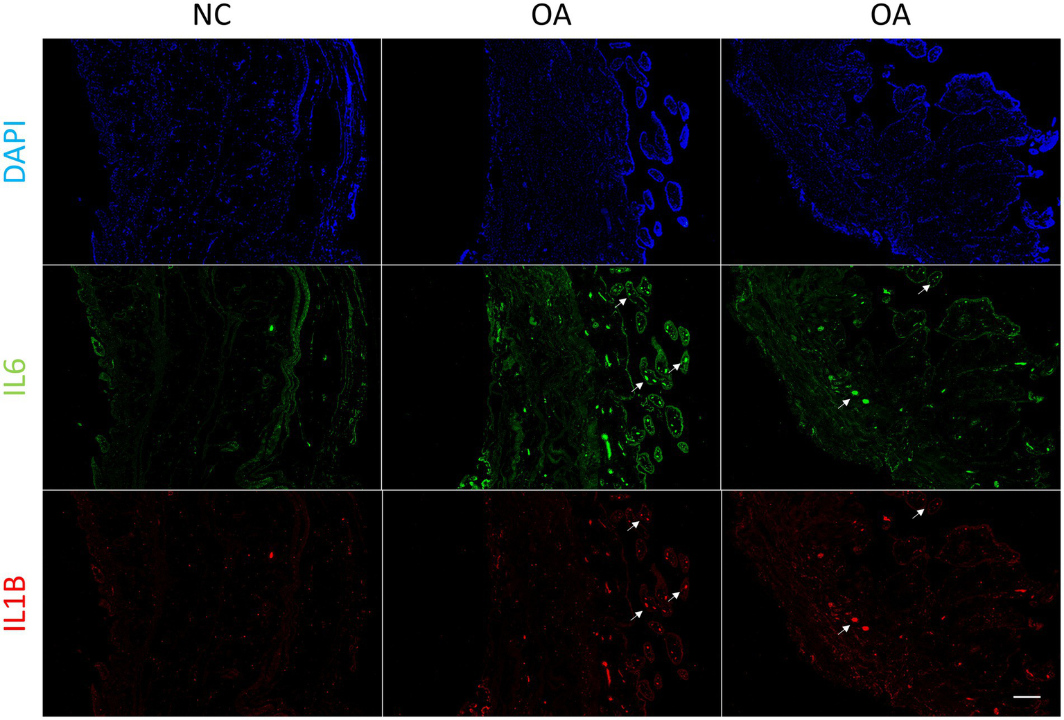
In order to verify whether synovial inflammation partly originated from blood, we performed immunofluorescence on synovium sections. Using synovium tissues from amputees as the control, control and OA synovium tissues were stained to detect IL6 and IL1B expression. In OA patients, there were more newly-formed micro-vessels, and the expression of IL6 and Il1B increased in OA blood (Figure 4). This indicated that inflammation in OA synovium might partly due to the elevated pro-inflammatory cytokines IL6 and IL1B in blood. Based on PPI molecular network and ligand-receptor interactions, a molecular interaction network between synovium and blood was further constructed (Figure 5). This map could lay the foundation for studying synovium-blood interactions and provide insights for developing drugs to simultaneously control synovial inflammation and neovascularization in OA.
Figure 4
Immunofluorescence of synovium tissue. Immunofluorescence of IL6 and IL1B in normal and OA synovium tissues. White arrows indicate the locations of some blood micro-vessels in the synovium. Human synovium samples were obtained from two patients with OA and one patient requiring amputation. Scale bars, 200 μm. DAPI, blue; IL6, green; IL1B, red. NC, control; OA, osteoarthritis.
Figure 5
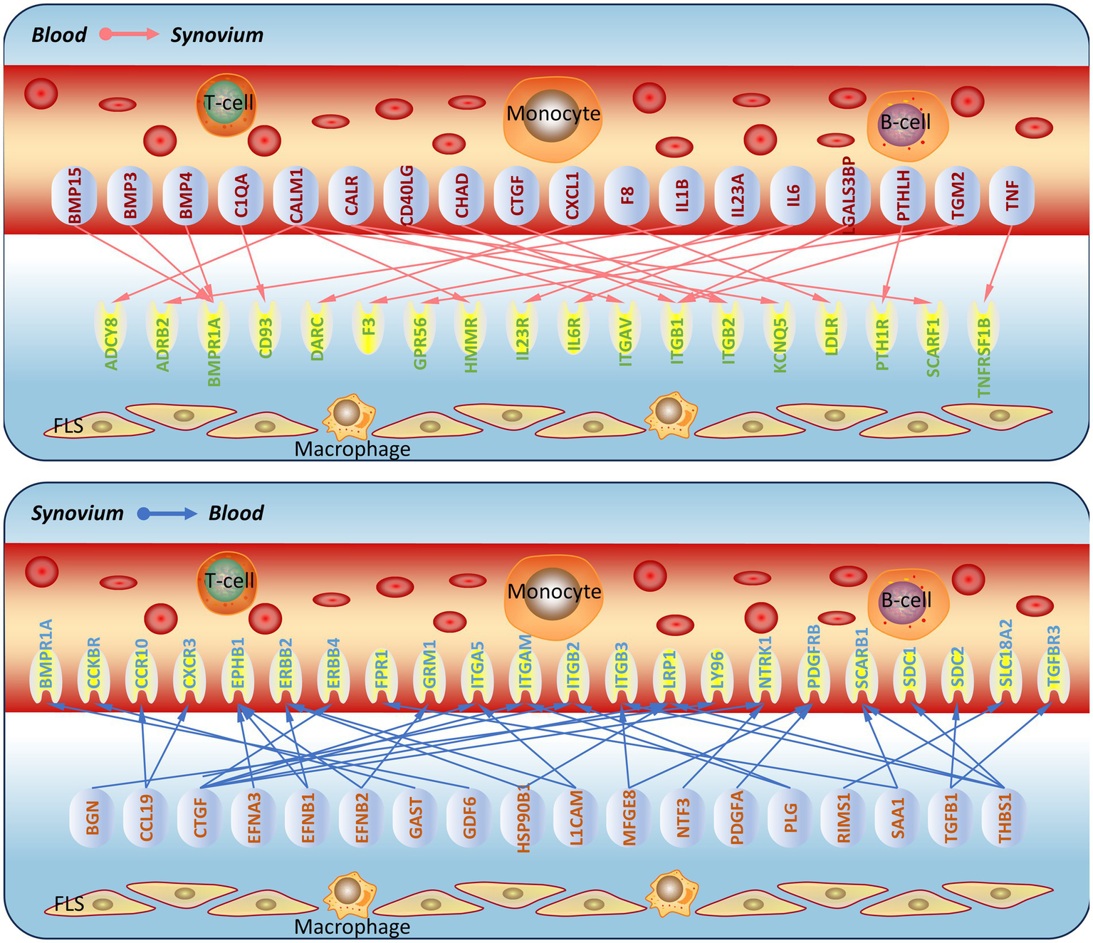
Prediction of molecular communication between blood and synovium in osteoarthritis. Based on the screened ligand molecular network and ligand-receptor interactions, a schematic of molecular communication between blood and synovium in osteoarthritis was constructed.
Discussion
Understanding the complex molecular interactions in OA progression is crucial for developing more effective therapeutic approaches. Synovitis is a key pathological feature of OA, contributing significantly to disease progression, which is accompanied by the formation of micro-vessels and pannus (26, 27). However, the molecular mechanisms underlying whether the newly formed pannus communicates with the synovium to further aggravate the progression of OA or whether synovial inflammation promotes angiogenesis remain unclear. Therefore, elucidating the tissue communication between the synovium and blood will help develop new therapeutic approaches for OA.
The differentially expressed genes in the blood and synovium transcriptomes were consistent with the results that reported in the original dataset. For example, the upregulation of HSPA1B, ADRB2, FAM43A, and ID3 and the downregulation of H3F3B, IL8, EGR1, and SNORD13 in the blood of OA patients (22), and the upregulation of ITGB2, RIMS1, and GRIN3A and the downregulation of ANXA10 in the synovium of OA patients (23), which further demonstrated the accuracy of our analysis results. Secondly, we found that many differentially expressed genes have been reported to play roles in OA. For example, HSPA1B is a member of the heat shock protein 70 family, and the RNA-binding protein ZFP36L1 regulates members of the HSP70 family (HSPA1A and HSPA1B), preventing OA onset by inhibiting chondrocyte apoptosis (28). ADRB2 (β2-adrenergic receptor) has been identified as one of the potential targets in the OA-related gene network (22), and its knockout leads to increased calcified cartilage thickness and subchondral bone remodeling in experimental OA in mice (29). H3F3B is a target of miR-10a-5p, which promotes IL-1β-induced chondrocyte apoptosis by regulating H3F3B (30). ITGB2 is a member of the integrin family, involved in cell adhesion and inflammatory responses, and studies have shown that high expression of ITGB2 in synovial fluid can assist in OA diagnosis and serve as a biomarker for OA severity (31). PTGER2 is the receptor for prostaglandin E2, and inhibition of prostaglandin E2 attenuates aberrant alteration of subchondral bone, articular cartilage degeneration and pain in osteoarthritis (32). Other unreported differentially expressed genes may also play a role in OA, and these findings can serve as a basis for future research.
The enrichment of differentially expressed ligands in the blood and synovium transcriptomes showed both similarities and differences. In terms of molecular function and cellular components, most of the enriched terms were similar, such as endoplasmic reticulum lumen, collagen-containing extracellular matrix, leukocyte migration, cell chemotaxis, and leukocyte chemotaxis. In terms of biological processes, the blood was more associated with the functions of leukocytes and the regulation of immune responses, while the synovium showed more involvement of cell signaling and regulation, as well as immune responses. These results also showed correlation with many previously reported findings. The increased expression of IL1B in peripheral blood leukocytes is associated with OA progression and pain (15). And the activated macrophages in the synovium produce pro-inflammatory mediators and various tissue-degrading enzymes that lead to the destruction of cartilage and subchondral bone, thereby aggravating OA progression (33). Among the other biological processes we identified in the synovium, such as morphogenesis of a branching structure, epithelial cell proliferation, and regulation of blood coagulation, suggested that the synovium might interact with the blood through these processes at the molecular level. Further molecular network construction revealed that the blood’s molecular network contained many molecules related to inflammation and immunity (such as IL6, IL1B), while the synovium molecular network contained many molecules related to angiogenesis (such as TGFB1, FGF7). This suggested that the blood may influence the synovium through inflammation and immunity, while the synovium may affect vascular regeneration through angiogenesis. It was known that IL-6 and IL-1β were pivotal pro-inflammatory cytokines in OA, which induced cartilage destruction and synovial inflammation, thereby accelerating OA progression (34). While TGF-β1, FGF7, and PDGFA modulated cartilage and synovium homeostasis through different mechanisms. Local inhibition of TGF-β1 signaling attenuated cartilage damage and osteophyte formation in mouse osteoarthritis (35). The role of FGF7 in OA was not clear, and the current evidence was related to bone-defect repair, where exogenous FGF7 enhanced bone formation in rat mandible defects (36). And exogenous PDGFA increased the proliferation of synovial mesenchymal stem cells (37), which might contribute to osteoarthritis through synovitis. These findings revealed the distinct molecular profiles of blood and synovium tissues in OA, highlighting the importance of targeting both inflammatory and angiogenic pathways for effective disease management.
This study also has many limitations. Initially, to obtain more robust data, we applied a stricter screening threshold (adj. P. Val < 0.05), which yielded 10 ligands and 24 receptors in blood, and 4 receptors and no ligands in synovium, leaving virtually no interacting pairs for downstream analysis. We therefore adopted p < 0.05 as the criterion, which allowed us to identify a larger set of potential tissue communications while still retaining statistical significance. Secondly, we focused on the communication between blood and synovium, but OA was a disease involving the coordinated pathology of multiple tissues, including cartilage, subchondral bone, synovium, meniscus, and ligaments, etc. Thirdly, the datasets we used were based on microarray platforms, which can only provide a macroscopic overview of the overall changes. Single-cell sequencing would offer better resolution and reveal more cell types. Fourthly, the datasets here were focused on PBMCs and SFs, while endothelial cells in the blood or macrophages in the synovium may also be involved. Whether these communications occur through intermediate cells requires more transcriptome data. Fifthly, the molecular communications we analyzed were based on ligand-receptor interactions; other undiscovered interactions may be overlooked, and these interactions need further experimental validation, such as Co-IP and Tarnswell assays. Finally, the differentially expressed genes we analyzed were mainly coding genes, while miRNAs, lncRNAs, and others may also participate in tissue interactions through exosomes. Further sequencing and analysis of miRNAs, lncRNAs, and others were needed.
In conclusion, our study provided novel insights into the molecular mechanisms underlying the communication between blood and synovium tissues in OA. The identification of key ligand-receptor pairs and their associated pathways offered potential therapeutic targets for modulating inflammation and angiogenesis. Future research should focus on validating these results in larger cohorts and exploring the therapeutic potential of targeting these molecular interactions. Additionally, further investigation into the role of systemic inflammation and local angiogenesis in OA progression may reveal new strategies for disease modification and prevention.
Statements
Data availability statement
The original contributions presented in the study are included in the article/supplementary material, further inquiries can be directed to the corresponding authors.
Ethics statement
The studies involving humans were approved by Ethical Committee of Affiliated Hospital of Shandong University of Traditional Chinese Medicine (AF/SC-08/03.0). The studies were conducted in accordance with the local legislation and institutional requirements. The participants provided their written informed consent to participate in this study.
Author contributions
MW: Formal analysis, Data curation, Writing – original draft, Conceptualization, Supervision. GT: Writing – original draft, Formal analysis, Methodology. SW: Methodology, Writing – review & editing. WL: Funding acquisition, Writing – review & editing, Supervision, Conceptualization.
Funding
The author(s) declared that financial support was received for this work and/or its publication. This work was supported by Project of China Association for the Promotion of Traditional Chinese Medicine Research (2021–18) and Shandong Provincial Archives Science and Technology Project (2024–42).
Conflict of interest
The author(s) declared that this work was conducted in the absence of any commercial or financial relationships that could be construed as a potential conflict of interest.
Generative AI statement
The author(s) declared that Generative AI was not used in the creation of this manuscript.
Any alternative text (alt text) provided alongside figures in this article has been generated by Frontiers with the support of artificial intelligence and reasonable efforts have been made to ensure accuracy, including review by the authors wherever possible. If you identify any issues, please contact us.
Publisher’s note
All claims expressed in this article are solely those of the authors and do not necessarily represent those of their affiliated organizations, or those of the publisher, the editors and the reviewers. Any product that may be evaluated in this article, or claim that may be made by its manufacturer, is not guaranteed or endorsed by the publisher.
References
1.
O’Sullivan O . Osteoarthritis: pathophysiology and classification of a common disabling condition In: BennettGGoodallE, editors. The Palgrave encyclopedia of disability. Cham: Springer (2024). 1–11.
2.
Tang S Zhang C Oo WM Fu K Risberg MA Bierma-Zeinstra SM et al . Osteoarthritis. Nat Rev Dis Prim. (2025) 11:1–22. doi: 10.1038/s41572-025-00594-6
3.
Abdel-Aziz MA Ahmed HM El-Nekeety AA Abdel-Wahhab MA . Osteoarthritis complications and the recent therapeutic approaches. Inflammopharmacology. (2021) 29:1653–67. doi: 10.1007/s10787-021-00888-7
4.
Magni A Agostoni P Bonezzi C Massazza G Menè P Savarino V et al . Management of osteoarthritis: expert opinion on NSAIDs. Pain Ther. (2021) 10:783–808. doi: 10.1007/s40122-021-00260-1,
5.
Alvarez-Soria M Herrero-Beaumont G Moreno-Rubio J Calvo E Santillana J Egido J et al . Long-term NSAID treatment directly decreases COX-2 and mPGES-1 production in the articular cartilage of patients with osteoarthritis. Osteoarthr Cartil. (2008) 16:1484–93. doi: 10.1016/j.joca.2008.04.022,
6.
Fidelix TS Macedo CR Maxwell LJ Trevisani VFM . Diacerein for osteoarthritis. Cochrane Database Syst Rev. (2014) 2014:CD005117. doi: 10.1002/14651858.CD005117.pub3
7.
Laine L . Gastrointestinal effects of NSAIDs and coxibs. J Pain Symptom Manag. (2003) 25:32–40. doi: 10.1016/S0885-3924(02)00629-2
8.
Mahmoudian A Lohmander LS Mobasheri A Englund M Luyten FP . Early-stage symptomatic osteoarthritis of the knee—time for action. Nat Rev Rheumatol. (2021) 17:621–32. doi: 10.1038/s41584-021-00673-4,
9.
Sanchez-Lopez E Coras R Torres A Lane NE Guma M . Synovial inflammation in osteoarthritis progression. Nat Rev Rheumatol. (2022) 18:258–75. doi: 10.1038/s41584-022-00749-9,
10.
Wang M Tan G Jiang H Liu A Wu R Li J et al . Molecular crosstalk between articular cartilage, meniscus, synovium, and subchondral bone in osteoarthritis. Bone Joint Res. (2022) 11:862–72. doi: 10.1302/2046-3758.1112.BJR-2022-0215.R1,
11.
Sellam J Berenbaum F . The role of synovitis in pathophysiology and clinical symptoms of osteoarthritis. Nat Rev Rheumatol. (2010) 6:625–35. doi: 10.1038/nrrheum.2010.159,
12.
Rahmati M Mobasheri A Mozafari M . Inflammatory mediators in osteoarthritis: a critical review of the state-of-the-art, current prospects, and future challenges. Bone. (2016) 85:81–90. doi: 10.1016/j.bone.2016.01.019
13.
Mukherjee A Das B . The role of inflammatory mediators and matrix metalloproteinases (MMPs) in the progression of osteoarthritis. Biomater Biosyst. (2024) 13:100090. doi: 10.1016/j.bbiosy.2024.100090
14.
Walsh DA Bonnet C Turner E Wilson D Situ M McWilliams D . Angiogenesis in the synovium and at the osteochondral junction in osteoarthritis. Osteoarthr Cartil. (2007) 15:743–51. doi: 10.1016/j.joca.2007.01.020
15.
Attur M Belitskaya-Lévy I Oh C Krasnokutsky S Greenberg J Samuels J et al . Increased interleukin-1β gene expression in peripheral blood leukocytes is associated with increased pain and predicts risk for progression of symptomatic knee osteoarthritis. Arthritis Rheum. (2011) 63:1908–17. doi: 10.1002/art.30360
16.
Lambert C Mathy-Hartert M Dubuc J-E Montell E Vergés J Munaut C et al . Characterization of synovial angiogenesis in osteoarthritis patients and its modulation by chondroitin sulfate. Arthritis Res Ther. (2012) 14:1–11. doi: 10.1186/ar3771
17.
Afuwape A Kiriakidis S Paleolog EM . The role of the angiogenic molecule VEGF in the pathogenesis of rheumatoid arthritis. Histol Histopathol. (2002) 17:961–72. doi: 10.14670/HH-17.961
18.
Bonnet C Walsh D . Osteoarthritis, angiogenesis and inflammation. Rheumatology. (2005) 44:7–16. doi: 10.1093/rheumatology/keh344,
19.
Leblond A Allanore Y Avouac J . Targeting synovial neoangiogenesis in rheumatoid arthritis. Autoimmun Rev. (2017) 16:594–601. doi: 10.1016/j.autrev.2017.04.005
20.
Elshabrawy HA Chen Z Volin MV Ravella S Virupannavar S Shahrara S . The pathogenic role of angiogenesis in rheumatoid arthritis. Angiogenesis. (2015) 18:433–48. doi: 10.1007/s10456-015-9477-2,
21.
Szekanecz Z Besenyei T Paragh G Koch AE . New insights in synovial angiogenesis. Joint Bone Spine. (2010) 77:13–9. doi: 10.1016/j.jbspin.2009.05.011,
22.
Ramos YF Bos SD Lakenberg N Böhringer S den Hollander WJ Kloppenburg M et al . Genes expressed in blood link osteoarthritis with apoptotic pathways. Ann Rheum Dis. (2014) 73:1844–53. doi: 10.1136/annrheumdis-2013-203405,
23.
Del Rey MJ Usategui A Izquierdo E Cañete JD Blanco FJ Criado G et al . Transcriptome analysis reveals specific changes in osteoarthritis synovial fibroblasts. Ann Rheum Dis. (2012) 71:275–80. doi: 10.1136/annrheumdis-2011-200281,
24.
Zhao Q Huang L Qin G Qiao Y Ren F Shen C et al . Cancer-associated fibroblasts induce monocytic myeloid-derived suppressor cell generation via IL-6/exosomal miR-21-activated STAT3 signaling to promote cisplatin resistance in esophageal squamous cell carcinoma. Cancer Lett. (2021) 518:35–48. doi: 10.1016/j.canlet.2021.06.009,
25.
Zou Y Zhang B Jiang K Zhou X Tang Q Chen S et al . GPR40 inhibits microglia-mediated neuroinflammation via the NLRP3/IL-1β/glutaminase pathway after subarachnoid hemorrhage. Biochem Pharmacol. (2025) 238:116971. doi: 10.1016/j.bcp.2025.116971,
26.
Mathiessen A Conaghan PG . Synovitis in osteoarthritis: current understanding with therapeutic implications. Arthritis Res Ther. (2017) 19:1–9. doi: 10.1186/s13075-017-1229-9
27.
Shibakawa A Aoki H Masuko-Hongo K Kato T Tanaka M Nishioka K et al . Presence of pannus-like tissue on osteoarthritic cartilage and its histological character. Osteoarthr Cartil. (2003) 11:133–40. doi: 10.1053/joca.2002.0871,
28.
Son Y-O Kim H-E Choi W-S Chun C-H Chun J-S . RNA-binding protein ZFP36L1 regulates osteoarthritis by modulating members of the heat shock protein 70 family. Nat Commun. (2019) 10:77. doi: 10.1038/s41467-018-08035-7,
29.
Rösch G Muschter D Taheri S El Bagdadi K Dorn C Meurer A et al . β2-adrenoceptor deficiency results in increased calcified cartilage thickness and subchondral bone remodeling in murine experimental osteoarthritis. Front Immunol. (2022) 12:801505. doi: 10.3389/fimmu.2021.801505
30.
Jiang H Pang H Wu P Cao Z Li Z Yang X . LncRNA SNHG5 promotes chondrocyte proliferation and inhibits apoptosis in osteoarthritis by regulating miR-10a-5p/H3F3B axis. Connect Tissue Res. (2021) 62:605–14. doi: 10.1080/03008207.2020.1825701
31.
Qian W Li Z . Expression and diagnostic significance of integrin beta-2 in synovial fluid of patients with osteoarthritis. J Orthop Surg. (2023) 31:10225536221147213. doi: 10.1177/10225536221147213
32.
Sun Q Zhang Y Ding Y Xie W Li H Li S et al . Inhibition of PGE2 in subchondral bone attenuates osteoarthritis. Cells. (2022) 11:2760. doi: 10.3390/cells11172760
33.
Zhang H Cai D Bai X . Macrophages regulate the progression of osteoarthritis. Osteoarthr Cartil. (2020) 28:555–61. doi: 10.1016/j.joca.2020.01.007,
34.
Wojdasiewicz P Poniatowski ŁA Szukiewicz D . The role of inflammatory and anti-inflammatory cytokines in the pathogenesis of osteoarthritis. Mediat Inflamm. (2014) 2014:561459. doi: 10.1155/2014/561459,
35.
Chen R Mian M Fu M Zhao JY Yang L Li Y et al . Attenuation of the progression of articular cartilage degeneration by inhibition of TGF-β1 signaling in a mouse model of osteoarthritis. Am J Pathol. (2015) 185:2875–85. doi: 10.1016/j.ajpath.2015.07.003,
36.
Poudel SB Bhattarai G Kim JH Kook SH Seo YK Jeon YM et al . Local delivery of recombinant human FGF7 enhances bone formation in rat mandible defects. J Bone Miner Metab. (2017) 35:485–96. doi: 10.1007/s00774-016-0784-5,
37.
Mizuno M Katano H Otabe K Komori K Matsumoto Y Fujii S et al . Platelet-derived growth factor (PDGF)-AA/AB in human serum are potential indicators of the proliferative capacity of human synovial mesenchymal stem cells. Stem Cell Res Ther. (2015) 6:243. doi: 10.1186/s13287-015-0239-2,
Summary
Keywords
blood, ligand-receptor interactions, osteoarthritis, synovium, transcriptome
Citation
Wang M, Tan G, Wang S and Lv W (2026) Potential molecular communication of blood and synovium through ligand-receptor interactions in osteoarthritis. Front. Med. 13:1719995. doi: 10.3389/fmed.2026.1719995
Received
27 October 2025
Revised
08 January 2026
Accepted
09 January 2026
Published
23 January 2026
Volume
13 - 2026
Edited by
Samah M. Elaidy, Suez Canal University, Egypt
Reviewed by
Cale Jacobs, Mass General Brigham, United States
Samar Imbaby, Suez Canal University, Egypt
Updates
Copyright
© 2026 Wang, Tan, Wang and Lv.
This is an open-access article distributed under the terms of the Creative Commons Attribution License (CC BY). The use, distribution or reproduction in other forums is permitted, provided the original author(s) and the copyright owner(s) are credited and that the original publication in this journal is cited, in accordance with accepted academic practice. No use, distribution or reproduction is permitted which does not comply with these terms.
*Correspondence: Maochun Wang, wangmaochun0815@163.com; Wenxue Lv, lvwenxue@163.com
†These authors have contributed equally to this work
Disclaimer
All claims expressed in this article are solely those of the authors and do not necessarily represent those of their affiliated organizations, or those of the publisher, the editors and the reviewers. Any product that may be evaluated in this article or claim that may be made by its manufacturer is not guaranteed or endorsed by the publisher.