 Wanessa S. Mota1
 Patricia Severino1
 Varsha Kadian2Rekha Rao2
 Aleksandra Zielińska3
 Amélia M. Silva4,5Sheefali Mahant6
 Eliana B. Souto7*
- 1Institute of Technology and Research (ITP), University of Tiradentes (UNIT), Aracaju, Brazil
- 2Department of Pharmaceutical Sciences, Guru Jambheshwar University of Science and Technology, Hisar, India
- 3Department of Pharmacology and Phytochemistry, Institute of Natural Fibres and Medicinal Plants, National Research Institute, Plewiska, Poland
- 4Department of Biology and Environment, University of Trás-os-Montes e Alto Douro, UTAD, Vila Real, Portugal
- 5Centre for Research and Technology of Agro-Environmental and Biological Sciences, CITAB, UTAD, Vila Real, Portugal
- 6Department of Pharmaceutical Sciences, Maharshi Dayanand University, Rohtak, India
- 7UCD School of Chemical and Bioprocess Engineering, University College Dublin, Dublin, Ireland
The in vitro and in vivo performance of nanoparticles is directly linked to their physicochemical attributes, i.e., their shape, size, crystal phase and surface properties. By definition, nanoparticles are particles with three external dimensions in the range of 1–100 nm, per ISO 80004-1:2023. They may be naturally occurring, incidental, or intentionally engineered, with their physicochemical properties influencing their biological interactions. Broadly, they have been classified as ultrafine nanoparticles if not intentionally produced, and as engineered nanoparticles produced in a systematic and controlled manner. The designation of “nano,” as affixed to particulate systems, is determined by the size of discrete particles. Therefore, rigorous analysis of particle size and accurate assessment of their properties with a special focus on their particle size distribution, morphology and surface chemistry, is of great importance for their interaction with the biological surroundings. In this review, we discuss the main analytical techniques used in particle size measurements and relate the outcomes with the cytotoxicity and genotoxic profile of nanoparticles commonly used for drug delivery.
1 Introduction
In recent decades, nanotechnology has been leading the research and development in the field of drug delivery and targeting, giving birth to materials with unique physicochemical characteristics that offer distinct biological properties (Ramakrishna and Rao, 2011; Yusuf et al., 2023).
According to the US National Nanotechnology Initiative, nanotechnology is the study and manipulation of matter at sizes ranging from 1 to 100 nm, where special properties allow for new and innovative uses (Bayda et al., 2019). According to the international standard ISO 80004-1:2023, a nanoparticle (NP) is defined as a material whose three external dimensions (length, width and height) are in the range of 1–100 nm (ISO, 2023). When the nanomaterial is a nanowire or nanofilm, the definition refers to two dimensions and one dimension, respectively. Broadly, NPs have been categorized as ultrafine NPs when not intentionally produced, and as engineered NPs if produced for a specific purpose (Gupta and Xie, 2018).
To be classified as “nano,” the accurate determination of their particle size and size distribution is required (Babick et al., 2016; Caputo et al., 2019). A systematic characterization of the particle size and polydispersity would also help to establish an authentic protocol for their quality control and quality-assurance, especially at the industrial level. More importantly, many of the crucial pharmaceutical (e.g., drug payload, dissolution and release profiles), pharmacokinetic and pharmacodynamic (e.g., half-life, drug distribution and interactions with cells and tissues) parameters of NPs and their stability are directly influenced by the particle size (Caputo et al., 2019; Krawczykowski, 2024). Aspects, such as dispersion, sedimentation, coagulation, light scattering and inertial phenomena, also depend on the particle motion. The motion of small dispersed NPs, in turn, is influenced by their size (Yahyaee et al., 2024). When it comes to the reporting of particle size, the average diameter is generally used, implying that such measurements are based on the assumption that NPs are spherical in shape. In fact, it is the simplest method, useful in the measurements of a monodisperse system (i.e., a system containing particles of uniform size and shape). However, NPs encountered in pharmaceutical applications are generally polydisperse, and particle size distribution analysis gives a measure of polydispersity of the sample (Mahant et al., 2019; Mahant et al., 2020). Based on the product quality requirements, size of particles may be described by its minimum or maximum length or its average diameter. A multitude of methods are available for the assessment of particle size, each based on a principle related to some property of the particle, such as surface area and sedimentation rate. In the context of nanosized particles, the volume is a significant parameter, along with the polydispersity index (PdI), that is used to define product specifications.
Besides the principles of the instrument, other factors that influence particle size include pH, ionic strength and temperature of the suspending medium. In case particle size determination of a sample is performed using a microscopic technique, the result for the same sample will depend on the type of microscopic analysis. Image analyses are based on the number distribution of particles within a size range, while laser diffraction can provide the mean particle size as number, volume or mass distribution. Thus, understanding the principle of the technique and the calculations underlying the reported results is very important for correct interpretation. Experts also emphasize that average particle size measurements should be performed separately after dispersion of NPs in biological media. The components of biological media have an enormous impact on aggregation and sedimentation behavior of the dispersed particles. Moreover, the complex environment of biological media can induce interactions between the NPs and the plasma proteins, generating a protein corona and modifying the NPs’ functionality, besides the variations in ionic strength and pH (Bashiri et al., 2023). In this review, we provide an in-depth discussion of the relevant physicochemical techniques that are commonly used for NPs size characterization, a comparison of their advantages and limitations, and highlight the effects of size on the toxicity risk of a variety of particles when used in drug delivery.
2 Nanoparticles sizing techniques
The fundamental principles of metrology (science of measurement), comprises practical and theoretical aspects of measurement, such as, measurement standards and methods, precision and accuracy, traceability, calibration, measurement uncertainty, quality control. Certified reference materials (CRMs, standards) and reference units must be used to ensure validation of the data collected, so that measurement results are reliable and comparable to internationally recognized standards, as defined by the International System of Units (SI, Système international d’unités). CRMs are essential in nanoparticle characterization, providing traceability to SI units and ensuring measurement accuracy by minimizing systematic bias. Measurement traceability is the property of a measurement result that enables its relation to a reference standard through a continuous chain of documented calibrations, each with a stated measurement uncertainty, ensuring comparability across laboratories which enables confidence in the result accuracy. While accuracy indicates how close a measured value is to the true value, precision indicates how close the measurement results are to each other (i.e., indicates the degree of repeatability or reproducibility). On the other hand, measurement uncertainty gives the range of values within which the true value of a measured quantity is estimated to be. The standards must be certified reference materials to provide reliable calibration and verification (Feltin et al., 2023).
Standard methods for NPs analysis are essential to ensure precision in nanoscale analyses and to guarantee the reproducibility of results. As example the Monte Carlo method that involves computational simulations based on repeated random sampling, is commonly used for high statistical accuracy (Carne et al., 2023). Thus, applying metrological principles to the measurement methods and techniques used to assess materials properties at the nanoscale (nanometrology) makes these measurements precise, reliable and traceable. Thus, advances in NP characterization and measurement methods contribute to the validity and quality of the results, allowing reliability of nanoscience studies.
2.1 Dynamic light scattering (DLS)
Dynamic light scattering (DLS), through detection of light scattering from matter, allows the measure of shape, size and size distribution profile, of suspended particles or polymers under Brownian motion. DLS measures the hydrodynamic diameter of nanoparticles, which includes the core particle, its solvation shell, and any adsorbed molecules. The technique is highly sensitive to aggregation, as larger aggregates scatter light more intensely, influencing size distribution results. DLS provides a rapid, non-destructive measurement but is less accurate for polydisperse or non-spherical particles.
The scattering fluctuations are then used to estimate the diffusion coefficient (D) applying the Stokes-Einstein equation (Equation 1) to calculate the hydrodynamic diameter (dH) of a particle. This calculation depends on the viscosity of suspending media (η), the absolute temperature (T), and the Boltzmann constant (k) as follows:
The obtained diffusion coefficient (D) relies on the assumption that particles are spherical in shape and that the viscosity of suspension is known. But, particles in suspension exhibit constant movement and are seldomly spherical. The hydrodynamic particle diameter determined by DLS represents the diffusion of particles in suspension, which means that the obtained diameter provides only an approximate indication of the particle true size. The hydrodynamic diameter, also known as the Stokes diameter, then refers to the size of a hypothetical sphere that has the same translational diffusion coefficient as the particle under analysis (Kastner and Perrie, 2016).
DLS measurement generally yields a distribution dependent on intensity, number or volume. Intensity-based measurements can be used to calculate the number and volume distributions, which are crucial for estimating the relative proportion of particles in a sample (Chu and Liu, 2000). The size distribution is determined by weighing the intensity-based distribution according to the scattered light intensity of each particle. The intensity of scattered light correlates with both the concentration of the analyte and its molecular weight. The absolute molecular weight can be ascertained by integrating light scattering measurements with concentration assessments, such as refractive index measurements. The dispersed light is unaffected by the scattering angle for molecules significantly smaller than the wavelength of the input light (Andersson et al., 2009). DLS is a rapid technique for analyzing particles of very small size, is a highly reproducible technique and with the advantage of not requiring substantial training of the operator. The limitations of DLS are related to its sensitivity to the presence of larger particles or aggregates which can cause intensity measurement distortion (Chu and Liu, 2000; Ito et al., 2004), As a result, this method is not suitable for characterizing particle size distributions that are heterogeneous or multimodal (Bell et al., 2012). Accurate dilution of the sample is also needed to minimize the ratio of signal to noise in the measurements and to prevent the occurrence of multiple light scattering. The quality of filtered medium and clean cuvettes are essential in order to prevent contamination (Kastner and Perrie, 2016).
Sikora et al. (2016) evaluated the size change of silica NPs due to the adsorption of proteins from serum which forms a “protein corona.” The authors reported an increase in particle with the adsorption of proteins at the outer particle layer (Sikora et al., 2016). However, in case of hydrophilicity and agglomeration, DLS might prove unable to accurately measure the particle size. In such cases, one can rely on other high-resolution techniques such as Differential Centrifugal Sedimentation (DCS).
2.2 Differential centrifugal sedimentation (DCS)
Differential Centrifugal Sedimentation (DCS), also known as disk centrifugal photo-sedimentation (DCP) or analytical disc centrifugation, is a novel method for the quantification of NP size distribution as small as 2 nm, and is being used to resolve multi-modal particle distributions and to detect minute variations in particle size (Plüisch et al., 2021). The sample is introduced at the base of a centrifuge chamber, rather than at the fluid interface, enabling particles with a lower density than the fluid to ascend to the fluid’s surface. DCS was used in a study by Kruse et al. (2024), to evaluate the size distribution of ultra-small gold NPs of 1.5 nm covalently conjugated with doxorubicin and a far-red-fluorescent dye. Authors highlighted that the hydrodynamic diameter is routinely underestimated by DCS for extremely small particles due to the unknown effective density of the metal core combined with the hydrated ligand shell (Kruse et al., 2024). Ultra-small metal NPs (mean size of 2 nm), made of silver, platinum, and others, stabilized with glutathione, were characterized by DCS (Wolff et al., 2024b), allowing to confirm the NPs dispersibility in water without signs of agglomeration.
Nakajima et al. (2024) used DCS to assess the adsorptive interactions between ceramic particles and polymeric binders in slurry. Under a centrifugal force, the particles settling induce viscous resistance on the adhered binder, resulting in separation governed by the particle size and density. The authors confirmed that DCS helped in the quantitative evaluation of adsorption forces in the nano-Newton range, translating the interactions between particles and binders (Nakajima et al., 2024).
2.3 Nanoparticle tracking analysis (NTA)
In Nanoparticle Tracking Analysis (NTA) technique, the size distribution of particles suspended in a liquid medium is assessed by analyzing their Brownian motion and light scattering (Monakhova et al., 2023). A beam of laser light passing through a prism encounters the suspended particles, which causes scattering of light. This light is focused by an optical microscope onto a camera that captures particle movement, creating a video file. This enables each particle in the frame to be tracked and its specific movement in the suspended medium to be studied (Filipe et al., 2010). It is a comparatively new special technique that can be used particularly for biological systems and molecules, such as DNA and proteins.
As in the case of DLS analysis, diffusion coefficient via NTA can be computed from the distance traveled by the particles, with the help of Stokes-Einstein equation, the distances travelled by each particle in the frame yields real-time information regarding particle size distribution. Besides, the data obtained can be used to display the number-based distribution, intensity and particle concentration (Kastner and Perrie, 2016).
The smallest particle size that can be estimated by this technique depends on the intensity of the scattered light. The intensity of the scattered light is affected by factors, such as particle size, laser power wavelength, angle and refractive index of the particle and suspending medium, and particle shape. Refractive index of the particles being analyzed determines the lower limit detected. The upper limit, on the other hand, is affected by Brownian movement of the particles which tends to decrease with increase in particle size. NTA measurements are based on the assessment of hydrodynamic diameter of each particle under study. Thus, this avoids errors due to the presence of large particles or their aggregates, being NTA a complementary technique to DLS. NTA usually analyses the motion of individual NPs rather than the sum of their motion. The technique allows higher resolution of sample containing mixed population of particles and analysis of fluorescent particles (Braeckmans et al., 2010). NTA also allows to investigate the size distribution of NPs of diameters in the range of 10–1,000 nm (Filipe et al., 2010). In comparison to DLS, NTA is found to be very accurate for size evaluation of mono- and polydisperse NP populations. NTA, however, requires well-trained personnel for precise measurement. User systematic error in selection of field of view cannot be ruled out. Comparing the time of experiment, NTA usually takes longer time to be carried out than DLS. In addition, the measurement chamber has to be cleaned manually after each measurement (Kastner and Perrie, 2016).
Gross et al. (2016) investigated the particle size, as well as the concentration of variable sized NPs in polymeric suspensions and protein samples. The authors confirmed the very accurate and reproducible particle size measurements and concentration in monodisperse systems, under well-defined and constant parameter conditions (Gross et al., 2016).
Hubert et al. (2020) showed that the NTA technique can potentially be used to characterize submicron-sized protein aggregates and particles in a range of lyophilized and in-solution pharmaceutical products. The authors also showed that the average size of standard 200 nm NPs was within 10% of the expected particle size (Hubert et al., 2020).
2.3 Microscopic techniques
Among various imaging techniques available for NPs size analysis, microscopic techniques, such as Scanning Electron Microscopy (SEM), Transmission Electron Microscopy (TEM) and Atomic Force Microscopy (AFM), represent the most important ones.
2.3.1 Scanning electron microscopy (SEM)
Scanning Electron Microscopy (SEM) is a technique used to measure the size of NPs, first detecting their edges. Generally, dimensional metrology of NPs by SEM focuses on particles larger than 10 nm (Feltin et al., 2024). The principle of SEM is the electron scanning, capable of performing resolutions as low as 1 nm, and of providing detailed information regarding the surface morphology of NPs (Khan et al., 2019). SEM can be used to study the morphological characteristics of the nanomaterials, in addition to the NPs dispersion in a matrix or bulk (Khalid et al., 2018).
Wang et al. (2013) described the quadripolar hollow shell structure of Co3O4 NPs (multi-shelled microspheres) using SEM. These NPs have been reported particularly active as anode in lithium-ion batteries (Wang et al., 2013). The size dispersion and morphology of cooper oxide (CuO) NPs have been studied by SEM; Khalid et al. (2018) reported a uniform distribution of the particles and shape of the synthesized oxide (Khalid et al., 2018). SEM was also used to characterize the morphology of silver NPs embedded in chitosan films used as wound dressings (Amaral et al., 2024). Aggregates and irregularities were noted on the material’s surface, showing SEM capacity in predicting chemical interactions between silver NPs and the polymer.
In the preparation of the sample for SEM analysis, interactions between the substrate grid and particles, as well as the kinetics of solvent drying, can influence the formation of nanostructures under microscopic analysis. SEM may not be very helpful to analyse sol-gel structures and explain adequately their localized surface plasmon resonance (LSPR) behaviour. This offers some disadvantage of SEM and TEM compared to DLS since this latter does not exposed NPs to the risk of clustering, during the preparation of sample due to solvent evaporation (Mourdikoudis et al., 2018).
2.3.2 Transmission electron microscopy (TEM)
Transmission Electron Microscopy (TEM) analyzes the crystalline structure and morphology of NPs with high precision (Neumann and Rafaja, 2024). TEM can also provide information about the bulk material, ranging from a too low magnification to a higher one. Further, it provides information about materials having two or more layers (Tomoda et al., 2020). It is one of the most important microscopic techniques that has been employed to characterize systems, such as super-lattice nanocomposites (Shevchenko et al., 2006). It has been reported beneficial for microstructural analysis of biopolymer membranes, including microporous structure developed during film fabrication process (which cannot be observed in SEM). It is also employed to characterize non-reinforcement dispersion in biopolymer composites (Tomoda et al., 2020).
During shelf-life, the structure and characteristics of NPs can change, as a result of their interactions with surroundings (Wiesner et al., 2011). TEM can be used to exploit the interaction taking place between NPs and an electron beam of uniform current density. When the electron beam reaches the particle, it is either scattered or transmitted (Kumar et al., 2019). The magnitude of the resulting interaction is influenced by its size.
TEM has been used to characterize NPs aggregates for numerous biomedical applications. As example, the aggregation of protein A-coated gold NPs in the presence of anti-protein A was monitored by Thanh and Rosenzweig (2002), confirming the aggregation of monodispersed particles as a function of protein concentration (Thanh and Rosenzweig, 2002). Gold NPs were also designed to aggregate in mild acidic intracellular environments by hydrolysis-susceptible citraconic amide surface (Nam J. et al., 2009). The temporal dynamics of the aggregates were analyzed by TEM measurements confirming the electrostatic attractions between gold NPs that have mixed surface charges during the hydrolysis of their surface molecules. However, some limitations are also associated with this technique, such as misleading images because of orientation effects or difficulty in quantification of a large number of particles. An extra care is needed when preparing the samples so that reliable results are obtained. Improper protocol may lead to erroneous results, e.g., formation of aggregates when the colloidal suspension is subject to drying.
An advantage of TEM is that this technique helps to assess the alteration in subcellular structures caused by NPs (Blanco-Andujar et al., 2016). DLS and TEM have been used to check the polymeric coating biodegradation in NPs, which may give rise to colloidal aggregation, hampering their mobility and causing cytotoxicity (Kirschling et al., 2011). Interaction of NPs and cells is commonly studied combining TEM with confocal laser scanning microscopy (CLSM) (Ostrowski et al., 2015). Frequently, these two techniques are used together, since CLSM permits live imaging of cells and fluorescent labelling of various components of cell, while TEM gives higher resolutions than other imaging techniques (Cutler et al., 2012). Hence, TEM frequently employed to determine the located NPs within a cell (Yue et al., 2017).
TEM was used to assess the size and shape needed to ensure high cellular uptake and vesicle escape of gold NPs from vesicles (Yue et al., 2017).
Hussain et al. (2020) synthesized gold NPs using different polarity indices of ethanol-water mixtures and carried out TEM imaging. The resulting images revealed nearly spherical NPs measuring 9.7 nm and 13.9 nm produced, respectively, by 20% and 50% volumetric percentage of ethanol to water. TEM images of gold NPs prepared using 80% volumetric percentage of ethanolic solution displayed however irregularly shaped and larger sized particles (approximately 53.1 nm), which were present as aggregates in the colloidal dispersion.
TEM helped to disclose the mechanism of cell death resulted from Magnetic field hyperthermia treatment of melanoma cells with iron oxide NPs, which led to the formation of vacuoles as a result of apoptosis (Blanco-Andujar et al., 2016).
2.3.2.1 High resolution transmission electron microscopy (HR-TEM)
High Resolution Transmission Electron Microscopy (HR-TEM) is based on the principle of phase contrast imaging, in which scattered and transmitted electrons create an image. This technique provides very high resolution and allows the detection of atoms in crystalline structures. Hence, HR-TEM is one of the most commonly used techniques to conduct characterization of internal structure of NPs (Mourdikoudis et al., 2018). However, if inner structure of NPs attain random orientation relative to the source of electrons, there may be regions where the atoms are misaligned, resulting in complex images (José-Yacamán et al., 2001; Khan et al., 2020). HR-TEM was found useful in studying the palladium nanoclusters (with size falling between 1 and 1.5 nm) prepared by colloidal methods (José-Yacamán et al., 2001). The study by José-Yacamán et al. (2001) revealed that the Pd mono constructions were a mixture of four differently shapes of structured crystals (FCC cubo-octahedral, icosahedral, decahedral and twinned), being those structures with comparable energy levels (José-Yacamán et al., 2001). The use of HR-TEM has been reported for the study of effect of ligands in the structure of platinum NPs, produced by organometallic synthesis (Axet et al., 2011).
Khan et al. (2020) combined two techniques, viz. bright-field TEM (BF-TEM) and phase-contrast HR-TEM, to record information about particle size, shape and crystallographic structure of silver NPs. The images were derived from films deposited on cooper TEM grids. The accelerating voltage was set to a value of 300 keV, while the distance between the sample and the detector was 11.8 mm. The layer of silver NPs on the surface exhibited a clear morphology and narrow size distribution, with a mean size of approximately 8 nm. When compared to SEM, a smaller particle size obtained by TEM was due to the smaller sample deposit on TEM grid, positioned 20 mm farther from the ablation point. Phase contrast imaging of the crystal lattice displayed fringe spacing, suggesting crystalline structure of silver NPs (Khan et al., 2020).
HR-TEM was also used to evaluate the degree to which NPs penetrate to different tissues (Gontier et al., 2008). Sunscreens containing TiO2 NPs of size range of 20–100 nm were tested on several skin samples (human skin grafted on immuno-deficient mice models, healthy human skin and porcine skin), with HR-TEM showing that the particles were retained in the upper corneocyte layers of the stratum corneum.
SEM and TEM provide high-resolution imaging of nanoparticles, offering structural and morphological insights. However, both techniques generate 2D projections, which may not fully represent the actual 3D shape of nanoparticles. TEM is particularly effective for high-resolution imaging at the atomic scale but requires careful sample preparation to minimize beam damage.
2.3.2.2 Cryo-Transmission electron microscopy (Cryo-TEM)
Cryo-Transmission Electron Microscopy (cryo-TEM) allows the near-unaltered samples to be visualized in their frozen-native environment by freezing them to cryogenic temperatures, using liquid nitrogen. Cryo-TEM calls for tedious sample preparation, but it enables direct imaging of the NPs in their native environment (Tkachenko et al., 2020).
Tkachenko et al. (2020) used cryo-TEM for the characterization of core-shell spherical NPs falling in sub-50 nm range prepared using poly(hydroxyethyl acrylate)–b–poly(styrene) (PHEA23–b–PS130) by polymerization-induced self-assembly method (Tkachenko et al., 2020). The average diameter of prepared NPs was 37.0 ± 5.5 nm. A close examination of sample derived at high magnification showed the existence of dark spots within the particles. These were ascribed to the presence of extended PHEA chains oriented parallel to the direction of electron beam (Tkachenko et al., 2020).
2.3.3 Atomic force microscopy (AFM)
Atomic Force Microscopy (AFM) is another microscopic technique employed to generate 3-D images of the surface, size and morphology of the sample at higher magnification. This technique is used in a wide range of fields, such as for the characterization of biological systems, materials and in physics, with the advantages of simple sample preparation and of high-resolution topography measurement. What limits its monitoring in relation to dynamic processes and subsequent applications is the low scanning speed of traditional AFM (Xu et al., 2022; Mulholland et al., 2024).
AFM is based upon measurement of interacting forces between the sample surface and a fine probe. The probe consists of a sharp tip (in the nm order) made of silicon nitride or silicon, which is coupled to one end of a cantilever. When AFM scans the sample, due to repulsive and attractive forces (between the sample surface and the probe), the cantilever gets deflected. A laser beam is present to quantify the deformation, which is reflected on the backside of the cantilever. The forces are computed by consolidating the data from non-cantilever stiffness and laser variation. AFM scans under three variable modes on the basis of the degree of proximity between the sample and probe (tapping, contact and non-contact mode) (Vilalta-Clemente et al., 2008; Hameed et al., 2018). The tapping mode is the most commonly used for characterization of NPs. AFM can be used for imaging any type of surface. Samples which exhibit poor contrast in electron microscopy can be characterized by AFM. Furthermore, when compared to electron microscopy (TEM and SEM), AFM is capable of providing comparable results for size analysis of NPs (Vilalta-Clemente et al., 2008). It also allows the evaluation of NPs’ height. It requires a small floor space, displays laser scanning times and provides comparable resolutions to TEM and SEM. It provide results similar to DLS, when sample is uniform and monodisperse (Oćwieja et al., 2013).
Hosseingholilou et al. (2020) prepared thin films of gold NPs and results of AFM surface analysis indicated that these thin films had an appropriate uniformity and similarity (Hosseingholilou et al., 2020). Khan et al. (2020) employed AFM to characterize films of silver NPs for their topography (2D and 3D surface landscapes). On average, over 20 surface line profiles were found in the region. The surface line profiles exhibited a variation of the order of 3–5 nm, confirming the presence of fine features. The mean particle size was 40 (22) nm, as derived from the distribution histogram. The value showed slight variation from SEM observation, which could be attributed to tip limitation and some other artifices that are a part of AFM measurements. In addition, a duplicate convoluted image was obtained with better contrast and reduced noise. This image enabled the discrimination of smaller features present on the surface (Khan et al., 2020).
2.3.4 Confocal laser scanning microscopy (CLSM)
Confocal Laser Scanning Microscopy (CLSM) comprises an epifluorescence microscope equipped with multiple laser sources and a confocal scan head that includes fluorescent filter sets, a galvanometer-based raster scanning system, multiple or adjustable pinhole apertures, and photomultiplier tube detectors for diverse wavelength ranges (Hard et al., 2014).
CLSM is commonly used for the tracking of NPs in biological media. Ruseska et al. (2025) used this technique for the elucidation of the cell uptake of nanostructured lipid carriers (NLC) bearing miRNA in two distinct cell lines (3T3-L1 and MCF-7). For the imaging of the cells, the actin cytoskeleton was stained with Alexa Fluor™ 488 Phalloidin, and the nuclei was stained with the blue-fluorescent DNA stain DAPI (4′,6-diamidino-2-phenylindole). The fluorescently labelled Cy3-NLC were seen to be taken up and internalized by the 2 cell lines (Ruseska et al., 2025).
Curcumin-loaded lyotropic liquid crystal NPs were characterized by CLSM to evaluate the three-dimensional formation of the lamellar structure (de Souza et al., 2020). It could be seen that, under low hydration or dried state, lipids adopted the typical crystalline lamellar phase.
2.4 X-ray diffraction (XRD)
X-Ray Diffraction (XRD) represents one of the primary techniques to describe the structural properties and crystallinity of NPs. This method provides a rough estimation regarding particle size, using Debye Scherrer formula (Khan et al., 2017; Kumar and Rao, 2019), besides allowing the identification of single and multiple NPs (Emery et al., 2016). However, for NPs with sizes lower than 100 of atoms, the proper measurement and acquisition of structural attributes may pose problems. Besides, the diffractograms can be influenced by NPs with amorphous properties and variable atom-atom bonds (Ingham, 2015).
X-ray diffraction is known to vary according to the atoms composing the crystal lattice and their arrangement. When a sample is exposed to a beam of X-ray and undergoes diffraction, the distances between the planes of atoms can be measured employing the Bragg’s Law (Equation 2):
where n is an integer in the order of the diffracted beam, λ represents the wavelength of the incident beam of X-ray, d denotes the distance between adjacent planes of atoms (the d-spacings), and θ is the angle formed by incidence of the X-ray beam. The d-spacings can also be calculated as the geometry of the XRD instrument is specifically designed to facilitate this measurement. The sample’s particular set of d-spacings is what allows the X-ray scan to provide a precise “fingerprint” of the material. XRD can also estimate crystallite size using Scherrer’s equation, making it a key tool in nanoparticle characterization. The interpretation of the scan entails comparison with the standard references patterns, on the basis of which the materials are identified (Pandian, 2014).
Statistically representative data are among the benefits of XRD. However, the diffractogram obtained for NPs with size less than 3 nm may be too broad. Moreover, the samples meant for XRD analysis need to be in powder form, which is usually obtained by drying the colloidal dispersion of NPs (Mourdikoudis et al., 2018), and this treatment can modify the polymorphism of the particles under study.
Upadhyay et al. (2016) checked the size of magnetized Fe3O4 particles by means of XRD with the Scherer formula using the broadening of the most intense peak. The size of Fe3O4 particles ranged from 9 nm to 14 nm when the synthesis temperature increased from room temperature to 100°C. The size increased from 27 nm to 53 nm with increasing the reaction period from 10 min to 2 h. The lattice parameter for the analyzed samples ranged from 0.8397 to 0.8414 nm (Upadhyay et al., 2016).
Nath et al. (2020) published a study on XRD peak broadening analysis by different models. Cadmium selenide (CdSe) NPs were prepared by first reducing selenium powder into sodium hydrogen selenide (NaHSe) with sodium borohydride (Nath et al., 2020). To this selenium precursor, a mixture of distilled water, 3-mercaptoacetic acid and cadmium chloride was added to finally obtain CdSe NPs. XRD was used for the analysis of their structural attributes and crystallinity. XRD peak broadening analysis was carried out to estimate the elastic properties of the CdSe NPs. Various properties studied with the help of Scherrer plot were stress, intrinsic strain and energy density. Models, such as Williamson–Hall plot, Halder-Wagner Method and Size-Strain Plot were employed, in addition to particle size determination. The values of average particle size of CdSe NPs determined by TEM and SEM analysis were found to be 36.23 nm and 36.2 nm, respectively. The authors confirmed that the values obtained from Halder-Wagner Method and Size Strain Plot for the particle size of CdSe NPs were close to those obtained from TEM, SEM and AFM analysis (Nath et al., 2020).
Similar to XRD technique, small-angle X-ray scattering (SAXS) method allows elastic scattering at a specific angle in a solid; however, the SAXS detector covers small angle of scattering (Modrow, 2005). Typically, this technique is used to measure the size, size distribution and shape of NPs.
Wang et al. (2008) utilized SAXS to assess the structural changes in platinum NPs as a function of temperature. Wide-angle X-ray scattering (WAXS) allows the diffraction maxima at larger angles because the distance between the detector and the NPs sample is smaller than in SAXS (Wang et al., 2008).
Navarro Oliva et al. (2024) used both SAXS and WAXS in the size characterization of nanocomposites based on Fe3O4 and polyvinylidene fluoride (PVDF) of 6, 9, and 14 nm. SAXS and WAXS allowed to distinguish α-phase into the β-phase of PVDF (Navarro Oliva et al., 2024). The scattering patterns yielded information regarding the size and structure of the NP aggregates, the lamellar structure of the PVDF (Navarro Oliva et al., 2024), and besides informing the type of α, β, γ, and δ polymorphs (Concha et al., 2024).
2.5 X-Ray absorption spectroscopy (XAS)
X-Ray Absorption Spectroscopy (XAS) ascertains the X-ray absorption of the sample as a function of energy. Every element has an exquisite set of characteristic absorption edges which correspond to different binding energies of its electrons, enabling element selectivity by XAS. This technique encompasses X-ray absorption near edge structure (XANES or NEXAFS) and extended X-ray absorption fine structure (EXAFS) methods (Mourdikoudis et al., 2018). It provides information regarding coordination number, bond distances and disorders of the neighboring atoms. As it does not need any long-range order, it can be widely employed for a variety of materials with some modifications in mechanical and optical design, entire XAS spectrum can be recorded with millisecond resolution. Further, it is one of the most appropriate tools to observe structural modification during a reaction process and the change in the oxidation state of metal cations in situ. Despite these advantages, one major challenge of all X-ray based techniques is that they yield ordinary information. Therefore, XAS can be complimentary to other techniques, such as XRD, UV-visible spectroscopy, Fourier Transformed Infra-Red (FTIR), Raman and mass spectroscopy, for broader view of the NPs and more consolidated results (Nayak et al., 2018). Wu et al. (2002) (Wu et al., 2002) employed XAS for detailed structural determination of titanium oxide NPs. de Oliveira et al. (2020) coupled XAS with X-ray diffraction and used these techniques to explore the bottom-up mechano-synthesis of gold NPs in the presence of different reducing agents. The authors demonstrated that these techniques provide new directions for mechanochemical research of advanced electronic substances (de Oliveira et al., 2020).
2.6 UV-visible spectroscopy (UV-Vis)
Ultraviolet visible (UV-Vis) spectroscopy is another low-cost characterization technique often employed for investigation of nanoscale materials (Carpentieri and Domenici, 2024). It is capable of handling a great number of NPs and the sample preparation is relatively easy (Tomoda et al., 2020). In this technique, intensity of light reflected from a sample is measured and compared to the intensity of reflected light from the reference standard (Tomaszewska et al., 2013). NPs possess optical characteristics, and their shape, concentration, size, state of agglomeration and refractive index are other features which make UV-Vis spectroscopy a vital technique to carry out for identification, characterization and study of NPs stability (Mourdikoudis et al., 2018). Sensitivity, simplicity and selectivity of this technique, together with the short duration of measurement and no requirement of calibration, are some merits of UV-Vis spectroscopy over other size determination techniques (Tomaszewska et al., 2013). This technique is also a non-destructive robust tool for quick recognition of the different types of NPs. Silver, copper and gold NPs exhibit typical UV-visible extinction spectra in the visible region. The absorbance (350–400 nm) can also be employed to determine the gold colloid concentration (Mourdikoudis et al., 2018).
UV-Vis spectroscopy was used to confirm the synthesis of silver NPs from fungal isolates (Santos et al., 2022) and from hydroalcoholic extract of leaves of Schinus terebinthifolius Raddi (de Oliveira et al., 2021). Haiss et al. (2007) (Haiss et al., 2007) published an interesting study on the use of UV-Vis spectra to assess the concentration and size of gold NPs, varying between 5 and 100 nm. Hussain et al. (2020) (Hussain et al., 2020) used UV–Vis spectroscopy to characterize gold NPs in varying ethanol-water volumetric ratios in the solvent mixture. The maximum surface plasmon resonance (SPR) absorption wavelength depends on the size and shape of NPs. The maximum wavelength value of the prepared gold NPs exhibited a shift, meaning that the size of NPs increased with the increase of ethanol-water volumetric ratio. The maximum wavelength of gold NPs produced in a solvent mixture with 20% and 50% volumetric percentage of ethanol was found to appear in the shorter absorption wavelength regions (514 nm and 520 nm), which suggested that gold NPs synthesized with lower ethanol percentage were smaller in size. Also, the maximum wavelength of NPs, prepared with ethanol-water volume percentage of 80%, displayed a shift towards higher wavelength regions. This implies that larger sized particles were formed, while a broad trend of the plots indicates unevenly shaped gold NPs (Hussain et al., 2020).
2.7 Nuclear magnetic resonance (NMR) spectroscopy
Nuclear Magnetic Resonance (NMR) Spectroscopy is a useful analytical method for structural and quantitative characterization of nanomaterials. NMR was used to study interaction between ligand and surface of magnetic NPs (Wolff et al., 2024a). This technique helps in the routine investigation of molecular scale NPs, in both solid and solution phases. Particularly, it is useful for analysis of the production and final concentration of metallic NPs (Mourdikoudis et al., 2018). NMR also provides a more convenient and advanced way of surface chemistry evaluations and size determination of NPs. All NMR reports have inherent low sensitivity of Zeeman splitting (Marbella and Millstone, 2015). Guo and Yarger (2018) employed NMR spectroscopy for thoroughly studying characterization of gold NPs for surface chemistry investigation, size determination and structural evaluations. The sizes of gold NPs assessed by NMR agreed with TEM outcomes (Guo and Yarger, 2018).
Other useful techniques for NP size measurement include ferromagnetic resonance (FMR), dynamic light scattering (DLS) or photon correlation spectroscopy (PCS), and matrix assisted laser desorption/ionization (MALDI). Inductively coupled plasma mass spectroscopy (ICP-MS) is a powerful elemental analysis technique used to determine the composition and concentration of nanoparticles. While it does not directly measure nanoparticle size, single-particle ICP-MS (spICP-MS) can estimate size distributions by analyzing individual nanoparticle ionization events.
Table 1 lists examples of size measurement techniques applied to NPs for different size ranges and purposes.
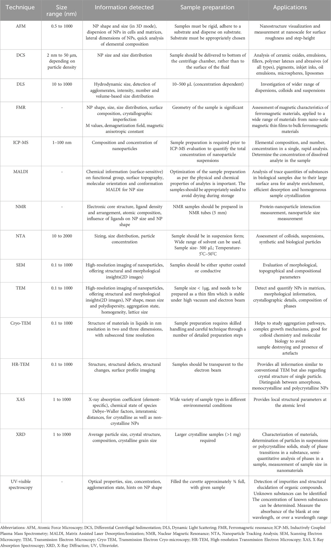
Table 1. Examples of techniques commonly used for the characterization of nanoparticles and purposes.
3 Effect of mean size on the toxicological profile of nanoparticles
Unique properties of NPs, such as shape, surface characteristics and size, need to be optimized in order to capitalize their potential for different biomedical applications (Khan et al., 2019). Thus, the risk of toxicity arising due to their nano size is largely overlooked (Yang et al., 2017). Due to their small size, NPs can be a source of toxicity, both when used in medical applications and when released into the environment. NPs toxicological characterization aims to elucidate the interaction between the physicochemical features of these NPs and their biological targets. Hence, thorough understanding of the interaction taking place between biological systems and NPs is needed. Studies correlating the size of NPs with toxicity using cells in cultures, as well as animal models, are being undertaken. These studies in particular, aim to assess the effect of size of NPs on their in vitro and in vivo toxicological profile, paving the way towards the fabrication of appropriate carriers for various therapeutic applications (Abbasi et al., 2023; Qamar et al., 2024).
3.1 In vitro toxicity evaluation of nanoparticles
The merits of in vitro toxicity assessment include relatively lower cost and fewer ethical concerns, when compared to the in vivo screening assays. In vitro cytotoxicity is mainly assessed through necrosis, apoptosis, proliferation, oxidative stress and DNA damage assays (Lama et al., 2020; Tirumala et al., 2021). Evaluation of in vitro toxicity using various cell lines has been used and reported in a number of publications [e.g., (Silva et al., 2019a; Silva et al., 2019b]. Cell viability and lethality are the two most common characteristics assessed for in vitro measurement of toxicity for different types of NPs (Silva et al., 2019a; Silva et al., 2019b; Mao et al., 2022).
An increased interest in investigating the effects of NPs size on targeting cellular uptake has been observed over the years (Augustine et al., 2020; Galindo-Camacho et al., 2023). Numerous research groups have conducted in vitro studies involving variable cell types and different NPs, and assessed the impact of size and surface charge on their uptake into the cells and on drug targeting.
3.1.1 Cellular uptake studies
Cellular processes in mammals, such as endocytosis, cellular uptake and inflammatory response, have been found to be closely related to particle size (Augustine et al., 2020). Most common mechanisms of NPs cytotoxicity are listed in Table 2.
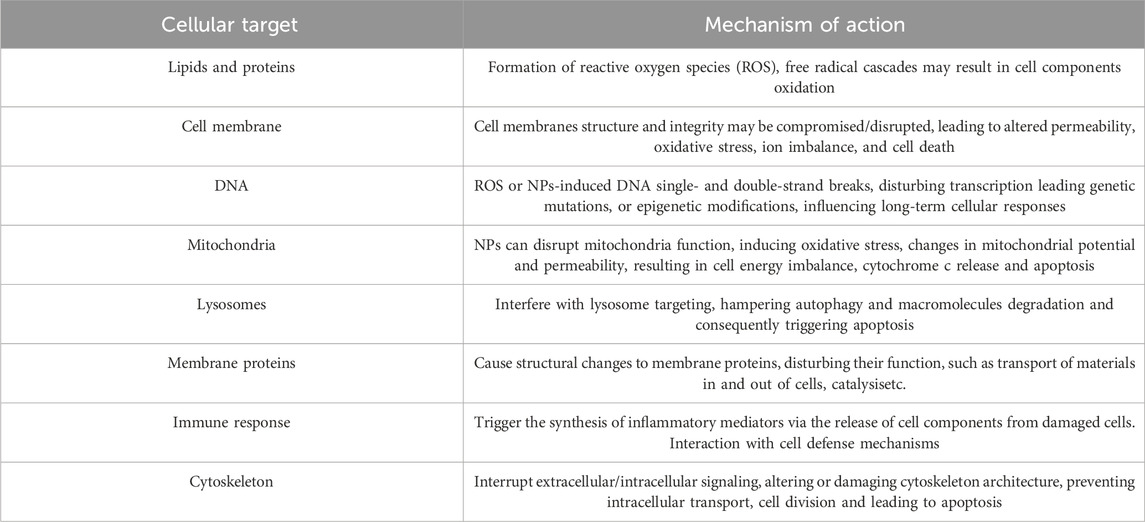
Table 2. Common cytotoxicity mechanisms promoted by nanoparticles. Summary of main cell targets and the putative mechanism of toxicity.
The NPs size determines their interactions with biological systems (Augustine et al., 2020). Although a great deal of work has been carried out to investigate the toxicological effects of NPs in a systematic manner based on their size, the field still demands scientific attention so that the internal fate of the nanosized materials may be disclosed and their toxic potential may be ascertained. This is especially important in case where studies of similar nature performed by different research groups bring out contrasting outcomes.
For a given geometric shape and composition, NPs dimensions are a determining factor of cell uptake (Augustine et al., 2020). NP size influences the entropic and enthalpic features, which impact their cellular uptake by governing the adhesion forces between the cellular receptors and NPs (Villanueva-Flores et al., 2020). Hoshyar et al. (2016) (Hoshyar et al., 2016) reported that most in vitro investigations have shown the highest cellular uptake of NPs with size ranging between 10–60 nm, regardless their composition and surface charge (Hoshyar et al., 2016). In case of materials with the same chemical composition but varying size, numerous physical characteristics and chemistries may be anticipated. NPs have large surface area, therefore, the particle surface is of paramount importance as a large number of atoms are located at the surface (Gnach et al., 2015). It has been reported that NPs with size falling below 5 nm are able to circumvent cell barriers by non-specific means (e.g., translocation). On the other hand, particles with larger size access the cells via phagocytosis, macro pinocytosis, and specific and nonspecific transport mechanisms. Pinocytosis, however, depends on cell type and size of particles, since a size of around 25 nm is optimal (Manzanares and Ceña, 2020; Sousa de Almeida et al., 2021).
For cellular uptake, NPs undergo membrane wrapping process via endocytosis of ligand coated NPs and this mechanism of action is size dependent (Li et al., 2022). NPs of size above 60 nm in diameter may lead to a large interaction with a number of receptors limiting the binding of other NPs in a competitive fashion. NPs with diameters below 30 nm attach to some receptors on the membrane surface but are unable to drive the membrane-wrapping process. NPs ranging between 30 and 60 nm may bind to membrane receptors, usefully driving the membrane-wrapping process.
Pan et al. (2007) studied the size dependent toxicity of gold NPs, sized in the range 0.8–15 nm. The study was conducted on fibroblasts, macrophages, epithelial cells and on melanoma cells, revealing that particles measuring 15 nm were around 60 times less toxic than those measuring 1.4 nm in size (Pan et al., 2007). The authors also found that 1.4 nm sized NPs led to cell necrosis (within a period of 12 h subsequent to the culture medium) (Pan et al., 2007).
In a study carried out by Jiang J. et al (2008) (Jiang W. et al., 2008) using gold NPs, it was reported that the particles measuring 40–50 nm displayed the highest cellular uptake in SKBR-3 cells (Jiang W. et al., 2008). The same results were obtained when the core of NPs was changed to silver (Jiang W. et al., 2008). Studies carried out with HeLa cells using mesoporous silica NPs showed a maximum cellular uptake for particles with 50-nm size (Lu et al., 2009). Zhang et al. (2009) (Zhang et al., 2009) reported the development of a thermodynamic model that exhibited optimum uptake within the cells when the diameter of a ligand-coated NPs was 50 nm (Zhang et al., 2009).
In a study by Huo et al. (2014) (Huo et al., 2014), it was found that gold NPs not greater than 6 nm effectively gained entry within the cell nucleus, while larger sized NPs, i.e., measuring 10 or 16 nm, only crossed the cell membrane and could be found only in the cytoplasm. These findings are particularly interesting in case ultrasmall gold NPs are aimed for gene therapy, having the DNA as their target (Huo et al., 2014).
Braakhuis et al. (2016) evaluated the cytotoxicity of inhaled silver NPs of different sizes. Although the primary determinant of whether NPs reach the alveoli and where they can induce toxicity was found to be their size, the authors also reported the need for a dose metric to assess the risk of nanomaterials of the same chemical composition but varying in size (Braakhuis et al., 2016). This study also described a concentration-dependent rise in lung toxicity markers, i.e., cell counts and pro-inflammatory cytokines, in the bronchoalveolar lavage fluid (Braakhuis et al., 2016).
Carnovale et al. (2019) performed a systematic evaluation of the impact of shape, size and surface capping by developing a set of gold NPs, on cytotoxic behavior and cellular uptake. A reduction in the size of gold NPs resulted in an enhanced cellular uptake, in terms of particle number (Carnovale et al., 2019). Additionally, gold NPs of the same size, stabilized using amino acids, displayed elevated cellular uptake levels, in comparison to citrate or cetyltrimethylammonium bromide (CTAB) coated gold NPs (Carnovale et al., 2019).
Lee et al. (2019) evaluated the cytotoxic effects of silica NPs on human endothelial cells. Unlike other size ranges, silica NPs of ca. 20 nm notably induced both necrosis and apoptosis; however, these two mechanisms occurred independently and via different pathways (Lee et al., 2019). Reactive oxygen species-mediated endoplasmic reticulum (ROS-mediated ER) stress was responsible for apoptotic cell death. Autophagy-mediated necrotic cell death was caused via pI3K/AKT/eNOS signaling axis (Lee et al., 2019).
Chakraborty et al. (2020) investigated the size dependent biological activity of gold NPs on osteosarcoma cells. Gold NPs of size range of 40–60 nm were synthesized by a Tris-assisted citrate-based method, whereas surface enhanced Raman scattering (SERS) was used to confirm size dependent apoptosis of osteosarcoma cells treated with these particles (Chakraborty et al., 2020).
3.1.2 Hemolytic activity
In vitro assays, particularly those assessing hemolytic activity, offer an efficient approach for preliminary toxicity screening; however, these methods often overlook key aspects, such as the influence of plasma proteins and mechanical stress on NP-hemocompatibility. NP toxicity tests should consider disease-specific red blood cells, such as diabetes and hemoglobinopathies (Yedgar et al., 2022). Additionally, the effect of NP size on the hemolytic activity of cells is also worth pondering upon. A study carried out with 100–600 nm mesoporous silica NPs on human erythrocytes established that a large specific surface area of NPs leads to their effective adsorption on cell surface. Particles measuring 100 nm in size were better adsorbed on the surface of human erythrocytes without leading to any alteration in the cell morphology, whereas particles measuring 600 nm entered the cells, causing membrane deformation and hemolysis (Sukhanova et al., 2018).
3.1.3 Administration routes
NPs have become a part of daily life of humans, where they may find access to the human body either in the form of pharmaceutical products, meant for therapeutic or diagnostic purpose, or as a constituent of cosmetic products or accidentally in the form of environmental pollutants (Gnach et al., 2015). Human exposure to nanoparticulate matter may occur via occupational exposure through inhalation and/or ingestion, but also through any drug administration route (Kumah et al., 2023). Investigations in this regard have correlated organ toxicity with pre-determined dose. However, exposure to NPs may have some non-dose dependent effects as well. Since the reduction of the particle size increases the surface area per unit volume, particles become more chemically reactive with the size reduction (Jiang J. et al., 2008).
The extent of toxicity is influenced by the route of administration, as well as by the sites of NP deposition (Lara et al., 2011). Depending upon the route of entry of the NPs, different cell lines have been employed for their in vitro testing. For example, for oral uptake of NPs, cell lines such as Caco-2, HT29, and SW480 have been used (Andreani et al., 2014; Silva et al., 2019a). When intended for intravascular route, HeLa, MCF-7, BT-474, PC3, C4-2, and SKBR-3 have been employed (Doktorovova and Silva, 2018; Souto et al., 2019). To study the transdermal route, keratinocytes (such as HaCaT cells (Carbone et al., 2019b), fibroblasts, and, rarely, sebocytes, have been proposed, while for inhalation uptake RAW 264.7, BEAS-2B, NHBE, 16-HBE, SAEC, and THP-1 cell lines have been used (Sukhanova et al., 2018). Conventional cell-based assays designed to evaluate chemicals may however yield unreliable and erroneous results when employed for the testing of nanosized materials. This is probably because NPs tend to interact with the read-out systems and assay components.
A great deal of research has already provided extensive information and a variety of data; however, a clear understanding of the results is still limited due to the absence of standardized assessment procedures and inconsistencies in the model employed, media used, reference standards adopted and analysis of collected data. Kroll et al. (2012) suggested that classical cytotoxicity assays need to be designed, so that they can be validated for every NP type, and the testing concentration of the NPs must be limited to levels below which interference is likely to occur (Kroll et al., 2012). Data comparison regarding SLN in vitro cytotoxicity (using various cell models), oxidative stress and hemocompatibility studies also revealed the inexistence of standard protocols and data analysis tools which may lead to contradictory results (Doktorovova et al., 2014b; Zielińska et al., 2020).
3.2 In vivo toxicity evaluation
In vivo toxicity evaluation brings NPs a step closer to its clinical relevance. Precision in the control of physicochemical characteristics, such as size, surface charge, shape and stability of NPs, is of utmost importance in in vivo studies since these properties govern their residence time in the blood stream and also their site specific targeting properties (Kumar et al., 2017; Sabourian et al., 2020; Yagublu et al., 2022). In vivo toxicological evaluation is generally performed using appropriate animal models. The choice of an adequate experimental model for estimating toxicity (in vitro and in vivo) is of vital significance. In vitro models (cell lines) easily help to analyze toxic effects of nanoparticulate matter on individual components of cells and tissues, but in vivo evaluation makes it possible to assess the NPs toxicity for individual organ and to the whole organism (Ajdary et al., 2018; Gnach et al., 2015). The assessment techniques for in vivo toxicity comprise studies regarding biodistribution, clearance, hematology, histopathology and serum chemistry.
The most commonly employed animal models, for in vivo studies, include mice and rats, due to their explicit genome, which makes them most appropriate for studying the histopathological changes and functions of various organs (such as hepatic, hematological, renal, nervous and immune systems). Besides these, rabbits, zebra fish, Drosophila melanogaster and Caenorhabdtis alegans also find use in toxicological investigations (Yang et al., 2017; Severino et al., 2023).
In vivo assessment studies may be focused on changes in the tissue structure (Jans and Huo, 2012), apoptosis (Mallidi et al., 2009; Sharma et al., 2009), inflammation and infiltration in major organs (spleen, kidney, brain, lungs and heart), but may also be aimed to investigate certain organ systems, which tend to concentrate NPs on account of their structures specificity (Yang et al., 2017). Elucidating the biodistribution of NPs in various organs may prove beneficial to guide the optimization of NPs, in all respects including their size. After animal sacrifice, the main organs or tissue are removed, commonly to explore distribution of NPs, but also to assess morphological and histological changes and signs of toxicity. The ultra-structural modification and effect of the NPs on tissue can be clearly revealed employing electron microscopy analyses. Functional injury can be detected with the help of diverse biomarkers and sophisticated high-tech instruments (Yang et al., 2017; Joseph et al., 2023). Furthermore, NPs can be detected in live or sacrificed animal models through radio labelling (Dai et al., 2021).
Gold NPs were found to cause apoptosis and acute inflammation in liver, accumulation in various organs, as well as penetration potential in sperm head and tail regions (Cho et al., 2009; Wiwanitkit et al., 2009). A study was carried out with the aim of checking the in vivo toxicity of zinc NPs at concentration of 14–20 μg/mL for 12 h in order to ascertain the impact of treatment on DNA damage, cell viability, apoptosis and ROS production (Sharma et al., 2012).
Silver NPs biodistribution and toxicity were investigated using CD-1 mice by intravenous injection of 10 mg/kg of silver NPs with different particle size, i.e. 10, 40, and 100 nm, possessing different shell coatings. Each type of synthesized NPs caused toxicity to tissues, but larger sized particles were less toxic, apparently because they are less prone to penetration (Recordati et al., 2015).
Jimeno-Romero et al. (2016) evaluated the biodistribution, bioaccumulation and biological effects of disodium laureth sulfosuccinate (DSLS) stabilized titanium dioxide (TiO2) NPs in marine mussels (Mytilus galloprovincallis). This in vivo study used 0.1, 1, and 10 mg titanium/L concentration, either in bulk or as TiO2 NPs (sized 60 and 180 nm). A considerable accumulation of NPs was noted in mussels dosed with TiO2 NPs, localized in lysosomes, endosomes and residual bodies of digestive cells, as well as in the lumen of digestive tubules. The NPs measuring 60 nm were internalized to a larger extent in lysosomes of digestive cells as compared to 180 nm ones. This was confirmed by quantitative analysis of black silver deposits following autometallography (Jimeno-Romero et al., 2016). The larger sized particles were found in the lumen of gut where they were present as large aggregates. This resulted in notably decreased stability of lysosomal membrane to both the types of NPs, though greater in extent upon exposure to those with 60 nm diameter. The membrane stability was also affected by exposure to TiO2 in bulk and DSLS-stabilized NPs. Therefore, it was concluded that membrane stability was dependent on the size of the NPs, and also on the biological effects caused by DSLS and TiO2 (Jimeno-Romero et al., 2016).
Balmuri et al. (2017), Balmuri et al. (2017) synthesized two different types of cadmium oxide NPs, by calcinations of Cd(OH)2 without any organic molecule (CdO-1) and with cadmium-citrate coordination polymer (CdO-2). The authors compared the toxicity of the two types of cadmium oxide NPs, on zebra fish showing those obtained by calcination with CdO-2 reduced toxicity, which was revealed by lower levels of oxidative stress, rescue of hepatic carboxylexterases and diminished metallothionein activity. These results were also supported by histopathological examination. The study articulated the toxic effects of cadmium oxide NPs in aquatic animals and also illustrated that the toxicity could be significantly reduced by carbon coverage (Balmuri et al., 2017).
Kang et al. (2020) designed infrared fluorophore conjugated polyethylene glycols (PEGs) in different sizes, and investigated their size-dependent enhanced permeability and retention (EPR) effect, and checked their pharmacokinetics, biodistribution and renal clearance in tumor-bearing mice. The targeting efficiency of PEGylated particles was confirmed by measuring tumor-to-background ratio. While the smaller sized variants of PEGs (≤20 kDa, 12 nm) displayed marked tumor targeting with either very low or non-specific uptakes, the larger sized variants (>20 kDa, 13 nm) were found to accumulate significantly in major organs, such as liver, lungs and pancreas (Kang et al., 2020).
3.2.1 Genotoxicity evaluation
Genotoxicity can appear directly at DNA, as well as at other chromosome components (Doktorovova et al., 2014a; Doktorovova et al., 2014b; Souto et al., 2020; Cardoso et al., 2021a). Modifications in the genetic material can result in serious health issues in humans and can be responsible for chronic diseases (Kohl et al., 2020). Genotoxicity agents affect DNA and non-DNA targets of cells via several modes of action. Poorly soluble particles have strong tendency to produce ROS, which are responsible for their genotoxicity (Oberdörster et al., 2005; Schins and Knaapen, 2007). Because of their high surface reactivity when compared to microparticles, NPs can produce high ROS levels when compared to larger sized particles (Knaapen et al., 2004; Schins and Knaapen, 2007; Gonzalez et al., 2008).
The potential risk of nanoscale particles in causing genetic damage is assessed through genotoxicity testing. This involves selecting a series of particles made of the same material, but with progressively smaller sizes, to evaluate the impact of size on DNA damage. Gel electrophoresis assay or comet assay are then used to quantify DNA single strand breaks in cells (Cardoso et al., 2021b; Menz et al., 2023). DNA damage results in gene mutation, altered protein synthesis, followed by altered mechanisms of cell cycle arrest which may lead to carcinogenesis (Singh et al., 2017).
The research conducted by Barnes et al. (2008) focused on the examination of genotoxicity by single cell gel electrophoresis or Comet assay performed on a single fibroblast cell line (3T3-L1) to examine five distinct commercial colloidal and laboratory-synthesized silica NPs. None of the tested NPs exhibited any notable genotoxicity. Remarkably, these findings were confirmed independently in two distinct research facilities, demonstrating that in vitro genotoxicity testing may be accurately replicated across different laboratories (Barnes et al., 2008). Nevertheless, caution is needed in the interpretation of the findings that indicate that amorphous silica NPs are not hazardous (Fubini et al., 2010).
Exposure conditions, such as dose and duration, are also significant in determining toxicity of NPs. In addition to size, shape, and the level of hydrophilicity/hydrophobicity, the regulation of particle absorption also significantly influences genotoxicity (Prior et al., 2002; Nam H. Y. et al., 2009). Research on the toxicity of nano-sized particles has demonstrated that these are more harmful than micrometer-sized particles of the same composition. This finding has prompted concerns about the potential influence of NPs on human health (Carbone et al., 2019a). For example, copper oxide NPs have significantly higher toxicity in comparison to copper oxide micrometer-sized particles. On the other hand, the small particles of TiO2 generated a greater amount of DNA damage compared to TiO2 NPs, most likely due to differences in their crystal structures. While the iron oxide NPs did not exhibit toxicity that varied with size, their toxicity was comparatively lower than that of other NPs (Gautam and van Veggel, 2013). Although significant advancements have been made, accurately predicting the toxicity behaviour of NPs remains challenging due to the diverse range of concentrations, sizes, shapes, surface coatings, exposure times, and other factors involved in in vitro and in vivo experiments. This complexity makes it highly difficult to anticipate the toxicity of NPs for a new formulation. Table 3 lists examples of studies reporting the in vitro and in vivo toxicity assessment of NPs.
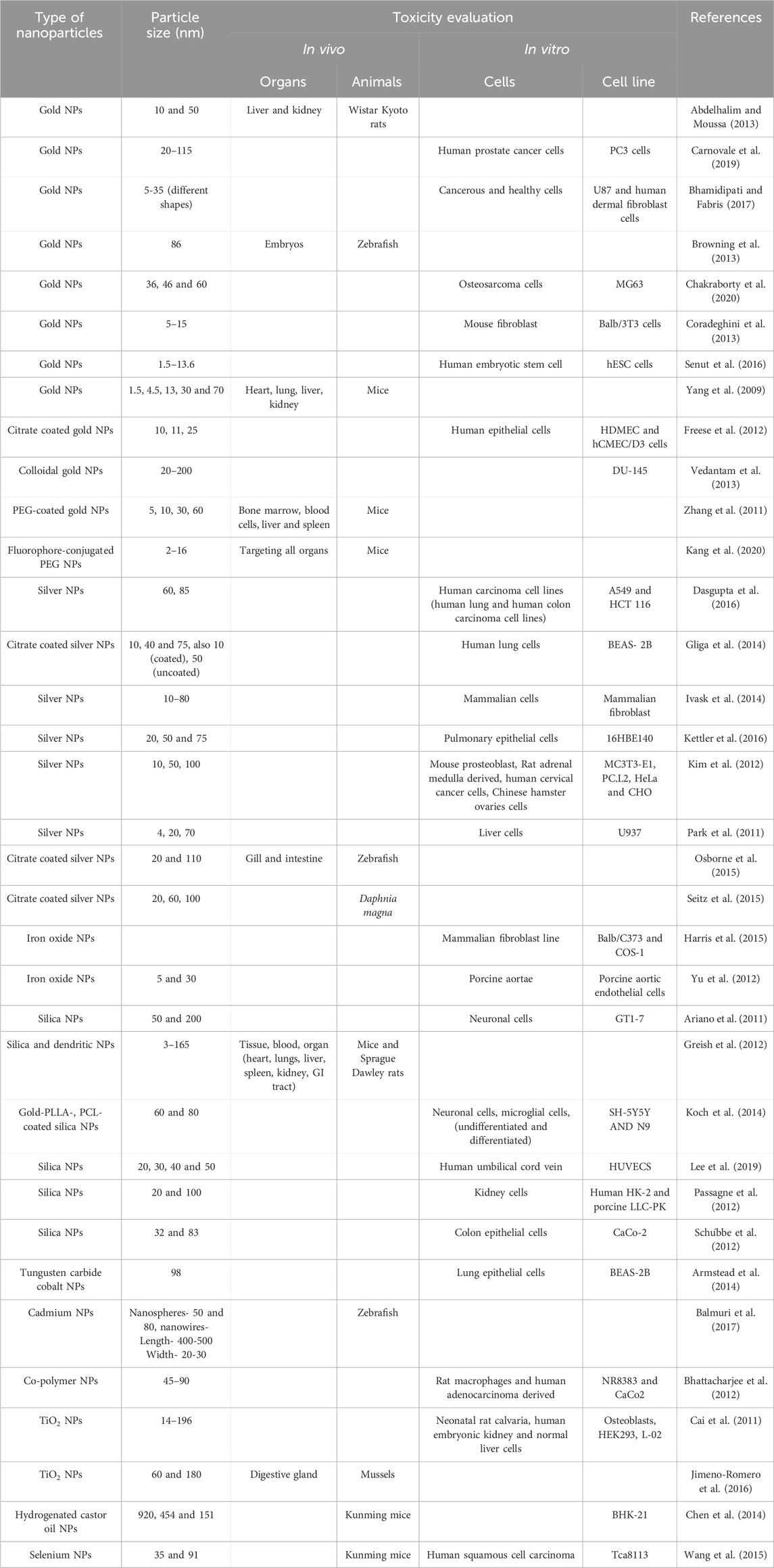
Table 3. Selected studies evaluating in vitro and in vivo toxicity of NPs.
4 Conclusion
Nanotechnology has offered exciting new alternatives for therapeutics and diagnostics in the past 2 decades attributed to the range of new types of nanoparticles (NPs) that were developed and physicochemical characterized using a number of analytical techniques. NPs are classified within the size range varying from 1 to 100 nm. Various types of NPs including gold NPs, citrate-coated gold NPs, colloidal gold NPs, PEG-coated gold NPs, polymeric NPs, silver NPs, citrate coated silver NPs, iron oxide NPs, silica NPs, gold-PLLA-coated silica NPs, PCL-coated silica NPs, tungsten carbide cobalt NPs, cadmium NPs, TiO2 NPs, hydrogenated castor oil NPs, and selenium NPs, have been discussed for their versatile application. Though these fabricated NPs have been a subject of extensive research, and are known for their utility, still several questions remain unanswered and ethical concerns surround their use. Their fabrication is based on the assumption that they are formulated using biodegradable and biocompatible ingredients. Studies on their action at the cellular and systemic levels, allowing for the generation of correlations between the physicochemical properties of NPs and biological responses, are still needed to ultimately serve as a blueprint for the development of future NPs. Computer simulation studies can also play an important role in designing NPs for a specific application. Further, to address the limited data available about safety and toxicity of NPs, it is important to incorporate toxicity research from the early stages of formulation development towards the generation of safer formulations, based on the comprehensive assessment of each NP-based formulation to establish complete toxicity safety profiles. This will help meet the regulatory agency requirements and expedite the approval process.
Author contributions
WSM: Data curation, Formal Analysis, Investigation, Methodology, Resources, Software, Validation, Visualization, Writing – original draft. PS: Data curation, Formal Analysis, Investigation, Methodology, Resources, Software, Validation, Visualization, Writing – original draft. VK: Conceptualization, Data curation, Formal Analysis, Investigation, Methodology, Resources, Software, Validation, Writing – original draft. RR: Conceptualization, Data curation, Formal Analysis, Investigation, Methodology, Resources, Software, Validation, Writing – original draft. AZ: Data curation, Formal Analysis, Investigation, Methodology, Resources, Software, Validation, Visualization, Writing – original draft. AMS: Supervision, Investigation, Methodology, Resources, Software, Validation, Visualization, Writing – review and editing. SM: Conceptualization, Data curation, Formal Analysis, Investigation, Methodology, Resources, Software, Validation, Writing – original draft. EBS: Conceptualization, Supervision, Data curation, Formal Analysis, Investigation, Methodology, Resources, Software, Validation, Visualization, Writing – original draft, Writing – original draft.
Funding
The author(s) declare that financial support was received for the research and/or publication of this article. Eliana B. Souto acknowledges University College Dublin for the Research Scheme fund 2024-2028 (82934-NP/R27885). Amélia M. Silva acknowledges FCT–Portuguese Foundation for Science and Technology, for the support of the project UID/04033:CITAB.
Conflict of interest
The authors declare that the research was conducted in the absence of any commercial or financial relationships that could be construed as a potential conflict of interest.
The author(s) declared that they were an editorial board member of Frontiers, at the time of submission. This had no impact on the peer review process and the final decision.
Publisher’s note
All claims expressed in this article are solely those of the authors and do not necessarily represent those of their affiliated organizations, or those of the publisher, the editors and the reviewers. Any product that may be evaluated in this article, or claim that may be made by its manufacturer, is not guaranteed or endorsed by the publisher.
Abbreviations
AFM, Atomic Force Microscopy; BF-TEM, Bright-Field Transmission Electron Microscopy; CdSe, Cadmium selenide; CLSM, Confocal Laser Scanning Microscopy; Cryo-TEM, Cryo-Transmission Electron Microscopy; DCS, Differential Centrifugal Sedimentation; DLS, Dynamic Light Scattering; DSLS, Disodium Laureth Sulfosuccinate; EPR, Enhanced Permeability and Retention; EXAFS, Extended X-ray Absorption Fine Structure; FMR, Ferromagnetic Resonance; ICP-MS, Inductively Coupled Plasma Mass Spectroscopy; LSPR, Localized Surface Plasmon Resonance; MALDI, Matrix Assisted Laser Desorption/Ionization; MPA, Mercaptoacetic acid; NP, Nanoparticle; NPs, Nanoparticles; NTA, Nanoparticle Tracking Analysis; PCS, Photon Correlation Spectroscopy; PdI, Polydispersity Index; PEG, Polyethylene Glycol; PHEA, Poly(hydroxyethyl Acrylate); PVDF, Polyvinylidene fluoride; SAXS, Small-Angle X-ray Scattering; TiO2, Titanium dioxide; UV-Vis, Ultraviolet-Visible; WAXS, Wide-Angle X-ray Scattering; XANES, X-ray Absorption Near Edge Structure; XAS, X-ray Absorption Spectroscopy.
References
Abbasi, R., Shineh, G., Mobaraki, M., Doughty, S., and Tayebi, L. (2023). Structural parameters of nanoparticles affecting their toxicity for biomedical applications: a review. J. Nanoparticle Res. 25 (3), 43. doi:10.1007/s11051-023-05690-w
Abdelhalim, M. A. K., and Moussa, S. A. A. (2013). The gold nanoparticle size and exposure duration effect on the liver and kidney function of rats: in vivo. Saudi J. Biol. Sci. 20 (2), 177–181. doi:10.1016/j.sjbs.2013.01.007
Ajdary, M., Moosavi, M. A., Rahmati, M., Falahati, M., Mahboubi, M., Mandegary, A., et al. (2018). Health concerns of various nanoparticles: a review of their in vitro and in vivo toxicity. Nanomaterials 8 (9), 634. doi:10.3390/nano8090634
Amaral, V. A., Santana, V. L., Lisboa, E. S., Martins, F. S., Chaud, M. V., de Albuquerque-Junior, R. L. C., et al. (2024). Chitosan membranes incorporating Aloe vera glycolic extract with joint synthesis of silver nanoparticles for the treatment of skin lesions. Drug Deliv. Transl. Res. 15, 1376–1392. doi:10.1007/s13346-024-01683-x
Andersson, R., Andersson, A., and Åman, P. (2009). Molecular weight distributions of water-extractable β-glucan and arabinoxylan, 203, 216. doi:10.1016/b978-1-891127-70-0.50019-4
Andreani, T., Kiill, C. P., Souza, A. L. R. d., Fangueiro, J. F., Fernandes, L., Doktorovová, S., et al. (2014). Surface engineering of silica nanoparticles for oral insulin delivery: characterization and cell toxicity studies. Colloids Surfaces B Biointerfaces 123, 916–923. doi:10.1016/j.colsurfb.2014.10.047
Ariano, P., Zamburlin, P., Gilardino, A., Mortera, R., Onida, B., Tomatis, M., et al. (2011). Interaction of spherical silica nanoparticles with neuronal cells: size-dependent toxicity and perturbation of calcium homeostasis. Small 7 (6), 766–774. doi:10.1002/smll.201002287
Armstead, A. L., Arena, C. B., and Li, B. (2014). Exploring the potential role of tungsten carbide cobalt (WC-Co) nanoparticle internalization in observed toxicity toward lung epithelial cells in vitro. Toxicol. Appl. Pharmacol. 278 (1), 1–8. doi:10.1016/j.taap.2014.04.008
Augustine, R., Hasan, A., Primavera, R., Wilson, R. J., Thakor, A. S., and Kevadiya, B. D. (2020). Cellular uptake and retention of nanoparticles: insights on particle properties and interaction with cellular components. Mater. Today Commun. 25, 101692. doi:10.1016/j.mtcomm.2020.101692
Axet, M. R., Philippot, K., Chaudret, B., Cabié, M., Giorgio, S., and Henry, C. R. (2011). TEM and HRTEM evidence for the role of ligands in the formation of shape-controlled platinum nanoparticles. Small 7 (2), 235–241. doi:10.1002/smll.201001112
Babick, F., Mielke, J., Wohlleben, W., Weigel, S., and Hodoroaba, V.-D. (2016). How reliably can a material be classified as a nanomaterial? Available particle-sizing techniques at work. J. Nanoparticle Res. 18 (6), 158–240. doi:10.1007/s11051-016-3461-7
Balmuri, S. R., Selvaraj, U., Kumar, V. V., Anthony, S. P., Tsatsakis, A. M., Golokhvast, K. S., et al. (2017). Effect of surfactant in mitigating cadmium oxide nanoparticle toxicity: implications for mitigating cadmium toxicity in environment. Environ. Res. 152, 141–149. doi:10.1016/j.envres.2016.10.005
Barnes, C. A., Elsaesser, A., Arkusz, J., Smok, A., Palus, J., Lesniak, A., et al. (2008). Reproducible comet assay of amorphous silica nanoparticles detects no genotoxicity. Nano Lett. 8 (9), 3069–3074. doi:10.1021/nl801661w
Bashiri, G., Padilla, M. S., Swingle, K. L., Shepherd, S. J., Mitchell, M. J., and Wang, K. (2023). Nanoparticle protein corona: from structure and function to therapeutic targeting. Lab. Chip 23 (6), 1432–1466. doi:10.1039/d2lc00799a
Bayda, S., Adeel, M., Tuccinardi, T., Cordani, M., and Rizzolio, F. (2019). The history of nanoscience and nanotechnology: from chemical-physical applications to nanomedicine. Molecules 25 (1), 112. doi:10.3390/molecules25010112
Bell, N. C., Minelli, C., Tompkins, J., Stevens, M. M., and Shard, A. G. (2012). Emerging techniques for submicrometer particle sizing applied to Stober silica. Langmuir 28 (29), 10860–10872. doi:10.1021/la301351k
Bhamidipati, M., and Fabris, L. (2017). Multiparametric assessment of gold nanoparticle cytotoxicity in cancerous and healthy cells: the role of size, shape, and surface chemistry. Bioconjugate Chem. 28 (2), 449–460. doi:10.1021/acs.bioconjchem.6b00605
Bhattacharjee, S., Ershov, D., Fytianos, K., van der Gucht, J., Alink, G. M., Rietjens, I. M., et al. (2012). Cytotoxicity and cellular uptake of tri-block copolymer nanoparticles with different size and surface characteristics. Part. Fibre Toxicol. 9 (1), 11–19. doi:10.1186/1743-8977-9-11
Blanco-Andujar, C., Ortega, D., Southern, P., Nesbitt, S. A., Thanh, N. T. K., and Pankhurst, Q. A. (2016). Real-time tracking of delayed-onset cellular apoptosis induced by intracellular magnetic hyperthermia. Nanomedicine 11 (2), 121–136. doi:10.2217/nnm.15.185
Braakhuis, H. M., Cassee, F. R., Fokkens, P. H., De La Fonteyne, L. J., Oomen, A. G., Krystek, P., et al. (2016). Identification of the appropriate dose metric for pulmonary inflammation of silver nanoparticles in an inhalation toxicity study. Nanotoxicology 10 (1), 63–73. doi:10.3109/17435390.2015.1012184
Braeckmans, K., Buyens, K., Bouquet, W., Vervaet, C., Joye, P., Vos, F. D., et al. (2010). Sizing nanomatter in biological fluids by fluorescence single particle tracking. Nano Lett. 10 (11), 4435–4442. doi:10.1021/nl103264u
Browning, L. M., Huang, T., and Xu, X.-H. N. (2013). Real-time in vivo imaging of size-dependent transport and toxicity of gold nanoparticles in zebrafish embryos using single nanoparticle plasmonic spectroscopy. Interface Focus 3 (3), 20120098. doi:10.1098/rsfs.2012.0098
Cai, K., Hou, Y., Hu, Y., Zhao, L., Luo, Z., Shi, Y., et al. (2011). Correlation of the cytotoxicity of TiO2 nanoparticles with different particle sizes on a sub-200-nm scale. Small 7 (21), 3026–3031. doi:10.1002/smll.201101170
Caputo, F., Clogston, J., Calzolai, L., Rösslein, M., and Prina-Mello, A. (2019). Measuring particle size distribution of nanoparticle enabled medicinal products, the joint view of EUNCL and NCI-NCL. A step by step approach combining orthogonal measurements with increasing complexity. J. Control. Release 299, 31–43. doi:10.1016/j.jconrel.2019.02.030
Carbone, C., Teixeira, M. D. C., Sousa, M. D. C., Martins-Gomes, C., Silva, A. M., Souto, E. M. B., et al. (2019a). Clotrimazole-loaded mediterranean essential oils NLC: a synergic treatment of Candida skin infections. Pharmaceutics 11 (5), 231. doi:10.3390/pharmaceutics11050231
Carbone, C., Teixeira, M. d.C., Sousa, M. d.C., Martins-Gomes, C., Silva, A. M., Souto, E. M. B., et al. (2019b). Clotrimazole-loaded mediterranean essential oils NLC: a synergic treatment of Candida skin infections. Pharmaceutics 11 (5), 231. doi:10.3390/pharmaceutics11050231
Cardoso, S., da Silva, C. F., Severino, P., Silva, A. M., Souto, S. B., Zielińska, A., et al. (2021a). Genotoxicity assessment of metal-based nanocomposites applied in drug delivery. Mater. (Basel) 14 (21), 6551. doi:10.3390/ma14216551
Cardoso, S., da Silva, C. F., Severino, P., Silva, A. M., Souto, S. B., Zielińska, A., et al. (2021b). Genotoxicity assessment of metal-based nanocomposites applied in drug delivery. Mater. (Basel) 14 (21), 6551. doi:10.3390/ma14216551
Carne, D., Peoples, J., Feng, D., Ruan, X. J. A. J. o.H., and Transfer, M. (2023). Accelerated prediction of photon transport in nanoparticle media using machine learning trained with Monte Carlo simulations. ASME J. Heat Mass Transf. 145 (5), 052502. doi:10.1115/1.4062188
Carnovale, C., Bryant, G., Shukla, R., and Bansal, V. (2019). Identifying trends in gold nanoparticle toxicity and uptake: size, shape, capping ligand, and biological corona. ACS Omega 4 (1), 242–256. doi:10.1021/acsomega.8b03227
Carpentieri, M. A., and Domenici, V. (2024). Introducing UV–visible spectroscopy at high school level following the historical evolution of spectroscopic instruments: a proposal for chemistry teachers. Found. Chem. 26 (1), 115–139. doi:10.1007/s10698-024-09501-5
Chakraborty, A., Das, A., Raha, S., and Barui, A. (2020). Size-dependent apoptotic activity of gold nanoparticles on osteosarcoma cells correlated with SERS signal. J. Photochem. Photobiol. B Biol. 203, 111778. doi:10.1016/j.jphotobiol.2020.111778
Chen, X., Wang, T., Lu, M., Zhu, L., Wang, Y., and Zhou, W. (2014). Preparation and evaluation of tilmicosin-loaded hydrogenated castor oil nanoparticle suspensions of different particle sizes. Int. J. Nanomedicine 9, 2655–2664. doi:10.2147/ijn.s58898
Cho, W.-S., Cho, M., Jeong, J., Choi, M., Cho, H.-Y., Han, B. S., et al. (2009). Acute toxicity and pharmacokinetics of 13 nm-sized PEG-coated gold nanoparticles. Toxicol. Appl. Pharmacol. 236 (1), 16–24. doi:10.1016/j.taap.2008.12.023
Chu, B., and Liu, T. (2000). Characterization of nanoparticles by scattering techniques. J. Nanoparticle Res. 2 (1), 29–41. doi:10.1023/a:1010001822699
Concha, V. O. C., Timóteo, L., Duarte, L. A. N., Bahú, J. O., Munoz, F. L., Silva, A. P., et al. (2024). Properties, characterization and biomedical applications of polyvinylidene fluoride (PVDF): a review. J. Mater. Sci. 59 (31), 14185–14204. doi:10.1007/s10853-024-10046-3
Coradeghini, R., Gioria, S., García, C. P., Nativo, P., Franchini, F., Gilliland, D., et al. (2013). Size-dependent toxicity and cell interaction mechanisms of gold nanoparticles on mouse fibroblasts. Toxicol. Lett. 217 (3), 205–216. doi:10.1016/j.toxlet.2012.11.022
Cutler, J. I., Auyeung, E., and Mirkin, C. A. (2012). Spherical nucleic acids. J. Am. Chem. Soc. 134 (3), 1376–1391. doi:10.1021/ja209351u
Dai, W., Zhang, J., Wang, Y., Jiao, C., Song, Z., Ma, Y., et al. (2021). Radiolabeling of nanomaterials: advantages and challenges. Front. Toxicol. 3, 753316–762021. doi:10.3389/ftox.2021.753316
Dasgupta, N., Ranjan, S., Rajendran, B., Manickam, V., Ramalingam, C., Avadhani, G. S., et al. (2016). Thermal co-reduction approach to vary size of silver nanoparticle: its microbial and cellular toxicology. Environ. Sci. Pollut. Res. 23 (5), 4149–4163. doi:10.1007/s11356-015-4570-z
de Oliveira, D. M., Menezes, D. B., Andrade, L. R., Lima, F. D. C., Hollanda, L., Zielinska, A., et al. (2021). Silver nanoparticles obtained from Brazilian pepper extracts with synergistic anti-microbial effect: production, characterization, hydrogel formulation, cell viability, and in vitro efficacy. Pharm. Dev. Technol. 26 (5), 539–548. doi:10.1080/10837450.2021.1898634
de Oliveira, P. F. M., Michalchuk, A. A. L., Buzanich, A. G., Bienert, R., Torresi, R. M., Camargo, P. H. C., et al. (2020). Tandem X-ray absorption spectroscopy and scattering for in situ time-resolved monitoring of gold nanoparticle mechanosynthesis. Chem. Commun. 56 (71), 10329–10332. doi:10.1039/d0cc03862h
de Souza, J. F., da Silva Pontes, K., Alves, T. F. R., Torqueti de Barros, C., Amaral, V. A., de Moura Crescencio, K. M., et al. (2020). Structural comparison, physicochemical properties, and in vitro release profile of curcumin-loaded lyotropic liquid crystalline nanoparticle: influence of hydrotrope as interface stabilizers. J. Mol. Liq. 306, 112861. doi:10.1016/j.molliq.2020.112861
Doktorovova, S., and Silva, A. M. (2018). Hansen solubility parameters (HSP) for prescreening formulation of solid lipid nanoparticles (SLN): in vitro testing of curcumin-loaded SLN in MCF-7 and BT-474 cell lines. Pharm. Dev. Technol. 23 (1), 96–105. doi:10.1080/10837450.2017.1384491
Doktorovova, S., Silva, A. M., Gaivao, I., Souto, E. B., Teixeira, J. P., and Martins-Lopes, P. (2014a). Comet assay reveals no genotoxicity risk of cationic solid lipid nanoparticles. J. Appl. Toxicol. 34 (4), 395–403. doi:10.1002/jat.2961
Doktorovova, S., Souto, E. B., and Silva, A. M. (2014b). Nanotoxicology applied to solid lipid nanoparticles and nanostructured lipid carriers - a systematic review of in vitro data. Eur. J. Pharm. Biopharm. 87 (1), 1–18. doi:10.1016/j.ejpb.2014.02.005
Emery, A. A., Saal, J. E., Kirklin, S., Hegde, V. I., and Wolverton, C. (2016). High-throughput computational screening of perovskites for thermochemical water splitting applications. Chem. Mater. 28 (16), 5621–5634. doi:10.1021/acs.chemmater.6b01182
Feltin, N., Crouzier, L., Delvallée, A., Pellegrino, F., Maurino, V., Bartczak, D., et al. (2023). Metrological protocols for reaching reliable and SI-traceable size results for multi-modal and complexly shaped reference nanoparticles. Nanomater. (Basel) 13 (6), 993. doi:10.3390/nano13060993
Feltin, N., Delvallée, A., and Crouzier, L. (2024). Strategy for ensuring the metrological traceability of nanoparticle size measurements by SEM. Nanomater. (Basel) 14 (11), 931. doi:10.3390/nano14110931
Filipe, V., Hawe, A., and Jiskoot, W. (2010). Critical evaluation of Nanoparticle Tracking Analysis (NTA) by NanoSight for the measurement of nanoparticles and protein aggregates. Pharm. Res. 27 (5), 796–810. doi:10.1007/s11095-010-0073-2
Freese, C., Uboldi, C., Gibson, M. I., Unger, R. E., Weksler, B. B., Romero, I. A., et al. (2012). Uptake and cytotoxicity of citrate-coated gold nanospheres: comparative studies on human endothelial and epithelial cells. Part. Fibre Toxicol. 9 (1), 23–11. doi:10.1186/1743-8977-9-23
Fubini, B., Ghiazza, M., and Fenoglio, I. (2010). Physico-chemical features of engineered nanoparticles relevant to their toxicity. Nanotoxicology 4 (4), 347–363. doi:10.3109/17435390.2010.509519
Galindo-Camacho, R. M., Haro, I., Gómara, M. J., Espina, M., Fonseca, J., Martins-Gomes, C., et al. (2023). Cell penetrating peptides-functionalized Licochalcone-A-loaded PLGA nanoparticles for ocular inflammatory diseases: evaluation of in vitro anti-proliferative effects, stabilization by freeze-drying and characterization of an in-situ forming gel. Int. J. Pharm. 639, 122982. doi:10.1016/j.ijpharm.2023.122982
Gautam, A., and van Veggel, F. C. (2013). Synthesis of nanoparticles, their biocompatibility, and toxicity behavior for biomedical applications. J. Mater. Chem. B 1 (39), 5186–5200. doi:10.1039/c3tb20738b
Gliga, A. R., Skoglund, S., Wallinder, I. O., Fadeel, B., and Karlsson, H. L. (2014). Size-dependent cytotoxicity of silver nanoparticles in human lung cells: the role of cellular uptake, agglomeration and Ag release. Part. Fibre Toxicol. 11 (1), 1–17. doi:10.1186/1743-8977-11-11
Gnach, A., Lipinski, T., Bednarkiewicz, A., Rybka, J., and Capobianco, J. A. (2015). Upconverting nanoparticles: assessing the toxicity. Chem. Soc. Rev. 44 (6), 1561–1584. doi:10.1039/c4cs00177j
Gontier, E., Ynsa, M.-D., Biro, T., Hunyadi, J., Kiss, B., Gaspar, K., et al. (2008). Is there penetration of titania nanoparticles in sunscreens through skin? A comparative electron and ion microscopy study. Nanotoxicology 2 (4), 218–231. doi:10.1080/17435390802538508
Gonzalez, L., Lison, D., and Kirsch-Volders, M. (2008). Genotoxicity of engineered nanomaterials: a critical review. Nanotoxicology 2 (4), 252–273. doi:10.1080/17435390802464986
Greish, K., Thiagarajan, G., Herd, H., Price, R., Bauer, H., Hubbard, D., et al. (2012). Size and surface charge significantly influence the toxicity of silica and dendritic nanoparticles. Nanotoxicology 6 (7), 713–723. doi:10.3109/17435390.2011.604442
Gross, J., Sayle, S., Karow, A. R., Bakowsky, U., and Garidel, P. (2016). Nanoparticle tracking analysis of particle size and concentration detection in suspensions of polymer and protein samples: influence of experimental and data evaluation parameters. Eur. J. Pharm. Biopharm. 104, 30–41. doi:10.1016/j.ejpb.2016.04.013
Guo, C., and Yarger, J. L. (2018). Characterizing gold nanoparticles by NMR spectroscopy. Magnetic Reson. Chem. 56 (11), 1074–1082. doi:10.1002/mrc.4753
Gupta, R., and Xie, H. (2018). Nanoparticles in daily life: applications, toxicity and regulations. J. Environ. Pathology, Toxicol. Oncol. 37 (3), 209–230. doi:10.1615/jenvironpatholtoxicoloncol.2018026009
Haiss, W., Thanh, N. T., Aveyard, J., and Fernig, D. G. (2007). Determination of size and concentration of gold nanoparticles from UV− Vis spectra. Anal. Chem. 79 (11), 4215–4221. doi:10.1021/ac0702084
Hameed, B. S., Bhatt, C. S., Nagaraj, B., and Suresh, A. K. (2018). “Chromatography as an efficient technique for the separation of diversified nanoparticles,” in Nanomaterials in chromatography (Elsevier), 503–518.
Hard, R., Hipp, J., Tangrea, M. A., and Tomaszewski, J. E. (2014). “Applications of image science in pathology and cell Biology,” in Pathobiology of human disease.
Harris, G., Palosaari, T., Magdolenova, Z., Mennecozzi, M., Gineste, J. M., Saavedra, L., et al. (2015). Iron oxide nanoparticle toxicity testing using high-throughput analysis and high-content imaging. Nanotoxicology 9 (Suppl. 1), 87–94. doi:10.3109/17435390.2013.816797
Hoshyar, N., Gray, S., Han, H., and Bao, G. (2016). The effect of nanoparticle size on in vivo pharmacokinetics and cellular interaction. Nanomedicine (Lond) 11 (6), 673–692. doi:10.2217/nnm.16.5
Hosseingholilou, S., Dorranian, D., and Ghoranneviss, M. (2020). Characterization of gold nanoparticle thin film prepared by electrophoretic deposition method. Gold Bull. 53 (1), 1–10. doi:10.1007/s13404-020-00268-z
Hubert, M., Yang, D. T., Kwok, S. C., Rios, A., Das, T. K., Patel, A., et al. (2020). A multicompany assessment of submicron particle levels by NTA and RMM in a wide range of late-phase clinical and commercial biotechnology-derived protein products. J. Pharm. Sci. 109 (1), 830–844. doi:10.1016/j.xphs.2019.10.025
Huo, S., Jin, S., Ma, X., Xue, X., Yang, K., Kumar, A., et al. (2014). Ultrasmall gold nanoparticles as carriers for nucleus-based gene therapy due to size-dependent nuclear entry. ACS Nano 8 (6), 5852–5862. doi:10.1021/nn5008572
Hussain, M. H., Bakar, N. F. A., Mustapa, A. N., Low, K.-F., Othman, N. H., and Adam, F. (2020). Synthesis of various size gold nanoparticles by chemical reduction method with different solvent polarity. Nanoscale Res. Lett. 15 (1), 1–10. doi:10.1186/s11671-020-03370-5
Ingham, B. (2015). X-ray scattering characterisation of nanoparticles. Crystallogr. Rev. 21 (4), 229–303. doi:10.1080/0889311x.2015.1024114
ISO (2023). ISO 80004-1:2023 nanotechnologies – vocabularyPart 1: core vocabulary. Available online at: https://www.iso.org/standard/79525.html.
Ito, T., Sun, L., Bevan, M. A., and Crooks, R. M. (2004). Comparison of nanoparticle size and electrophoretic mobility measurements using a carbon-nanotube-based coulter counter, dynamic light scattering, transmission electron microscopy, and phase analysis light scattering. Langmuir 20 (16), 6940–6945. doi:10.1021/la049524t
Ivask, A., Kurvet, I., Kasemets, K., Blinova, I., Aruoja, V., Suppi, S., et al. (2014). Size-dependent toxicity of silver nanoparticles to bacteria, yeast, algae, crustaceans and mammalian cells in vitro. PloS one 9 (7), e102108. doi:10.1371/journal.pone.0102108
Jans, H., and Huo, Q. (2012). Gold nanoparticle-enabled biological and chemical detection and analysis. Chem. Soc. Rev. 41 (7), 2849–2866. doi:10.1039/c1cs15280g
Jiang, J., Oberdörster, G., Elder, A., Gelein, R., Mercer, P., and Biswas, P. (2008a). Does nanoparticle activity depend upon size and crystal phase? Nanotoxicology 2 (1), 33–42. doi:10.1080/17435390701882478
Jiang, W., Kim, B. Y., Rutka, J. T., and Chan, W. C. (2008b). Nanoparticle-mediated cellular response is size-dependent. Nat. Nanotechnol. 3 (3), 145–150. doi:10.1038/nnano.2008.30
Jimeno-Romero, A., Oron, M., Cajaraville, M., Soto, M., and Marigómez, I. (2016). Nanoparticle size and combined toxicity of TiO2 and DSLS (surfactant) contribute to lysosomal responses in digestive cells of mussels exposed to TiO2 nanoparticles. Nanotoxicology 10 (8), 1168–1176. doi:10.1080/17435390.2016.1196250
Joseph, T. M., Kar Mahapatra, D., Esmaeili, A., Piszczyk, Ł., Hasanin, M. S., Kattali, M., et al. (2023). Nanoparticles: taking a unique position in medicine. Nanomater. (Basel) 13 (3), 574. doi:10.3390/nano13030574
José-Yacamán, M., Marın-Almazo, M., and Ascencio, J. (2001). High resolution TEM studies on palladium nanoparticles. J. Mol. Catal. A Chem. 173 (1-2), 61–74. doi:10.1016/s1381-1169(01)00145-5
Kang, H., Rho, S., Stiles, W. R., Hu, S., Baek, Y., Hwang, D. W., et al. (2020). Size-dependent EPR effect of polymeric nanoparticles on tumor targeting. Adv. Healthc. Mater. 9 (1), 1901223. doi:10.1002/adhm.201901223
Kastner, E., and Perrie, Y. (2016). “Particle size analysis of micro and nanoparticles,” in Analytical techniques in the pharmaceutical sciences (Springer), 677–699.
Kettler, K., Krystek, P., Giannakou, C., Hendriks, A. J., and de Jong, W. H. (2016). Exploring the effect of silver nanoparticle size and medium composition on uptake into pulmonary epithelial 16HBE14o-cells. J. Nanoparticle Res. 18 (7), 182–211. doi:10.1007/s11051-016-3493-z
Khalid, S., Afzal, N., Khan, J. A., Hussain, Z., Qureshi, A. S., Anwar, H., et al. (2018). Antioxidant resveratrol protects against copper oxide nanoparticle toxicity in vivo. Naunyn-Schmiedeberg's Archives Pharmacol. 391 (10), 1053–1062. doi:10.1007/s00210-018-1526-0
Khan, I., Ali, S., Mansha, M., and Qurashi, A. (2017). Sonochemical assisted hydrothermal synthesis of pseudo-flower shaped Bismuth vanadate (BiVO4) and their solar-driven water splitting application. Ultrason. Sonochemistry 36, 386–392. doi:10.1016/j.ultsonch.2016.12.014
Khan, I., Saeed, K., and Khan, I. (2019). Nanoparticles: properties, applications and toxicities. Arabian J. Chem. 12 (7), 908–931. doi:10.1016/j.arabjc.2017.05.011
Khan, T. M., Khan, S. U. D., Khan, S. U. D., Ahmad, A., Abbasi, S. A., Khan, E. M., et al. (2020). Silver nanoparticle films by flowing gas atmospheric pulsed laser deposition and application to surface-enhanced Raman spectroscopy. Int. J. Energy Res. 44 (14), 11443–11452. doi:10.1002/er.5767
Kim, T. H., Kim, M., Park, H. S., Shin, U. S., Gong, M. S., and Kim, H. W. (2012). Size-dependent cellular toxicity of silver nanoparticles. J. Biomed. Mater. Res. Part A 100 (4), 1033–1043. doi:10.1002/jbm.a.34053
Kirschling, T. L., Golas, P. L., Unrine, J. M., Matyjaszewski, K., Gregory, K. B., Lowry, G. V., et al. (2011). Microbial bioavailability of covalently bound polymer coatings on model engineered nanomaterials. Environ. Sci. and Technol. 45 (12), 5253–5259. doi:10.1021/es200770z
Knaapen, A. M., Borm, P. J., Albrecht, C., and Schins, R. P. (2004). Inhaled particles and lung cancer. Part A: mechanisms. Int. J. Cancer 109 (6), 799–809. doi:10.1002/ijc.11708
Koch, F., Möller, A.-M., Frenz, M., Pieles, U., Kuehni-Boghenbor, K., and Mevissen, M. (2014). An in vitro toxicity evaluation of gold-PLLA-and PCL-coated silica nanoparticles in neuronal cells for nanoparticle-assisted laser-tissue soldering. Toxicol. Vitro 28 (5), 990–998. doi:10.1016/j.tiv.2014.04.010
Kohl, Y., Rundén-Pran, E., Mariussen, E., Hesler, M., El Yamani, N., Longhin, E. M., et al. (2020). Genotoxicity of nanomaterials: advanced in vitro models and high throughput methods for human hazard assessment—a review. Nanomaterials 10 (10), 1911. doi:10.3390/nano10101911
Krawczykowski, D. (2024). Unification of particle size analysis results, part 1 − comparison of particle size distribution functions obtained by various measurement methods. Measurement 238, 115403. doi:10.1016/j.measurement.2024.115403
Kroll, A., Pillukat, M. H., Hahn, D., and Schnekenburger, J. (2012). Interference of engineered nanoparticles with in vitro toxicity assays. Archives Toxicol. 86 (7), 1123–1136. doi:10.1007/s00204-012-0837-z
Kruse, B., Dash, B. S., Kostka, K., Wolff, N., Prymak, O., Loza, K., et al. (2024). Doxorubicin-loaded ultrasmall gold nanoparticles (1.5 nm) for brain tumor therapy and assessment of their biodistribution. ACS Appl. Bio Mater. 7 (10), 6890–6907. doi:10.1021/acsabm.4c00999
Kumah, E. A., Fopa, R. D., Harati, S., Boadu, P., Zohoori, F. V., and Pak, T. (2023). Human and environmental impacts of nanoparticles: a scoping review of the current literature. BMC Public Health 23 (1), 1059. doi:10.1186/s12889-023-15958-4
Kumar, P. S., Pavithra, K. G., and Naushad, M. (2019). “Characterization techniques for nanomaterials,” in Nanomaterials for solar cell applications (Elsevier), 97–124.
Kumar, S., and Rao, R. (2019). Analytical tools for cyclodextrin nanosponges in pharmaceutical field: a review. J. Inclusion Phenom. Macrocycl. Chem. 94 (1), 11–30. doi:10.1007/s10847-019-00903-z
Kumar, V., Sharma, N., and Maitra, S. (2017). In vitro and in vivo toxicity assessment of nanoparticles. Int. Nano Lett. 7 (4), 243–256. doi:10.1007/s40089-017-0221-3
Lama, S., Merlin-Zhang, O., and Yang, C. (2020). In vitro and in vivo models for evaluating the oral toxicity of nanomedicines. Nanomater. (Basel) 10 (11), 2177. doi:10.3390/nano10112177
Lara, Y., Nguyen, T., Marilena, L., and Alexander, M. (2011). Toxicology and clinical potential of nanoparticles. Nano Today 6, 585–607. doi:10.1016/j.nantod.2011.10.001
Lee, K., Lee, J., Kwak, M., Cho, Y.-L., Hwang, B., Cho, M. J., et al. (2019). Two distinct cellular pathways leading to endothelial cell cytotoxicity by silica nanoparticle size. J. Nanobiotechnology 17 (1), 24–14. doi:10.1186/s12951-019-0456-4
Li, Y., Zhang, M., Niu, X., and Yue, T. (2022). Selective membrane wrapping on differently sized nanoparticles regulated by clathrin assembly: a computational model. Colloids Surfaces B Biointerfaces 214, 112467. doi:10.1016/j.colsurfb.2022.112467
Lu, F., Wu, S. H., Hung, Y., and Mou, C. Y. (2009). Size effect on cell uptake in well-suspended, uniform mesoporous silica nanoparticles. Small 5 (12), 1408–1413. doi:10.1002/smll.200900005
Mahant, S., Kumar, S., Pahwa, R., Kaushik, D., Nanda, S., and Rao, R. (2019). Solid lipid nanoparticles in drug delivery for skincare. Nanoparticulate Drug Deliv. Syst. 337, 337–368. doi:10.1201/9781351137263-10
Mahant, S., Rao, R., Souto, E. B., and Nanda, S. (2020). Analytical tools and evaluation strategies for nanostructured lipid carrier-based topical delivery systems. Expert Opin. Drug Deliv. 17 (7), 963–992. doi:10.1080/17425247.2020.1772750
Mallidi, S., Larson, T., Tam, J., Joshi, P. P., Karpiouk, A., Sokolov, K., et al. (2009). Multiwavelength photoacoustic imaging and plasmon resonance coupling of gold nanoparticles for selective detection of cancer. Nano Lett. 9 (8), 2825–2831. doi:10.1021/nl802929u
Manzanares, D., and Ceña, V. (2020). Endocytosis: the nanoparticle and submicron nanocompounds gateway into the cell. Pharmaceutics 12 (4), 371. doi:10.3390/pharmaceutics12040371
Mao, B.-H., Luo, Y.-K., Wang, B., Chen, C.-W., Cheng, F.-Y., Lee, Y.-H., et al. (2022). Use of an in silico knowledge discovery approach to determine mechanistic studies of silver nanoparticles-induced toxicity from in vitro to in vivo. Part. Fibre Toxicol. 19 (1), 6. doi:10.1186/s12989-022-00447-0
Marbella, L. E., and Millstone, J. E. (2015). NMR techniques for noble metal nanoparticles. Chem. Mater. 27 (8), 2721–2739. doi:10.1021/cm504809c
Menz, J., Götz, M. E., Gündel, U., Gürtler, R., Herrmann, K., Hessel-Pras, S., et al. (2023). Genotoxicity assessment: opportunities, challenges and perspectives for quantitative evaluations of dose-response data. Archives Toxicol. 97 (9), 2303–2328. doi:10.1007/s00204-023-03553-w
Modrow, H. (2005). X-Ray methods for the characterization of nanoparticles. Nanofabrication Towards Biomed. Appl. Tech. Tools, Appl. Impact, 163–196. doi:10.1002/3527603476.ch7
Monakhova, P. A., Shalaev, P. V., and Gorev, I. N. (2023). Rapid characterization of synthesized nanoparticles’ liquid dispersions using nanoparticle tracking analysis. Mater. Proc. 14 (1), 65. doi:10.3390/iocn2023-14528
Mourdikoudis, S., Pallares, R. M., and Thanh, N. T. (2018). Characterization techniques for nanoparticles: comparison and complementarity upon studying nanoparticle properties. Nanoscale 10 (27), 12871–12934. doi:10.1039/c8nr02278j
Mulholland, G. W., Duelge, K. J., Hackley, V. A., Farkas, N., Kramar, J. A., Zachariah, M. R., et al. (2024). Measurement of 100 nm monodisperse particles by four Accurate methods: traceability and uncertainty. Aerosol Sci. Technol. 58 (3), 323–333. doi:10.1080/02786826.2024.2310548
Nakajima, H., Ogura, T., Kato, Y., Kondo, N., Usukawa, R., Watanabe, R., et al. (2024). Quantitative evaluation of particle-binder interactions in ceramic slurries via differential centrifugal sedimentation. Sci. Rep. 14 (1), 18508. doi:10.1038/s41598-024-68420-9
Nam, H. Y., Kwon, S. M., Chung, H., Lee, S.-Y., Kwon, S.-H., Jeon, H., et al. (2009a). Cellular uptake mechanism and intracellular fate of hydrophobically modified glycol chitosan nanoparticles. J. Control. Release 135 (3), 259–267. doi:10.1016/j.jconrel.2009.01.018
Nam, J., Won, N., Jin, H., Chung, H., and Kim, S. (2009b). pH-induced aggregation of gold nanoparticles for photothermal cancer therapy. J. Am. Chem. Soc. 131 (38), 13639–13645. doi:10.1021/ja902062j
Nath, D., Singh, F., and Das, R. (2020). X-ray diffraction analysis by Williamson-Hall, Halder-Wagner and size-strain plot methods of CdSe nanoparticles-a comparative study. Mater. Chem. Phys. 239, 122021. doi:10.1016/j.matchemphys.2019.122021
Navarro Oliva, F. S., Murthy, S. N., Lenglet, L., Ospina, A., Weigand, S., and Bedoui, F. (2024). In-situ WAXS and SAXS microstructural investigation of Poly(vinylidene fluoride)- Fe3O4 coaxial-electrospun nanocomposites under thermal and mechanical loading: nanoparticles size effects on fibers molecular orientation, mechanical properties and crystalline polymorphs. Polymer 308, 127406. doi:10.1016/j.polymer.2024.127406
Nayak, C., Jha, S., and Bhattacharyya, D. (2018). “In situ X-ray absorption spectroscopy to study growth of nanoparticles,” in In-situ characterization techniques for nanomaterials. Springer, 189–222.
Neumann, S., and Rafaja, D. (2024). Correlative multi-scale characterization of nanoparticles using transmission electron microscopy. Powders 3 (4), 531–549. doi:10.3390/powders3040028
Oberdörster, G., Oberdörster, E., and Oberdörster, J. (2005). Nanotoxicology: an emerging discipline evolving from studies of ultrafine particles. Environ. Health Perspect. 113 (7), 823–839. doi:10.1289/ehp.7339
Oćwieja, M., Morga, M., and Adamczyk, Z. (2013). Self-assembled silver nanoparticles monolayers on mica-AFM, SEM, and electrokinetic characteristics. J. Nanoparticle Res. 15 (3), 1460–1514. doi:10.1007/s11051-013-1460-5
Osborne, O. J., Lin, S., Chang, C. H., Ji, Z., Yu, X., Wang, X., et al. (2015). Organ-specific and size-dependent Ag nanoparticle toxicity in gills and intestines of adult zebrafish. ACS Nano 9 (10), 9573–9584. doi:10.1021/acsnano.5b04583
Ostrowski, A., Nordmeyer, D., Boreham, A., Holzhausen, C., Mundhenk, L., Graf, C., et al. (2015). Overview about the localization of nanoparticles in tissue and cellular context by different imaging techniques. Beilstein J. Nanotechnol. 6 (1), 263–280. doi:10.3762/bjnano.6.25
Pan, Y., Neuss, S., Leifert, A., Fischler, M., Wen, F., Simon, U., et al. (2007). Size-dependent cytotoxicity of gold nanoparticles. Small 3 (11), 1941–1949. doi:10.1002/smll.200700378
Pandian, M. (2014). X-Ray diffraction analysis: principle, instrument and applications, 34. Pondicherry University Puducherry, INDIA.
Park, J., Lim, D.-H., Lim, H.-J., Kwon, T., Choi, J.-s., Jeong, S., et al. (2011). Size dependent macrophage responses and toxicological effects of Ag nanoparticles. Chem. Commun. 47 (15), 4382–4384. doi:10.1039/c1cc10357a
Passagne, I., Morille, M., Rousset, M., Pujalté, I., and L’azou, B. (2012). Implication of oxidative stress in size-dependent toxicity of silica nanoparticles in kidney cells. Toxicology 299 (2-3), 112–124. doi:10.1016/j.tox.2012.05.010
Plüisch, C. S., Stuckert, R., and Wittemann, A. (2021). Direct measurement of sedimentation coefficient distributions in multimodal nanoparticle mixtures. Nanomaterials 11 (4), 1027. doi:10.3390/nano11041027
Prior, S., Gander, B., Blarer, N., Merkle, H. P., Subira, M. L., Irache, J. M., et al. (2002). In vitro phagocytosis and monocyte-macrophage activation with poly (lactide) and poly (lactide-co-glycolide) microspheres. Eur. J. Pharm. Sci. 15 (2), 197–207. doi:10.1016/s0928-0987(01)00218-4
Qamar, W., Gulia, S., Athar, M., Ahmad, R., Imam, M. T., Chandra, P., et al. (2024). An insight into impact of nanomaterials toxicity on human health. PeerJ 12, e17807. doi:10.7717/peerj.17807
Recordati, C., De Maglie, M., Bianchessi, S., Argentiere, S., Cella, C., Mattiello, S., et al. (2015). Tissue distribution and acute toxicity of silver after single intravenous administration in mice: nano-specific and size-dependent effects. Part. Fibre Toxicol. 13 (1), 12–17. doi:10.1186/s12989-016-0124-x
Ruseska, I., Tucak-Smajic, A., and Zimmer, A. (2025). Elucidating the uptake and trafficking of nanostructured lipid carriers as delivery systems for miRNA. Eur. J. Pharm. Sci. 204, 106973. doi:10.1016/j.ejps.2024.106973
Sabourian, P., Yazdani, G., Ashraf, S. S., Frounchi, M., Mashayekhan, S., Kiani, S., et al. (2020). Effect of physico-chemical properties of nanoparticles on their intracellular uptake. Int. J. Mol. Sci. 21 (21), 8019. doi:10.3390/ijms21218019
Santos, T. S., de Souza Varize, C., Sanchez-Lopez, E., Jain, S. A., Souto, E. B., Severino, P., et al. (2022). Entomopathogenic fungi-mediated AgNPs: synthesis and insecticidal effect against plutella xylostella (Lepidoptera: plutellidae). Mater. (Basel) 15 (21), 7596. doi:10.3390/ma15217596
Schins, R. P., and Knaapen, A. M. (2007). Genotoxicity of poorly soluble particles. Inhal. Toxicol. 19 (Suppl. 1), 189–198. doi:10.1080/08958370701496202
Schübbe, S., Schumann, C., Cavelius, C., Koch, M., Müller, T., and Kraegeloh, A. (2012). Size-dependent localization and quantitative evaluation of the intracellular migration of silica nanoparticles in Caco-2 cells. Chem. Mater. 24 (5), 914–923. doi:10.1021/cm2018532
Seitz, F., Rosenfeldt, R. R., Storm, K., Metreveli, G., Schaumann, G. E., Schulz, R., et al. (2015). Effects of silver nanoparticle properties, media pH and dissolved organic matter on toxicity to Daphnia magna. Ecotoxicol. Environ. Saf. 111, 263–270. doi:10.1016/j.ecoenv.2014.09.031
Senut, M. C., Zhang, Y., Liu, F., Sen, A., Ruden, D. M., and Mao, G. (2016). Size-dependent toxicity of gold nanoparticles on human embryonic stem cells and their neural derivatives. Small 12 (5), 631–646. doi:10.1002/smll.201502346
Severino, P., Dias-Ferreira, J., Andrade, L. N., Lima, D. M., de Hollanda, L. M., da Silva, C. F., et al. (2023). “Chapter 21 - toxicological screening of nanoparticles for biological applications: Drosophila melanogaster as a representative toxicological model,” in Nanotechnology and regenerative medicine. Editors M. H. Santana, E. B. Souto, and R. Shegokar (Academic Press), 551–573.
Sharma, V., Park, K., and Srinivasarao, M. (2009). Colloidal dispersion of gold nanorods: historical background, optical properties, seed-mediated synthesis, shape separation and self-assembly. Mater. Sci. Eng. R Rep. 65 (1-3), 1–38. doi:10.1016/j.mser.2009.02.002
Sharma, V., Singh, P., Pandey, A. K., and Dhawan, A. (2012). Induction of oxidative stress, DNA damage and apoptosis in mouse liver after sub-acute oral exposure to zinc oxide nanoparticles. Mutat. Research/Genetic Toxicol. Environ. Mutagen. 745 (1-2), 84–91. doi:10.1016/j.mrgentox.2011.12.009
Shevchenko, E. V., Talapin, D. V., Kotov, N. A., O'Brien, S., and Murray, C. B. (2006). Structural diversity in binary nanoparticle superlattices. Nature 439 (7072), 55–59. doi:10.1038/nature04414
Sikora, A., Shard, A. G., and Minelli, C. (2016). Size and ζ-potential measurement of silica nanoparticles in serum using tunable resistive pulse sensing. Langmuir 32 (9), 2216–2224. doi:10.1021/acs.langmuir.5b04160
Silva, A. M., Alvarado, H. L., Abrego, G., Martins-Gomes, C., Garduño-Ramirez, M. L., García, M. L., et al. (2019a). In vitro cytotoxicity of oleanolic/ursolic acids-loaded in PLGA nanoparticles in different cell lines. Pharmaceutics 11 (8), 362. doi:10.3390/pharmaceutics11080362
Silva, A. M., Carlos, M., Tatiana, A., and Souto, E. B. (2019b). Comparison of antiproliferative effect of epigallocatechin gallate when loaded into cationic solid lipid nanoparticles against different cell lines. Pharm. Dev. Technol. 24 (10), 1243–1249. doi:10.1080/10837450.2019.1658774
Singh, N., Nelson, B. C., Scanlan, L. D., Coskun, E., Jaruga, P., and Doak, S. H. (2017). Exposure to engineered nanomaterials: impact on DNA repair pathways. Int. J. Mol. Sci. 18 (7), 1515. doi:10.3390/ijms18071515
Sousa de Almeida, M., Susnik, E., Drasler, B., Taladriz-Blanco, P., Petri-Fink, A., and Rothen-Rutishauser, B. (2021). Understanding nanoparticle endocytosis to improve targeting strategies in nanomedicine. Chem. Soc. Rev. 50 (9), 5397–5434. doi:10.1039/d0cs01127d
Souto, E. B., Campos, J. R., Da Ana, R., Martins-Gomes, C., Silva, A. M., Souto, S. B., et al. (2020). Ocular cell lines and genotoxicity assessment. Int. J. Environ. Res. Public Health 17 (6), 2046. doi:10.3390/ijerph17062046
Souto, E. B., Doktorovova, S., Campos, J. R., Martins-Lopes, P., and Silva, A. M. (2019). Surface-tailored anti-HER2/neu-solid lipid nanoparticles for site-specific targeting MCF-7 and BT-474 breast cancer cells. Eur. J. Pharm. Sci. 128, 27–35. doi:10.1016/j.ejps.2018.11.022
Sukhanova, A., Bozrova, S., Sokolov, P., Berestovoy, M., Karaulov, A., and Nabiev, I. (2018). Dependence of nanoparticle toxicity on their physical and chemical properties. Nanoscale Res. Lett. 13 (1), 44–21. doi:10.1186/s11671-018-2457-x
Thanh, N. T. K., and Rosenzweig, Z. (2002). Development of an aggregation-based immunoassay for anti-protein A using gold nanoparticles. Anal. Chem. 74 (7), 1624–1628. doi:10.1021/ac011127p
Tirumala, M. G., Anchi, P., Raja, S., Rachamalla, M., and Godugu, C. (2021). Novel methods and approaches for safety evaluation of nanoparticle formulations: a focus towards in vitro models and adverse outcome pathways. Front. Pharmacolology 12, 612659. doi:10.3389/fphar.2021.612659
Tkachenko, V., Vidal, L., Josien, L., Schmutz, M., Poly, J., and Chemtob, A. (2020). Characterizing the core-shell architecture of block copolymer nanoparticles with electron microscopy: a multi-technique approach. Polymers 12 (8), 1656. doi:10.3390/polym12081656
Tomaszewska, E., Soliwoda, K., Kadziola, K., Tkacz-Szczesna, B., Celichowski, G., Cichomski, M., et al. (2013). Detection limits of DLS and UV-Vis spectroscopy in characterization of polydisperse nanoparticles colloids. J. Nanomater. 2013. doi:10.1155/2013/313081
Tomoda, B. T., Yassue-Cordeiro, P. H., Ernesto, J. V., Lopes, P. S., Péres, L. O., da Silva, C. F., et al. (2020). “Chapter 3 - characterization of biopolymer membranes and films: physicochemical, mechanical, barrier, and biological properties,” in Biopolymer membranes and films. Editors M. A. de Moraes, C. F. da Silva, and R. S. Vieira (Elsevier).
Upadhyay, S., Parekh, K., and Pandey, B. (2016). Influence of crystallite size on the magnetic properties of Fe3O4 nanoparticles. J. Alloys Compd. 678, 478–485. doi:10.1016/j.jallcom.2016.03.279
Vedantam, P., Huang, G., and Tzeng, T. J. (2013). Size-dependent cellular toxicity and uptake of commercial colloidal gold nanoparticles in DU-145 cells. Cancer Nanotechnol. 4 (1-3), 13–20. doi:10.1007/s12645-013-0033-8
Vilalta-Clemente, A., Gloystein, K., and Frangis, N. (2008). “Principles of atomic force microscopy (AFM),” in Proceedings of physics of advanced materials winter School.
Villanueva-Flores, F., Castro-Lugo, A., Ramírez, O. T., and Palomares, L. A. (2020). Understanding cellular interactions with nanomaterials: towards a rational design of medical nanodevices. Nanotechnology 31 (13), 132002. doi:10.1088/1361-6528/ab5bc8
Wang, J., Yang, N., Tang, H., Dong, Z., Jin, Q., Yang, M., et al. (2013). Accurate control of multishelled Co3O4 hollow microspheres as high-performance anode materials in lithium-ion batteries. Angew. Chem. 125 (25), 6545–6548. doi:10.1002/ange.201301622
Wang, W., Chen, X., Cai, Q., Mo, G., Jiang, L., Zhang, K., et al. (2008). In situ SAXS study on size changes of platinum nanoparticles with temperature. Eur. Phys. J. B 65 (1), 57–64. doi:10.1140/epjb/e2008-00322-7
Wang, Y., Chen, P., Zhao, G., Sun, K., Li, D., Wan, X., et al. (2015). Inverse relationship between elemental selenium nanoparticle size and inhibition of cancer cell growth in vitro and in vivo. Food Chem. Toxicol. 85, 71–77. doi:10.1016/j.fct.2015.08.006
Wiesner, M. R., Lowry, G. V., Casman, E., Bertsch, P. M., Matson, C. W., Di Giulio, R. T., et al. (2011). Meditations on the ubiquity and mutability of nano-sized materials in the environment. ACS Nano 5 (11), 8466–8470. doi:10.1021/nn204118p
Wiwanitkit, V., Sereemaspun, A., and Rojanathanes, R. (2009). Effect of gold nanoparticles on spermatozoa: the first world report. Fertil. Steril. 91 (1), e7–e8. doi:10.1016/j.fertnstert.2007.08.021
Wolff, N., Beuck, C., Schaller, T., and Epple, M. (2024a). Possibilities and limitations of solution-state NMR spectroscopy to analyze the ligand shell of ultrasmall metal nanoparticles. Nanoscale Adv. 6 (13), 3285–3298. doi:10.1039/d4na00139g
Wolff, N., Bialas, N., Loza, K., Heggen, M., Schaller, T., Niemeyer, F., et al. (2024b). Increased cytotoxicity of bimetallic ultrasmall silver-platinum nanoparticles (2 nm) on cells and bacteria in comparison to silver nanoparticles of the same size. Mater. (Basel) 17 (15), 3702. doi:10.3390/ma17153702
Wu, Z., Zhang, J., Ibrahim, K., Xian, D., Li, G., Tao, Y., et al. (2002). Structural determination of titanium-oxide nanoparticles by x-ray absorption spectroscopy. Appl. Phys. Lett. 80 (16), 2973–2975. doi:10.1063/1.1470699
Xu, K., An, Q., and Li, P. (2022). High-speed atomic force microscope Technology: a review. Curr. Nanosci. 18 (5), 545–553. doi:10.2174/1573413717666210706113844
Yagublu, V., Karimova, A., Hajibabazadeh, J., Reissfelder, C., Muradov, M., Bellucci, S., et al. (2022). Overview of physicochemical properties of nanoparticles as drug carriers for targeted cancer therapy. J. Funct. Biomaterials 13 (4), 196. doi:10.3390/jfb13040196
Yahyaee, A., Vatankhah, P., and Sørensen, H. (2024). Effects of nanoparticle size on surface dynamics and thermal performance in film boiling of Al2O3 water-based nanofluids. Colloids Surfaces A Physicochem. Eng. Aspects 696, 134267. doi:10.1016/j.colsurfa.2024.134267
Yang, W., Shen, C., Ji, Q., An, H., Wang, J., Liu, Q., et al. (2009). Food storage material silver nanoparticles interfere with DNA replication fidelity and bind with DNA. Nanotechnology 20 (8), 085102. doi:10.1088/0957-4484/20/8/085102
Yang, Y., Qin, Z., Zeng, W., Yang, T., Cao, Y., Mei, C., et al. (2017). Toxicity assessment of nanoparticles in various systems and organs. Nanotechnol. Rev. 6 (3), 279–289. doi:10.1515/ntrev-2016-0047
Yedgar, S., Barshtein, G., and Gural, A. (2022). Hemolytic activity of nanoparticles as a marker of their hemocompatibility. Micromachines 13 (12), 2091. doi:10.3390/mi13122091
Yu, M., Huang, S., Yu, K. J., and Clyne, A. M. (2012). Dextran and polymer polyethylene glycol (PEG) coating reduce both 5 and 30 nm iron oxide nanoparticle cytotoxicity in 2D and 3D cell culture. Int. J. Mol. Sci. 13 (5), 5554–5570. doi:10.3390/ijms13055554
Yue, J., Feliciano, T. J., Li, W., Lee, A., and Odom, T. W. (2017). Gold nanoparticle size and shape effects on cellular uptake and intracellular distribution of siRNA nanoconstructs. Bioconjugate Chem. 28 (6), 1791–1800. doi:10.1021/acs.bioconjchem.7b00252
Yusuf, A., Almotairy, A. R. Z., Henidi, H., Alshehri, O. Y., and Aldughaim, M. S. (2023). Nanoparticles as drug delivery systems: a review of the implication of nanoparticles' physicochemical properties on responses in biological systems. Polym. (Basel) 15 (7), 1596. doi:10.3390/polym15071596
Zhang, S., Li, J., Lykotrafitis, G., Bao, G., and Suresh, S. (2009). Size-dependent endocytosis of nanoparticles. Adv. Mater. 21 (4), 419–424. doi:10.1002/adma.200801393
Zhang, X.-D., Di Wu, X. S., Liu, P.-X., Yang, N., Zhao, B., Zhang, H., et al. (2011). Size-dependent in vivo toxicity of PEG-coated gold nanoparticles. Int. J. Nanomedicine 6, 2071–2081. doi:10.2147/ijn.s21657
Keywords: nanoparticles, particle sizing, nanotoxicity, cytotoxicity, genotoxicity, drug delivery
Citation: Mota WS, Severino P, Kadian V, Rao R, Zielińska A, Silva AM, Mahant S and Souto EB (2025) Nanometrology: particle sizing and influence on the toxicological profile. Front. Nanotechnol. 7:1479464. doi: 10.3389/fnano.2025.1479464
Received: 12 August 2024; Accepted: 21 April 2025;
Published: 06 May 2025.
Edited by:
Fernando De Andrés, University of Castilla La Mancha, SpainReviewed by:
Jose Mauro Granjeiro, National Institute of Metrology, Quality and Technology, BrazilMithun Rudrapal, Technology and Research, India
Copyright © 2025 Mota, Severino, Kadian, Rao, Zielińska, Silva, Mahant and Souto. This is an open-access article distributed under the terms of the Creative Commons Attribution License (CC BY). The use, distribution or reproduction in other forums is permitted, provided the original author(s) and the copyright owner(s) are credited and that the original publication in this journal is cited, in accordance with accepted academic practice. No use, distribution or reproduction is permitted which does not comply with these terms.
*Correspondence: Eliana B. Souto, ZWxpYW5hLnNvdXRvQHVjZC5pZQ==