 Barbara Sartori
 Benedetta Marmiroli
- Institute of Inorganic Chemistry, Graz University of Technology, Graz, Austria
Raman spectroscopy is a versatile method to investigate the chemical properties of matter. Thanks to extensive technical developments that lead to analytical devices with high sensitivity and ease of use, it currently finds application in both research and industry. Surface Enhanced Raman Spectroscopy (SERS) in particular, overcomes the low sensitivity of traditional Raman and allows to measure very low concentrations of analyte, even down to single molecule detection. This is obtained through the functionalization of the surface with metal nanoparticles, that generate a strong localized surface plasmon resonance when irradiated at a suitable wavelength, greatly increasing the sensitivity of the devices. Nevertheless, the production of substrates with high sensitivity and reproducibility that avoid high costs, are flexible to adapt to samples with irregular surfaces, and are possibly regenerable and reusable, still remains a challenge. Due to its unique mechanical characteristics, its biodegradability and its low SERS response, cellulose in its various forms represents an ideal substrate for developing SERS sensors with the characteristics mentioned above. This review summarizes 21 studies from 2020 to 2025, that describe cellulose based SERS sensors with remarkable high enhancement factor up to 1011. We focused on the functionalization with both metal and non-metal nanostructures, including metal oxides and innovative materials as metal-organic frameworks. This work aims to emphasize how, through the appropriate pre-treatment of the substrate material, it is possible to obtain a better and more homogeneous plasmonic surface, with metal nanoparticles, to improve the sensitivity and the performance of the device.
1 Introduction
Raman spectroscopy originates from the inelastic scattering of a photon by a molecule, in which the frequency change corresponds to the difference between the vibrational and rotational frequencies of the scattering molecule (the Raman effect). The frequency changes (shifts), are not dependent on the wavelength of the incident light, and are typical and unique for each scattering molecule. This analytical technique was developed in the first decades of 1900, starting from the pioneering work of Adolf Smekal (Smekal, 1923), that predicted that when a light of a certain wavelength irradiates matter, it is partially scattered with a wavelength which is different from the one of the incident beam. In 1928, almost contemporary, two working groups, specifically Raman and Krishnan, and Landsberg and Mandelstam, independently performed the first experiments on liquids and crystalline systems respectively. Their results demonstrated that the secondary radiation scattered by atoms corresponds to “the fluctuation from their normal state” induced by their interaction with light (Landsberg and Mandelstam, 1928; Raman and Krishnan, 1928). After almost a century, thanks to major technological improvements that allowed to develop portable devices, Raman spectroscopy is widely used for scientific studies, as well as for daily applications, such as the detection of food contaminants or dangerous substances like explosives or illegal drugs (Farquharson et al., 2019; Sun et al., 2020; Jiang et al., 2021).
Potentially, Raman spectroscopy allows both qualitative and quantitative studies on inorganic and organic systems. Unfortunately, it suffers from two important limitations. First, the sensitivity is very low, because approximately 1 every 106 photons is scattered inelastically. Second, fluorescence emission, which depends both on the incident radiation wavelength and on the chemical nature of the sample, is more intense than Raman scattering and can eventually completely hide the signal. The sensitivity of standard Raman scattering is in a concentration range between 1% and 10%, thus limiting its usage for low concentration samples.
Compared to traditional Raman technique, SERS takes advantage of the ability of some substrates to increase the sensitivity as well as to reduce the interference due to fluorescence.
Already in the ‘70s, it was demonstrated that adsorbing the sensitive analyte Pyridine to a rough metallic surface, it was possible to induce fluorescence quenching (Fleischmann et al., 1974; Albrecht and Creighton, 1977; Jeanmaire and Duyne, 1977) and to intensify the Raman scattering signal up to 1011. This value was questioned for single molecule SERS (SM-SERS) in the early 2000, reducing the enhancement factor (EF) in standard conditions of about 1 order of magnitude (Le Ru et al., 2007), but still high compared to traditional Raman Technique. In fact, when an electromagnetic wave interacts with a rough nanometallic surface, the magnetic field generated in its vicinity can be enhanced, due to the excitation of Localized Surface Plasmons Resonance (LSPR). As a result of the enhanced electric field originating from the LSPR of the metallic nanostructures, the incident laser radiation is intensified, as well as the one scattered by the analyte on the surface (Stiles et al., 2008). SERS is a suitable technique to detect single molecules as demonstrated in the late 90s (Kneipp et al., 1997; Nie and Emory, 1997). Single molecule SERS was successively extensively developed (Qiu et al., 2022), and applied to the design of molecular sensor for applications e.g., in molecular diagnostics (Almehmadi et al., 2019).
High performance SERS substrates consist of a rough nanometallic surface of noble metals like silver, gold, copper or platinum, deposited on a support, typically glass. (Vo-Dinh, 1995). Noble metal nanostructures attached to the support create sensitive regions called “hot spots” which enhance the electromagnetic field locally on the surface, increasing the device detection efficiency. Controlling the interspace between metal nanostructures (“nanogaps”) (Akil-Jradi et al., 2012) and the uniform distribution of hot spots is important to guarantee the SERS efficiency, thus the performance of the produced devices. Considering the greater sensitivity of SERS if compared to traditional Raman spectroscopy, a big effort was posed in the study of new, performing substrates. An ideal SERS substrate should be economical, easy to produce, and eventually show no signal when excited by the laser light. In this respect, cellulose is an interesting candidate. Reactive surfaces obtained coupling cellulose films with plasmonic nanoparticles would allow to obtain flexible devices with a significative enhancement of the scattering signal (Wang et al., 2020) as schematically shown in Figure 1.
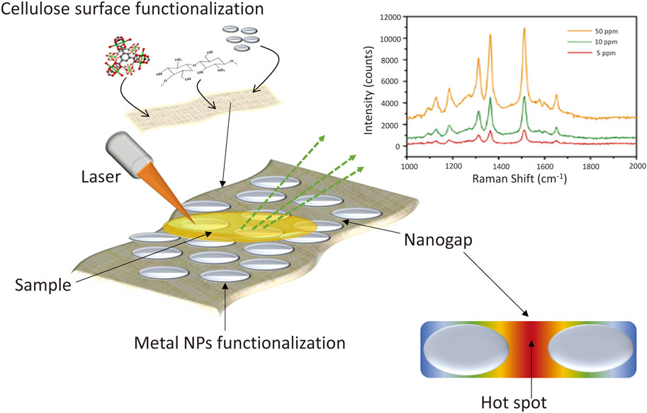
Figure 1. Scheme of cellulose based SERS devices preparation. The nanoparticles (NPs) interspace (nanogaps) and the hot spots are indicated.
Cellulose is constituted by building blocks of nanocellulose, that can be categorized as nanofibres, crystals, or bacterial cellulose, according to the production source and morphology (Trache et al., 2020; Prilepskii et al., 2023). Nanocellulose is available from a variety of sources, mainly plant material (Phanthong et al., 2018), but also from recycling of previously used paper and agriculture waste (Ogundare and van Zyl, 2019). It is a cheap material, abundant and easy to manipulate. It can be used to produce films to be decorated with functional groups or nanostructures, and employed for the production of sensors, including flexible substrates for SERS (Soriano and Dueñas-Mas, 2018), which can be shaped around substrates of various configurations. This characteristic is particularly beneficial for field applications, for example, for the detection of food contaminants (Zhang et al., 2021; Martins et al., 2023).
Recently, some comprehensive reviews concerning the design and applications of cellulose-based substrates for SERS were published (Ogundare and van Zyl, 2019; Hu B. et al., 2021; Eskandari et al., 2022; Lin et al., 2023). Nevertheless, the increasing interest in the field for both scientific research and foreseen real-life applications, highlights the importance -of sensitive and reliable platforms for SERS analysis. Thus, we believe there is the need to summarize the latest results obtained on their production. In the following sections, we will outline the most recent achievements (2020–2025) in the fabrication and performance of cellulose-based SERS devices, focusing on different strategies for an efficient cellulose functionalization with metallic nanostructures. Most of the reported research focus on deposition of metal nanoparticles. Some hints will be also given on the use of non metallic nanostructures such as metal-organic frameworks (MOFs) and metal oxides.
2 SERS electromagnetic enhancement and chemical enhancement
SERS exploits the particular properties of plasmonic nanoparticles to obtain an amplification of Raman signals by several orders of magnitude if compared to classical Raman spectroscopy. This signal enhancement effect is determined by two mechanisms, namely, electromagnetic and chemical enhancement. When plasmonic nanoparticles interact with an electromagnetic field, a dipolar oscillation is generated, namely, the localized surface plasmon (LSPs); the localized surface plasmon resonance (LSPR) therefore corresponds to the resonance at which the LSP arises. LSPR creates intense localized electromagnetic fields, known as “hot spots,” near the surface, that increase the Raman signal. The chemical enhancement relies on the chemical interaction between the metal NPs and the analyte molecules, their interaction with the incident light wavelength, and on charge transfer between the adsorbed molecule and the metal NPs (Jensen et al., 2008; Langer et al., 2020).
A common method to evaluate the performance of a SERS sensor is the Enhancement Factor (EF), There is a variety of possible definition of the EF of a SERS, raising from the diversity of conditions like the detection of single or multiple molecules, the spatial distribution, the time average. For example, EF for single molecule corresponds to the intensity of the SERS signal of a molecule divided by the Raman signal obtained from the same molecule without the metallic substrate. Once the definition is selected, attention must be paid to the accuracy of the experimental procedure because this value depends on several factors. They include the type of substrate, e.g., colloidal NPs suspension or immobilized nanostructures (Rodrigues et al., 2015), the dimension of hot-spots, the number of analyte molecules in the probed areas, the analyte affinity for the substrate, the laser power, the efficiency of the detector (Etchegoin and Le Ru, 2008; Bell et al., 2020; Le Ru and Auguié, 2024). The calculation approach and the experimental procedure chosen to evaluate the EF may lead to huge changes in its values (Le Ru et al., 2007), and to inter-laboratory variations up to 40% (Bell et al., 2020). This underlines the need of a standardization of both the EF measurement procedure and the use of known substances like Rhodamine 6G as calibrants.
3 Cellulose as SERS substrate
Cellulose consists of alternating crystalline (nanocellulose crystals) and amorphous regions, which give the flexibility and mechanical resistance to the material. These mechanical properties are advantageous because the sensors can be wrapped around objects if needed.
All the several forms of cellulose show remarkable characteristics of sustainability and non-toxicity (Gabrielli and Frasconi, 2022). Cellulose as SERS substrate has some advantages: it shows low Raman signal thus it produces negligible interferences during SERS measurements (Ogundare and van Zyl, 2019). The surface of nanocellulose is rich in hydroxyl groups, that may act as reducing and stabilizing agents to induce the condensation of metallic NPs (Kaushik and Moores, 2016) favoring functionalization. Moreover, paper can efficiently trap liquids, increasing the concentration of the analyte, leading to SERS signal enhancement.
Cellulose-based substrates for SERS devices can be fabricated from several sources, including chromatography paper, standard paper sheets, filter paper, ultrathin nanocellulose films deposited on solid surfaces (Lee et al., 2011; Carapeto et al., 2015; Kontturi and Spirk, 2019; Jones et al., 2020), or from the deposition and subsequent acid hydrolysis of the organic soluble precursor trimethylsilyl cellulose (TMSC) (Cooper et al., 1981).
Cellulose nano- and microfibres are produced by mechanical or chemical treatment, or are synthesized by some types of bacteria (bacterial nanocellulose). These fibres can be used to build three dimensional networks for the production of SERS sensors with very high sensitivity. Metal nanoparticles can be deposited around the entire fibres, which results in a homogeneous distribution of hot spots. This is particularly beneficial for complex applications such as the production of microfluidic chips, because functionalized microfilaments can be inserted in the device allowing the molecules of the analyte to access a larger plasmonic area compared to conventional deposition, where the plasmonic surface is limited to the sides of the microfluidic channel (Yao et al., 2024).
A few drawbacks must however be addressed. For example, some forms of cellulose supports, such as Cellulose nanocrystal (CNC) aerogels, lack flexibility due to the rigidity of the crystalline region (Zhou et al., 2019). Cellulose roughness might affect the distribution of metal NPs, leading to non uniform and therefore non reproducible SERS signal over the surface (Giuffrida et al., 2025). Moreover, SERS sensors made of cellulose cannot be easily recycled or reused several times because the support might degrade during wet regeneration treatments.
4 Functionalization with metal nanostructures
In order to be used as SERS substrate, cellulose must be functionalized with plasmonic nanostructures. The most commonly used materials used for this purpose, are noble metals, in particular silver and gold. Although Ag and Au have approximately the same crystal structure, the choice of the metal to use for SERS relies on the respective properties, both in terms of plasmonic efficiency and on toxicity if biomedical applications are foreseen. Silver is less expensive, and more efficient in terms of plasmonic resonance properties for SERS, in fact Ag NPs exhibit stronger localized plasmonic resonance, leading to higher EF. On the other hand, they have lower chemical stability and are less biocompatible than gold. Moreover, the stronger Raman signal enhancement induced by Ag NPs is observed in a wavelenght range below IR and near-IR, which are preferable for most biomedical applications. Thus, Au nanoparticles (NPs) would be a better choice as gold does not interfere with biological molecules properties. Hybrid Ag/Au core shell nanoparticles couple the advantages of both metals and allow to tune the plasmonic properties of the surface changing the thickness of the Ag shell with respect to the Au core. (Chaffin et al., 2016; Sha et al., 2021; Park M. et al., 2025; Sirgedaite et al., 2025).
The homogeneity of the plasmonic layer is crucial for the efficiency of SERS sensors: the deposition method plays an important role, as it was demonstrated that interparticle distance increase reduces the SERS enhancement factor. Top-down methods such as electron beam lithography or ion beam etching allow to fabricate metal nanostructures with controlled interparticle spacing. Unfortunately, these techniques are expensive, time consuming, and their resolution is limited to approximately 10 nm, (Liu et al., 2017), while smaller nanogaps between 5 and 1 nm increase the enhancement factor by 103 (Xu et al., 2000).
In-situ self-assembly of metal NPs is in general obtained via mild chemical conditions, is cheaper, and more efficient than direct attachment of pre-synthesized nanoparticles to the surface. While pre-synthesized NPs can be tailored for specific functionalities, their chemical affinity with the substrate might be critical. In-situ self-assembly of metallic NPs allows a better control over the interaction between the NPs and the substrate. Moreover, it enables to grow superstructures with low interparticle distances, and to obtain a relatively homogeneous distribution of closely located hot spots that leads to increased SERS enhancement. Nonetheless it requires careful tuning of the synthesis parameters, to avoid aggregation and optimize homogeneity. (Navvabpour et al., 2024).
In the following, different functionalization approaches will be described.
4.1 Addition of already synthesized nanostructures
Attaching a positively charged compound to a cellulose substrate is a simple way to favour the deposition of negatively charged Ag NPs to the surface, to which they bind via electrostatic interaction. A SERS device produced first grafting Glycidyl-trimethylammonium chloride (GTAC) to recycled cellulose textile fibre, and then functionalizing with citrate coated Ag NPs, was recently developed for the detection of low concentrations of BPA, a contaminant present in water that can be toxic to the endocrine system. The obtained device showed excellent mechanical resistance and flexibility. It also showed a very high sensitivity, with a detection limit as low as 0.01 ppm (Liu et al., 2020). Recently, cellulose-Ag nanocomposites SERS substrates were prepared mixing proper amounts of cellulose nanofibres with citrate capped Ag NPs: the obtained nanocomposite was deposited on a gold slide and dried at 45°C to obtain a substrate that allowed to effectively detect residues of the pesticide flusilazole in Oolong tea leaves extract (Chen et al., 2020). The homogeneous distribution of Ag NPs on the cellulose nanofibres substrate was evaluated treating the sensor with a 10 mg L-1 flusilazole solution, detecting its signal in several spots randomly located on the surface: the standard deviation of the intensity of one of the characteristic flusilazole peak (at 828 cm-1) from spot to spot on the surface was calculated to be around 3.5%. When methanol-water flusilazole solutions at different concentrations were tested, an increase in the bands intensity correlated to the analyte concentration could be observed, thus confirming the reliability of the substrate for the detection of the pesticide. The measurement of an extract from Oolong tea leaves containing increasing amounts of flusilazole, from 0 to 10 mg/kg of leaves, resulted in increasing intensity of three peaks attributed to the contaminant, demonstrating the possibility to employ this SERS substrate for quantitative detection.
The insecticide λ-cyhalothrin, a pyrethroid largely used in agriculture, poses severe risks to the health of humans and animals if used in excess or for extensive time. SERS devices based on bacterial nanocellulose and Ag NPs were successfully used to detect λ-cyhalothrin in very low concentration (Zhang et al., 2022). Transparent films of bacterial nanocellulose obtained by filtration of aqueous nanocellulose solution were decorated with layers of Ag nanoparticles with different thickness by magnetron sputtering technology. After assessing the homogenous distribution of Ag NPs on the cellulose surface via SEM EDS, the SERS devices were successfully tested for sensitivity and stability over time with Crystal Violet, and afterwards, used to detect λ-cyhalothrin in decreasing concentrations. The detection limit was assessed to be lower than 10–7 mol L-1, at which the characteristic signal at 1,394 nm-1 was still visible. Magnetron sputtering technology is a rapid, low cost technology, thus this method for the fabrication of high sensitivity SERS substrates is promising for large scale production.
SERS devices designed to detect environmental pollutants such as plastic nanoparticles in water, overcome the disadvantageous strong water absorption observed in FT-IR. Recently, regenerated cellulose hydrogel obtained from ashless cellulose pulp was functionalized with Ag nanowires and Au nanorods and subsequently dried to a thin film, to obtain a SERS device for the detection of polystyrene nanoplastics (Jeon et al., 2021). A comparison of the SERS intensity at 1,117 cm-1 of 10 μmol L-1 Crystal Violet in regenerated cellulose films obtained respectively incorporating the two metals nanoparticles, showed that the Ag-functionalized device was 6 times more efficient than the Au-functionalized one. Both functionalizations were used to test the efficiency of the sensors for the detection of nanoplastic in solution. Commercial polystyrene (PS) nanoparticles of different diameters were detected to a minimum concentration of 0.1 mg mL-1 on Ag functionalized devices, while Au functionalized SERS allowed to detect PS nanoparticles with a minimum concentration of 0.5 mg mL-1.
The SERS EF (LeRu et al., 2007) was calculated as one order of magnitude higher for Ag-functionalized sensors. These results confirm the larger plasmonic effect of silver. Moreover they reflect the possibility that Ag nanowires network induce the formation of a bigger number of hot spots on the substrate, with respect to gold nanorods (Starowicz et al., 2018).
Aiming to produce a SERS sensor for field applications where a high confidence level in detection is desirable, citrate coated colloidal Au NPs were deposited both by drop casting or inkjet printing on filter paper. The dimension of the synthesized NPs was of approximately 80 nm: these large particles were chosen to improve the LSPR of the sensors. The deposition method proved to be important with respect to the distribution of the hot spots on the surface: the printed devices were much more homogeneous compared to drop casting, although several printing cycles were necessary to obtain a suitable metal thickness. Inkjet printed devices were used to test low concentration solution of pyridine and benzenethiol, obtaining a detection limit of 0.75 μmol L-1 for pyridine, and 0.1 nmol L-1 for benzenethiol. The sensors were also tested against benzenethiol in aerosol, but due to the lower amount of sample captured by the paper substrate, and the low time of exposure, a quantitative detection of contaminant was not feasible. (Tay et al., 2020).
In a recent experiment, Au functionalization was used to produce SERS devices for contemporary detection of multiple chemicals such as food contaminants or environmental pollutants (Kim et al., 2021a). Au nanorods (Au NRs) were embedded in a cellulose hydrogel by mixing commercial cellulose pulp, LiOH and urea. The mixture was poured in a silicon mould, to obtain a 1 mm layer that was subsequently coagulated and regenerated into a cellulose hydrogel. A silicon rubber dotted mask attached to the cellulose hydrogel substrate was used as a template for the deposition of a suitable amount of Au NRs, to create an array of separated functionalized spots on the surface. 3 μL of different chemicals including pesticides and model compounds were deposited in various concentrations on each SERS dot, and subsequent Raman experiments demonstrated the detection of several peaks characteristic for each analyte.
Paper substrates were recently functionalized with silver nanowires, in order to obtain a conductive surface for subsequent electrodeposition of mesoporous gold film of controllable thickness. Cellulose powder was initially oxidized with (2,2,6,6-Tetramethylpiperidin-1-yl)oxyl (TEMPO) and converted into nanofibrils via high pressure homogenization. The dispersion of TEMPO-nanocellulose fibrils was then deposited on a filter membrane, a suspension of Ag nanowires was added on top and attached to the TEMPO-nanocellulose membrane via vacuum filtration. The resulting silver layer acted as electrode in the subsequent gold electrodeposition. After filtration, the membrane was dried, the functionalized nanocellulose was peeled off from the filter membrane, and used as a substrate for electrochemical deposition of a gold nanolayer, as shown in Figure 2 (Kim et al., 2021b). The obtained device showed a high density of electromagnetic hot spots, that exploited a dramatic increase of SERS signal when it was tested with Rhodamine 6G. SERS signals from samples in gas-phase delivered to the surface by a nitrogen stream were also evaluated, demonstrating the effective absorption of the analyte in the mesoporous metal framework produced by electrodeposition. The gas phase samples showed significative differences with the exposure time, and eventually led to sensor saturation.
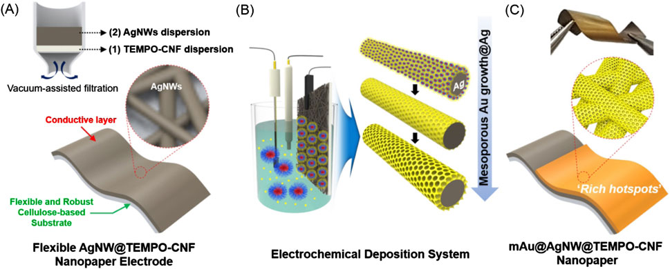
Figure 2. Preparation of a cellulose substrate functionalized with Ag nanowires (Ag NW) to be used as an electrode for the electrodeposition of mesoporous gold. (A) Scheme showing the preparation of Ag NW functionalized (shown in the inset) cellulose nanofibre substrate. (B) Electrochemical deposition of a gold mesoporous nanolayer on the functionalized surface. (C) Final SERS sensor. Reproduced with permission from (Kim et al., 2021b).
4.2 Self-assembly of NPs on cellulose substrates
Metal NPs can be grown directly on the cellulose substrate, to obtain a better distribution of the metal layer on the paper surface, that results in a more homogeneous responsivity of the sensor. The Successive Ionic Layer Adsorption and Reaction method (SILAR) (Nicolau, 1985) is a deposition technique that can be used to produce layers of metallic nanostructures with controlled structure and properties on paper surface. Both the reactant composition and the deposition protocol must be carefully optimized, because aggregation might occur if i) the concentration of the precursors is too high, and ii) the deposition process is not controlled. If the rinsing step between each respective layer deposition is missing, fast but uncontrollable growth is obtained. Moreover, if the ionic layer deposition is done via dip coating, the first precursor could be released in the following solution, provoking precipitation, thus a change in the stoichiometry of the reaction and as a consequence, in the quality of the deposited film (Ratnayake et al., 2021).
SILAR was used to grow Ag NPs on ultra smooth (2,2,6,6-tetramethylpiperidin-1-yl)oxyl-oxidized Nano Fibrillated Cellulose (NFC) nanocellulose paper substrate (TO-nanopaper). The substrate was submerged in a carefully optimized concentrated solution of AgNO3, and subsequently in the reductant solution of NaBH4. Successive SILAR cycles induced the formation of a dense metal layer composed of Ag NPs with an average size of 60 nm, and of bigger clusters. A multi well plate device with the TO-nanopaper Ag NPs base was fabricated. Two model chemicals, Rhodamine B and 2-naphthalenethiol, were used to test the SERS activity, displaying a concentration limit sensitivity of 1 pmol L-1, and an SERS enhancement factor (EF) of approximately 1.5 × 10⁹ (Chen et al., 2019). A scheme of the fabrication procedure is shown in Figure 3.
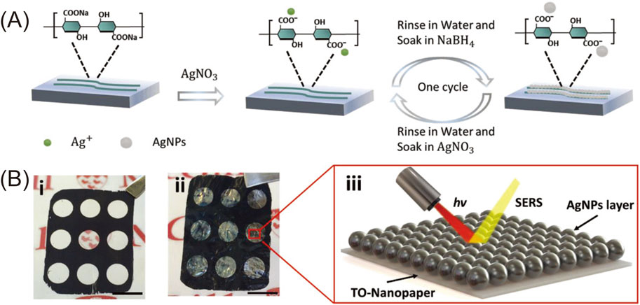
Figure 3. In-situ synthesis of Ag NPs on NFC paper. (A) Scheme of the SILAR deposition of Ag NPs by successive reduction of AgNO3. (B) pictures of the multiwell plate device before (i) and after (ii) one cycle of Ag NPs deposition. (iii) cartoon of the SERS device. Reproduced with permission from (Chen et al., 2019).
Functionalized cellulose fibre substrates modified with silver nanostructures of various morphologies have been utilized for label-free SERS detection of specific compounds, further emphasizing the versatility of cellulose-based substrates in molecular detection.
The large specific surface area of sulfonated cellulose nanofibres promotes the binding of a large number of metal NPs and the formation of a homogeneous reactive surface. In a recent experiment, Ag ions were attached on the hydroxyl and sulfonic acid residues present on sulfonated cellulose nanofibres, to produce a SERS sensor to detect a single molecule of NADH - a biomarker for cellular metabolism - in serum. AgNO3 was mixed at high temperature with sulfonated cellulose nanofibres and the mixture was subsequently reduced adding Na-citrate. The deposited Ag seeds were stable and tightly bound to the cellulose substrate. The resulting SERS sensor showed a more homogeneous surface and an increased sensitivity, as demonstrated by test detection of Rhodamine 6G. The cellulose-Ag substrate was then used to produce an innovative “on-off” system for selective identification of NADH, triggering the redox reaction that converts 4-mercaptophenol (4-MP) to 4-mercaptocyclohexane-2,5-dienone (4-MC): binding 4-MP to the Ag-absorbed surface, and subsequently oxidizing it to 4-MC via Fe3+ addition, a SERS inactive (“off”) sensor was produced: since NADH can reduce 4-MC back to 4-MP, once applied to the surface it was acting as a reducing agent, producing a SERS signal (“on”) (Wang W. et al., 2023).
Recently, raspberry-like Ag nanostructures were grown on a hybrid surface obtained coupling cellulose fibres (CF) with silver nanostructures in a silver mirror reaction. The resulting layered surface showed an enhanced roughness and denser distribution of silver nanostructures, that provided a better SERS performance both in an estimation test on the standard Raman probe 4-mercaptobenzoic acid, and on the detection of malathion, a commonly used organophosphate insecticide (Serebrennikova et al., 2023).
Highly sensitive SERS sensors for the detection of the fungicide Carbendazim on irregular surfaces were recently produced soaking bacterial nanocellulose fibres in a silver ammonia solution that was subsequently reduced to obtain Ag NPs (Zhang et al., 2024). The three dimensional distribution of Ag NPs around the cellulose fibres creates a superstructure with an elevated number of metal hot spots, that allows the sensor to detect very low concentration of the target molecules as well as a remarkable high EF, in the order of 1010. A similar three dimensional functionalization approach was used to develop a microfluidic device able to overcome the low sensitivity of SERS in solution (Yao et al., 2024). Single microcellulose filaments were treated with AgNO3 and then silver was reduced with sodium borohydride, to obtain plasmonic cellulose microfilaments homogeneously coated with silver NPs. The functionalized filaments were embedded in a PDMS microfluidic channel and tested under continuous flow, to detect traces of Methylene Blue, Rhodamine 6G, Nile Blue and urea in solution.
SERS devices that can be used for quantitative analysis of chemicals in traces, are suitable for applications also in the medical field. A recent experiment demonstrated that a low cost SERS device based on cellulose decorated with Ag NPs could be used to detect the antiviral drug acyclovir, with a concentration detection limit of 1 pmol L-1. (Eskandari et al., 2023). Acyclovir is an antiviral drug which might have toxic effects, thus its concentration needs to be carefully controlled when administered to patients. To functionalize the structure, AgNO3 solution was treated with ammonia in controlled pH conditions until silver oxide precipitation and subsequent dissolution occurred. The addition of a reducing agent induced the condensation of Ag NPs and its homogeneous deposition on filter paper stripes soaked in the solution. Droplets of acyclovir solutions in a concentration range between 100 mmol L-1 to 1 pmol L-1 were deposited on the prepared devices, and the Raman spectra were collected. At all concentrations, distinct Raman peaks at 981 cm−1, 1,223 cm−1 and 1,409 cm−1 were evident, corresponding to the molecular vibration of acyclovir.
One-pot reactions are highly desirable to obtain flexible functionalized substrates in a fast, economical way. For example, cellulose substrates doped with Au NPs could be produced mixing cellulose pulp dissolved in alkaline NaOH solution, in presence of urea and HAuCl4−: the resulting solution was drop casted on a suitable support, regenerated to obtain a cellulose-Au doped nanocomposite film by soaking in ethylene glycol, and finally air-dried at RT. The obtained substrate proved to be effective for SERS detection of both Rhodamine 6G and Melamine, a food additive that poses severe issues to health when in excess (Figure 4). The homogeneity of Au NPs distribution on the surface was tested detecting the 617 nm-1 peak of Rhodamine 6G in several different spots, with a relative standard deviation of approximately 5.4%. The specificity of the substrate was evaluated detecting the presence of Melamine in milk. During the test, a distinct peak at 695 nm-1, attributed to the change in polarity of the molecule, was visible at concentration of 1 mg/kg (Hu X. et al., 2021).
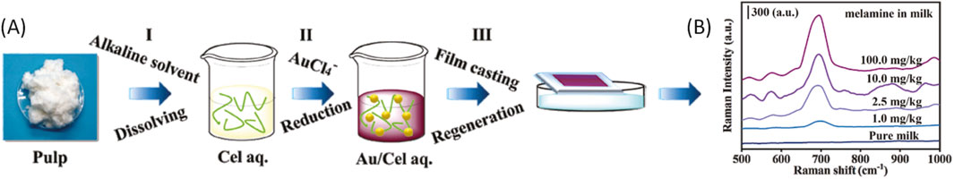
Figure 4. (A) Three-step procedure to produce the nanocomposite: (I) Dissolving cellulose pulp in a NaOH/urea/H2O system; (II) in-situ reduction of AuCl4− in the dissolved cellulose solution; (III) film casting and regeneration. (B) SERS spectra of melamine in milk using the Au/RCF-4 substrate. Reproduced with permission from (Hu X. et al., 2021)
4.3 Cellulose pre-treatment to promote metal NPs functionalization
Chemical modification of cellulose is an efficient way to improve the immobilization of metal NPs. Moreover, it improves not only their sensitivity, but also the mechanical resistance (Restaino and White, 2019). Different treatment protocols are reported in the following.
4.3.1 Chitosan
In order to improve the hot spot distribution due to the homogeneous deposition of metal nanostructures on the surface, a mixture of Chitosan (CH) and silver nanoparticles was deposited on stripes of standard chromatography paper in several multi step dipping cycles. In solution, chitosan is electrostatically attracted by cellulose. CH-treated paper stripes were then immersed in a basic solution, as at high pH chitosan becomes insoluble. A subsequent treatment with AgNO4 and a NaBH4 reductant solution containing a capping agent induced the formation of an Ag NPs network on the paper surface. Control (blank) substrates containing reduced silver nanoparticles but without chitosan were produced and tested in parallel, by submerging the SERS substrates in a 0.1 mmol L-1 ethanolic solution of 4-aminothiophenol (4-ATP). The test results indicated a high sensitivity of the chitosan-treated device. From the Raman spectra collected from chitosan doped paper in comparison with untreated cellulose paper substrate, no notable interference from chitosan was detected. Besides this, the optical micrograph analysis demonstrated the effect of chitosan in preventing the Ag nanoparticles aggregation. Moreover, the tensile strength of the CH-treated paper stripes was comparable to the one of native paper, in contrast with the stripes prepared without chitosan that were significantly less resistant. This behaviour is attributed to the effect of chitosan, that prevents the hydrolysis of cellulose fibres induced by AgNO3, and by the subsequent treatment with the reductant solution (Kang et al., 2022).
Chitosan and its quaternary ammonium derivative N,N,N-trimethyl chitosan (TMC) were also used to prepare SERS substrates successively functionalized with Ag NPs (Martins et al., 2023). TMC is positively charged, has a higher number of cationic groups and remains soluble at high pH, thus it can favor the binding of negatively charged citrate coated metal nanoparticles. In the described experiment, an ink incorporating Ag NPs was used to print on both CH- and TMC-loaded paper, to obtain a SERS substrate that was subsequently tested with decreasing concentrations of Crystal Violet. Both the substrates demonstrated great homogeneity of hot spots distribution, as well as high sensitivity, although CH-paper was more efficient, being able to detect the analyte to a concentration as low as 0.1 μmol L-1. The authors speculate that TMC favours the formation of larger Ag-NPs clusters, that according to previously reported results, may worsen the SERS sensitivity.
Chitosan can further be used as reducing agent to incorporate Au NPs on paper substrates, to induce the direct growth of NPs. Applying a solution of chloroauric acid and low concentration chitosan to the surface of filter paper and incubating the substrates at high temperature in controlled humidity conditions, allowed to obtain a fast in situ reduction of gold nanoparticles on the surface. The resulting SERS devices showed enhanced response to Rhodamine 6G. Moreover, the thermal treatment speeds up the reduction of chloroauric acid, consequently the time necessary for their synthesis (Srivastava et al., 2023).
4.3.2 Silane and heterocycles
A non-invasive, in situ time monitoring of the healing process cascade is beneficial for the management of patients that suffer of chronic wounds: a cellulose-based SERS biosensor to detect several molecules involved in wound healing process was recently proposed. Pre-treatment with APTES was employed to optimize the deposition via sputtering of thin Ag NPs layers on cellulose membrane filters (Perumal et al., 2021). The SERS performance was first evaluated in an efficiency test measuring the characteristic band of 2-naphthalenethiol (1,066 nm-1). As expected, devices with higher Ag layer thickness produced increasing SERS signal upon irradiation. Interestingly, if the metal layer thickness exceeded 60 nm, the signal intensity dropped. This was attributed to a loss of the spatial distancing between the nanoparticles that aggregate at the surface, reducing the SERS signal (Perumal et al., 2014). The optimized sensor was then functionalized with a number of proteins including recombinant antibodies against specific biomarkers, aiming to monitor the tissue repair process in those patients where it is impaired, e.g., due to clinical conditions like diabetes. Simulated biological fluid containing proteins and other species derived from plasma, which are naturally involved in wound tissue repair, was then applied to the SERS device to test its efficiency. The sensor produced highly accurate results at low analytes concentration for all the molecules involved in the reported study.
Recently, Au NPs coated with citrate were successfully immobilized on chromatography paper grafted with imidazole, dithiol (ane), or silanized with APTES. As demonstrated by extinction spectra and SEM analysis of the Au NPs functionalized surface, imidazole groups are less effective in promoting Au NPs binding than dithiol groups. Moreover, the latter are more efficient when attached to cellulose previously functionalized with APTES, producing a surface with more NPs per unit area. Cellulose paper substrates functionalized with Au NPs coated with citrate, DHLA or GSH, could be immobilized on the treated paper surface, and tested for SERS signal after soaking in solution of 1,2-di (4-pyridyl)ethylene. The enhancement factor was evaluated after either spotting or soaking the sample in the solution, and was similar for both methods (Kim et al., 2020).
4.3.3 Carboxymethylation
Bacterial cellulose surface can be chemically modified to obtain an enrichment of hydroxyl and carboxyl groups, that promote the formation of metallic hotspots. Recently, a commercial bacterial cellulose membrane treated with sodium chloroacetate and NaOH was used to produce SERS sensors to monitor the metabolites in human urine. To attach Ag NPs to the surface, the carboxymethylated bacterial cellulose was immersed in a solution of AgNO3. The negatively charged groups on the surface attract the Ag ions, that were subsequently reduced by ascorbic acid, creating a stable layer of Ag NPs. A Limit of Detection (LoD), namely, the lowest concentration of analyte that can be reliably detected, in the order of μmol L-1 was measured for urea and uric acid in human urine samples, demonstrating the sensibility of the sensor to detect metabolites in solution (Yang et al., 2024).
5 Functionalization with non metal nanostructures
Besides noble metals, other compounds were successfully used for the fabrication of SERS sensors. In the following section, some examples of the functionalization of cellulose-based substrates with metal oxides and metal-organic frameworks will be described.
5.1 Metal oxides
Differently from noble metals, where the increasing SERS intensity relies prevalently on the electromagnetic mechanism, metal oxides nanostructures induce strong signal enhancement mainly via chemical mechanism based on charge transfer between the sensor surface and the probe. Moreover, they display better chemical stability, biocompatibility, and are less prone to laser-induced aggregation than metal NPs. This allows to reach detection limits and EF which are consistent with those generated by noble metals hot spots. However, SERS substrates based on metal oxides are often less homogenous, reducing the overall sensor performance by some orders of magnitude with respect to Ag or Au nanostructures. The chemical composition and the oxidation state of the metal, may as well affect the SERS performance. (Chen et al., 2023; Wang Z. et al., 2023; Jin et al., 2024).
Two dimensional Molybdenum oxide (MoO3-x) nanosheets were recently used to fabricate a highly sensitive device. A MoO3-x nanosheets ink was synthesized and used in a traditional inkjet printer, to obtain a SERS array on a paper substrate previously made hydrophobic by surface treatment. The device was tested with decreasing concentration of Rhodamine 6G solution, and featured a detection limit of 10–7 mol L-1, while the EF was calculated as approximately 3.3 × 10⁵. Interestingly, the MoO3-x nanosheets ink SERS array printed on several types of paper, from the cheap one to more expensive cellulose filter or chromatography paper, maintained the same efficiency, demonstrating that low cost supports can be successfully used for the production of these devices (Lan et al., 2020).
5.2 Metal-organic frameworks
MOFs are highly porous hybrid materials, consisting of ionic or metallic clusters connected by organic linkers, that create a 3D structure. They exhibit excellent catalytic activity and due to the tunability of porosity are suitable compounds for the immobilization of small molecules. In SERS devices, they can be coupled with metal NPs to improve their stability and prevent NPs aggregation. MOFs are promising for the production of SERS sensors for the detection of gaseous compounds. Gas molecules trapped into the MOFs pores increase their concentration for better detection (Allegretto and Dostalek, 2024). MOFs functionalized sensors may reach a gas detection efficiency, in terms of low detection limit and EF, which is comparable with that obtained from liquid probes Their drawback is, that some common MOFs are highly sensitive to humidity, that induces an irreversible collapse of the structure reducing porosity and permeation properties (Li et al., 2022). This might preclude some field applications.
MOFs were successfully integrated in cellulose-MOFs nanocomposites, to obtain hybrid materials containing electrochemically active molecules that can be used for chemical sensing, or functionalized with proteins for biosensing. Details on the several strategies to produce cellulose-MOFs composites and on the production of MOF functionalized paper-based sensors are reported in two recent reviews (Tu et al., 2022; Huang et al., 2024).
MOFs functionalized SERS sensors are potentially effective for the detection of volatile compounds, e.g., those arising from early food decay, due to the capability of MOFs to trap small gaseous molecules. In example, Zeolitic Imidazolate Frameworks (ZIF) were successfully combined with gold NPs impregnated cellulose to obtain highly sensitive SERS devices for gas detection (Kim H. et al., 2021). Standard filter paper was treated with HAuCl4 ethanolic solution, dried and subsequently the gold reduction was obtained via plasma treatment in Argon. The Au-impregnated paper was submerged in the ZIF-8 precursor solution containing Zn acetate and 2-methyl imidazole dissolved in methanol at a molar ratio of 1:40. The resulting device was functionalized with 4-mercaptobenzaldehyde, a specific receptor for volatile amines, and used for the selective detection of gaseous putrescine and cadaverine molecules derived from spoiled food. From the analysis of the ratio between the intensity of two characteristic bands, namely, 1,638 and 1,074 cm-1, versus the concentration of the respective analyte, the lower detection limit was estimated around 77 ppb for putrescine, and around 116 ppb for cadaverine. The estimated EF was in the order of 106.
Recently, SERS sensors based on necklace-like structures composed of highly hydrophilic cellulose nanocrystals coupled with ZIF-8, and subsequently functionalized with Ag NPs were produced, to test the presence of a pesticide in green tea leaves (Luo et al., 2025). The porosity of the MOFs nanostructures allowed to reach a sensitivity to concentration of the SERS standard 4-mercaptobenzoic acid and of the pesticide diquat dibromide in the order of 10–9 mol L-1. The calculated EF was around 6*104. Interestingly, since ZIF-8 is not degraded in presence of water, the sensor could be used to detect the target dissolved in aqueous solution. Reproducibility test ensured the consistency of SERS response from 20 different batches.
5.3 MXenes
MXenes are a class of two dimensional transition metals carbide and nitrides. They are hydrophilic, biocompatible compounds with large surface area and excellent electrical conductivity (Nashim and Parida, 2022). Coupled with plasmonic nanostructures, MXenes can be used to produce SERS sensors with high electromagnetic and chemical enhancement. Hybrid nanostructures composed of Ti3C2Tx nanosheets and Au NPs attached via vacuum filtration technique to TEMPO-oxidized nanocellulose (TC) in a hierarchical layer structure, were used to produce highly sensitive, flexible devices to detect the pesticide Thiram on fruit surface (Figure 5). Tested with Rhodamine 6G, these devices showed a LoD of 10–11 mol L-1 and an EF of 9.9*109. This performance is due to the amplified chemical enhancement induced by the electron exchange between Ti3C2Tx and the analyte (Park J. et al., 2025).
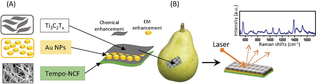
Figure 5. (A) Scheme of the sandwich-like SERS sensor based on hybrid Ti3C2Tx nanosheets–Au NPs attached on flexible TEMPO-NCF support.(B) SERS sensor application to the curved surface for the detection of pesticide on fruits.
Table 1 summarizes the functionalization treatments of cellulose substrates for SERS reported in this review. The respective calculated EF, and the Relative Standard Deviation (RSD) on characteristic peaks intensity measured on different spots on the surface are shown. It should be underlined, that the EF calculated in the reported experiments should be compared with care, because the methodology applied to calculate them was not always clarified. Where available, the calculation method was reported.
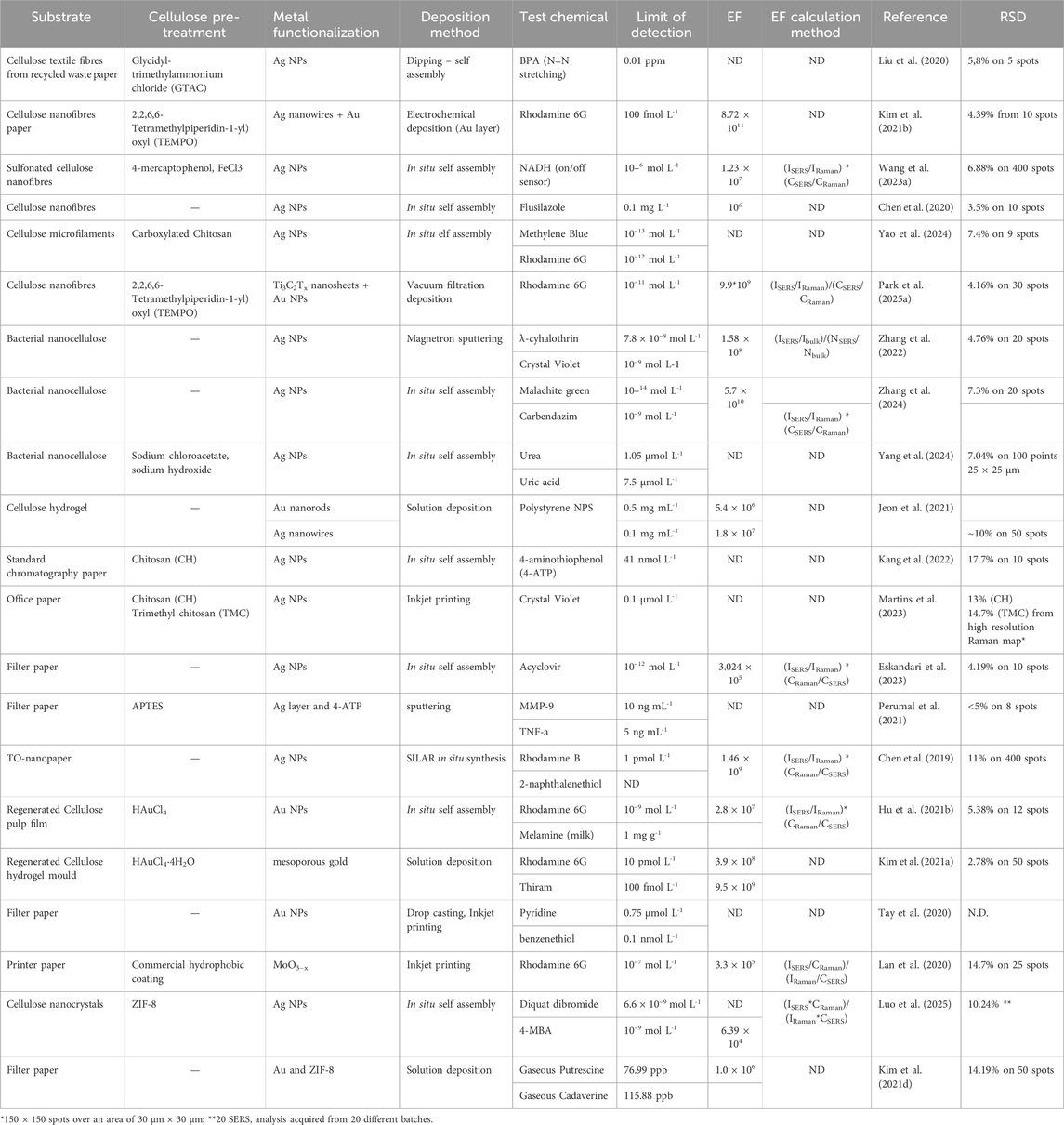
Table 1. Summary of the experiments reported in the present review. I: intensity of the selected peak during the SERS measurement (ISERS) and in absence of SERS substrate (IRaman); C: concentration of the analyte used for SERS (CSERS) and Raman (CRaman) measurement; N: average number of analyte molecules in the scattering volume during SERS (NSERS) and Raman (Nbulk) detection.
6 Discussion
6.1 Sensor efficiency and reproducibility
Different substrate preparation protocols and the use of EF as an evaluation method can give rise to large variations in the estimation of the sensor efficiency. This is particularly challenging in SERS quantitative analysis (Sloan-Dennison et al., 2024). To overcome this problem, it would be important to develop a standard protocol for the evaluation of the efficiency of SERS sensors. From a practical point of view, however, this is not simple because the SERS signal depends also on the binding efficiency of the analyte molecule to the substrate, which is due to both the chemical characteristics of the analyte and to the functionalization of the surface. In a recent review, Bell et al. report a list of potential “standard probes” that, thanks to their binding affinity, can be used as standards to evaluate the efficiency of substrates functionalized with different molecules (Bell et al., 2020). However, this approach does not allow to determine how much of the analyte actually sticks to the substrate, leading to potential incorrect estimation of the signal. In this case, it would be necessary to use a reference sample and a rigorous measurement protocol, to maximize the reproducibility of the test.
The ideal calibrant should be non toxic, not expensive, show a strong electronic resonance and a well distinguished SERS spectrum in order to be easily recognizable and reproducible, to prevent false positives even in presence of contaminations, especially if the sensor should be used on complex media for field applications. Among the others, Rhodamine 6G and Purines derivatives (Cheng et al., 2023) are commonly used as reference samples for SERS.
The homogeneous distribution of hot spots on the substrate can also affect the reliability and reproducibility of SERS response: in fact, comparing the characteristic peak signal of a standard molecule collected in several position across the sensors functionalized area, the RSD of the peak intensity varies from values as low as 3.5% (Chen et al., 2020) to 17.7% (Kang et al., 2022). In this respect, embedding an internal standard for the calibration of the intensity of the analyte in the functionalization would be beneficial, as recently demonstrated by Jie et al. Their work reports the use of carbon nanotubes (CNTs)/Ag NPs hybrids where CNTs are used as internal standard to evaluate the hot spot distribution on the sensor surface (Jie et al., 2018). Since the localized electromagnetic field enhancement is the same both for CNTs and for the analyte, measuring their respective Raman intensity at the same time it is possible to calibrate the differences in intensity across the substrate, which are due to the inhomogeneous distribution of metal hot spots.
Interlaboratory reproducibility is also an issue for quantitative SERS. It might be addressed using the SERS signal of a reference molecule to estimate a standard intensity, which can be compared to the analyte intensity in order to determine the EF variation (Bell et al., 2020). A reliable calibration model should be independent from the acquisition instrument. Batch to batch reproducibility could be ensured by finding a method that allows the comparison of different substrates. An extensive study involving 15 laboratories was recently published, in which the authors suggest a detailed protocol for the standardization of SERS substrates performance (Fornasaro et al., 2020).
6.2 Sensors resistance and specificity
Field applications require that SERS sensors based on cellulose should resist to swelling and friction, and the metal nanostructures attached to the surface should not detach over time. This is favored by surface functionalization. Bending resistance test are routinely performed during the devices production, to guarantee the stability of the intensity signal after multiple bending cycles (see i.e., the experiments of (Jeon et al., 2021; Kim et al., 2021a; 2021b; Kang et al., 2022; Zhang et al., 2022) reported in this work), showing that the performance of the sensor is not affected by mechanical stress like, e.g., in case of wearable patches for continuous health monitoring.
Cross selectivity of specific molecules is a critical issue in real life application of SERS. Especially when analyzing complex mixtures like body fluids, wastewater, or food, interference from co-existing molecules may hinder the Raman signal of the target. Thus, non-specific binding of interfering molecules should be avoided. This can be obtained tailoring the functionalization attaching to the surface specific functional groups with high affinity for the target (López-Puente et al., 2015), or combining appropriate detection techniques in one sensor, for simultaneous detection of several molecules (Kim E. J. et al., 2021; Wei et al., 2023).
7 Conclusions and future challenges
We have described some recent advances in the fabrication of SERS devices based on cellulose. Either produced from nanocellulose paper which features more densely packed cellulose fibrils and smaller pores, or from standard paper treated to obtain a homogeneous surface for better distribution of functional nanoparticles, such devices promote the application of Raman spectroscopy in real life, where enhanced Raman signal, ease of use, affordability are of pivotal importance. In this respect, it must be emphasized that traditional SERS devices are designed to be used in the clean, protected environment of a laboratory, but field applications require more robust substrates, that are not damaged by bending or scratching. To make a further step forward in the direction of Point of Care (PoC) and field applications, several challenges still remain to be addressed.
So far, the production of monodispersed metal nanoparticles is challenging. The sensitivity and reproducibility of SERS depend on the homogeneous distribution of the plasmonic coating on the support, which can be achieved only via chemical modification of the surface. The thickness of the plasmonic layer is as well critical for SERS sensitivity. Techniques like inkjet printing and magnetron sputtering seem promising but further investigations are needed to improve the monodispersity of the particles and the hot spot uniformity, especially for production at industrial scale.
Cost control and scalability are pivotal for mass production of SERS sensors that would be used for e.g., environmental pollution control, or in PoC diagnostics. In this respect, roll to roll printing is a high throughput, cost effective technique. Metal NPs can be deposited on large paper sheets via inkjet printing in an automated process, reducing the manufacturing costs, still obtaining a LoD in the order of 100 nmol L-1. Single NPs deposition technique such as flame spray deposition on paper pre-treated with graphite carbon is also attractive, because it allows to reduce the amount of silver needed to obtain a NPs coating that produce a reasonable SERS signal, with a LoD in the order of 1 μmol L-1. (Saarinen et al., 2017).
The possibility to regenerate the active surface with mild chemical methods like solvent washing (Zhang et al., 2023) would promote the reusability of the devices, thus reducing waste, metal nanoparticles pollution, and costs. As previously demonstrated on other SERS devices, the photocatalytic activity of metal oxides such as TiO2 or ZnO coupled with UV light exposure can be exploited to selectively degrade the analyte molecules. Moreover, the combined charge transfer effect of the metal oxide layer and the noble metal hot spots enhance the SERS signal. (Ge et al., 2019; Wang Z. et al., 2023; Zhang et al., 2023). In the future, photocatalytic compounds might be used to functionalize cellulose before decorating it with noble metals nanostructures.
Since paper-based substrates proved to be effective when the sample is deposited in sufficient quantity on the sensor, enough material should be collected to allow a reliable detection and quantitative evaluation of the analyte. This is particularly true for aerosolized samples. In fact, the pores of the support should be able to trap enough material, and to let the analyte interact for a time sufficient to develop a stable signal. Moreover, selective sensors would be necessary for applications where several components can hinder the contaminating molecule, as in example food or water pollution. For aerosolized samples we consider the use of structures like MOFs as notably attractive once the sensitivity to water is properly addressed.
Paper based microfluidic devices have gained attention for analytical devices, and can be fabricated by various methods (Whitesides, 2006; Martinez et al., 2010; Anushka et al., 2023). Coupling cellulose and microfabrication is a promising research field for the production of next-generation labs on chip. For instance, patterning of the precursor TMSC (trimethylsilyl cellulose) is a suitable technique to obtain microdevices that contain hydrophilic and hydrophobic areas. Thin films of TMSC have been irradiated with UV light in presence of a photoacid generator (Wolfberger et al., 2014), with focused electron beam (Ganner et al., 2016), or with X-rays (Andreev et al., 2022) and then converted to cellulose, to create high resolution microstructures. Paper based microdevices could be further modified by functionalization, to facilitate the binding of metal nanoparticles. Moreover, they could be coupled to innovative materials for the production of complex multifunctional devices with tailored properties on selected areas (Münch et al., 2018).
SERS sensors based on cellulose substrates can be embedded in wearable patches to create efficient devices for health monitoring, e.g., for the identification of certain indicators in body fluids like sweat. These devices should be designed to be easy to use, safe, and comfortable for the patient. If long term use if foreseen, the sensors must be suited for skin contact: double layer patches with biocompatible surface on the skin side and metal-functionalized active layer on the other were recently developed (Puravankara et al., 2025). Wearable patches should maintain the performance over time for continuous health monitoring. This could be achieved by means of microfluidics, which allows to selectively collect sweat samples “on demand” (Yang et al., 2025). For example, microcellulose fibres can be inserted in biocompatible polymer microfluidic devices, to enhance the SERS signal and avoid skin contact with the plasmonic surface.
Complex matrices analysis is a big challenge for field applications of SERS. Environmental control applications such as food composition, water contaminants, and pesticide detection, as well as forensic and biomedical monitoring are difficult tasks, because they involve the analysis of mixed samples where many components might have partially overlapping SERS spectra which are difficult to distinguish. Machine learning (ML) models could be trained from large libraries of Raman spectra, to improve the identification of specific molecules in complex matrices. Properly trained ML algorithms would detect signal interferences due to contaminants, discriminate between background noise and sample signal, and generally speed up the readout process for fast, direct molecule detection (Lussier et al., 2020; Goel et al., 2024).
As a whole, flexible SERS sensors made of cellulose are cheap, resistant, can be wrapped around the probe, regardless its shape, and do not require special storage conditions. Besides being used in standard or portable spectrometers, they may be integrated in complex systems such as PoC tools, environment pollutant control devices or packaging for continuous in-situ monitoring of contaminants or of biological processes. The results reported in the present review demonstrate the potential of cellulose as a support for the design and facile production of SERS devices, with a focus on future field applications.
Author contributions
BS: Conceptualization, Writing – original draft. BM: Writing – review and editing, Conceptualization.
Funding
The author(s) declare that financial support was received for the research and/or publication of this article. TU Graz Open Access Publishing Fund contributed to cover the publication fees.
Conflict of interest
The authors declare that the research was conducted in the absence of any commercial or financial relationships that could be construed as a potential conflict of interest.
The author(s) declared that they were an editorial board member of Frontiers, at the time of submission. This had no impact on the peer review process and the final decision.
Generative AI statement
The author(s) declare that no Generative AI was used in the creation of this manuscript.
Publisher’s note
All claims expressed in this article are solely those of the authors and do not necessarily represent those of their affiliated organizations, or those of the publisher, the editors and the reviewers. Any product that may be evaluated in this article, or claim that may be made by its manufacturer, is not guaranteed or endorsed by the publisher.
References
Akil-Jradi, S., Jradi, S., Plain, J., Adam, P.-M., Bijeon, J.-L., Royer, P., et al. (2012). Micro/Nanoporous polymer chips as templates for highly sensitive SERS sensors. RSC Adv. 2, 7837. doi:10.1039/c2ra21186f
Albrecht, M. G., and Creighton, J. A. (1977). Anomalously intense raman spectra of pyridine at a silver electrode. J. Am. Chem. Soc. 99, 5215–5217. doi:10.1021/ja00457a071
Allegretto, J. A., and Dostalek, J. (2024). Metal–organic frameworks in surface enhanced raman spectroscopy–based analysis of volatile organic compounds. Adv. Sci. 11, 2401437. doi:10.1002/advs.202401437
Almehmadi, L. M., Curley, S. M., Tokranova, N. A., Tenenbaum, S. A., and Lednev, I. K. (2019). Surface enhanced raman spectroscopy for single molecule protein detection. Sci. Rep. 9, 12356. doi:10.1038/s41598-019-48650-y
Andreev, M., Marmiroli, B., Schennach, R., and Amenitsch, H. (2022). Patterning a cellulose based dual-tone photoresist via deep X-ray lithography. Microelectron. Eng. 256, 111720–111726. doi:10.1016/j.mee.2022.111720
Anushka, , Bandopadhyay, A., and Das, P. K. (2023). Paper based microfluidic devices: a review of fabrication techniques and applications. Eur. Phys. J. Spec. Top. 232, 781–815. doi:10.1140/epjs/s11734-022-00727-y
Bell, S. E. J., Charron, G., Cortés, E., Kneipp, J., De La Chapelle, M. L., Langer, J., et al. (2020). Towards reliable and quantitative surface-enhanced raman scattering (SERS): from key parameters to good analytical practice. Angew. Chem. Int. Ed. 59, 5454–5462. doi:10.1002/anie.201908154
Carapeto, A. P., Ferraria, A. M., Brogueira, P., Boufi, S., and Do Rego, A. M. B. (2015). Cellulose films: designing template-free nanoporous cellulose films on semiconducting surfaces. Microsc. Microanal. 21, 102–107. doi:10.1017/S1431927614001706
Chaffin, E., O’Connor, R. T., Barr, J., Huang, X., and Wang, Y. (2016). Dependence of SERS enhancement on the chemical composition and structure of Ag/Au hybrid nanoparticles. J. Chem. Phys. 145, 054706. doi:10.1063/1.4960052
Chen, L., Ying, B., Song, P., and Liu, X. (2019). A nanocellulose-paper-based SERS multiwell plate with high sensitivity and high signal homogeneity. Adv. Mater. Interfaces 6. doi:10.1002/admi.201901346
Chen, X., Lin, H., Xu, T., Lai, K., Han, X., and Lin, M. (2020). Cellulose nanofibers coated with silver nanoparticles as a flexible nanocomposite for measurement of flusilazole residues in oolong tea by surface-enhanced raman spectroscopy. Food Chem. 315, 126276. doi:10.1016/j.foodchem.2020.126276
Chen, Y., Hu, Y., and Li, G. (2023). A review on non-noble metal substrates for surface-enhanced raman scattering detection. Chemosensors 11, 427. doi:10.3390/chemosensors11080427
Cheng, H.-W., Tsai, H.-M., and Wang, Y.-L. (2023). Exploiting purine as an internal standard for SERS quantification of purine derivative molecules released by bacteria. Anal. Chem. 95, 16967–16975. doi:10.1021/acs.analchem.3c03259
Cooper, G. K., Sandberg, K. R., and Hinck, J. F. (1981). Trimethylsilyl cellulose as precursor to regenerated cellulose fiber. J. Appl. Polym. Sci. 26, 3827–3836. doi:10.1002/app.1981.070261129
Eskandari, V., Sahbafar, H., Karooby, E., Heris, M. H., Mehmandoust, S., Razmjoue, D., et al. (2023). Surface-enhanced raman scattering (SERS) filter paper substrates decorated with silver nanoparticles for the detection of molecular vibrations of acyclovir drug. Spectrochim. Acta - Part Mol. Biomol. Spectrosc. 298, 122762. doi:10.1016/j.saa.2023.122762
Eskandari, V., Sahbafar, H., Zeinalizad, L., Marashipour, R., and Hadi, A. (2022). A review of paper-based substrates as surface-enhanced raman spectroscopy (SERS) biosensors and microfluidic paper-based SERS platforms. J. Comput. Appl. Mech. 53, 132–146. doi:10.22059/jcamech.2022.322373.705
Etchegoin, P. G., and Le Ru, E. C. (2008). A perspective on single molecule SERS: current status and future challenges. Phys. Chem. Chem. Phys. 10, 6079. doi:10.1039/b809196j
Farquharson, S., Brouillette, C., Smith, W., and Shende, C. (2019). A surface-enhanced raman spectral library of important drugs associated with point-of-care and field applications. Front. Chem. 7, 706. doi:10.3389/fchem.2019.00706
Fleischmann, M., Hendra, P. J., and Mcquillan, A. J. (1974). Raman spectra of pyridine adsorbed at a silver electrode. Chem. Phys. Lett. 26, 163–166. doi:10.1016/0009-2614(74)85388-1
Fornasaro, S., Alsamad, F., Baia, M., Batista de Carvalho, L. A. E., Beleites, C., Byrne, H. J., et al. (2020). Surface enhanced raman spectroscopy for quantitative analysis: results of a large-scale European multi-instrument interlaboratory study. Anal. Chem. 92, 4053–4064. doi:10.1021/acs.analchem.9b05658
Gabrielli, V., and Frasconi, M. (2022). Cellulose-based functional materials for sensing. Chemosensors 10, 352. doi:10.3390/chemosensors10090352
Ganner, T., Sattelkow, J., Rumpf, B., Eibinger, M., Reishofer, D., Winkler, R., et al. (2016). Direct-write fabrication of cellulose nano-structures via focused electron beam induced nanosynthesis. Sci. Rep. 6, 32451–11. doi:10.1038/srep32451
Ge, F., Chen, Y., Liu, A., Guang, S., and Cai, Z. (2019). Flexible and recyclable SERS substrate fabricated by decorated TiO 2 film with Ag NPs on the cotton fabric. Cellulose 26, 2689–2697. doi:10.1007/s10570-018-2209-1
Giuffrida, D., Spadaro, D., Strano, V., Trusso, S., Saladino, M. L., Armetta, F., et al. (2025). Eco-sustainable and flexible SERS platform based on waste cellulose decorated by Ag nanoparticles. Mater. Chem. Phys. 329, 130061. doi:10.1016/j.matchemphys.2024.130061
Goel, R., Chakraborty, S., Awasthi, V., Bhardwaj, V., and Kumar Dubey, S. (2024). Exploring the various aspects of surface enhanced raman spectroscopy (SERS) with focus on the recent progress: SERS-Active substrate, SERS-instrumentation, SERS-Application. Sens. Actuators Phys. 376, 115555. doi:10.1016/j.sna.2024.115555
Hu, B., Pu, H., and Sun, D.-W. (2021a). Multifunctional cellulose based substrates for SERS smart sensing: principles, applications and emerging trends for food safety detection. Trends Food Sci. Technol. 110, 304–320. doi:10.1016/j.tifs.2021.02.005
Hu, X., Yang, B., Wen, X., Su, J., Jia, B., Fu, F., et al. (2021b). One-pot synthesis of a three-dimensional Au-Decorated cellulose nanocomposite as a surface-enhanced raman scattering sensor for selective detection and in situ monitoring. ACS Sustain. Chem. Eng. 9, 3324–3336. doi:10.1021/acssuschemeng.0c09296
Huang, J., Pan, J., Song, Y., Lin, Q., Xu, Y., Dai, Z., et al. (2024). MOF-Functionalized paper-based biosensors: fabrications, mechanisms and applications. Trac. Trends Anal. Chem. 173, 117619. doi:10.1016/j.trac.2024.117619
Jeanmaire, D. L., and Duyne, R. P. V. (1977). Surface raman spectroelectrochemistry. J. Electroanal. Chem. 84, 1–20. doi:10.1016/S0022-0728(77)80224-6
Jensen, L., Aikens, C. M., and Schatz, G. C. (2008). Electronic structure methods for studying surface-enhanced raman scattering. Chem. Soc. Rev. 37, 1061. doi:10.1039/b706023h
Jeon, Y., Kim, D., Kwon, G., Lee, K., Oh, C. S., Kim, U. J., et al. (2021). Detection of nanoplastics based on surface-enhanced raman scattering with silver nanowire arrays on regenerated cellulose films. Carbohydr. Polym. 272, 118470. doi:10.1016/j.carbpol.2021.118470
Jiang, L., Hassan, M. M., Ali, S., Li, H., Sheng, R., and Chen, Q. (2021). Evolving trends in SERS-Based techniques for food quality and safety: a review. Trends Food Sci. Technol. 112, 225–240. doi:10.1016/j.tifs.2021.04.006
Jie, Z., Zenghe, Y., Xiaolei, Z., and Yong, Z. (2018). Quantitative SERS by electromagnetic enhancement normalization with carbon nanotube as an internal standard. Opt. Express 26, 23534. doi:10.1364/oe.26.023534
Jin, S., Zhang, D., Yang, B., Guo, S., Chen, L., and Jung, Y. M. (2024). Noble metal-free SERS: mechanisms and applications. Analyst 149, 11–28. doi:10.1039/D3AN01669B
Jones, A. O. F., Resel, R., Schrode, B., Machado-Charry, E., Röthel, C., Kunert, B., et al. (2020). Structural order in cellulose thin films prepared from a trimethylsilyl precursor. Biomacromolecules 21, 653–659. doi:10.1021/acs.biomac.9b01377
Kang, Y., Kim, H. J., Lee, S. H., and Noh, H. (2022). Paper-based substrate for a surface-enhanced raman spectroscopy biosensing platform—A silver/chitosan nanocomposite approach. Biosensors 12, 266. doi:10.3390/bios12050266
Kaushik, M., and Moores, A. (2016). Review: nanocelluloses as versatile supports for metal nanoparticles and their applications in catalysis. Green Chem. 18, 622–637. doi:10.1039/C5GC02500A
Kim, D., Gwon, G., Lee, G., Jeon, Y., Kim, U.-J., Alothman, Z. A., et al. (2021a). Surface-enhanced raman scattering-active AuNR array cellulose films for multi-hazard detection. J. Hazard. Mater. 402, 123505. doi:10.1016/j.jhazmat.2020.123505
Kim, D., Kim, J., Henzie, J., Ko, Y., Lim, H., Kwon, G., et al. (2021b). Mesoporous Au films assembled on flexible cellulose nanopaper as high-performance SERS substrates. Chem. Eng. J. 419, 129445. doi:10.1016/j.cej.2021.129445
Kim, E. J., Kim, H., Park, E., Kim, T., Chung, D. R., Choi, Y.-M., et al. (2021c). Paper-based multiplex surface-enhanced raman scattering detection using polymerase chain reaction probe codification. Anal. Chem. 93, 3677–3685. doi:10.1021/acs.analchem.0c05285
Kim, H., Tran, M. V., Petryayeva, E., Solodova, O., Susumu, K., Oh, E., et al. (2020). Affinity immobilization of semiconductor quantum dots and metal nanoparticles on cellulose paper substrates. ACS Appl. Mater. Interfaces 12, 53462–53474. doi:10.1021/acsami.0c14559
Kim, H., Trinh, B. T., Kim, K. H., Moon, J., Kang, H., Jo, K., et al. (2021d). Au@ZIF-8 SERS paper for food spoilage detection. Biosens. Bioelectron. 179, 113063. doi:10.1016/j.bios.2021.113063
Kneipp, K., Wang, Y., Kneipp, H., Perelman, L. T., Itzkan, I., Dasari, R. R., et al. (1997). Single molecule detection using surface-enhanced raman scattering (SERS). Phys. Rev. Lett. 78, 1667–1670. doi:10.1103/PhysRevLett.78.1667
Kontturi, E., and Spirk, S. (2019). Ultrathin films of cellulose: a materials perspective. Front. Chem. 7, 488. doi:10.3389/fchem.2019.00488
Lan, L., Hou, X., Gao, Y., Fan, X., and Qiu, T. (2020). Inkjet-printed paper-based semiconducting substrates for surface-enhanced raman spectroscopy. Nanotechnology 31, 055502. doi:10.1088/1361-6528/ab4f11
Landsberg, G., and Mandelstam, L. (1928). Eine neue Erscheinung bei der Lichtzerstreuung in Krystallen. Naturwissenschaften 16, 557–558. doi:10.1007/BF01506807
Langer, J., Jimenez De Aberasturi, D., Aizpurua, J., Alvarez-Puebla, R. A., Auguié, B., Baumberg, J. J., et al. (2020). Present and future of surface-enhanced raman scattering. ACS Nano 14, 28–117. doi:10.1021/acsnano.9b04224
Lee, C. H., Hankus, M. E., Tian, L., Pellegrino, P. M., and Singamaneni, S. (2011). Highly sensitive surface enhanced raman scattering substrates based on filter paper loaded with plasmonic nanostructures. Anal. Chem. 83, 8953–8958. doi:10.1021/ac2016882
Le Ru, E. C., and Auguié, B. (2024). Enhancement factors: a central concept during 50 years of surface-enhanced raman spectroscopy. ACS Nano 18, 9773–9783. doi:10.1021/acsnano.4c01474
Le Ru, E. C., Blackie, E., Meyer, M., and Etchegoin, P. G. (2007). Surface enhanced raman scattering enhancement factors: a comprehensive study. J. Phys. Chem. C 111, 13794–13803. doi:10.1021/jp0687908
Li, C., Chandresh, A., Zhang, Z., Moulai, S., and Heinke, L. (2022). Stability and degradation of Metal–Organic-Framework films under ambient air explored by uptake and diffusion experiments. Adv. Mater. Interfaces 9, 2101947. doi:10.1002/admi.202101947
Lin, D.-Y., Yu, C.-Y., Ku, C.-A., and Chung, C.-K. (2023). Design, fabrication, and applications of SERS substrates for food safety detection: review. Micromachines 14, 1343. doi:10.3390/mi14071343
Liu, R., Li, S., and Liu, J.-F. (2017). Self-assembly of plasmonic nanostructures into superlattices for surface-enhanced raman scattering applications. Trac. Trends Anal. Chem. 97, 188–200. doi:10.1016/j.trac.2017.09.003
Liu, S., Cui, R., Ma, Y., Yu, Q., Kannegulla, A., Wu, B., et al. (2020). Plasmonic cellulose textile fiber from waste paper for BPA sensing by SERS. Spectrochim. Acta - Part Mol. Biomol. Spectrosc. 227, 117664. doi:10.1016/j.saa.2019.117664
López-Puente, V., Angelomé, P. C., Soler-Illia, G. J. A. A., and Liz-Marzán, L. M. (2015). Selective SERS sensing modulated by functionalized mesoporous films. ACS Appl. Mater. Interfaces 7, 25633–25640. doi:10.1021/acsami.5b10543
Luo, K., Chen, A., Liu, Y., Yang, J., Cai, N., and Li, J. (2025). Constructing a highly sensitive SERS sensor based on necklace-like CNC/ZIF-8/Ag to detect and photo-degrade diquat in green tea leaves. Ind. Crops Prod. 225, 120453. doi:10.1016/j.indcrop.2024.120453
Lussier, F., Thibault, V., Charron, B., Wallace, G. Q., and Masson, J.-F. (2020). Deep learning and artificial intelligence methods for raman and surface-enhanced raman scattering. Trac. Trends Anal. Chem. 124, 115796. doi:10.1016/j.trac.2019.115796
Martinez, A. W., Phillips, S. T., Whitesides, G. M., and Carrilho, E. (2010). Diagnostics for the developing world: Microfluidic paper-based analytical devices. Anal. Chem. 82, 3–10. doi:10.1021/ac9013989
Martins, N. C. T., Fateixa, S., and Trindade, T. (2023). Chitosan coated papers as sustainable platforms for the development of surface-enhanced raman scattering hydrophobic substrates. J. Mol. Liq. 375, 121388. doi:10.1016/j.molliq.2023.121388
Münch, A. S., Wölk, M., Malanin, M., Eichhorn, K. J., Simon, F., and Uhlmann, P. (2018). Smart functional polymer coatings for paper with anti-fouling properties. J. Mater. Chem. B 6, 830–843. doi:10.1039/c7tb02886e
Nashim, A., and Parida, K. (2022). A glimpse on the plethora of applications of prodigious material MXene. Sustain. Mater. Technol. 32, e00439. doi:10.1016/j.susmat.2022.e00439
Navvabpour, M., Adam, P.-M., Jradi, S., and Akil, S. (2024). Self-assembled Pd nanocomposites into a monolayer for enhanced sensing performance. Coatings 14, 934. doi:10.3390/coatings14080934
Nicolau, Y. F. (1985). Solution deposition of thin solid compound films by a successive ionic-layer adsorption and reaction process. Appl. Surf. Sci. 22–23, 1061–1074. doi:10.1016/0378-5963(85)90241-7
Nie, S., and Emory, S. R. (1997). Probing single molecules and single nanoparticles by surface-enhanced raman scattering. Science 275, 1102–1106. doi:10.1126/science.275.5303.1102
Ogundare, S. A., and van Zyl, W. E. (2019). A review of cellulose-based substrates for SERS: fundamentals, design principles, applications. Cellulose 26, 6489–6528. doi:10.1007/s10570-019-02580-0
Park, J., Kim, J., Kim, J., Kim, K., Kim, J., You, J., et al. (2025a). Highly sensitive thin SERS substrate by sandwich nanoarchitecture using multiscale nanomaterials for pesticide detection on curved surface of fruit. J. Hazard. Mater. 494, 138450. doi:10.1016/j.jhazmat.2025.138450
Park, M., Kim, Y.-S., Kim, S., and Lim, J. Y. (2025b). Ultra-sensitive, on-site pesticide detection for environmental and food safety monitoring using flexible cellulose nano fiber/Au nanorod@Ag SERS sensor. J. Hazard. Mater. 487, 137197. doi:10.1016/j.jhazmat.2025.137197
Perumal, J., Kong, K. V., Dinish, U. S., Bakker, R. M., and Olivo, M. (2014). Design and fabrication of random silver films as substrate for SERS based nano-stress sensing of proteins. RSC Adv. 4, 12995. doi:10.1039/c3ra44867c
Perumal, J., Lim, H. Q., Attia, A. B. E., Raziq, R., Leavesley, D. I., Upton, Z., et al. (2021). Novel cellulose fibre-based flexible plasmonic membrane for point-of-care sers biomarker detection in chronic wound healing. Int. J. Nanomedicine 16, 5869–5878. doi:10.2147/IJN.S303130
Phanthong, P., Reubroycharoen, P., Hao, X., Xu, G., Abudula, A., and Guan, G. (2018). Nanocellulose: extraction and application. Carbon Resour. Convers. 1, 32–43. doi:10.1016/j.crcon.2018.05.004
Prilepskii, A., Nikolaev, V., and Klaving, A. (2023). Conductive bacterial cellulose: from drug delivery to flexible electronics. Carbohydr. Polym. 313, 120850. doi:10.1016/j.carbpol.2023.120850
Puravankara, V., Morayi, A., Hanosh, S., and George, S. D. (2025). Plasmonic wearable adhesive patch for a SERS-Based sweat sensor. RSC Adv. 15, 12152–12161. doi:10.1039/D5RA00529A
Qiu, Y., Kuang, C., Liu, X., and Tang, L. (2022). Single-molecule surface-enhanced raman spectroscopy. Sensors 22, 4889. doi:10.3390/s22134889
Raman, C. V., and Krishnan, K. S. (1928). A new type of secondary radiation. Nature 121, 501–502. doi:10.1038/121501c0
Ratnayake, S. P., Ren, J., Colusso, E., Guglielmi, M., Martucci, A., and Della Gaspera, E. (2021). SILAR deposition of metal oxide nanostructured films. Small 17, 2101666. doi:10.1002/smll.202101666
Restaino, S. M., and White, I. M. (2019). A critical review of flexible and porous SERS sensors for analytical chemistry at the point-of-sample. Anal. Chim. Acta 1060, 17–29. doi:10.1016/j.aca.2018.11.057
Rodrigues, D. C., De Souza, M. L., Souza, K. S., Dos Santos, D. P., Andrade, G. F. S., and Temperini, M. L. A. (2015). Critical assessment of enhancement factor measurements in surface-enhanced raman scattering on different substrates. Phys. Chem. Chem. Phys. 17, 21294–21301. doi:10.1039/C4CP05080K
Saarinen, J. J., Valtakari, D., Sandén, S., Haapanen, J., Mäkelä, J. M., Salminen, T., et al. (2017). Roll-to-roll manufacturing of disposable surfaceenhanced raman scattering (SERS) sensors on paper based substrates. Nord. Pulp Pap. Res. J. 32, 222–228. doi:10.3183/npprj-2017-32-02-p222-228
Serebrennikova, K. V., Komova, N. S., Aybush, A. V., Zherdev, A. V., and Dzantiev, B. B. (2023). Flexible substrate of cellulose fiber/structured plasmonic silver nanoparticles applied for label-free SERS detection of malathion. Materials 16, 1475. doi:10.3390/ma16041475
Sha, P., Su, Q., Dong, P., Wang, T., Zhu, C., Gao, W., et al. (2021). Fabrication of ag@Au (core@shell) nanorods as a SERS substrate by the oblique angle deposition process and sputtering technology. RSC Adv. 11, 27107–27114. doi:10.1039/D1RA04709D
Sirgedaite, G., Talaikis, M., Drabavicius, A., Niaura, G., and Mikoliunaite, L. (2025). Synthesis and characterization of au@Ag nanoparticles for multiwavelength SERS biosensing. Spectrochim. Acta. A. Mol. Biomol. Spectrosc. 338, 126160. doi:10.1016/j.saa.2025.126160
Sloan-Dennison, S., Wallace, G. Q., Hassanain, W. A., Laing, S., Faulds, K., and Graham, D. (2024). Advancing SERS as a quantitative technique: challenges, considerations, and correlative approaches to aid validation. Nano Converg. 11, 33. doi:10.1186/s40580-024-00443-4
Smekal, A. (1923). Zur Quantentheorie der Dispersion. Naturwissenschaften 11, 873–875. doi:10.1007/BF01576902
Soriano, M. L., and Dueñas-Mas, M. J. (2018). “Promising sensing platforms based on nanocellulose,” in Carbon-based nanosensor technology (Springer Series on Chemical Sensors and Biosensors), 273–301. doi:10.1007/5346_2018_26
Srivastava, S. K., Oggu, G. S., Rayaprolu, A., Adicherla, H., Rao, C. M., Bhatnagar, I., et al. (2023). Chitosan reduced in-situ synthesis of gold nanoparticles on paper towards fabricating highly sensitive, stable uniform SERS substrates for sensing applications. Int. J. Biol. Macromol. 239, 124240. doi:10.1016/j.ijbiomac.2023.124240
Starowicz, Z., Wojnarowska-Nowak, R., Ozga, P., and Sheregii, E. M. (2018). The tuning of the plasmon resonance of the metal nanoparticles in terms of the SERS effect. Colloid Polym. Sci. 296, 1029–1037. doi:10.1007/s00396-018-4308-9
Stiles, P. L., Dieringer, J. A., Shah, N. C., and Van Duyne, R. P. (2008). Surface-enhanced raman spectroscopy. Annu. Rev. Anal. Chem. 1, 601–626. doi:10.1146/annurev.anchem.1.031207.112814
Sun, J., Gong, L., Wang, W., Gong, Z., Wang, D., and Fan, M. (2020). Surface-enhanced raman spectroscopy for on-site analysis: a review of recent developments. Luminescence 35, 808–820. doi:10.1002/bio.3796
Tay, L.-L., Poirier, S., Ghaemi, A., Hulse, J., and Wang, S. (2020). Paper-based surface-enhanced raman spectroscopy sensors for field applications. J. Raman Spectrosc. 52, 563–572. doi:10.1002/jrs.6017
Trache, D., Tarchoun, A. F., Derradji, M., Hamidon, T. S., Masruchin, N., Brosse, N., et al. (2020). Nanocellulose: from fundamentals to advanced applications. Front. Chem. 8, 392. doi:10.3389/fchem.2020.00392
Tu, K., Ding, Y., and Keplinger, T. (2022). Review on design strategies and applications of metal-organic framework-cellulose composites. Carbohydr. Polym. 291, 119539. doi:10.1016/j.carbpol.2022.119539
Vo-Dinh, T. (1995). SERS chemical sensors and biosensors: new tools for environmental and biological analysis. Sens. Actuators B Chem. 29, 183–189. doi:10.1016/0925-4005(95)01681-3
Wang, W., Ruan, S., Su, Z., Xu, P., Chen, Y., Lin, Z., et al. (2023a). A novel “on–off” SERS nanoprobe based on sulfonated cellulose nanofiber-Ag composite for selective determination of NADH in human serum. Microchim. Acta 190, 254. doi:10.1007/s00604-023-05809-9
Wang, X., Huang, S. C., Hu, S., Yan, S., and Ren, B. (2020). Fundamental understanding and applications of plasmon-enhanced raman spectroscopy. Nat. Rev. Phys. 2, 253–271. doi:10.1038/s42254-020-0171-y
Wang, Z., Liao, C., Yang, C., Mao, X., Zhang, H., He, G., et al. (2023b). Plasma synthesized molybdenum derived nano-fiber films: the influence of surface composition on their SERS effect. J. Phys. Chem. Solids 175, 111207. doi:10.1016/j.jpcs.2022.111207
Wei, Q., Dong, Q., and Pu, H. (2023). Multiplex surface-enhanced raman scattering: an emerging tool for multicomponent detection of food contaminants. Biosensors 13, 296. doi:10.3390/bios13020296
Whitesides, G. M. (2006). The origins and the future of microfluidics. Nature 442, 368–373. doi:10.1038/nature05058
Wolfberger, A., Kargl, R., Griesser, T., and Spirk, S. (2014). Photoregeneration of trimethylsilyl cellulose as a tool for microstructuring ultrathin cellulose supports. Molecules 19, 16266–16273. doi:10.3390/molecules191016266
Xu, H., Aizpurua, J., Käll, M., and Apell, P. (2000). Electromagnetic contributions to single-molecule sensitivity in surface-enhanced raman scattering. Phys. Rev. E - Stat. Phys. Plasmas Fluids Relat. Interdiscip. Top. 62, 4318–4324. doi:10.1103/PhysRevE.62.4318
Yang, J., Zhang, B., Nie, Q., Zheng, R., Lin, P., Wang, C., et al. (2024). Preparation of carboxymethyl cellulose membrane flexible SERS substrate and its application in the detection of metabolites and pH in urine. Int. J. Biol. Macromol. 283, 137821. doi:10.1016/j.ijbiomac.2024.137821
Yang, K., Wang, Z., Zhu, K., Zhao, Y., Wu, L., Zong, S., et al. (2025). A wearable SERS-Microfluidic patch for continuous monitoring of kidney health-related biomarkers in sweat. Talanta 293, 128039. doi:10.1016/j.talanta.2025.128039
Yao, K., Xie, X., Jiao, J., Liu, A., and Huang, Y. (2024). Plasmonic cellulose microfilament assisted SERS detection in microfluidics. Spectrochim. Acta. A. Mol. Biomol. Spectrosc. 308, 123631. doi:10.1016/j.saa.2023.123631
Zhang, D., Pu, H., Huang, L., and Sun, D. W. (2021). Advances in flexible surface-enhanced raman scattering (SERS) substrates for nondestructive food detection: fundamentals and recent applications. Trends Food Sci. Technol. 109, 690–701. doi:10.1016/j.tifs.2021.01.058
Zhang, S., Xu, J., Liu, Z., Huang, Y., and Jiang, S. (2022). Rapid and scalable preparation of flexible Ag nanoparticle-decorated nanocellulose SERS sensors by magnetron sputtering for trace detection of toxic materials. Cellulose 29, 9865–9879. doi:10.1007/s10570-022-04871-5
Zhang, S., Xu, J., Xu, J., Sun, Z., Ding, L., Liu, X., et al. (2024). In-situ growth of silver nanoparticles on 3D cellulose nanofibrous network for SERS sensing of pesticide residues on uneven surfaces. J. Food Compos. Anal. 134, 106564. doi:10.1016/j.jfca.2024.106564
Zhang, Z., Yang, S., Zhao, R., Chen, J., Wang, S., Choo, J., et al. (2023). Recyclable surface-enhanced raman scattering substrate-based sensors for various applications. ACS Sustain. Chem. Eng. 11, 1278–1293. doi:10.1021/acssuschemeng.2c05291
Keywords: SERS (surface-enhanced Raman spectroscopy), cellulose substrate, functionalization, plasmonic nanoparticles, sensor efficiency
Citation: Sartori B and Marmiroli B (2025) Recent advances in the functionalization of cellulose substrates for SERS sensors with improved performance. Front. Nanotechnol. 7:1599944. doi: 10.3389/fnano.2025.1599944
Received: 25 March 2025; Accepted: 10 July 2025;
Published: 21 July 2025.
Edited by:
Virginie Ponsinet, UPR8641 Centre de Recherche Paul Pascal (CRPP), FranceReviewed by:
Gustavo F. S. Andrade, Juiz de Fora Federal University, BrazilSuzanna Akil, Université de Lorraine, France
Dhanpal Naidu, Kalasalingam University, India
Copyright © 2025 Sartori and Marmiroli. This is an open-access article distributed under the terms of the Creative Commons Attribution License (CC BY). The use, distribution or reproduction in other forums is permitted, provided the original author(s) and the copyright owner(s) are credited and that the original publication in this journal is cited, in accordance with accepted academic practice. No use, distribution or reproduction is permitted which does not comply with these terms.
*Correspondence: Barbara Sartori, YmFyYmFyYS5zYXJ0b3JpQHR1Z3Jhei5hdA==