- 1Orthopedics Research Institute of Zhejiang University, Hangzhou, Zhejiang, China
- 2Key Laboratory of Motor System Disease Research and Precision Therapy of Zhejiang Province, Hangzhou, Zhejiang, China
- 3Clinical Research Center of Motor System Disease of Zhejiang Province, Hangzhou, China
- 4Zhejiang Chinese Medical University, Hangzhou, Zhejiang, China
- 5Hangzhou Philosopher’s Stone Biotechnology Co., Ltd., Hangzhou, Zhejiang, China
Introduction: Poly-L-lactic acid (PLLA) has gained prominence as an injectable dermal filler as it can both stimulate collagen regeneration and deliver long-lasting effects. However, its application is often hampered by delayed therapeutic onset and adverse events, particularly nodule formation, likely due to uneven distribution caused by the easy formation of small clumps during PLLA reconstitution.
Methods: In this study, PLLA porous microspheres were administered at six dorsal sites on one flank of New Zealand white rabbits, with six contralateral sites receiving Löviselle as control. At predetermined experimental time points, subjects were humanely euthanized. Post-euthanasia, tissue sections underwent histological examination through hematoxylin and eosin (H&E) staining and Masson’s trichrome technique, with subsequent statistical analysis of the observational data.
Results: The PLLA porous microsphere group demonstrated significantly faster onset of action compared to the control, with observable collagen deposition as early as week 2, enhanced inflammatory cell infiltration, more homogeneous tissue distribution, and substantially fewer microaggregates upon histological examination.
Discussion: Structural modification of PLLA microspheres has successfully accelerated their onset of action while reducing nodule formation, thereby providing novel insights for clinical applications.
Introduction
As individuals age, a marked decline in skin elasticity and moisture levels occurs, leading to an increase in folds and wrinkles. This phenomenon is associated with epidermal thinning, dermal atrophy, a reduction in elastic tissue within the dermis, and a loss of dermal collagen. Injectable fillers have garnered increasing attention in the field of aesthetic medicine due to their simplicity and minimally invasive application. Injectable dermal fillers typically restore lost volume through two principal mechanisms: physical augmentation and stimulation of neocollagenesis (Guo et al., 2023). Physically augmenting fillers, such as hyaluronic acid (HA) and collagen, demonstrate immediate volumizing effects upon injection, though this effect gradually diminishes over time. In contrast, collagen-stimulating injectable fillers enhance long-term aesthetic outcomes by promoting collagen synthesis through the activation of macrophages and fibroblasts. These fillers commonly include materials such as polylactic acid (PLA), polycaprolactone (PCL), hydroxyapatite (CaHA), and polymethyl methacrylate (PMMA), which serve as the matrix for collagen deposition (Guo et al., 2023; Nowag et al., 2024; Eppley and Dadvand, 2006; Christen and Vercesi, 2020). However, a significant disadvantage is that the accumulation of these agents can lead to nodule formation or local calcification, posing risks of adverse effects.
Poly-L-lactic acid (PLLA) is a synthetic, biodegradable, and biocompatible polymer, approved by the FDA for use in various medical devices, including implants and injectable fillers. It is classified within the family of alpha-hydroxy acid polymers and has recently been utilized for the correction of fine wrinkles in the cheek area (Narins et al., 2010; Fabi et al., 2024). The mechanism of action of PLLA involves a regulated inflammatory response initiated by the immune system’s recognition of PLLA particles as foreign. This response initiates monocyte differentiation into macrophages, and the formation of foreign body giant cells. These cells then attract fibroblasts, which increase the levels of transforming growth factor-beta 1 (TGF-β1) and tissue inhibitors of metalloproteinases-1 (TIMP1), thereby promoting the synthesis of type I and type III collagen (Nowag et al., 2024; Sun et al., 2023; Oh et al., 2023; Bohnert et al., 2019). The first PLLA-based dermal filler Sculptra was approved by the FDA in 2004 and initially used to treat facial fat loss in HIV patients and later expanded its application to include the cosmetic correction of fine wrinkles in the cheek area, demonstrating broad clinical impact over time (Fabi et al., 2024; Carey et al., 2007; Moyle et al., 2004; Lam et al., 2006; Valantin et al., 2003). One of the most common adverse effects observed with Sculptra is the formation of subcutaneous papules and nodules, often due to factors such as improper reconstitution, uneven distribution of the product, superficial injection techniques, or insufficient post-treatment massage (Carey et al., 2007; Valantin et al., 2003; Signori, 2024; Vleggaar, 2006; Engelhard et al., 2005; Bachmann et al., 2009; Kadouch, 2017). Ensuring an even distribution of the PLLA product is crucial because technical challenges such as needle clogging, uneven dispersal, and micro-aggregation contribute to nodule formation (Bass, 2015; Narins, 2008; Lin et al., 2022; Vleggaar et al., 2014; Lin and Lin, 2021; Chen et al., 2020). To address these issues, research and development are focusing on the next-generation of PLLA fillers, incorporating porous microsphere technology to facilitate smoother injections and reduce the incidence of nodules.
In this study, we fabricated porous PLLA microspheres via a double emulsion solvent evaporation method, employing NH4HCO3 as the pore-forming agent due to its efficiency in producing uniform and moderately sized pores. The distinguishing features of these porous microspheres lie in their reduced weight and increased surface area. Porosity levels were adjusted to investigate their impact on suspension stability, injectability, and biological activity. These attributes enable porous microspheres, in contrast to solid microspheres, to undergo degradation more rapidly, thereby initiating their functional effects earlier. Additionally, they allow the porous microspheres to remain suspended in water upon reconstitution, facilitating a more uniform distribution and mitigating the formation of nodules (Figure 1). Our experimental analysis revealed that microspheres with medium porosity not only maintained suspension up to 7 h post-reconstitution but also required lower injection forces similar to those in the low porosity group. Advanced imaging and in vivo staining, such as SEM, Hematoxylin and Eosin (HE), and Masson’s trichrome, indicated these medium porosity microspheres supported faster degradation, enhanced immune cell infiltration, and increased collagen production compared to their solid counterparts. Overall, optimization of porous PLLA microspheres via NH4HCO3 assisted double emulsion approach significantly improves their clinical application potential by reducing coagulation, promoting sustained product distribution, and accelerating effective outcomes in tissue integration and regeneration.
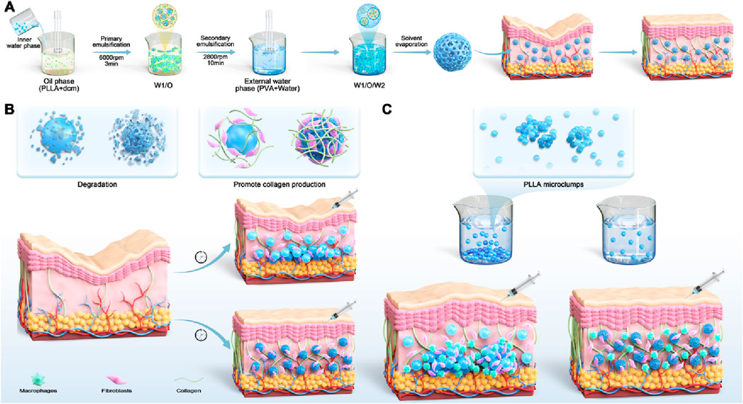
Figure 1. Schematic illustration of the fabrication and function of porous microspheres. (A) The PLLA porous microspheres are crafted using the double emulsion-solvent evaporation technique, which promotes collagen regeneration and combats skin sagging. (B) In comparison with solid microspheres, PLLA porous microspheres exhibit a quicker degradation rate and stimulate the production of increased amounts of collagen, rendering them more efficacious. (C) Compared to solid microspheres, PLLA porous microspheres exhibit a more uniform distribution upon reconstitution, which diminishes the aggregation of particles and thereby prevents the formation of nodules.
Results
Effect of different porogens on microsphere characteristics
We successfully developed porous PLLA microspheres using a double emulsion method to optimize their porosity, essential for enhancing their functionality in anti-aging applications. The porosity of these microspheres facilitated suspension in liquids, simplifying injection processes, and promoting greater cell attachment and collagen synthesis. Porous particles were fabricated via double emulsion solvent evaporation as shown in Figure 1A (Rosca et al., 2004; Kumar et al., 2022; Nishimura and Murakami, 2021; Li et al., 2024; Liu et al., 2005). To assess the impact of different pore-forming agents on porous PLLA microspheres, four agents were explored: a combination of 6% gelatin with 30% MgCl2, 30% MgCl2 alone, 6% gelatin alone, and 4% NH4HCO3. Each was incorporated into the internal aqueous phase, with an oil-water ratio set at 150:30 for the PLLA solution to internal aqueous phase solution volume. The morphological traits of these microspheres were then scrutinized using scanning electron microscopy (SEM). The analysis revealed that microspheres from the 6% Gelatin +30% MgCl2 group presented pores of varying sizes and irregular shapes, while those solely from the 30% MgCl2 exhibited consistently round pores but had a diminished rate of pore formation. In contrast, the 6% Gelatin group produced microspheres with high porosity, characterized by uniformly round pores, limited internal structure, and enlarged internal voids. Meanwhile, the 4% NH4HCO3 group’s microspheres were distinguished by their circular pores, consistent sizing, extensive porosity, and intricate internal honeycomb-like structure (Figure 2A).
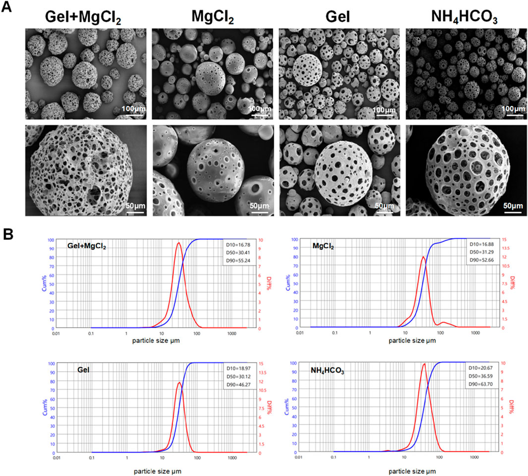
Figure 2. The influence of different pore forming agents on PLLA porous microspheres. (A) Surface morphology of the PLLA porous microspheres under different inner water phase, 6%Gel+30%MgCl2, 30%MgCl2, 6%Gel, 4%NH4HCO3. (B) Particle size distribution of the PLLA porous microspheres under different inner water phase, 6%Gel+30% MgCl2, 30%MgCl2, 6%Gel, 4%NH4HCO3.
Subsequent analysis focused on the diameter distribution of porous microspheres within various experimental groups, employing D10, D50, and D90 values to represent the particle size distribution. D10, D50, and D90 denote the percentile diameters where 10%, 50%, and 90% of particles in the population are smaller in size, respectively. In the 6% Gelatin +30% MgCl2 group, the D10, D50, and D90 values were recorded as 16.78 μm, 30.41 μm, and 55.24 μm, respectively. Comparatively, the 30% MgCl2 group showed slightly different distributions with D10, D50, and D90 values of 16.88 μm, 31.29 μm, and 52.66 μm, respectively. The 6% Gelatin group presented values of 18.97 μm, 30.12 μm, and 46.27 μm for D10, D50, and D90, respectively. Notably, the 4% NH4HCO3 group exhibited wider size distributions with D10, D50, and D90 values of 20.67 μm, 36.59 μm, and 63.70 μm, respectively, as illustrated in Figure 2B. Given its higher porosity, regular surface morphology, and distinctive internal honeycomb structure that enhances cell adhesion, NH4HCO3 was selected for further use as the optimal pore-forming agent in our ongoing experiments.
Effect of different oil-water ratios on porosity and particle size
The investigation into the effects of 4% NH4HCO3 as a pore-forming agent on the porosity and particle size of PLLA microspheres under varying oil-water ratios yielded insightful observations regarding the structural characteristics of the microspheres. SEM analysis was pivotal in revealing that an increase in the oil-water ratio correlated with a reduction in porosity. Lower porosity was found to potentially reduce bioactivity due to a decreased surface area for cell interaction. Despite this decrease, the microspheres maintained their distinctive internal honeycomb structure, which was crucial for their functionality in biomedical applications (Figure 3A). This structure was particularly valued for its potential to enhance interconnectivity within the spheres, facilitating better integration with biological tissues and improved delivery of therapeutic agent (Li et al., 2024; McCarthy et al., 2024; Yuan et al., 2021).
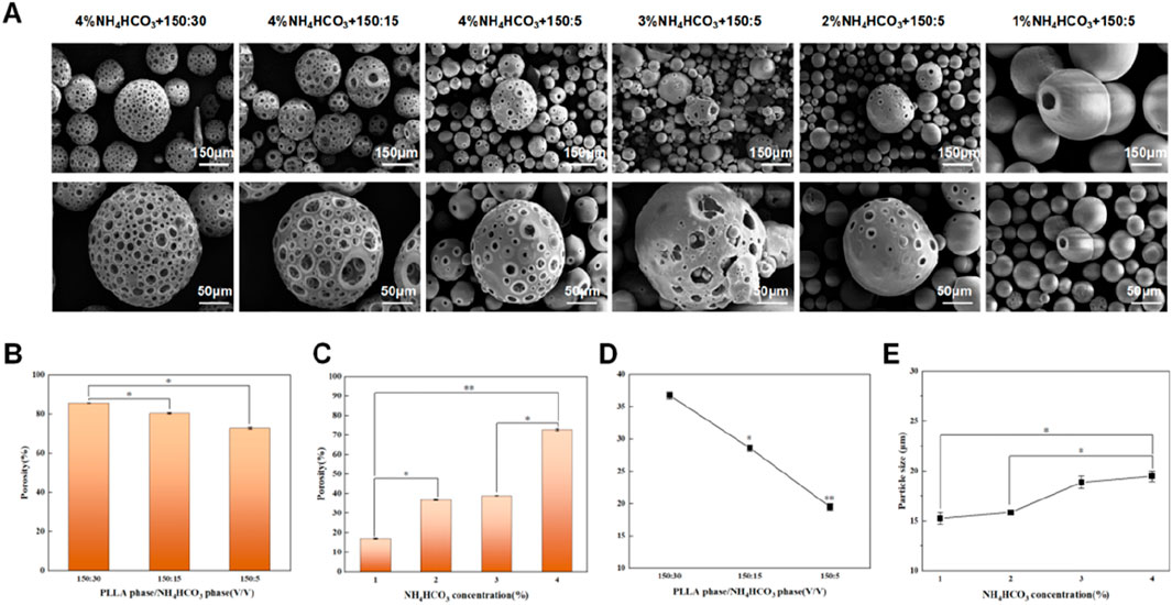
Figure 3. Evaluation of porous microspheres and regulation of porosity with NH4HCO3 as porogen. (A) Morphology of porous microspheres with different porosity. From left to right are 85.69%, 80.43%, 72.84%, 39.03%, 36.89%, 16.98%. (B) Effect of PLLA phase/NH4HCO3 phase on porosity (150:30, 150:15, 150:5). (C) Effect of NH4HCO3 concentration on porosity (4%, 3%, 2%, 1%). (D) Effect of PLLA phase/NH4HCO3 phase on particle size (150:30, 150:15, 150:5). (E) Effect of NH4HCO3 concentration on particle size (4%, 3%, 2%, 1%). The particle size refers to D50 values. Data are presented as mean ± SD (n = 3) (*p < 0.05; **p < 0.01).
These results underscore the critical relationship between oil-water ratios and the structural characteristics of PLLA microspheres. The decreased porosity with higher oil concentrations suggests the agent cannot effectively induce porosity under these conditions. Meanwhile, the reduction in particle size associated with increasing oil-water ratios could be beneficial for applications requiring finer particles for improved dispersion and injectability in medical applications (Bentkover, 2009; Cohen et al., 2013; Alqahtani et al., 2020; Doshi and Mitragotri, 2010; Attenello and Maas, 2015; Lee and Kim, 2015; Herrmann et al., 2018). Thus, adjusting the oil-water ratio provides an adjustable processing parameter for customizing microsphere performance to optimizing the physical properties of PLLA microspheres for specific clinical uses.
Effect of NH4HCO3 concentration on porosity and particle size
Next, we systematically analyzed the impact of varying NH4HCO3 concentrations (4%, 3%, 2%, 1%) on the porosity and particle size of porous microspheres while maintaining an oil-water ratio of 150:5. Utilizing scanning electron microscopy (SEM), we observed a significant reduction in porosity as NH4HCO3 concentration decreased (Figure 3A). Micropore formation on the surface was notably infrequent. The quantitative analysis showed a stark decrease in porosity from 72.84% at the highest concentration to 16.98% at the lowest (Figure 3C). Interestingly, the porosity levels between the 2% and 3% NH4HCO3 concentrations exhibited minimal variation, registering porosities of 36.89% and 39.03% respectively. In terms of particle size, an inverse relationship was observed with NH4HCO3 concentration; as the concentration increased, the median particle size (D50) also increased, ranging from 15.26 μm at 1% NH4HCO3 to 19.48 μm at 4% NH4HCO3 (Figure 3E). This correlation suggests that the concentration of NH4HCO3 not only influences porosity but also affects the overall dimensional growth of the microspheres, underscoring the critical role of NH4HCO3 in tuning both the structural and physical properties of these biomaterials for specific applications.
Injectability and sedimentation rates of porous microspheres with different porosity
Due to the importance of injectability and sedimentation rate of microspheres when used for dermal void fillers, we next explored the relationship between the porosity levels of microspheres synthesized with varying concentrations of NH4HCO3 (1%, 3%, and 4%) and their influences on the force required for administration. Utilizing an oil-water ratio of 150:5, the microspheres were categorized into three groups based on porosity: low (<30%, with actual porosity of 16.98%), medium (30%–60%, with actual porosity of 39.03%), and high (>60%, with actual porosity of 72.84%). Injection force experiments were conducted using 26G and 27G needles on reconstituted poly-L-lactic acid porous particles. Results indicated that injection forces for the 26G needle across the low, medium, and high porosity groups were 1.597 N, 1.740 N, and 2.151 N, respectively. For the 27G needle, forces were 2.098 N, 2.172 N, and 2.591 N, respectively (Figure 4A). Additionally, we evaluated the sedimentation behavior of these groups over 7 h post-reconstitution. The low porosity group demonstrated significant particle precipitation and minimal suspension, contrasting with the medium porosity group, which showed the highest level of suspended particles and moderate precipitation. The high porosity group displayed significantly reduced precipitation and suspended particles, with an increase in floating liquid (Figure 4B).
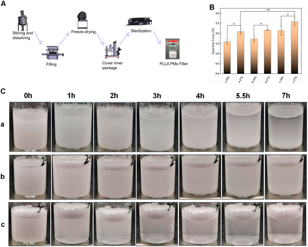
Figure 4. Performance evaluation of fillers prepared by PLLA porous microspheres with different porosity. (A) Schematic diagram showing the preparation of PLLA porous particle fillers. (B) Evaluation of injection properties of fillers with different porosity by two kinds of injection needles (26G, 27G). Data are presented as mean ± SD (n = 3) (*p < 0.05; **p < 0.01). (C) Suspension stability pictures during 7 h, a is low porosity group (<30%, 16.98%), b is medium porosity group (30%–60%, 39.03%), c is high porosity group (>60%, 72.84%).
These findings demonstrated that microspheres with medium porosity not only required less injection force but also maintained a more stable suspension state post-reconstitution. This stability promoted homogeneous in vivo distribution of the microspheres upon injection, potentially enhancing the therapeutic efficacy of the delivered product (Zhao et al., 2023; Ginter et al., 2023). The results underscored the importance of optimizing microsphere porosity to balance ease of injection with effective distribution and stability in suspension, which were critical for achieving desired clinical outcomes in injectable application.
Porous structure accelerated microspheres degradation and improved their performance in vivo
Next, we conducted microsphere degradation experiments to evaluate the influence of porous structure on the degradation behaviors of different microspheres: porous microspheres vs. solid microspheres. Both groups of microspheres were exposed to a soaking solution and incubated for varying durations (1, 3, 5, 7, and 10 days) to assess degradation rates at an elevated temperature to accelerate the degradation of PLLA. Degradation was quantitatively assessed through weight loss measurements and qualitatively through changes in surface integrity using Scanning Electron Microscopy (SEM). Solid microspheres exhibited microfractures visible via SEM, while porous microspheres degraded more uniformly, retaining their internal honeycomb structure even after the outer layer decomposed (Figures 5A–D). By day 10, weight loss was 4.46% for solid microspheres and 6.33% for porous ones. The pH of the soaking solution for solid microspheres was 7.45, compared to 7.18 for porous microspheres, indicating a more acidic environment due to faster degradation of the latter (Figures 5E,F). These findings demonstrate faster degradation kinetics in porous formulations and the porous microspheres are capable of preserving their internal honeycomb structure throughout the degradation process.
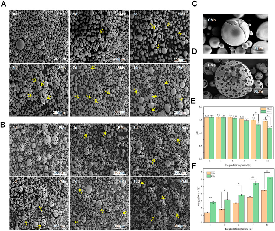
Figure 5. Evaluation of solid porous microspheres and porous microspheres in vitro. (A) SEM images of solid porous microspheres after different degradation periods (0, 1, 3, 5, 7, and 10 days), Yellow Arrow: solid porous microspheres with cracked surface. (B) SEM images of porous microspheres after different degradation periods (0, 1, 3, 5, 7, and 10 days), Yellow Arrow: the porous edge began to degrade until the porous microspheres collapsed. (C) Enlarged image of the cracking process of solid microspheres. (D) Enlarged image of the cracking process of porous microspheres (E) Weight loss of solid porous microspheres and porous microspheres after different time (1, 3, 5, 7, and 10 days). (F) pH values of solid porous microspheres and porous microspheres after different time (0, 1, 3, 5, 7, and 10 days). Data are presented as mean ± SD (n = 3) (*p < 0.05; **p < 0.01).
We employed a rabbit intradermal injection model to evaluate the in vivo performance of solid versus porous PLLA microspheres, assessing tissue response by administering medium-porosity porous PLLA microsphere fillers to the experimental group and treating the control group with LöviselleTM (a commercially available PLLA-based filler). Histopathological analysis revealed markedly enhanced inflammatory reactions in the porous microsphere group from week 2 through week 12, whereas the solid microsphere group exhibited only mild inflammation that became discernible at weeks 8 and 12 (Figure 6A). Quantitative histomorphometric analysis of inflammatory cell infiltration demonstrated significantly higher cell counts in porous microspheres throughout the observation period, reaching an approximate 30:5 ratio compared to solid microspheres by week 12 (Figure 6C). This pronounced inflammatory response may be attributed to synergistic effects of the porous architecture promoting cellular adhesion and accelerated degradation kinetics generating pro-inflammatory byproducts. Furthermore, robust collagen deposition was observed in porous microspheres at weeks 8 and 12, indicating enhanced extracellular matrix remodeling as evidenced by increased subdermal layer thickness (Figure 6B). Statistical evaluation of collagen area ratios revealed persistent yet temporally attenuated differences between groups, with porous microspheres showing a statistically significant 7% greater collagen deposition at week 2 (p < 0.05, Figure 6D), suggesting accelerated tissue regeneration kinetics. These comprehensive findings demonstrate that porous PLLA microspheres facilitate superior tissue integration through enhanced bioactivity and faster matrix deposition, establishing their potential as next-generation injectable dermal fillers with improved therapeutic efficacy.
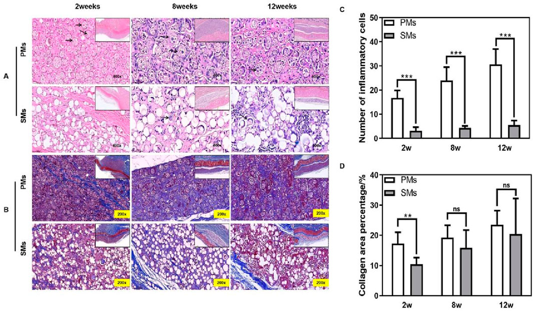
Figure 6. Solid microspheres vs. porous microspheres in vivo. (A) H&E staining (×400) at the 2nd, 8th and 12th week after injection. Black arrows: inflammatory cells, Solid boxes: zoom-in images of each sample. (B) Masson staining (×200) at the 2nd, 8th and 12th week after injection. Solid boxes: zoom-in images of each sample. (C) Histological evaluation of inflammatory cells. (D) Histological evaluation of collagen area ratio.
Porous microspheres significantly reduced nodule formation after implantation
In clinical practice, the aggregation of solid microspheres, such as nodule formation, can result in localized inflammation and discomfort for patients. This aggregation can disrupt the uniform distribution of the therapeutic agents, leading to concentrated areas of high dosage that may not only decrease the efficacy of the treatment but also increase the risk of adverse reactions. In this set of experiments, we aimed to compare the performance and side effects of a commercial product, Löviselle™, with our optimized porous microspheres with proper porosity and good injectability. The objective was to evaluate the potential advantages of the porous microspheres in reducing side effects commonly associated with solid microsphere formulations, such as nodule formation and localized inflammation. Our results demonstrate that in vivo, porous microspheres achieve significantly more uniform subcutaneous distribution in New Zealand white rabbits compared to their solid counterparts, which exhibit a pronounced tendency toward aggregation and may consequently induce nodule formation. Quantitative analysis of microcluster formation further revealed a markedly lower incidence of small clusters in the porous microsphere group relative to the solid microsphere group, with a ratio of 7.67:1.33, underscoring the superior tissue distribution homogeneity of porous microspheres. Complementing these in vivo observations, in vitro experiments demonstrated that porous microspheres remained suspended significantly longer (7 h vs. 3 h; Figure 7B). These collective findings indicate that porous microspheres may confer substantial clinical advantages by ensuring more predictable therapeutic outcomes while simultaneously mitigating the potential for adverse effects.

Figure 7. Injection of PLLA fillers may cause nodules in vivo and in vitro evaluation. (A) Masson staining (200×) is performed on the 12th week after subcutaneous implantation, the distribution of solid porous microspheres is disordered and shows aggregation and precipitation, while the distribution of porous microspheres is orderly and no aggregation and precipitation. (B) Evaluation of suspensibility of PLLA solid and porous microsphere fillers after redissolution. A large number of microspheres are precipitated in solid microspheres after 3 h, but the stability of porous microsphere is better in 7 h. (C) Histological evaluation of the number of micro clumps.
Discussion
We constructed porous poly (L-lactic acid) (PLLA) microspheres with an internal honeycomb-like structure in this study using a double-solvent evaporation method, employing ammonium bicarbonate (NH4HCO3) as a porogen. This design aimed to reduce the microspheres’ density while increasing their surface area, thereby reducing the lag phase for clinical efficacy. Additionally, the improved suspension stability upon reconstitution facilitates more uniform tissue distribution post-injection, minimizing nodule formation.
The selection of NH4HCO3 as a porogen was based on its ability to generate a homogeneous porous architecture, which not only enhances interfacial contact but also maintains structural integrity during degradation. Unlike alternative porogens such as sodium bicarbonate (NaHCO3) or calcium carbonate (CaCO3), NH4HCO3 decomposes entirely into gaseous byproducts (NH3, CO2, and H2O), eliminating risks associated with inorganic salt residues and pH fluctuations that could compromise product safety and performance (Sanchez-Herencia et al., 2021; Peng et al., 2017; Tewes et al., 2013).
Comparative evaluations of PLLA microspheres with varying porosities revealed that intermediate porosity (30%–60%) optimized both suspension duration and injection force (Figures 4B,C). While this range offers initial evidence, further refinement is required to determine the precise porosity threshold that maximizes clinical applicability.
In vitro and in vivo studies confirmed the accelerated efficacy of porous microspheres, though their long-term performance warrants careful evaluation. While collagen-mediated effects may mitigate potential disparities in prolonged outcomes, current animal data remain limited to a 12-week observation period, leaving long-term effects such as fibrotic remodeling or delayed immune reactions unexplored. Extended studies are necessary to assess degradation kinetics and therapeutic persistence beyond this timeframe.
Our research indicates that the degradation kinetics of PLLA microspheres are at a moderate level: they degrade faster than slowly degrading polycaprolactone (PCL, primarily degraded via ester hydrolysis with a degradation time of 2–4 years) but slower than cell-mediated calcium hydroxyapatite resorption (CaHA, typically 12–18 months). Currently, we have not conducted comparative studies between porous PLLA microspheres and other dermal fillers. We believe that performing such comparative studies will enable us to explore more potential application scenarios for porous PLLA microspheres.
Although PLLA porous microspheres accelerate the onset of action, a waiting period is still required to observe the full effect. To address this limitation, researchers have developed composite materials by combining PLLA with other substances. For instance, Su et al. synthesized a suspension of polylactic acid microspheres and hyaluronic acid (PLLA-b-PEG/HA), which demonstrated improved immediate filling effects while maintaining the long-term benefits of PLLA (Su et al., 2024). However, volume reduction after injection remains an issue due to the degradation of HA before PLLA achieves its complete efficacy.
Clinical studies have documented that the incidence of skin/subcutaneous nodules and granulomas, the most common adverse reactions associated with PLLA treatment, ranges from 1% to 44% (Ao et al., 2024). While current preventive measures primarily rely on refined injection techniques and post-treatment massage protocols, structural modifications of PLLA microspheres represent a novel approach to simultaneously address two key clinical challenges: accelerating the onset of therapeutic effects while reducing the incidence of nodules.
Experimental section
Preparation of porous microspheres
The PLLA, with an intrinsic viscosity of 1.8 dL/g, was sourced from Shenzhen Jusheng Biotechnology Co., Ltd. Porous microspheres were prepared using the double emulsion-solvent evaporation technique. Initially, the internal aqueous phase containing a pore-forming agent was introduced into the oil phase (composed of 150 mL of a 6% PLLA solution in dichloromethane) at a specific oil-to-water volume ratio. The biphasic mixture was then subjected to shear forces using a high-speed emulsifying shear machine at 6,000 rpm for 3 min to form a primary water-in-oil (w/o) emulsion. Subsequently, this primary emulsion was incorporated into an external aqueous phase of 1,500 mL of 1% polyvinyl alcohol (PVA), which had been pre-cooled to a temperature range of 2°C–8°C and sheared at 2,800 rpm for 10 min to form a complex water-in-oil-in-water (w/o/w) emulsion. The emulsion was then stirred in a fume hood using a stirrer at 100 rpm for 16 h (overnight). After allowing the mixture to stand for stratification, the obtained microspheres were washed twice with purified water at 40°C and subsequently washed twice with anhydrous ethanol. Finally, porous microspheres were dried in a forced-air oven at 40°C for 12 h.
Scanning electron microscope
The microsphere sample was adhered on an aluminum substrate using carbon tape and sprayed a small amount of gold for 60 s. Each microsphere morphology and surface conditions were observed under scanning electron microscopy (SEM, Scios2Hivac) at an accelerating voltage of 3 kV.
Particle diameter measurement
About 0.1 g of microspheres were weighed and thoroughly mixed with 10 mL of purified water. One to two drops of Triton X-100 solution were then added to create a homogeneous suspension. The laser diffraction particle size analyzer (Bettersize2600) was configured with an obscuration range set between 7% (upper limit) and 3% (lower limit), an ultrasonic frequency of 20 W, a rotational speed of 800 revolutions per minute, a sampling frequency of 7,200 times, a refractive index for the product particles of 1.451, and a refractive index for the dispersion medium of 1.333. The suspension was slowly introduced into the circulation cell and subjected to ultrasonic treatment for approximately 1 minute before measuring the D10, D50, and D90 particle size distributions.
Determination of porosity
The formula for calculating porosity was given by Porosity =
Preparation of PLLA filler
In clinical applications, the PLLA injection filler often contains additives such as mannitol and sodium carboxymethyl cellulose. To minimize the influence of these additives on our experimental outcomes, we formulated a specific PLLA filler composition comprising 30 mg/mL of PLLA porous microspheres, 29 mg/mL of mannitol, and 9 mg/mL of sodium carboxymethyl cellulose in a 5 mL volume. This precisely formulated mixture was then dispensed into a vial, freeze-dried to remove moisture, and subsequently subjected to electron beam irradiation at a dose of 20 kGy for sterilization and stabilization purposes.
Injection force test
The PLLA porous microspheres were formulated into a PLLA filler according to the aforementioned recipe. A 5 mL aliquot of the PLLA filler was reconstituted with 5 mL of purified water, followed by vigorous shaking for 1 min to ensure complete dissolution. The mixture was then allowed to stand for 2 h to promote stability. Subsequently, a 1 mL sample was aspirated using a syringe and placed into the mold of a servo tensile testing machine. The servo tensile testing machine was configured with a detection speed of 30 mm/min and a gauge length of 20 mm. The injection force was then measured and recorded for analysis.
Static stratification experiment
The PLLA porous microspheres were formulated into a PLLA filler according to the previously established recipe. Each vial was filled with 5 mL of the PLLA filler, and subsequently, 5 mL of purified water was added. The mixture was vigorously shaken for 1 min and then allowed to stand. Photographic observations were conducted at specified time points according to the experimental setup.
Accelerated degradation experiment
A soaking solution was prepared using 0.067 M potassium dihydrogen phosphate and 0.067 M disodium hydrogen phosphate, adjusted to maintain a pH of 7.4 ± 0.2. Porous microspheres were created using 3% NH4HCO3 as a pore-forming agent with an oil-to-water ratio of 150:5, achieving a porosity of 39.24%. For the control, a 6% (w/v) PLLA solution was emulsified into a 1% (w/v) PVA solution and subsequently solidified. Thirty centrifuge tubes were divided into five groups, each containing tubes loaded with 0.5 g of microspheres and 15 mL of the soaking solution, and were subjected to accelerated aging at 70°C ± 1°C for durations of 1, 3, 5, 7, and 10 days. Post-treatment, tubes were centrifuged, and the supernatant was analyzed for pH changes. Residues were dried to a constant weight for mass loss determination, providing a standardized method to assess material stability under accelerated conditions.
Animal preparation
Four Male New Zealand white rabbits weighing 2–2.5 kg was provided by the Animal Experiment Center of Zhejiang University. Treatment of experimental animals was in accordance with the guidance of the Animal Care and Use Committee of the Medical College of Zhejiang University and all National Institutes of Health animal handling procedures (Approval number: AIRB-2023-0348). The animals had free access to both sterile water and food in a light and temperature-controlled environment. Rabbits were anesthetized by intramuscular injection of a mixture of zoletil (30 mg/kg) and rompun (10 mg/kg). Euthanasia was performed via intravenous administration of pentobarbital sodium at a dose of 100–200 mg/kg body weight.
Histological analysis
The experimental group consisted of PLLA filler, which was synthesized using porous microspheres based on the previous formula. In contrast, the control group was administered Löviselle (Changchun Sheng Boma Biomaterials Co., LTD.). The dorsal fur of the white rabbits was carefully shaved, and subsequently, six experimental group samples were injected subcutaneously on one side, while six control group samples were injected subcutaneously on the opposite side, with each injection containing 0.2 mL. At the predetermined time for the experiment, the white rabbits were humanely euthanized. Following euthanasia, the specimens were dehydrated, embedded, and sectioned into 4 μm-thick paraffin slices. These slices were subsequently stained using Hematoxylin and Eosin (H&E) as well as Masson’s trichrome technique.
Statistical analysis method
All data were presented as mean ± standard deviation. Statistical analyses were conducted using SPSS version 19.0 (IBM, United States) and involved univariate analysis of variance (ANOVA) followed by Tukey’s multiple comparison tests to evaluate inter-group differences. A P-value of less than 0.05 was considered statistically significant (*p < 0.05, **p < 0.01). Mapping was performed using Origin 2021 (OriginLab, United States).
Data availability statement
The original contributions presented in the study are included in the article/supplementary material, further inquiries can be directed to the corresponding authors.
Ethics statement
The animal study was approved by the Animal Care and Use Committee of the Medical College of Zhejiang University. The study was conducted in accordance with the local legislation and institutional requirements.
Author contributions
QC: Data curation, Formal Analysis, Investigation, Methodology, Software, Visualization, Writing – original draft. JC: Formal Analysis, Investigation, Methodology, Software, Visualization, Writing – original draft. ZZ: Data curation, Formal Analysis, Investigation, Methodology, Validation, Writing – original draft. YX: Data curation, Formal Analysis, Methodology, Validation, Writing – original draft. JM: Funding acquisition, Project administration, Resources, Writing – original draft. WS: Data curation, Formal Analysis, Investigation, Methodology, Visualization, Writing – original draft. XC: Formal Analysis, Investigation, Software, Visualization, Writing – original draft. QL: Data curation, Investigation, Methodology, Software, Writing – original draft. KT: Investigation, Methodology, Software, Validation, Writing – original draft. FL: Conceptualization, Methodology, Resources, Supervision, Writing – review and editing. YZ: Investigation, Project administration, Software, Supervision, Validation, Writing – review and editing. XY: Conceptualization, Funding acquisition, Project administration, Resources, Visualization, Writing – review and editing.
Funding
The author(s) declare that financial support was received for the research and/or publication of this article. This work was supported by the National Natural Science Foundation of China (82372381).
Conflict of interest
Authors YX, JM, WS, XC, QL, KT, and XY were employed by Hangzhou Philosopher’s Stone Biotechnology Co., Ltd.
The remaining authors declare that the research was conducted in the absence of any commercial or financial relationships that could be construed as a potential conflict of interest.
Generative AI statement
The author(s) declare that no Generative AI was used in the creation of this manuscript.
Publisher’s note
All claims expressed in this article are solely those of the authors and do not necessarily represent those of their affiliated organizations, or those of the publisher, the editors and the reviewers. Any product that may be evaluated in this article, or claim that may be made by its manufacturer, is not guaranteed or endorsed by the publisher.
References
Alqahtani, M. S., Syed, R., and Alshehri, M. (2020). Size-dependent phagocytic uptake and immunogenicity of gliadin nanoparticles. Polymers 12, 2576. doi:10.3390/polym12112576
Ao, Y.-J., Yi, Y., and Wu, G.-H. (2024). Application of PLLA (Poly-L-Lactic acid) for rejuvenation and reproduction of facial cutaneous tissue in aesthetics: a review. Medicine 103, e37506. doi:10.1097/md.0000000000037506
Attenello, N., and Maas, C. (2015). Injectable fillers: review of material and properties. Facial Plast. Surg. 31, 029–034. doi:10.1055/s-0035-1544924
Bachmann, F., Erdmann, R., Hartmann, V., Wiest, L., and Rzany, B. (2009). The spectrum of adverse reactions after treatment with injectable fillers in the glabellar region: results from the injectable filler safety study. Dermatol. Surg. 35, 1629–1634. doi:10.1111/j.1524-4725.2009.01341.x
Bass, L. S. (2015). Injectable filler techniques for facial rejuvenation, volumization, and augmentation. Facial Plast. Surg. Clin. N. Am. 23, 479–488. doi:10.1016/j.fsc.2015.07.004
Bentkover, S. (2009). The biology of facial fillers. Facial Plast. Surg. 25, 073–085. doi:10.1055/s-0029-1220646
Bohnert, K., Dorizas, A., Lorenc, P., and Sadick, N. S. (2019). Randomized, controlled, multicentered, double-blind investigation of injectable Poly-l-Lactic acid for improving skin quality. Dermatol. Surg. 45, 718–724. doi:10.1097/dss.0000000000001772
Carey, D. L., Baker, D., Rogers, G. D., Petoumenos, K., Chuah, J., Easey, N., et al. (2007). A randomized, multicenter, open-label study of Poly-L-Lactic acid for HIV-1 facial lipoatrophy. JAIDS J. Acquir. Immune Defic. Syndr. 46, 581–589. doi:10.1097/qai.0b013e318158bec9
Chen, S.-Y., Lin, J.-Y., and Lin, C.-Y. (2020). Micro-fisheyes of carboxymethyl cellulose: the cause of micro-clumps in the suspension of injectable Poly-l-Lactic acid. Aesthet. Surg. J. 40, NP409–NP411. doi:10.1093/asj/sjaa040
Christen, M.-O., and Vercesi, F. (2020). Polycaprolactone: how a well-known and futuristic polymer has become an innovative collagen-stimulator in esthetics. Clin. Cosmet. Investig. Dermatol. 13, 31–48. doi:10.2147/ccid.s229054
Cohen, J. L., Dayan, S. H., Brandt, F. S., Nelson, D. B., Axford-Gatley, R. A., Theisen, M. J., et al. (2013). Systematic review of clinical trials of Small- and large-gel-particle hyaluronic acid injectable fillers for aesthetic soft tissue augmentation. Dermatol. Surg. 39, 205–231. doi:10.1111/dsu.12036
Doshi, N., and Mitragotri, S. (2010). Macrophages recognize size and shape of their targets. PLoS ONE 5, e10051. doi:10.1371/journal.pone.0010051
Engelhard, P., Humble, G., and Mest, D. (2005). Safety of sculptra®: a review of clinical trial data. J. Cosmet. Laser Ther. 7, 201–205. doi:10.1080/14764170500451404
Eppley, B. L., and Dadvand, B. (2006). Injectable soft-tissue fillers: clinical overview. Plast. Reconstr. Surg. 118, 98e–106e. doi:10.1097/01.prs.0000232436.91409.30
Fabi, S., Hamilton, T., LaTowsky, B., Kazin, R., Marcus, K., Mayoral, F., et al. (2024). Effectiveness and safety of sculptra Poly-L-Lactic acid injectable implant in the correction of cheek wrinkles. J. Drugs Dermatol. 23, 1297–1305. doi:10.36849/jdd.7729
Ginter, A., Lee, T., and Woodward, J. (2023). How much does filler apparatus influence ease of injection (and hence, potential safety)? Ophthal. Plast. Reconstr. Surg. 39, 76–80. doi:10.1097/iop.0000000000002247
Guo, J., Fang, W., and Wang, F. (2023). Injectable fillers: current status, physicochemical properties, function mechanism, and perspectives. RSC Adv. 13, 23841–23858. doi:10.1039/d3ra04321e
Herrmann, J. L., Hoffmann, R. K., Ward, C. E., Schulman, J. M., and Grekin, R. C. (2018). Biochemistry, physiology, and tissue interactions of contemporary biodegradable injectable dermal fillers. Dermatol. Surg. 44, S19–S31. doi:10.1097/dss.0000000000001582
Kadouch, J. A. (2017). Calcium hydroxylapatite: a review on safety and complications. J. Cosmet. Dermatol. 16, 152–161. doi:10.1111/jocd.12326
Kumar, A., Kaur, R., Kumar, V., Kumar, S., Gehlot, R., and Aggarwal, P. (2022). New insights into water-in-oil-in-water (W/O/W) double emulsions: properties, fabrication, instability mechanism, and food applications. Trends Food Sci. Technol. 128, 22–37. doi:10.1016/j.tifs.2022.07.016
Lam, S. M., Azizzadeh, B., and Graivier, M. (2006). Injectable Poly-L-Lactic acid (sculptra): Technical considerations in soft-tissue contouring. Plast. Reconstr. Surg. 118, 55S–63S. doi:10.1097/01.prs.0000234612.20611.5a
Lee, J. M., and Kim, Y. J. (2015). Foreign body granulomas after the use of dermal fillers: pathophysiology, clinical appearance, histologic features, and treatment. Arch. Plast. Surg. 42, 232–239. doi:10.5999/aps.2015.42.2.232
Li, X., Li, L., Wang, D., Zhang, J., Yi, K., Su, Y., et al. (2024). Fabrication of polymeric microspheres for biomedical applications. Mater. Horiz. 11, 2820–2855. doi:10.1039/d3mh01641b
Lin, J. Y., Hsu, N. J., and Lin, C. Y. (2022). Retaining Even distribution of biostimulators microscopically and grossly: key to prevent non-inflammatory nodules formation. J. Cosmet. Dermatol. 21, 6461–6463. doi:10.1111/jocd.15101
Lin, J.-Y., and Lin, C.-Y. (2021). Adjusting thickness before injection: a new trend for preparing collagen-stimulating fillers. Plast. Reconstr. Surg. Glob. Open 9, e3653. doi:10.1097/gox.0000000000003653
Liu, R., Ma, G.-H., Wan, Y.-H., and Su, Z.-G. (2005). Influence of process parameters on the size distribution of PLA microcapsules prepared by combining membrane emulsification technique and double emulsion-solvent evaporation method. Colloids Surf. B Biointerfaces 45, 144–153. doi:10.1016/j.colsurfb.2005.08.004
McCarthy, A. D., Hartmann, C., Durkin, A., Shahriar, S., Khalifian, S., and Xie, J. (2024). A morphological analysis of calcium hydroxylapatite and poly- l -Lactic acid biostimulator particles. Skin. Res. Technol. 30, e13764. doi:10.1111/srt.13764
Moyle, G., Lysakova, L., Brown, S., Sibtain, N., Healy, J., Priest, C., et al. (2004). A randomized open-label study of immediate versus delayed polylactic acid injections for the cosmetic management of facial lipoatrophy in persons with HIV infection. HIV Med. 5, 82–87. doi:10.1111/j.1468-1293.2004.00190.x
Narins, R. S. (2008). Minimizing adverse events associated with Poly-l-lactic acid injection. Dermatol. Surg. 34, S100–S104. doi:10.1097/00042728-200806001-00021
Narins, R. S., Baumann, L., Brandt, F. S., Fagien, S., Glazer, S., Lowe, N. J., et al. (2010). A randomized study of the efficacy and safety of injectable poly-L-lactic acid versus human-based collagen implant in the treatment of nasolabial fold wrinkles. J. Am. Acad. Dermatol. 62, 448–462. doi:10.1016/j.jaad.2009.07.040
Nishimura, S., and Murakami, Y. (2021). Precise control of the surface and internal morphologies of porous particles prepared using a spontaneous emulsification method. Langmuir 37, 3075–3085. doi:10.1021/acs.langmuir.0c03311
Nowag, B., Schäfer, D., Hengl, T., Corduff, N., and Goldie, K. (2024). Biostimulating fillers and induction of inflammatory pathways: a preclinical investigation of macrophage response to calcium hydroxylapatite and poly-L lactic acid. J. Cosmet. Dermatol. 23, 99–106. doi:10.1111/jocd.15928
Oh, S., Lee, J. H., Kim, H. M., Batsukh, S., Sung, M. J., Lim, T. H., et al. (2023). Poly-L-Lactic acid fillers improved dermal collagen synthesis by modulating M2 macrophage polarization in aged animal skin. Cells 12, 1320. doi:10.3390/cells12091320
Peng, T., Zhang, X., Huang, Y., Zhao, Z., Liao, Q., Xu, J., et al. (2017). Nanoporous mannitol carrier prepared by non-organic solvent spray drying technique to enhance the aerosolization performance for dry powder inhalation. Sci. Rep. 7, 46517. doi:10.1038/srep46517
Rosca, I. D., Watari, F., and Uo, M. (2004). Microparticle formation and its mechanism in single and double emulsion solvent evaporation. J. Control. Release 99, 271–280. doi:10.1016/j.jconrel.2004.07.007
Sanchez-Herencia, A. J., Gonzalez, Z., Rodriguez, A., Molero, E., and Ferrari, B. (2021). Operational variables on the processing of porous titanium bodies by gelation of slurries with an expansive porogen. Mater. Basel Switz. 14, 4744. doi:10.3390/ma14164744
Signori, R. (2024). Efficacy and safety of Poly-L-Lactic acid in facial aesthetics. doi:10.20944/preprints202407.1452.v1
Su, D., Yang, W., He, T., Wu, J., Zou, M., Liu, X., et al. (2024). Clinical applications of a novel Poly-L-lactic acid microsphere and hyaluronic acid suspension for facial depression filling and rejuvenation. J. Cosmet. Dermatol. 23, 3508–3516. doi:10.1111/jocd.16446
Sun, L., Sun, X., Ruan, W., Che, G., Zhu, F., Liu, C., et al. (2023). Mechanism of remodeling and local effects in vivo of a new injectable cosmetic filler. Sci. Rep. 13, 9599. doi:10.1038/s41598-023-36510-9
Tewes, F., Paluch, K. J., Tajber, L., Gulati, K., Kalantri, D., Ehrhardt, C., et al. (2013). Steroid/Mucokinetic hybrid nanoporous microparticles for pulmonary drug delivery. Eur. J. Pharm. Biopharm. Off. J. Arbeitsgemeinschaft Pharm. Verfahrenstechnik EV 85, 604–613. doi:10.1016/j.ejpb.2013.03.020
Valantin, M.-A., Aubron-Olivier, C., Ghosn, J., Laglenne, E., Pauchard, M., Schoen, H., et al. (2003). Polylactic acid implants (New-Fill) to correct facial lipoatrophy in HIV-infected patients: results of the open-label study VEGA. 17, 2471–2477. doi:10.1097/00002030-200311210-00009
Vleggaar, D. (2006). Soft-Tissue augmentation and the role of Poly-L-Lactic acid. Plast. Reconstr. Surg. 118, 46S–54S. doi:10.1097/01.prs.0000234846.00139.74
Vleggaar, D., Fitzgerald, R., Lorenc, Z. P., Andrews, J. T., Butterwick, K., Comstock, J., et al. (2014). Consensus recommendations on the use of injectable poly-L-lactic acid for facial and nonfacial volumization. J. Drugs Dermatol. JDD 13, s44–s51.
Yuan, S., Shen, Y., and Li, Z. (2021). Injectable Cell- and growth factor-free Poly(4-hydroxybutyrate) (P4HB) microspheres with open porous structures and great efficiency of promoting bone regeneration. ACS Appl. Bio Mater. 4, 4432–4440. doi:10.1021/acsabm.1c00188
Keywords: porous microspheres, poly-l-lactic acid, injectable filler, prevention of nodules, collagen regeneration, aesthetic medicine
Citation: Cao Q, Chen J, Zhang Z, Xiong Y, Ma J, Sun W, Chen X, Lou Q, Tang K, Lin F, Zhu Y and Yu X (2025) Faster efficacy and reduced nodule occurrence with PLLA (poly-l-lactic acid) porous microspheres. Front. Bioeng. Biotechnol. 13:1571820. doi: 10.3389/fbioe.2025.1571820
Received: 06 February 2025; Accepted: 14 July 2025;
Published: 29 July 2025.
Edited by:
Alina Kirillova, Iowa State University, United StatesReviewed by:
Sharanabasava V. Ganachari, KLE Technological University, IndiaSammar Elhabal, Modern University for Information and Technology, Egypt
Copyright © 2025 Cao, Chen, Zhang, Xiong, Ma, Sun, Chen, Lou, Tang, Lin, Zhu and Yu. This is an open-access article distributed under the terms of the Creative Commons Attribution License (CC BY). The use, distribution or reproduction in other forums is permitted, provided the original author(s) and the copyright owner(s) are credited and that the original publication in this journal is cited, in accordance with accepted academic practice. No use, distribution or reproduction is permitted which does not comply with these terms.
*Correspondence: Feng Lin, bGluZmVuZzIwMjNAemp1LmVkdS5jbg==; Yueliang Zhu, emh1MTIzQHpqdS5lZHUuY24=; Xiaohua Yu, eGlhb2h1YS55dUB6anUuZWR1LmNu
†ORCID: Qihua Cao, orcid.org/0000-0003-0656-8193
‡These authors have contributed equally to this work
 Qihua Cao1,2,3†‡
 Jiayu Chen
 Kaijia Tang
 Xiaohua Yu