- 1Department of Cardiology, Affiliated Jinhua Hospital, Zhejiang University School of Medicine, Jinhua, Zhejiang, China
- 2Department of Pharmacy, Affiliated Jinhua Hospital, Zhejiang University School of Medicine, Jinhua, Zhejiang, China
- 3Department of Gastroenterology, Affiliated Jinhua Hospital, Zhejiang University School of Medicine, Jinhua, Zhejiang, China
Background: The increasing prevalence of cardiovascular mortality is becoming a significant worry for individuals who have survived cancer. The aim of this study is to investigate the dynamic trend of cardiovascular death in patients with gastric cancer (GC) and identify the risk factors associated with cardiovascular disease (CVD)-specific mortality in non-metastatic GC patients.
Methods: In the present study, 29,324 eligible patients diagnosed with primary GC were collected from the Surveillance, Epidemiology, and End Results (SEER) database. Standardized mortality ratios (SMRs) adjusted by age, gender, calendar year, and race were calculated. Fine-Gray's competing risk models were taken to identify the prognostic factors of cardiovascular death in GC patients.
Results: There were 1083 (5.2%) cardiovascular deaths among 20,857 patients with local/regional GC, and 76 (0.9%) cardiovascular deaths among 8,467 patients with metastatic GC. The SMRs of CVD-specific mortality continuously increased since the 1975s throughout the 2015s. The competing risk models showed that age (>75 years vs. 0–50 years, HR: 6.602, 95% CI: 4.356–10.006), T stage (T4 vs. T1, HR:0.524, 95% CI: 0.370–0.741), N stage (N3 vs. N0, HR: 0.557, 95% CI: 0.343–0.903), surgery (Yes vs. No, HR: 0.551, 95% CI: 0.461–0.659), and radiotherapy (Yes vs. No, HR: 1.011, 95% CI: 1.011–1.437) were predictive of CVD-specific mortality. Furthermore, based on the results of the competing risk analyses, a nomogram was constructed to predict the probability of CVD-specific mortality for local/regional GC patients.
Conclusion: Our study demonstrated the dynamic trend of cardiovascular death in GC patients, and identified prognostic risk predictors, highlighting the importance cardio-oncology teams in offering comprehensive care and long-term follow-up for GC patients.
1 Introduction
Gastric cancer (GC) ranks as the fifth most prevalent malignancy and the third deadliest cancer worldwide, with over 1,000,000 new cases and 783,000 deaths annually, accounting for approximately 1 in every 12 mortalities globally (1, 2). Owing to the advancements in early detection and innovative treatments, the life expectancy of GC patients has significantly improved since the 1990s and early 2000s (3–5). With the growing population of GC survivors, understanding the precise causes of mortality among these individuals is vital for prioritizing healthcare interventions during survivorship.
Previous research on the causes of mortality among cancer patients has revealed that cancer survivors face an elevated risk of cardiovascular diseases (CVDs), stemming either from the toxicities of cancer treatments or from shared lifestyle (6–8). Several studies have highlighted the close correlation between increased cardiovascular mortality risk and cancer treatments in GC patients (9–11). Therefore, ensuring proper cardiology care for GC survivors is becoming increasingly crucial. However, the prognostic risk factors for predicting CVD-specific death in GC patients remain unknown, and effective clinical guidelines are still lacking (9).
Underestimating the increased risk of CVD faced by GC survivors may lead to missed opportunities for early intervention. To gain a deeper understanding of cardiovascular mortality among GC survivors, we conducted a comprehensive investigation based on data from the Surveillance, Epidemiology, and End Results (SEER) database. Our goals were to elucidate historical trends in cardiovascular mortality and identify prognostic risk factors for cardiovascular mortality in GC patients.
2 Methods
2.1 Data source
The SEER database encompasses approximately 28% of the general US population and collects demographic, clinical information, and survival data (12). Permission to access the database was obtained by signing and submitting a SEER Research Data Agreement form via email. The SEER∗Stat software was used to access the 18 Registry Research Datasets (2000–2015, with additional treatment fields; November 2018 sub). As all data extracted from the SEER database were de-identified and anonymized before release, local ethics approval was not required for this study.
2.2 Study population and definition
All cases diagnosed with GC as their first primary malignancy between January 1, 2000, and December 31, 2015, were included. Patients diagnosed solely through death certificates or autopsy, with missing information on SEER Summary Stage 2000 (local/regional or distant), unknown information on pathological grade, surgery status, adjuvant treatment status, or cause of death were excluded. Follow-up time was defined as the period from diagnosis to the death date, the end of the study period (December 31, 2020), or the date of last contact, whichever occurred first. Cardiovascular mortality encompassed mortality caused by aortic aneurysm/dissection, atherosclerosis, heart diseases (including acute myocardial infarction or other ischemic heart diseases), cerebrovascular diseases, other diseases of capillaries, arteries, and arterioles, as well as hypertension without heart disease (13, 14). Demographic and clinical information of interest, including age at diagnosis, year of diagnosis, gender (male and female), ethnicity, SEER histologic stage (local/regional and distant), grade, histology type, treatment (surgery, chemotherapy, and radiotherapy), and survival months, were collected for analysis.
2.3 Statistical analysis
Statistical analyses were performed using SPSS (version 25.0) or R (version 3.6.1). Descriptive statistics were used to describe the distribution of baseline characteristics. Categorical variables were presented as percentages and compared using Fisher's exact test, while continuous variables were summarized as median values and evaluated using the Kruskal–Wallis rank sum test. Standardized mortality ratios (SMRs), adjusted by age, gender, calendar year, and race, were calculated to compare cardiovascular death rates in our study population with those of the general population. SMRs were defined as the ratios of observed-to-expected deaths due to CVD, with 95% confidence intervals (95% CIs) calculated using exact methods (15, 16). Univariate and multivariate Fine-Gray's competing risk models were employed to calculate CVD-specific hazard ratios (HRs) to evaluate the relative association between prognostic factors and the risk of cardiovascular death (with non-cardiovascular mortality as a competing risk) (13). All tests were two-tailed, with a p-value of <0.05 considered statistically significant.
3 Results
3.1 Baseline characteristics
A total of 29,324 patients diagnosed with primary GC were included in our study, with 10,688 female patients and 18,636 male patients. Of the included patients, the majority were diagnosed with GC at age >50 (n = 25,699, 87.6%) and white (n = 20,213, 68.9%). Most patients (n = 20,857, 71.1%) were diagnosed with local/regional GC, while only 28.9% (n = 8,467) were diagnosed with distant GC. The majority of patients received surgical treatment, due to which a reduction in death was achieved compared to patients receiving non-surgical treatment (death rate: 60.8% vs. 90.9%). Given that most patients with distant GC had different oncological characteristics and received different treatment strategies compared to those with local/regional GC, we analyze the cohort of distant GC separately. Table 1 summarized the baseline clinical characteristics of the included patients by tumor stage.
3.2 Cause-specific mortality among patients with GC
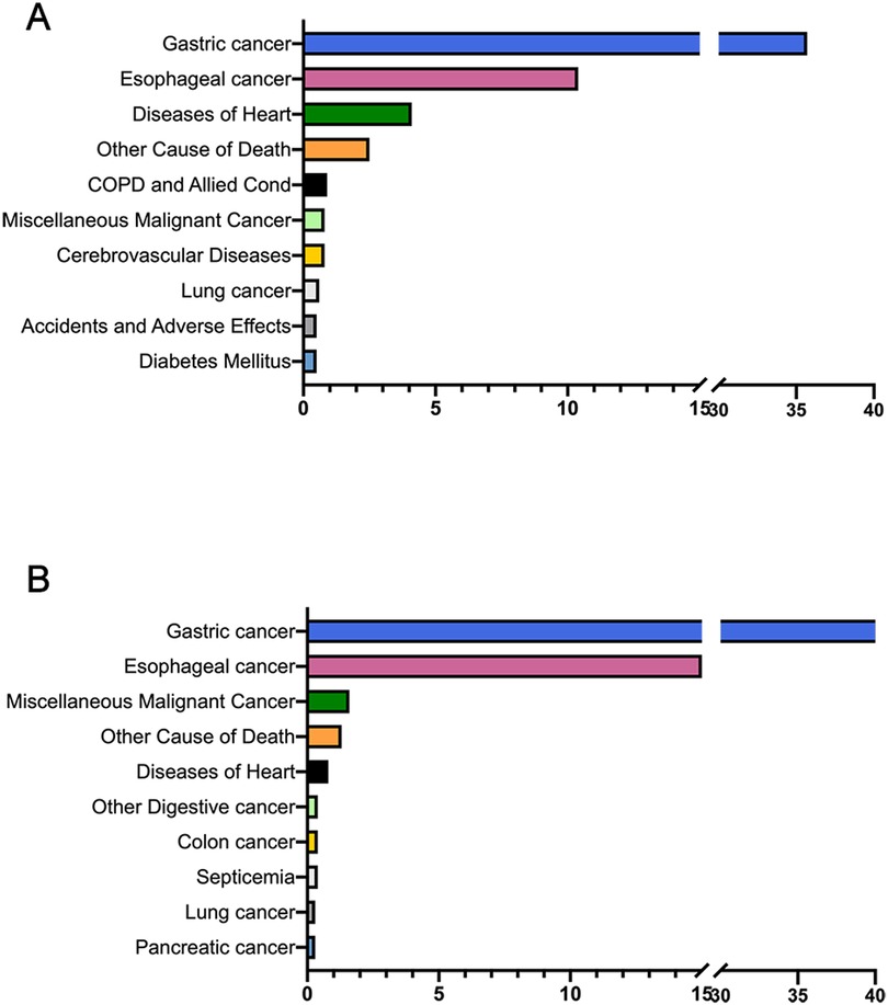
The main causes of mortality among patients with local/regional GC and patients with distant GC were summarized (Figure 1). Specifically, for patients with local/regional GC, GC was still the leading cause of mortality (n = 7,438, 35.7%), followed by esophageal cancer (n = 2,170, 10.4%) and heart diseases (n = 846, 4.1%). It is noteworthy that plurality (77.4%) of cardiovascular deaths in patients with GC were caused by heart diseases. For patients with distant GC, mortality from GC (n = 5,529, 65.3%) composed the majority of deaths, while non-cancer causes and other cancers were less common.
As evidenced in Table 1, there is a striking disparity in surgical intervention rates between distant and local/regional GC patients, with only 35.3% of distant GC patients undergoing surgery compared to 80.1% of local/regional cases. Furthermore, the proportion of distant GC patients not receiving any medical treatment (16.7%) is notably higher than that of local/regional patients (8.0%). Recognizing the substantial disparities in treatment approaches and survival outcomes between patients with distant metastatic GC and those with local/regional advanced disease (Table 1 and Figure 1), we excluded the distant metastatic cohort from subsequent SMR analyses to ensure more accurate and clinically relevant comparisons. We used SMRs to compared CVD-specific death in our study population with that of the general population (Table 2). Overall, the SMR between patients with local/regional GC was 2.10 (95% CI: 2.03–2.17), with 2.86 (95% CI: 2.70–3.03) for female patients, 3.52 (95% CI: 3.36–3.70) for male patients; 4.38 (95% CI: 3.98–4.81) for black ethnicity, 2.94 (95% CI: 2.81–3.07) for white ethnicity, 3.80 (95% CI: 3.44–4.19) for other (Asian/Pacific Islander, American Indian/Alaska Native). The SMRs of older adult patients were gradually decreased compared to those of young patients (<50 years, SMR: 53.64, 95% CI: 34.71–79.19; >75 years, SMR: 2.54, 95% CI: 2.43–2.65).

Table 2. Standardized mortality ratios among local/regional GC patients by demographic and clinic characteristics.
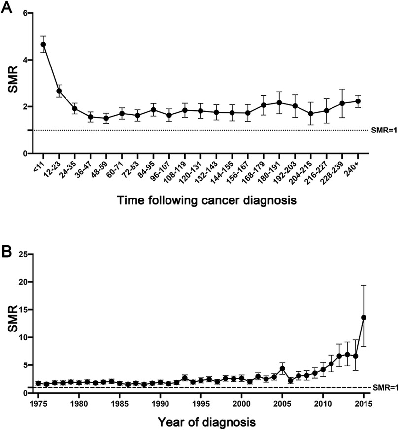
Notably, the CVD-specific death risk among local/regional GC patients was high within the first year following GC diagnosis (<12 months, SMR: 4.65, 95% CI: 4.31–5.01), and it remained elevated compared to that of the general population throughout the entail follow-up period (12–60 months, SMR: 1.94, 95% CI: 1.83–2.06; >120 months, SMR: 1.93, 95% CI: 1.82–2.04, Figure 2A). In addition, we observed that the risk of CVD-specific mortality continuously increased since the 1975s throughout the 2015s (the 1975s, SMR: 1.73, 95% CI: 1.41–2.09; the 2000s, SMR: 2.61, 95% CI: 2.08–3.23; the 2015s, SMR: 13.04, 95% CI: 8.36–19.41, Figure 2B).

Figure 2. The dynamic trend of SMRs of cardiovascular death among local/regional GC at different latency periods (A), and at different year of diagnosis (B).
3.3 Risk factors for CVD-associated mortality
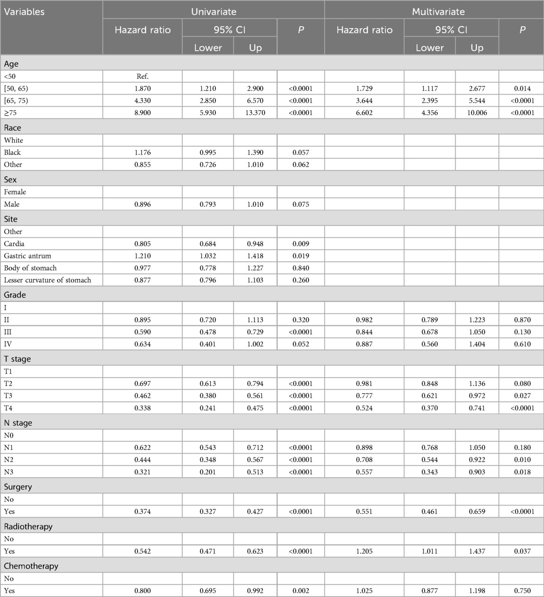
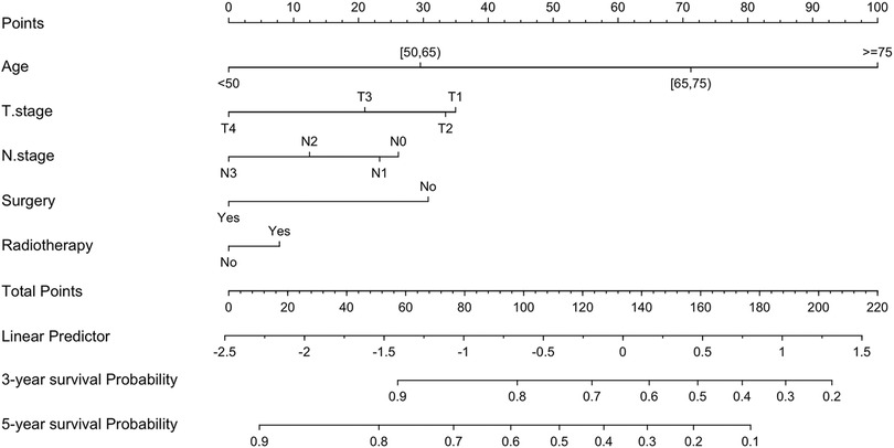
Fine-Gray's competing risk analyses were applied to identify the risk predictors for cardiovascular deaths among local/regional GC patients (Table 3). The results of the univariate competing risk model indicated that age, differentiation grade, T stage, N stage, surgery of the primary tumor, radiotherapy, and chemotherapy were significantly related to the prognosis of CVD-specific mortality. Subsequently, these factors were assessed using a multivariate competing risk model, and found that except for differentiation grade and chemotherapy, age at diagnosis (>75 years old vs. 0–50 years old, HR: 6.602, 95% CI: 4.356–10.006, p < 0.001), T stage (T4 vs. T1, HR:0.524, 95% CI: 0.370–0.741, p < 0.0001), N stage (N3 vs. N0, HR: 0.557, 95% CI: 0.343–0.903, p = 0.018), surgery (Yes vs. No, HR: 0.551, 95% CI: 0.461–0.659, p < 0.0001), and radiotherapy (Yes vs. No, HR: 1.011, 95% CI: 1.011–1.437, p = 0.037) were predictive of CVD-specific mortality. Detailed information regarding the predictors of CVD-specific death in the study cohort is presented in Table 3. Further, based on the results of Fine-Gray's competing risk analyses, a nomogram was constructed to predict the probability of CVD-specific mortality for local/regional GC patients (Figure 3).

Table 3. Fine-Gray's competing risk model analysis for CVD-specific death in patients with local/regional gastric cancer.
4 Discussion
To our knowledge, this population-base cohort study represents the most comprehensive and largest characterization of cardiovascular death among patients with GC. Our findings corroborate that cardiovascular mortality remains a challenge for individuals diagnosed with GC. Additionally, the Fine-Gray's competing risk analyses were utilized to identify several predictors for CVD-specific mortality, including age, gender, T stage, N stage, primary site, differentiation grade, surgery, radiotherapy, and chemotherapy. Our findings align with previous reports on cardiovascular risks in other malignancies (7, 17–19), highlighting the critical need for sustained cardiovascular care throughout the survivorship period.
With the widespread implementation of GC screening and advancements in cancer treatment, the survival rates of GC are improving, which indicates the importance of the management of comorbidities for survivors (19, 20). Recently, Lou et al. conducted a retrospective analysis of the causes of death among 42,813 GC patients. The results revealed that GC (66.7%) was the primary cause of death among these patients, followed by other types of cancer (17.6%). Additionally, among non-cancer causes of death, heart disease (5.7%) ranked first, with cerebrovascular disease (1.4%) closely following (21). Similarly, in the present study, GC was still the main cause of death for patients with GC, especially for those with metastatic status. Notably, among patients with local/regional GC, CVD ranked third among all the causes of death, with its proportion increasing over the follow-up period.
Evaluating SMRs offers important population-level data to screen GC patients who are at risk for elevated CVD-specific death (7, 17). In line with previous studies, a higher SMR was observed among patients with younger age of diagnosis, both historically and in the era of modern treatment (7). The risk for CVD-specific death occurred was greatest within the first year of GC diagnosis and decreased year by year during follow-up, which had been reported in other cancer studies (7, 22) and this may be likely due to aggressive cancer treatment shortly after GC diagnosis (23). This trend may also stem from the fact that patients with the most severe co-existing cardiovascular diseases are at higher risk of cancer treatment and are more likely to die early after cancer discovery (24, 25). Furthermore, compared to patients diagnosed in previous years, those recently diagnosed with GC faced a higher risk of CVD-specific death. This could be attributed to the shorter follow-up time for recently diagnosed patients. Since SMRs of cardiovascular death tended to be highest within the first few years of cancer discovery, SMRs for recently diagnosed patients were partially skewed and higher than those for patients diagnosed in prior years. Additionally, studies revealed that the emerging novel treatments (e.g., targeted therapy, immune checkpoint inhibitors) could cause severe cardiac toxicity (26, 27), which also contributed to the explanation of the elevated SMRs of cardiovascular mortality in recent years.
In the present study, Fine-Gray's competing risk analyses were conducted to identify risk factors for CVD-specific mortality among patients with local/regional GC. Specifically, age at diagnosis emerged as the predominant risk predictor for cardiovascular death, and the older adult patients faced higher risks of cardiovascular death (28). Regarding the histological features of tumor, patients with advanced tumor stage (i.e., T3/T4 status or N3 status) exhibited a lower risk of cardiovascular mortality. A possible explanation of this finding was that those with advance tumor stage were more likely to die of cancer shortly after GC diagnosis.
In terms of cancer treatment, patients who underwent surgery (such as subtotal or total gastrectomy) had lower risks of cardiovascular death in our study. The possible mechanisms explaining the decreased risks of CVD mortality after surgery were various, including reductions in body weight, subcutaneous fat area, and visceral fat area, and the improvement of glycemic control and metabolic profile (29, 30). Chemotherapy and radiotherapy were corroborated to be effective for GC treatment in various respects, such as delaying the metastasis of GC, decreasing the risk of local recurrence, and so on (31, 32). Nevertheless, in this study, we observed that radiotherapy was associated with the elevated risks of CVD-specific mortality among GC patients, indicating the necessity of detailed cardiovascular evaluation before radiotherapy. Moreover, the accelerated development of innovative therapeutic approaches, such as immunotherapy and targeted therapies, has significantly enhanced clinical outcomes for numerous gastric cancer (GC) patients. Further research is anticipated to delve deeply into how these contemporary treatments influence cardiovascular mortality rates.
Our study has several inherent limitations that merit careful consideration. The retrospective nature of the SEER database analysis inherently restricts its capacity to establish definitive causal relationships. A particularly notable limitation pertains to the evaluation of radiotherapy's association with increased cardiovascular mortality, as the absence of detailed dosimetric parameters and specific radiation technique information significantly impedes the formulation of clinically actionable conclusions. Other limitations align with previous comprehensive evaluations of the SEER database's inherent biases and constraints (33). Specifically, potential misclassification of cardiovascular disease-related mortality in death certificate data may lead to inaccuracies in cardiovascular disease estimation. Furthermore, the unavailability of comprehensive comorbidity data, including conditions such as diabetes mellitus, precluded our ability to assess their potential impact on cardiovascular mortality risk.
Notwithstanding these limitations, our findings provide valuable insights that underscore the importance of early cardiology involvement in patient care. Future research should focus on two critical areas: (1) establishing optimal protocols for early cardiology assessment in gastric cancer patients, and (2) determining the appropriate intensity of cardiology care in this patient population. These investigations would significantly enhance our understanding of cardiovascular risk management in gastric cancer patients undergoing treatment.
5 Conclusion
In summary, this study demonstrated the elevated risk of dying from CVDs in patients with GC and identified age at diagnosis, T stage, N stage, surgery of the primary site, and radiotherapy as potential risk factors for cardiovascular mortality using Fine-Gray's competing risk model. Our study underscores the importance cardio-oncology teams in offering comprehensive care and long-term follow-up for GC patients. However, the optimal integration of cardiovascular care into standard oncology treatment protocols remains an area requiring consensus. In this context, Bonaca et al. have proposed an innovative approach through the establishment of collaborative think tanks to systematically evaluate cardiovascular safety in cancer therapy trials (19). Most importantly, there is an urgent need to establish comprehensive, evidence-based guidelines for standardized cardiovascular care specifically tailored for GC patients. These guidelines should address critical gaps in current practice, including optimal screening protocols, risk stratification methods, and integrated care pathways throughout the cancer treatment continuum. The development of such standards requires collaborative efforts between oncologists and cardiologists to ensure both cancer treatment efficacy and cardiovascular safety.
Data availability statement
The original contributions presented in the study are included in the article/Supplementary Material, further inquiries can be directed to the corresponding author.
Author contributions
QZha: Data curation, Software, Writing – original draft, Writing – review & editing, Methodology. QZho: Data curation, Writing – review & editing. JD: Methodology, Writing – original draft. QT: Data curation, Software, Writing – review & editing.
Funding
The author(s) declare that no financial support was received for the research and/or publication of this article.
Conflict of interest
The authors declare that the research was conducted in the absence of any commercial or financial relationships that could be construed as a potential conflict of interest.
Generative AI statement
The author(s) declare that no Generative AI was used in the creation of this manuscript.
Publisher's note
All claims expressed in this article are solely those of the authors and do not necessarily represent those of their affiliated organizations, or those of the publisher, the editors and the reviewers. Any product that may be evaluated in this article, or claim that may be made by its manufacturer, is not guaranteed or endorsed by the publisher.
References
1. Bray F, Ferlay J, Soerjomataram I, Siegel RL, Torre LA, Jemal A. Global cancer statistics 2018: GLOBOCAN estimates of incidence and mortality worldwide for 36 cancers in 185 countries. CA Cancer J Clin. (2018) 68(6):394–424. doi: 10.3322/caac.21492
2. Siegel RL, Miller KD, Jemal A. Cancer statistics, 2020. CA Cancer J Clin. (2020) 70(1):7–30. doi: 10.3322/caac.21590
3. Welch HG, Kramer BS, Black WC. Epidemiologic signatures in cancer. N Engl J Med. (2019) 381(14):1378–86. doi: 10.1056/NEJMsr1905447
4. Joshi SS, Badgwell BD. Current treatment and recent progress in gastric cancer. CA Cancer J Clin. (2021) 71(3):264–79. doi: 10.3322/caac.21657
5. Smyth EC, Nilsson M, Grabsch HI, van Grieken NCT, Lordick F. Gastric cancer. Lancet. (2020) 396(10251):635–48. doi: 10.1016/s0140-6736(20)31288-5
6. Handy CE, Quispe R, Pinto X, Blaha MJ, Blumenthal RS, Michos ED, et al. Synergistic opportunities in the interplay between cancer screening and cardiovascular disease risk assessment: together we are stronger. Circulation. (2018) 138(7):727–34. doi: 10.1161/CIRCULATIONAHA.118.035516
7. Sturgeon KM, Deng L, Bluethmann SM, Zhou S, Trifiletti DM, Jiang C, et al. A population-based study of cardiovascular disease mortality risk in US cancer patients. Eur Heart J. (2019) 40(48):3889–97. doi: 10.1093/eurheartj/ehz766
8. Koene RJ, Prizment AE, Blaes A, Konety SH. Shared risk factors in cardiovascular disease and cancer. Circulation. (2016) 133(11):1104–14. doi: 10.1161/CIRCULATIONAHA.115.020406
9. Ha TK, Seo YK, Kang BK, Shin J, Ha E. Cardiovascular risk factors in gastric cancer patients decrease 1 year after gastrectomy. Obes Surg. (2016) 26(10):2340–7. doi: 10.1007/s11695-016-2085-4
10. Lee JE, Shin DW, Lee H, Son KY, Kim WJ, Suh YS, et al. One-year experience managing a cancer survivorship clinic using a shared-care model for gastric cancer survivors in Korea. J Korean Med Sci. (2016) 31(6):859–65. doi: 10.3346/jkms.2016.31.6.859
11. Abdelgawad IY, Sadak KT, Lone DW, Dabour MS, Niedernhofer LJ, Zordoky BN. Molecular mechanisms and cardiovascular implications of cancer therapy-induced senescence. Pharmacol Ther. (2021) 221:107751. doi: 10.1016/j.pharmthera.2020.107751
12. Surveillance Research Program, National Cancer Institute SEER*Stat software. Available online at: www.seer.cancer.gov/seerstat. version 8.3.6. (accessed December 30, 2019). doi: 10.1016/s0140-6736(20)32381-3
13. Sun JY, Zhang ZY, Qu Q, Wang N, Zhang YM, Miao LF, et al. Cardiovascular disease-specific mortality in 270,618 patients with non-small cell lung cancer. Int J Cardiol. (2021) 330:186–93. doi: 10.1016/j.ijcard.2021.02.025
14. Henson KE, McGale P, Taylor C, Darby SC. Radiation-related mortality from heart disease and lung cancer more than 20 years after radiotherapy for breast cancer. Br J Cancer. (2013) 108(1):179–82. doi: 10.1038/bjc.2012.575
15. Weiner AB, Li EV, Desai AS, Press DJ, Schaeffer EM. Cause of death during prostate cancer survivorship: a contemporary, US population-based analysis. Cancer. (2021) 127(16):2895–904. doi: 10.1002/cncr.33584
16. Ury HK, Wiggins AD. Another shortcut method for calculating the confidence interval of a poisson variable (or of a standardized mortality ratio). Am J Epidemiol. (1985) 122(1):197–8. doi: 10.1093/oxfordjournals.aje.a114083
17. Du B, Wang F, Wu L, Wang Z, Zhang D, Huang Z, et al. Cause-specific mortality after diagnosis of thyroid cancer: a large population-based study. Endocrine. (2021) 72(1):179–89. doi: 10.1007/s12020-020-02445-8
18. Mo X, Zhou M, Yan H, Chen X, Wang Y. Competing risk analysis of cardiovascular/cerebrovascular death in T1/2 kidney cancer: a SEER database analysis. BMC Cancer. (2021) 21(1):13. doi: 10.1186/s12885-020-07718-z
19. Bonaca MP, Lang NN, Chen A, Amiri-Kordestani L, Lipka L, Zwiewka M, et al. Cardiovascular safety in oncology clinical trials: jACC: cardioOncology primer. JACC CardioOncol. (2025) 7(2):83–95. doi: 10.1016/j.jaccao.2024.09.014
20. Kim YI, Kim YW, Choi IJ, Kim CG, Lee JY, Cho SJ, et al. Long-term survival after endoscopic resection versus surgery in early gastric cancers. Endoscopy. (2015) 47(4):293–301. (4):391. Commentaire du travail de Kim YI et al., pp. 293. doi: 10.1055/s-0035-1547028. doi: 10.1055/s-0034-1391284
21. Lou T, Hu X, Lu N, Zhang T. Causes of death following gastric cancer diagnosis: a population-based analysis. Med Sci Monit. (2023) 29:e939848. doi: 10.12659/MSM.939848
22. Yeh TL, Hsu MS, Hsu HY, Tsai MC, Jhuang JR, Chiang CJ, et al. Risk of cardiovascular diseases in cancer patients: a nationwide representative cohort study in Taiwan. BMC Cancer. (2022) 22(1):1198. doi: 10.1186/s12885-022-10314-y
23. Cats A, Jansen EPM, van Grieken NCT, Sikorska K, Lind P, Nordsmark M, et al. Chemotherapy versus chemoradiotherapy after surgery and preoperative chemotherapy for resectable gastric cancer (CRITICS): an international, open-label, randomised phase 3 trial. Lancet Oncol. (2018) 19(5):616–28. (2). doi: 10.1016/j.ijrobp.2018.05.026. doi: 10.1016/S1470-2045(18)30132-3
24. Scott JM, Nilsen TS, Gupta D, Jones LW. Exercise therapy and cardiovascular toxicity in cancer. Circulation. (2018) 137(11):1176–91. doi: 10.1161/CIRCULATIONAHA.117.024671
25. Beyer AM, Bonini MG, Moslehi J. Cancer therapy-induced cardiovascular toxicity: old/new problems and old drugs. Am J Physiol Heart Circ Physiol. (2019) 317(1):H164–7. doi: 10.1152/ajpheart.00277.2019
26. Varricchi G, Galdiero MR, Marone G, Criscuolo G, Triassi M, Bonaduce D, et al. Cardiotoxicity of immune checkpoint inhibitors. ESMO Open. (2017) 2(4):e000247. doi: 10.1136/esmoopen-2017-000247
27. Cautela J, Lalevee N, Ammar C, Ederhy S, Peyrol M, Debourdeau P, et al. Management and research in cancer treatment-related cardiovascular toxicity: challenges and perspectives. Int J Cardiol. (2016) 224:366–75. doi: 10.1016/j.ijcard.2016.09.046
28. Rodgers JL, Jones J, Bolleddu SI, Vanthenapalli S, Rodgers LE, Shah K, et al. Cardiovascular risks associated with gender and aging. J Cardiovasc Dev Dis. (2019) 6(2):19. doi: 10.3390/jcdd6020019
29. Brown JD, Buscemi J, Milsom V, Malcolm R, O'Neil PM. Effects on cardiovascular risk factors of weight losses limited to 5–10. Transl Behav Med. (2016) 6(3):339–46. doi: 10.1007/s13142-015-0353-9
30. Shin DW, Suh B, Park Y, Lim H, Suh YS, Yun JM, et al. Risk of coronary heart disease and ischemic stroke incidence in gastric cancer survivors: a nationwide study in Korea. Ann Surg Oncol. (2018) 25(11):3248–56. doi: 10.1245/s10434-018-6635-y
31. Ng J, Lee P. The role of radiotherapy in localized esophageal and gastric cancer. Hematol Oncol Clin North Am. (2017) 31(3):453–68. doi: 10.1016/j.hoc.2017.01.005
32. Choi AH, Kim J, Chao J. Perioperative chemotherapy for resectable gastric cancer: MAGIC and beyond. World J Gastroenterol. (2015) 21(24):7343–8. doi: 10.3748/wjg.v21.i24.7343
Keywords: cardiovascular mortality, gastric cancer, risk predictor, competing risk models, cardio-oncology teams
Citation: Zhao Q, Zhou Q, Dong J and Tong Q (2025) Risk analysis of cardiovascular mortality after gastric cancer diagnosis: a large population-based study. Front. Cardiovasc. Med. 12:1459151. doi: 10.3389/fcvm.2025.1459151
Received: 2 October 2024; Accepted: 20 March 2025;
Published: 22 April 2025.
Edited by:
Raul Zamora-Ros, Institut d'Investigacio Biomedica de Bellvitge (IDIBELL), SpainReviewed by:
Aditya Yashwant Sarode, Columbia University, United StatesWataru Shioyama, Shiga University of Medical Science, Japan
Copyright: © 2025 Zhao, Zhou, Dong and Tong. This is an open-access article distributed under the terms of the Creative Commons Attribution License (CC BY). The use, distribution or reproduction in other forums is permitted, provided the original author(s) and the copyright owner(s) are credited and that the original publication in this journal is cited, in accordance with accepted academic practice. No use, distribution or reproduction is permitted which does not comply with these terms.
*Correspondence: Qiang Zhao, cHFyc3R1MjAxNEAxNjMuY29t
 Qiang Zhao
 Qiaohong Zhou2
 Qiang Tong

