 Kathryn Reusch1,2*
- 1Department of Sociology and Anthropology, Metropolitan State University of Denver, Denver, CO, United States
- 2Department of Anthropology, University of Colorado Denver, Denver, CO, United States
Introduction: Human castration has a long history. Reasons for castration varied, but one continuous driver was the understanding that castration made individuals born with testes infertile. This manufactured infertility made castrates ideal guards in elite households, especially in cultures such as the Ottoman, Persian, and Chinese Empires. The retention of the childlike high voice, gave another reason for castration -the production of elite high-voiced singers for Catholic Church choirs.
Methods: Documentation related to the process of castration and medical studies of castrates were examined to determine what was known about the effects of castration, what medical treatment castrates received, and what modern doctors learned from castrates' bodies.
Results: The production of castrates was an important process, one which was medically understood to different degrees in different places, causing high mortality rates in some areas and practically none in others. Cultures which used castrates noted certain physical characteristics and medical conditions, but little specific care was given to castrates' medical needs beyond their castration. As medicine became increasingly professionalized, doctors became interested in castrates. Several doctors examined groups of living castrates, while others examined their bodies to understand the processes of castration and why certain bodies exhibited the same effects even when they had not been castrated. These studies contributed to the birth of endocrinology, letting us understand not only of the effects of castration, but a range of hormonal conditions.
Discussion: As some castrates had been enslaved far away from their families and others had been disowned by their natal families, they frequently joined together to provide proper burial for each other. Others who were wealthy would endow monasteries to ensure their burial in their chosen manner, retiring to those establishments in their later years. Other castrates would retire to these to serve as caretakers and guards, understanding that they would be buried in the grounds. In this way, castrates created for themselves communities of care in both life and death. The medical knowledge provided by studying castrates has given the ability to treat some problems intersex people face, allowed trans individuals to safely transition, and created treatments for diseases such as prostate cancer. Thus, castration remains relevant today, and we see that castrates have contributed to wider aspects of public health in a way that many do not know or even acknowledge, while they rarely received this same level of care in return.
1 Introduction
Castration as a practice likely dates back to the Secondary Products Revolution1 and the need to retain male animals for wool and traction without interrupting breeding programs. Human castration has a murkier origin, potentially also dating back to the Secondary Products Revolution, but the earliest definitive written references to it date back circa 4000 BCE, in hymns to the goddess Ishtar from Uruk (Ringrose, 2003; Tougher, 2008; Reusch, 2013a; Taylor, 2002). Since this time, castration has been found in one or more strongly hierarchical civilizations across Afro-Eurasia, with castrated individuals fulfilling multiple roles within these societies (Tougher, 2008; Ringrose, 2003; Patterson, 1982; Reusch, 2013a; Hopkins, 1978, 1963). While the view of castrates2 varied across time and space, they tended to occupy fairly liminal sociocultural positions due to the perception of their physical bodies, making them desirable servants and slaves and leading to a large market for castrated individuals. However, the medical practice of castration was rarely discussed, other than to ban its use within the borders of an empire, and the resulting medical needs of castrates were rarely, if ever, mentioned within the various writings of intact males, who frequently disparaged castrates (Reusch, 2013a,b). Despite this, castrates were often seen to band together to accommodate for their medical, parochial, and funerary needs (Reusch, 2020).
Human castration was an established practice across Afro-Eurasia by the 16th Century CE (Reusch, 2016, 2013a). Castrates had been serving in imperial palaces since the Achaemenid Empire, and continued fulfilling that role through the Roman, Sassanid, Byzantine, Ottoman, and Mamluk Empires, among others (Reusch, 2016; Tougher, 2008; Ayalon, 1988; Hathaway, 2005; Reusch, 2013a). In addition, potentially driven by Byzantine choirs which employed castrated singers, the Vatican Choir admitted two castrated singers to its number by papal bull in 1599 CE (Moran, 2002). China had vast numbers of castrated palace servants during the Ming Dynasty (1368–1644 CE), and a reduced number under the Qing Dynasty (1644–1912 CE) (Dale, 2018; Bi and Xiao, 2009; Tsai, 1996). In Africa, besides the Mamluks, several kingdoms were employing castrated individuals as servants and guards (Holt, 1975; Walthall, 2008; Ware, 2011) (Figure 1). Despite knowing which places employed castrates, we have little record of what the castration process entailed in many places, and what medical care castrated individuals, many of whom were also enslaved, would have received.
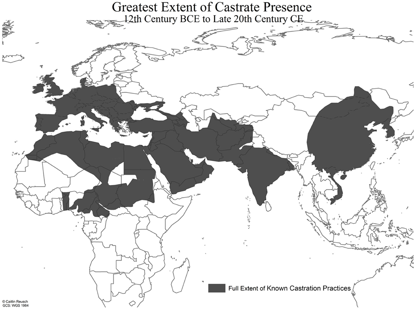
Figure 1. Map of the greatest known extent of castrate presence in Afro-Eurasia from the 12th Century BCE to the late 20th Century CE. Castration was practiced by many of the cultures in Western Asia, especially the Fertile Crescent region, from at least the 5th Millennium BCE, overlapping in space through time. Adapted with permission from Reusch (2020). Edited by Author.
This paper will examine the medical knowledge around the process of castration from ancient times to the early 20th Century CE, then will lay out the way in which studies of castrate bodies from the 18th to 20th Centuries CE contributed to the modern understanding of hormones, growth, and development, and laid the groundwork for the field of Endocrinology.3
2 Materials and methods
Documentation related to castration from ancient times to the 20th Century CE will be examined to understand what knowledge was available on the effects of castration, the dangers of the process of castrating individuals, and what medical treatments may have been available to castrates during their lives. This will include Greek and Roman treatises, medieval documents, early modern medical studies, and studies from the 19th and 20th Centuries CE.
3 Results
3.1 The process of castration
At its base, castration involves the removal of the testicles (Reusch, 2013a). In some societies, this may not have been the surgical cutting away of the testes and scrotum, but rather the crushing of the testes within the scrotum (Reusch, 2013b). In other societies, castration was not considered complete unless the penis had been removed as well, and, in many places, it was seen as fastest and most efficient to remove everything with “one stroke of the razor” (Skuse, 2020; Reusch, 2016, 2013a). However, this process leads to an immediate medical issue: there are a number of large vessels which feed the penis and testicles, and removal of either the gonads or both the gonads and penis can cause these vessels, which are normally held under tension, to retreat into the body, making it more difficult to close them, and increasing the chances that the castrated individual would bleed to death (Reusch, 2013b; Francis, 1926; Heeren, 2009). This process has led to some speculation that artifacts found in Roman deposits may in fact be human castration clamps, created and used in an attempt to hold the vessels in place long enough for suture, or at least cautery, though this has been disputed (Francis, 1926; Heeren, 2009).
It is unclear what castration methods were most commonly used in the ancient period, though there are some references. Texts from Uruk and the later Assyrian, Achaemenid (550–330 BCE), Parthian (238 BCE-236 CE), and Sassanid (205–651 CE) Empires only refer to castrates, not the methods by which they were made (Taylor, 2002; Tougher, 2008; Llewellyn-Jones, 2002; Patterson, 1982). The Classical Greeks (500–323 BCE) seem to have been uninterested in using castrates, but they were happy to enslave and castrate individuals to sell to the Persians. Several texts refer to castrated slaves in the Greek period taking revenge on their slavers, who were frequently the ones who had castrated them, but no reference is made to the process of castration (Hornblower, 2003). The use of castrates increased in the Hellenistic world (323–30 BCE) after Alexander the Great's empire was divided (323 BCE), possibly due to the influence of Persian subjects, though it is likely that castrates were only employed in any real numbers in the Ptolemaic (305–30 BCE) and Seleucid (312–63 BCE) Empires (Tougher, 2008).
Rome, while horrified by the practice of castration, quickly found that castrated slaves were lucrative to sell to their Persian neighbors, and, potentially influenced by this practice and the use of castrates by the Ptolemies and Seleucids, began using castrated servants and slaves in the imperial palace (Tougher, 2008, 2013; Patterson, 1982). In addition, the Phrygian goddess Cybele, whose cult was brought to Rome in 204 BCE, had castrated priests (galli) who served her, in reference to her lover, Attis, who had jilted the goddess, causing her to drive him mad so that he self-castrated (Beard, 1996, 2012; Bowden, 2010; Tougher, 2013). In our first clear reference to the actual process of castration, descriptions of the cult celebrations of Cybele indicated that Roman men, whipped into a frenzy by the atmosphere, would pick up a sharp shard of pottery or rock from the side of the road and use it to remove both penis and testicles. The newly castrated individual was said to throw their removed genitalia into a house they passed by, which was then required to provide new, female garments for the individual, so that they could join the priesthood of the goddess (Tougher, 2013; Bowden, 2010; Beard, 1996, 2012). No reference is made to the medical care that these individuals would have direly required, nor to how many individuals both carried out the process and survived.
Post-Roman Empire and into the Byzantine period, the type of castration performed seems to have taken on a specific geographical patterning, with removal of only the testes being performed on individuals north and west of Anatolia (mostly Europe and portions of North and Central Asia), and removal of both the testes and penis being performed on individuals south and east of Anatolia (Africa and Southwest, South, and East Asia) (Reusch, 2013b). Byzantine castrates could hold many social roles, from Patriarch of the Church or Chamberlain to prostitutes and artisans (Tougher, 2013, 2004, 1997, 1999, 2001, 2008; Ringrose, 1994, 2003). While many of their contemporaries wrote about them, and some discussion was given to who was castrated, no clear explanation of the Byzantine method of castration has survived to the present day.
Byzantine castrates, employed by the church, made up members of the choir when antiphony, the multirange form of singing common today, was introduced. The ability of castrated singers to retain high voices (soprano and alto) post-puberty was used within Byzantine choirs to create these higher vocal sounds while adhering to the Pauline edict for women not to speak in church (Kuefler, 1996; Moran, 2002). The use of castrated singers in Byzantine-controlled Sicilian choirs may have influenced the Normans to include castrated singers to their own choirs after they conquered Sicily, which may have led to the introduction of castrated singers to the choirs of the Roman Catholic Church in the early modern period (Moran, 2002). A papal bull issued by Sixtus V in 1589 officially included four individuals explicitly named as castrates within the Sistine Chapel choir for the first time, though earlier “falsettists” from France and Spain may also have been castrated (Gerbino, 2004; Sherr, 1980; Jenkins, 1998; Milner, 1973). While it is unclear exactly how boys were being castrated in Europe during the post-medieval and early modern periods, there are accounts of surgical removal of the testes from within the scrotum, surgical removal of the testes and scrotum, and crushing of the testicles within the scrotum, sometimes performed by individuals claiming to specialize in castration (Jenkins, 1998; Peschel and Peschel, 1987; Rosselli, 1988).
The fall of Constantinople to the Ottomans in 1453 CE changed the roles of castrates within Anatolia, with many castrates being employed specifically in the palace. We also begin to see a racial and physical differentiation in the types of castration performed. In the sultan's palace, domestic administration and the training and care of royal pages were the responsibilities of enslaved white castrates, who would frequently only have had their testicles removed. The care of the women and royal children was undertaken by enslaved African castrates, who would have had both their testicles and penis removed and thus were seen as “safer” to have around vulnerable individuals (Segal, 2001; Hathaway, 2005).
African Ottoman eunuchs are also where we begin to get our first in-depth information on how castration was performed. There exist two main methods for castration from the 19th−20th Centuries CE, one recorded by Europeans in Africa, and one from China. In the African example, boys would be captured in sub-Saharan Africa, brought up the Nile to Coptic monasteries in Southern Egypt, castrated with removal of testes and penis, and buried immediately in the Nile mud for 3 days, during which they would receive no food or liquid. After 3 days, they were unburied and allowed to eat. Due to the generally unhygienic procedure, up to 75% of people castrated in this way were said to have died either during the process or in the days afterward (Meinardus, 1969; Burckhardt, 1819). Lortet gives another two methods of castration for boys being brought up the Nile at the end of the 19th Century CE (Lortet, 1896). He states that boys being castrated at Coptic monasteries to be sold into Cairene and Ottoman households would, in a similar manner to that described above, have the penis and testicles cut off close to the pelvis with a razor and then be buried nearly to the neck in fine, dry sand that had been sitting under the sun, which he considered aseptic. After 4 or 5 days, the boy would be unburied, and a rag soaked in oil would be applied to the wound. The second method of castration described was to linearly crush the penis and “cords” (presumably the spermatic cords) with a fine string secured by two wooden boards. It is unclear if this was essentially severing the organs or actively crushing them between the boards. Lortet describes this method as avoiding the danger of hemorrhage that accompanied the cutting method, so presumably it was similar to the crushing method that some castrati underwent. Lortet records that because hemorrhage was not as great a fear with this method, the boys were not buried in the sand but instead were bandaged with bark from the Acacia nilotica, which he notes is rich in tannin. Despite this, he reports that up to two-thirds of the boys subjected to either method died shortly after the procedure. Survivors would constantly wear a nail made of lead in the urethral opening to prevent closure of the passage due to scarring (Lortet, 1896).
Hickmet and Regnault describe a method practiced on enslaved boys in Suakim and Tripolitania (Hickmet and Régnault, 1901), in which the child was held down in a chair, the penis was tied with a rope and pulled away from the body, and the penis and testicles were cut off as close to the body as possible. The bleeding was stopped with boiling oil and then a balm of wax, tallow, and mastic was applied to the wound and left there until it detached on its own. According to Hikmet and Regnault, up to nine-tenths of the boys who underwent this procedure died, which increased the value of the survivors from 200 to 2,000 francs (Hickmet and Régnault, 1901).
In China, the process of castration had been practiced in large or small numbers for thousands of years, with the first references to castration coming from oracle bones inscribed in 1300 BCE (Jay, 1993). These inscriptions indicate that prisoners of war were being castrated, and by the Zhou Dynasty (1045–256 BCE), castrates were serving in the government (Kutcher, 2010). By the time the Chinese method of castration was recorded by Stent in 1876 and Matignon in 1896, the process was well-understood (Matignon, 1896; Stent, 1876), and the job of castrator was even passed from father to son in the Qing (1644–1912 CE) Dynasty (Stent, 1876; Wilson and Roehrborn, 1999).
According to Stent, during the Qing Dynasty, castrations were performed just outside the inner Hsi-‘hua gate of the palace, within the imperial city (presumably what is now known as the Forbidden City) itself, in a building known as the Chang-tzû, or shed. The person to be castrated was laid down in a reclining position and held down around the waist and legs, with the legs held open. The castrator would ask whether the individual to be castrated was sure they would not repent, and, if he received consent, would tie ligatures or bandages around the lower belly and upper thighs to prevent hemorrhage. The penis and testicles would be bathed three times with hot pepper water (an antiseptic), and then, with a sharp knife, everything would be removed in one cut. After the castration, the ones performing the surgery would insert a pewter needle or straw (Stent uses the term “spigot”) into the urethral opening, then cover the wound in paper soaked in cold water and bind the wound. The patient would then be made to walk around the room for 2–3 h with the help of two assistants, after which he would be allowed to lay down. He would not be allowed to drink anything for 3 days. After the 3 days, the bandages and nail were removed, and the castrate would be allowed to urinate. If he was able to urinate, the operation was considered a success, but if he could not, he was expected to die, as his urethral passage had become blocked and would cause sepsis and death. After a hundred days, the castrate would be considered fully cured and sent to work in a prince's palace for a year, after which he would be transferred to the imperial palace. Stent had only heard of one death from the operation in “many years” (Stent, 1876).
Matignon's account does not differ much from Stent's, except that he does not mention the binding to prevent hemorrhage or the washing of the surgical area with pepper water before the cutting, and he stated that the castrator would either tie a string around the base of the penis and testicles before excision or grab and twist them to minimize blood flow to them. The wound would be heavily washed with pepper water, a nail of tin or wood would be placed in the urethral opening to prevent strictures, and the wound was tightly bound for 3 days. After the 3 days, the wound would be unwrapped and the nail removed, and if the patient passed clear liquid from the urethra, the operation would be considered a success and they would be sent away with their testicles and penis preserved in a small jar, so that they could present it as proof that they had been castrated. Matignon had only heard of deaths in three or four out of one hundred operations, and the main medical issue he had heard was that of urinary incontinence, something that was also said of the Ottoman eunuchs (Matignon, 1896).
What becomes clear from these accounts is that it was known that castration was a dangerous process that could lead to the death of the individuals being castrated. However, in many cases, such as the boys and young men being brought up the Nile, it was clear that profit outweighed the cost in lives, as castrated slaves could sell for four times what the next most valuable slave would bring in (Meinardus, 1969). What was also known was that the removal of the penis along with the testes, a common practice south and east of Anatolia, would result in the closure of the urethra unless it was held open by some object (frequently a nail made of lead, but potentially also of silver, or a hollow tube made of silver which could be inserted into the urethral opening and then plugged to reduce incontinence) (Wilson and Roehrborn, 1999; Matignon, 1896; Burckhardt, 1819; Lortet, 1896; Stent, 1876). However, this seems to be where the specific care for castrates as related to their physical condition ends. There seems to have been no particular attention paid to the incontinence of those who had lost the penis aside from using a nail or plug to close the urethral opening; the overlengthening of limbs that boys growing without the typical gonadal hormones would have experienced, other than to note that it occurred; nor any care taken about the osteopenia and osteoporosis that castrates would have developed as they aged. While this may be because there was no way to treat any of these conditions, there also seems to not have been any particular attention paid to them, beyond noting that castrates became frail in old age.
3.2 Castration and modern medicine
While not much attention seems to have been paid to castrates' health during their lives, other than what was necessary to keep them in service, as medicine became a recognized, specialized profession in the 18th-early 20th Centuries CE, doctors began to study living and dead castrates to understand why their bodies took on such different shapes to non-castrated males. The initial medical studies of castrated individuals were attempts to understand the mechanisms behind the anatomical changes to the soft and hard tissues which had been known since Aristotle (Reusch, 2013b). Medical practitioners attempted to describe, understand, and quantify what these changes were, using the knowledge that they and their contemporaries were compiling as they studied the natural world around them. The next steps were to look at how castration related to conditions that were not caused by physical castration, such as primary hypogonadism4 and cryptorchidism5. As medical knowledge grew to include hormones, castrates and castration were used to understand the mechanisms by which hormones directed growth and development in organisms, especially mammals.
The first focused medical discussions appear in the late 18th Century CE, with Dupuytren examining the shortening of the laryngeal cartilages in an individual who had been castrated before puberty. What had been clear before this point was that prepubertal castration caused the voice to remain high, allowing males to continue singing soprano and alto parts even after puberty, as was the reason for the creation of the castrati, but what was unclear were the mechanisms that caused the vocal cords to remain small. This is what Dupuytren investigated, followed by Gruber's studies of the genital organs and larynx of a member of the Skoptsy religious sect in Russia, according to (Bilharz 1860). A similar study was performed by John Hunter in 1786 (Bilharz, 1860).
In 1860, Bilharz published a study of the shape and configuration of the preserved genitals of six castrated males, ranging from boys to adults, trying to understand what changes the genitourinary and prostatic structures underwent as a result of castration, and determining which structures in the pubic area were related to the emission of semen and which were related to the urinary tract. He later gave a pelvis and a complete skeleton of two castrates to Ecker, who studied them to describe the effects of castration on the development of the skeleton (Ecker, 1865).
Pelikan studied the castrated members of the Skoptsy, a radical Christian sect that practiced castration of either only the testicles or both the testicles and penis for males and ablation of the breasts for females (Pelikan, 1876). The sect originated in Russia, but facing persecution from the government, some sect members moved to Romania and settled in a few villages near the Black Sea or worked as cab drivers in Bucharest. Pelikan's goal was to outline the external signs of castration in living individuals, to better allow the Russian government to identify and prosecute members of the sect. In order to understand what signs to direct doctors, police, and prosecutors to look for, Pelikan and his team performed experiments on animals and human cadavers, and then compared the results to living Skoptsy that had been examined previously, as well as completing a differential diagnosis of conditions that might mimic castration but were not the result of castration, such as cryptorchidism, anorchidism6, and testicular atrophy (Pelikan, 1876). His main results were that castration performed in early childhood would result in a penis that remained childlike in size as it would not grow after the castration; that the Skoptsy males who had been castrated were infertile, though those who retained their penis were capable of intercourse; that the voice would remain higher, as the larynx would not elongate at puberty; that facial hair would not grow and that axial hair would either not grow or would be thin, short, soft, and downy, while head hair would be less likely to fall out; that the onset of puberty was not obvious and that prepubertal castrates' physical forms would appear more feminine than masculine. In the appendix, one of Pelikan's assistants, Merschejewsky, laid out measurements for several castrated sect members, and noted that castrates grew taller than their non-castrated compatriots, with longer legs, but with narrower shoulders and wider hips, more closely approaching a female than male form (Pelikan, 1876).
Becker examined the skeleton of one castrate of Dinka7 heritage and the pelvis of another of unknown heritage to understand the effects of castration on the human skeleton (Becker, 1899). Both skeletons had previously been examined by Ecker and Bilharz (Ecker, 1865; Bilharz, 1859). Becker compared the two castrated individuals' remains to two non-castrated Ethiopians. All four sets of remains were held in the anatomical collection in Freiburg im Breigau. Becker's findings echoed those of Ecker and Bilharz (Becker, 1899).
Lortet examined the skeleton of a Shilluk8 castrate who had been enslaved, castrated, and sold into a Cairene household, where he died aged 24–26 (the age at death is unclear, as Lortet claims he died at 24 or 25, but writing on the skull claims the individual died at 26) (Lortet, 1896). Lortet performed this examination at least in part due to pure happenstance—he was a friend of the head of the medical school in Cairo and was invited to observe the autopsy of the castrate. In the article, Lortet was mostly focused on the social aspects of castration practiced in Egypt at the end of the 19th Century, but he noted that the skull was small with pronounced prognathism, and the skeleton's torso and legs were disproportionate due to the elongation of the long bones. He went on to mention that the skeletal disproportion between legs and torso seen in this individual was also visible in living castrates. He noted that the elongation of the long bones was more extreme in the more distal elements such as the metacarpals, metatarsals, and phalanges, and that all of the long bones were slim and gracile. He compared the elongation of the long bones in this individual to castrated animals, noting that prepubertal castration caused excessive long bone growth across species (Lortet, 1896). Through some unspecified means, he brought the skeleton to the medical school where he taught in Lyon, the only surviving castrate skeleton in an anatomical collection (Reusch, 2013a,b).
Tandler and Grosz, interested in the effects of the gonads on the development of organisms, began to study castration (Tandler and Grosz, 1909b). They examined the body of a castrate from Zanzibar, who had died of tuberculosis at age 28 in the Rudolfsspitale in Vienna, Austria. The penis and testicles had been removed, and the castrate's skeleton was described as “delicate,” with weak, emaciated musculature. He had a long and narrow neck, a “wide and bulging” thorax, and an underdeveloped larynx, more similar to a child's than an adult's. His skeleton was delicate, without major development of the muscle attachment sites, and a noted disproportion between the torso length (81.5 cm) and the leg length (95.5 cm), with an overall stature of 179 cm. The skull was described as small, with a strongly developed maxilla and mandible, though Tandler and Grosz described the mental eminence as weakly developed and the mandibular angle as childlike. They also felt that the pelvis was small and delicate, with clearly visible, open epiphyses, though the subpubic angle and pelvic outlet clearly displayed a masculine shape. The long bones were gracile and retained open epiphyses in the later fusing epiphyses, such as the proximal humerus and distal radius and ulna. Tandler and Grosz summarized the effects of castration as being the elongation of the long bones and thus disproportion between the torso and legs, but a stalling of the development of secondary sexual characteristics, rather than a switch from masculine to feminine (Tandler and Grosz, 1909a).
Tandler and Grosz followed their study of the castrate's body with a visit to the Skoptsy who were living in Romania at the time (Tandler and Grosz, 1910a). They began their investigation with references to Pelikan's work (Pelikan, 1876), then took anthropometric measurements of five Skoptsy men in Bucharest. They observed disproportional limb-to-torso lengths and “knock-knees” (genu valgum) on most of the men, and radiographs showed open epiphyses on long bones. Because the Skoptsy had been deeply persecuted by the Russian (and to a certain extent Romanian) governments, including doctors, as seen in Pelikan's work, they had a deep distrust of medical examinations, so Tandler and Grosz were only able to obtain a few measurements (Tandler and Grosz, 1910a).
Tandler and Grosz went on to study the skeleton of a eunuchoid individual [someone who today would likely be described as intersex or having a difference of sexual development (DSD)] with small, underdeveloped testes (Tandler and Grosz, 1910b). This individual had also died at age 28, and their overall stature was 181 cm, with a lower body length of 100 cm, displaying the same disproportion between the torso and lower legs. The eunuchoid individual also had a small, childlike larynx; delicate skeleton; and open epiphyses at both ends of the clavicle, the proximal ends of the humeri, tibiae, fibulae, the distal ends of the radii, ulnae, femora, and across the iliac crests and ischial tuberosities, showing that disruptions to the sex hormones before puberty resulted in delayed epiphyseal fusion. Within this study, Tandler and Grosz were attempting to highlight two body types that they had found were prevalent amongst castrated individuals: tall growth and fat growth. By examining the eunuchoid individual, they noted that the two types of growth were not mutually exclusive, and that a range of variation between the two types could be found in castrates. They also wanted to highlight the disproportion between the torso and leg lengths which they had found in all three groups they had examined, which they ascribed to the unfused epiphyses. This series of articles by Tandler and Grosz, especially the comparison between the castrates and the eunuchoid individual, definitively showed that the disproportionate appearance of eunuchoid individuals was due to a lack of testes, as it was so similar to the changes seen in castrated individuals (Tandler and Grosz, 1910b).
During the German occupation of Romania during World War I, Walter Koch took a few skull radiographs and the anthropometric measurements of 13 Skoptsy men (Koch, 1921), observing that some, especially those castrated before eleven, demonstrated thinning of the bones of the skull and kyphosis of the spine. He also noted that the men's torsos were in disproportion to their limbs, with those who had been castrated at an earlier age displaying longer limbs proportionally to their torsos, as had been seen in earlier studies (Koch, 1921).
Following World War I, Wagenseil examined 11 Ottoman palace eunuchs who were of Abyssinian and Sudanese descent and one eunuchoid servant of Turkish descent in Istanbul (Wagenseil, 1927). At the same time, he examined a non-castrated Sudanese male as a comparison. One of the African castrates and the Turkish servant had died in hospital and had been dissected, at which point it was discovered that the Turkish individual was a congenital eunuch (cryptorchia with hypoplastic genitals), rather than a surgical eunuch, as had been previously assumed. Wagenseil noted that his measurements were similar to Tandler and Grosz and Koch's, with the castrates possessing tall, relatively thin or fat bodies and torso to limb length disproportions. He also found that the effects of castration were strongly affected by the age at which the men had been castrated, even though all the individuals he examined had been castrated before puberty, with those castrated at a younger age showing stronger effects. Overall, the castrates displayed elongated limbs; wider bodies, especially through the pelvis; longer heads; and taller faces and noses (Wagenseil, 1927).
Wagenseil then went to Peking (modern Beijing), where he was able to study 31 castrates who had served in the imperial palace (Wagenseil, 1933). He took measurements and radiographs of the individuals, who were between 45 and 57 years old and had been castrated before, during, and after puberty, leading to a wide range of effects on their bodies. Twenty-one of the individuals were kyphotic, and all of the tallest individuals had been castrated prepubertally, though not all of the prepubertal castrates were tall. All of the castrates' torso heights were shorter than contemporaneous non-castrated Chinese males, with the effect strengthened in prepubertal castrates. All of the castrates' pelves were enlarged, and both upper and lower extremities were elongated, with genu valgum present. Their faces were described as taller and narrower than contemporaneous Chinese males, and their head hair was well-developed, but body hair and beards were reduced (Wagenseil, 1933).
Pittard worked with the Skoptsy in Romania before and after World War I, taking the measurements of thirty Skoptsy men from a single village on the Black Sea coast (Pittard, 1934). He noted that they were generally taller than their non-castrated peers and had disproportionate limbs to their torsos due to limb elongation, which he strongly emphasized throughout his analysis. Like Wagenseil, he found that not all of the prepubertal castrates were tall, but that everyone who was extremely tall had been castrated at a very young age, well before puberty (Pittard, 1934).
At the same time that these studies on human castrates were being done, studies on animal castrates confirmed the presence and functions of the hormones, especially androgens and estrogens, giving rise to the field of Endocrinology (Reusch, 2013b). After the Second World War and associated horror at the result of eugenics policies, as well as the deaths of many living castrates, much of the interest in castration, especially the medical effects, died out. Interest in the practice has recently revived, beginning with historical studies (Mitamura, 1970; Hathaway, 2005; Kutcher, 2010; Tougher, 2008; Ringrose, 2003; Patterson, 1982), followed by the study of castration as it relates to hormonal and cancer studies (Wassersug and Gray, 2011; Warkentin et al., 2006; Wassersug et al., 2007; Gray et al., 2005; Walker and Robinson, 2010; Wilson and Roehrborn, 1999), then modern paleopathological studies of castrated skeletons as they are discovered or exhumed (Reusch, 2013b; Belcastro et al., 2011, 2014; Eng et al., 2010; Zanatta et al., 2016).
4 Discussion and conclusions
We have seen that castrates experienced a dearth of care toward their specific medical needs beyond what was necessary to help them survive the procedure. This lack of particular care toward the needs of castrates seems to have caused them to at least partially band together to ensure that their parochial and funerary needs were being met (Reusch, 2020). We have records of the castrati forming burial clubs to ensure that they and their friends would be buried in more than pauper's graves. Chinese eunuchs in the Ming and Qing Dynasties who were too old or in too poor physical condition to fulfill their assigned duties were often retired from imperial service at the palaces (Reusch, 2020; Jay, 1993). Some had been able to save fair amounts of money or were backed by imperial consorts, concubines, or even sometimes the emperor himself and would endow monasteries at which they could become monks and later be buried, living out the end of their lives in ascetic, but fairly comfortable, conditions. Those eunuchs who had not managed to save as much before they left the palace could also retire to these monasteries and care for the monastery and any of the associated gravesites in return for room, board, and a burial site when they passed away (Jay, 1993; Reusch, 2020). In these ways, castrates banded together to ensure their survival, relative good health, and secure burial after death.
As castration waned as a large-scale practice and the medical field became increasingly professionalized, greater attention turned to castrates in an attempt to understand the processes of castration and its effects on the bodies of those castrated. The earliest studies focused on describing the effects of castration, attempting to differentiate between castrated and non-castrated males. Once it was felt that these effects were fairly well-understood, more attention was paid to specific aspects of castration, such as the effects on the hard tissues and the difference between individuals with congenital conditions that mimicked castration and those individuals who had undergone surgical castration. However, these studies highlighted the dearth of knowledge of the microbiological aspects of health, requiring more biochemical research. The studies that sprang from this realization used castrated animals to further understand the hormonal and metabolic processes which drove the effects of castration, giving rise to the field of Endocrinology as our knowledge of hormones improved.
The greater understanding of hormones led to a true understanding of the effects of castration as being driven by a lack of androgens and estrogens (Reusch, 2013b). Investigations into the sex hormones has allowed others, such as intersex or transgender individuals, to receive necessary treatments to improve their health. In this way, while castrates may not have received much medical care during life, and their bodies were abused after death, they have contributed to the greater health of larger populations globally.
Data availability statement
The original contributions presented in the study are included in the article/supplementary material, further inquiries can be directed to the corresponding author.
Author contributions
KR: Conceptualization, Data curation, Formal analysis, Investigation, Methodology, Software, Writing – original draft, Writing – review & editing.
Funding
The author(s) declare that no financial support was received for the research and/or publication of this article.
Conflict of interest
The author declares that the research was conducted in the absence of any commercial or financial relationships that could be construed as a potential conflict of interest.
Generative AI statement
The author(s) declare that no Gen AI was used in the creation of this manuscript.
Any alternative text (alt text) provided alongside figures in this article has been generated by Frontiers with the support of artificial intelligence and reasonable efforts have been made to ensure accuracy, including review by the authors wherever possible. If you identify any issues, please contact us.
Publisher's note
All claims expressed in this article are solely those of the authors and do not necessarily represent those of their affiliated organizations, or those of the publisher, the editors and the reviewers. Any product that may be evaluated in this article, or claim that may be made by its manufacturer, is not guaranteed or endorsed by the publisher.
Footnotes
1. ^The Secondary Products Revolution is a theory proposed by (Sherratt 1983) and expanded by several archaeologists (Greenfield, 2010; Marciniak, 2011) to explain a change in herd management strategies that begins approximately 6–8,000 years ago, in which animals are no longer kept only for meat or milk, but also for what are called “secondary products”, such as wool and traction labor. These secondary products led to the retention of male animals that would otherwise have been slaughtered but also required a change in herd management strategies to stop these males from breeding with herd females and interrupting ongoing breeding programs. These changes resulted in the widespread use of castration on herd animals in Afro-Eurasia and later American colonies, a practice which is still in use today.
2. ^There have historically been many terms to refer to castrated individuals. The two most common that have survived to the modern period are eunuch and castrato/i. Both are culturally specific terms that do not cover the full range of individuals who were castrated in the past and in some cases did not always apply to individuals who were castrated. Because of this confusion and lack of specificity, castrated individuals will be referred to as “castrates” in this paper, as it is specific to their physical condition, i.e., having had the testicles destroyed or removed (Reusch, 2013a, 2016).
3. ^Many more cultures than are explicitly named in this paper practiced castration. However, as the focus of the paper is on the process of castration and any care that castrates received immediately prior to, during, and after castration, those cultures are not discussed in this paper. For a broader historical perspective on castration, please see (Ringrose 2003), (Tougher 2008), (Reusch 2013a,b), and (Reusch 2016).
4. ^Hypogonadism is a condition in which the testes fail to produce testosterone or sperm and may be caused by problems in the testes, the anterior pituitary gland, or the hypothalamus. Primary hypogonadism is when this failure is due to a problem with the testicles themselves (Plymate, 2003). The failure to produce testosterone will result in a body that is physically very similar to that of a castrate's, as the same root cause, a lack of testosterone, is present in both conditions.
5. ^Cryptorchidism is a developmental condition in which the testes do not descend into the scrotal sack through the inguinal wall of the abdomen after birth. This condition occurs in between 2-4% of the modern male population and can be either unilateral (one-sided) or bilateral (both testes remain undescended). When the testes remain undescended, especially in the case of bilateral cryptorchidism, it can cause damage to the internal structures of the testes, including the Leydig cells, which generate testosterone (Virtanen et al., 2007). A lack of testosterone creates similar physical features to individuals who have been physically castrated.
6. ^Anorchidism is a condition in which the testes appear to have formed but are not found in the abdominal cavity or scrotum; it can be either bilateral or unilateral (Hegarty et al., 2007). The etiology is currently unclear, but it has been shown that the same genes which can cause cryptorchidism do not seem to be contributing to anorchidism (Vinci et al., 2004). The absence of both testes would result in growth and development similar to someone who has undergone castration.
7. ^The Dinka are a Nilotic ethnic group that have traditionally lived in the central Nile basin in Southern Sudan and are closely associated with the Nuer ethnic group (Lienhardt, 1970; Roberts, 1989).
8. ^The Shilluk are a Nilotic ethnic group that have traditionally lived in the Southern Sudan and are related to the Dinka (Lienhardt, 1965; Westermann, 1970).
References
Ayalon, D. (1988). Outsiders in the Lands of Islam: Mamluks, Mongols and Eunuchs. London: Variorum Reprints.
Beard, M. (1996). The Roman and the Foreign: The Cult of the ‘Great Mother' in Imperial Rome, eds. N. Thomas and C. Humphrey. Ann Arbor, MI: University of Michigan Press, 164–190. Available online at: http://books.google.co.uk/books?id=3_inrj3puRQC (Accessed September 04, 2013).
Beard, M. (2012). The Cult of the ‘Great Mother' in Imperial Rome, eds. J. Rasmus Brandt and J. W. Iddeng. Oxford: Oxford University Press, 323–362. Available online at: http://www.oxfordscholarship.com/view/10.1093/acprof:oso/9780199696093.001.0001/acprof-9780199696093-chapter-13 (Accessed September 04, 2013).
Becker, P. (1899). Über Das Knochensystem Eines Castraten. Archiv Für Anatomie Und Physiologie [Anat. Abth.] 1–2:83.
Belcastro, M. G., Mariotti, V., Bonfiglioli, B., Todero, A., Bocchini, G., Bettuzzi, M., et al. (2014). Dental status and 3D reconstruction of the malocclusion of the famous Singer Farinelli (1705–1782). Int. J. Paleopathol. 7, 64–69. doi: 10.1016/j.ijpp.2014.06.006
Belcastro, M. G., Todero, A., Fornaciari, G., and Mariotti, V. (2011). Hyperostosis frontalis interna (hfi) and castration: the case of the famous Singer Farinelli (1705–1782). J. Anat. 219, 632–637. doi: 10.1111/j.1469-7580.2011.01413.x
Bi, H., and Xiao, Q. (2009). Eunuchism of imperial China from the perspective of world history. J. Cambridge Stud. 4, 105–110. doi: 10.17863/CAM.1591
Bilharz, A. (1860). “Die Genitalorgane Schwarzer Eunuchen.” Zeitschrift Für Wissenschaftliche Zoologie 10:281.
Bowden, H. (2010). Mystery Cults of the Ancient World. Princeton, NJ: Princeton University Press. Available online at: http://books.google.co.uk/books?id=7MklAQAAMAAJ (Accessed September 04, 2013).
Dale, M. S. (2018). Inside the World of the Eunuch: A Social History of the Emperor's Servants in Qing China. Hong Kong: Hong Kong University Press. doi: 10.1515/9789888455607
Ecker, A. (1865). Zur Kenntniss Des Körperbaues Schwarzer Eunuchen: Ein Beitrag Zur Ethnographie Afrika's; Frankfurt am Mainz.
Eng, J. T., Zhang, Q., and Zhu, H. (2010). Skeletal effects of Castration on two Eunuchs of Ming China. Anthropol. Sci. 118, 107–116. doi: 10.1537/ase.090430
Francis, A. G. (1926). “On a Romano-British Castration Clamp Used in the Rights of Cybele.” Proc. R. Soc. Med. 19, 95–110. doi: 10.1177/003591572601901705
Gerbino, G. (2004). The quest for the Soprano voice: castrati in renaissance Italy. Studi Musicali 33, 303–357. Available online at: https://studimusicali.santacecilia.it/articles/239
Gray, R. E., Wassersug, R. J., Sinding, H., Barbara, A. M., Trosztmer, C., and Fleshner, N. (2005). The experiences of men receiving androgen deprivation treatment for prostate cancer: a qualitative study. Can. J. Urol. 12, 2755–2763. Available online at: https://mail.canjurol.com/abstract?ArticleID=1277&PMID=&version=1.0
Greenfield, H. S. (2010). The secondary products revolution: the past, the present, and the future. World Archaeol. 42, 29–54. doi: 10.1080/00438240903429722
Heeren, S. (2009). New views on the forfex of virilis the Veterinarian: shears, emasculator or twitch? J. Archaeol. Low Countries 1, 87–95. Available online at: https://hdl.handle.net/1871.1/a997fadc-f610-4377-8a59-d653113c202f
Hegarty, P., Mushtaq, I., and Sebire, N. (2007). Natural history of testicular regression syndrome and consequences for clinical management. J. Pediatr. Urol. 3, 206–208. doi: 10.1016/j.jpurol.2006.08.007
Hickmet, A., and Régnault, F. (1901). Les eunuques de constantinople. Bulletins de La Société d'Anthropologie de Paris 2, 234–240. doi: 10.3406/bmsap.1901.5957
Holt, P. M. (1975). Egypt, the Funj and Darfur, ed. R. Gray. Cambridge: Cambridge University Press, 14–57. doi: 10.1017/CHOL9780521204132.003
Hopkins, K. (1978). “The political power of Eunuchs,” in Conquerors and Slaves: Sociological Studies in Roman History, Volume 1 (Cambridge: Cambridge University Press), 172–196.
Hopkins, K. M. (1963). Eunuchs in politics in the later Roman empire. Proc. Cambridge Philol. Soc. 189, 62–80. doi: 10.1017/S1750270500001408
Hornblower, S. (2003). Panionios of Chios and Hermotimos of Pedasa (Hdt. 8. 104-6), eds. P. Derow and R. Parker. Oxford: Oxford University Press, 37–57. doi: 10.1093/acprof:oso/9780199253746.003.0003
Jay, J. (1993). Another side of Chinese Eunuch history: castration, marriage, adoption, and burial - ProQuest. Can. J. Hist. 28, 459–478. doi: 10.3138/cjh.28.3.459
Jenkins, J. S. (1998). The voice of the castrato. Lancet 351, 1877–1880. doi: 10.1016/S0140-6736(97)10198-2
Koch, W. (1921). “Über die russisch-rumänische kastratensekte der Skopzen.” Veroffentlichungen Kreigs Konstitutionspathologie 7, 1–39. Available online at: http://books.google.co.uk/books?id=BAkOAQAAMAAJ (Accessed February 04, 2012).
Kuefler, M. S. (1996). “Castration and Eunuchism in the middle ages,” in Handbook of Medieval Sexuality, eds. V. L. Bullough and J. A. Brundage (New York, NY: Garland), 279–306.
Kutcher, N. A. (2010). Unspoken collusions: the empowerment of Yuanming Yuan Eunuchs in the Qianlong period. Harvard J. Asiatic Stud. 70, 449–495. doi: 10.1353/jas.2010.0012
Lienhardt, G. (1965). “The Shilluk of the upper Nile,” in African Worlds: Studies in the Cosmological Ideas and Social Values of African Peoples, ed. D. Forde (London: Oxford University Press), 138–163.
Lienhardt, G. (1970). “The Western. Dinka,” in Tribes Without Rulers: Studies in African Segmentary Systems, eds. J. Middleton and D. Tait (New York, NY: Humanities Press), 97–135.
Llewellyn-Jones, L. (2002). “Eunuchs and the royal Harem in Achaemenid Persia (599-331 B.C.),” in Eunuchs in Antiquity and Beyond, ed. S. Tougher (London: Classical Press of Wales and Duckworth), 19–49. doi: 10.2307/j.ctv1n35846.6
Lortet, L.-C. (1896). Allongment Des Membres Infèrieurs Du a La Castration. Arch. d'Anthropol. Criminelle 64, 361–364.
Marciniak, A. (2011). The Secondary products revolution: empirical evidence and its current zooarchaeological critique. J. World Prehist. 24, 117–130. doi: 10.1007/s10963-011-9045-7
Matignon, J.-J. (1896). “La castration industrielle en Chine.” Gazette hebdomadaire des sciences médicales de Bordeaux 17, 403–404. Available online at: http://books.google.co.uk/books?id=yEflAAAAMAAJ (Accessed February 04, 2012).
Meinardus, O. (1969). The upper Egyptian practice of the making of Eunuchs in the XVIIIth and XIXth Century. Zeitschrift Für Ethnol. 94, 47–58.
Mitamura, T. (1970). Chinese Eunuchs: The Structure of Intimate Politics. Rutland, VT: C.E. Tuttle Co.
Moran, N. (2002). Byzantine Castrati. Plainsong Medieval Music 11, 99–112. doi: 10.1017/S0961137102002073
Patterson, O. (1982). “The ultimate slave,” in Slavery and Social Death: A Comparative Study. Cambridge, MA; London: Harvard University Press, 299–333.
Pelikan, E. (1876). Gerichtlich-Medicinische Untersuchungen Über Das Skopzenthum in Russland, Nebst Historischen Notizen von E. Pelikan. Aus Dem Russischen in's Deutsche Übersetzt von Dr Nicolaus Iwanoff. Giessen: J. Ricker.
Peschel, E. R., and Peschel, R. E. (1987). Medical insights into the Castrati in Opera. Am. Sci. 75, 578–583.
Pittard, E. (1934). La castration chez l'homme et les modifications morphologiques qu'elle entraîne. Recherches sur les adeptes d'une secte d'eunuques mystiques. Les Skoptzy. Paris: Masson et Cie.
Plymate, S. (2003). “Hypogonadism in men: an overview,” in Androgens in Health and Disease, eds. C. Bagatell and W. Bremmer (Totowa, NJ: Humana Press), 45–76. doi: 10.1385/1-59259-388-7:45
Reusch, K. (2013a). “Raised voices: the archaeology of castration,” in Castration and Culture in the Middle Ages, ed. L. Tracy (Woodbridge, UK: D. S. Brewer), 29–47.
Reusch, K. (2013b). That Which Was Missing: The Archaeology of Castration. Oxford: Oxford University.
Reusch, K. (2016). “Reading between the lines: disparate data and castration studies,” in Beyond the Bones: Engaging with Disparate Datasets, eds. M. Mant and A. Holland (London: Academic Press), 61–79. doi: 10.1016/B978-0-12-804601-2.00005-3
Reusch, K. (2020). “Dependent deviance: castration and deviant burial,” in A Bioarchaeological Perspective of Atypical Mortuary Practices: A Geographical and Temporal Investigation, ed. K. Tracy (Gainsville, FL: University Press of Florida) 376–396.
Ringrose, K. M. (1994). “Living in the shadows: Eunuchs and Gender in Byzantium,” in Third Sex, Third Gender: Beyond Sexual Dimorphism in Culture and History, ed. G. H. Herdt (New York, NY: Zone Books), 85–110. doi: 10.2307/j.ctv16t6n2p.5
Ringrose, K. M. (2003). The Perfect Servant : Eunuchs and the Social Construction of Gender in Byzantium. Chicago, Ill.; London: University of Chicago Press. doi: 10.7208/chicago/9780226720166.001.0001
Rosselli, J. (1988). The Castrati as a professional group and a social phenomenon, 1550-1850. Acta Musicol. 60, 143–179. doi: 10.2307/932789
Segal, R. (2001). Islam's Black Slaves: The Other Black Diaspora. New York, NY: Farrar, Straus and Giroux. Available online at: http://www.amazon.com/dp/0374527970 (Accessed February 09, 2012).
Sherr, R. (1980). Gugliemo Gonzaga and the Castrati. Renaissance Quart. 33, 33–56. doi: 10.2307/2861534
Sherratt, A. (1983). The secondary exploitation of animals in the old world. World Archaeol. 15, 90–104. doi: 10.1080/00438243.1983.9979887
Skuse, A. (2020). ‘One Stroak of His Razour': tales of self-gelding in early modern England. Soc. Hist. Med. 33, 377–393. doi: 10.1093/shm/hky100
Tandler, J., and Grosz, S. (1909a). Über Den Einfluß Der Kastration Auf Den Organismus - I. Beschreibung Eines Eunuchenskelets. Archiv Für Entwicklungsmechanik Der Organismen 27, 35–61.
Tandler, J., and Grosz, S. (1909b). Über Den Einfluss Der Kastration Auf Den Organismus I. Beschreibung Eines Eunuchen Skeletes. Archiv Für Entwicklungsmechanik Der Organismen 27, 35–45.
Tandler, J., and Grosz, S. (1910a). Über Den Einfluss Der Kastration Auf Den Organismus II. Die Skopzen. Archiv Für Entwicklungsmechanik Der Organismen 28, 236–253. doi: 10.1007/BF02263810
Tandler, J., and Grosz, S. (1910b). Über Den Einfluss Der Kastration Auf Den Organismus III. Die Eunuchoide. Archiv Für Entwicklungsmechanik Der Organismen 29, 290–324. doi: 10.1007/BF02161743
Taylor, G. (2002). Castration : An Abbreviated History of Western Manhood. New York, NY: Routledge. doi: 10.4324/9780203904558
Tougher, S. (1997). “Byzantine Eunuchs: an overview, with special reference to their creation and origin,” in Women, Men, and Eunuchs: Gender in Byzantium, ed. L. James (London: Routledge), 168–199.
Tougher, S. (1999). Images of Effeminate Men: The Case of the Byzantine Eunuchs, ed. D. M. Hadley. London: Longman, 89–100.
Tougher, S. (2001). “Eunuch Saints.” XXe congres international des etudes byzantines. Pre-Actes 3:165.
Tougher, S. (2004). Social Transformation, Gender Transformation? The Court Eunuch, 300-900. Cambridge: Cambridge University Press, 70–82.
Tougher, S. (2008). The Eunuch in Byzantine History and Society. Abingdon: Routledge. doi: 10.4324/9780203866207
Tougher, S. (2013). The Aesthetics of Castration: The Beauty of Roman Eunuchs, ed. L. Tracy. Woodbridge: Boydell and Brewer. doi: 10.1515/9781782041108-007
Vinci, G., Anjot, M., Trivin, C., Lottmann, H., Brauner, R., and McElreavey, K. (2004). An analysis of the genetic factors involved in testicular descent in a cohort of 14 male patients with Anorchia. J. Clin. Endocrinol. Metab. 89, 6282–6285. doi: 10.1210/jc.2004-0891
Virtanen, H., Bjerknes, R., Cortes, D., Jørgensen, N., Rajpert-De Meyts, E., Thorsson, A., et al. (2007). Cryptorchidism: classification, prevalence, and long-term consequences. Acta Paediatr. 96, 611–616. doi: 10.1111/j.1651-2227.2007.00241.x
Wagenseil, F. (1927). Beiträge Zur Kenntnis Der Kastrationsfolgen Und Des Eunchoidismus Beim Mann. Zeitschrift Für Morphologie Und Anthropologie 26, 264–304.
Wagenseil, F. (1933). Chinesische Eunuchen. (Zugleich Ein Beitrag Zur Kenntnis Der Kastrationsfolgen Und Der Rassialen Und Körperbaulichen Bedeutung Der Anthropologischen Merkmale).” Zeitschrift Für Morphologie Und Anthropologie 32, 415–468.
Walker, L. M., and Robinson, J. W. (2010). The unique needs of couples experiencing androgen deprivation therapy for prostate cancer. J. Sex Marital Ther. 36, 154–165. doi: 10.1080/00926230903554552
Walthall, A. (2008). Servants of the Dynasty. Berkeley, CA; Los Angeles, CA; London: University of California Press. Available online at; http://books.google.co.uk/books?id=QXHbhsfaJAYC (Accessed April 01, 2013).
Ware, R. T. (2011). “Slavery in Islamic Africa, 1400-1800,” in The Cambridge World History of Slavery, Vol. 3, eds. D. Eltis and L. Stanley Engerman (Cambridge: Cambridge University Press), 47–80. doi: 10.1017/CHOL9780521840682.005
Warkentin, K. M., Gray, R. E., and Wassersug, R. J. (2006). Restoration of satisfying sex for a castrated cancer patient with complete impotence: a case study. J. Sex Marital Ther. 32, 389–399. doi: 10.1080/00926230600835346
Wassersug, R., Gray, R. E., Barbara, A., Trosztmer, C., Raj, R., and Sinding, C. (2007). Experiences of transwomen with hormone therapy. Sexualities 10, 101–122. doi: 10.1177/1363460707072957
Wassersug, R. J., and Gray, R. (2011). The health and well-being of prostate cancer patients and male-to-female transsexuals on androgen deprivation therapy: a qualitative study with comments on expectations and estrogen. Psychol. Health Med. 16, 39–52. doi: 10.1080/13548506.2010.516364
Westermann, D. (1970). The Shilluk People: Their Language and Folklore. Westport, CT: Negro Universities Press.
Wilson, J. D., and Roehrborn, C. (1999). Long-term consequences of castration in men: lessons from the Skoptzy and the Eunuchs of the Chinese and Ottoman courts. J. Clin. Endocrinol. Metab. 84, 4324–4331. doi: 10.1210/jcem.84.12.6206
Keywords: castration, hormones, China, Ottoman, castrati
Citation: Reusch K (2025) Creating and caring for castrates. Front. Environ. Archaeol. 4:1665353. doi: 10.3389/fearc.2025.1665353
Received: 14 July 2025; Accepted: 03 September 2025;
Published: 07 October 2025.
Edited by:
Elena Fiorin, Sapienza University of Rome, ItalyReviewed by:
Alan David Rogol, University of Virginia, United StatesTracy Betsinger, SUNY Oneonta, United States
Copyright © 2025 Reusch. This is an open-access article distributed under the terms of the Creative Commons Attribution License (CC BY). The use, distribution or reproduction in other forums is permitted, provided the original author(s) and the copyright owner(s) are credited and that the original publication in this journal is cited, in accordance with accepted academic practice. No use, distribution or reproduction is permitted which does not comply with these terms.
*Correspondence: Kathryn Reusch, ay5sLnJldXNjaEBnbWFpbC5jb20=