 David Roofeh
 J. Michelle Kahlenberg
- Department of Internal Medicine, Division of Rheumatology, Michigan Medicine, University of Michigan, Ann Arbor, MI, United States
A female patient with undifferentiated connective tissue disease and no documented history of photosensitivity fastidiously adhered to the rheumatologist's recommendation to avoid UV light exposure. Due to unexpected UV exposure, she developed multiple erythematous/violaceous macules, concerning a cutaneous lupus erythematosus reaction with a chilblain-like appearance. This study highlighted the importance of screening for unintentional photo-provocation in those who are suspected of having or being at risk for developing systemic lupus erythematosus.
Introduction
Ultraviolet (UV) light (wavelength <400 nm) can induce an immunosuppressive, reparative milieu in healthy skin. Paradoxically, a proinflammatory state and subsequent cutaneous damage are observed in response to the same stimulus in those with systemic and cutaneous lupus (1–3). Hence, photosensitivity represents a critical diagnostic feature in patients suspected of having these diseases.
Controlled exposure to UV light in lupus patients has been used as an objective tool to evaluate photosensitivity dating since the 1980s (4, 5). A multicenter European study of cutaneous lupus erythematosus (CLE) patients demonstrated exposure to UV light-induced photosensitivity in 47% of those with CLE and none in normal subjects, providing a proof of principle that this measure may be valuable in distinguishing between those with and without CLE (6).
Patients may unknowingly subject themselves to photo-provocation (7). The case presented in this study adds to the recent report of two patients who developed periungual CLE lesions after having gel manicures dried under UV nail lamps (8).
Case description
A 24-year-old previously healthy woman reported to our rheumatology clinic with concerns about a 3-year history of fatigue, exacerbated arthralgia during rainy weather, and the development of mild Raynaud's phenomenon. She reported no joint swelling, joint tenderness, digital ulcers on her fingers, pleuritic chest discomfort, alopecia, mucocutaneous concerns, or reported photosensitivity. Her only medication was oral contraception, and she never smoked. Her primary care physician detected a positive antinuclear antibody (ANA) titer of 1:640 by immunofluorescence.
Upon presentation to our rheumatology clinic, she had no unexplained fever, alopecia, malar rash, skin photosensitivity, nasal and oral ulcers, pleuritic chest discomfort, inflammatory arthritis, or neurologic disease. Her laboratory evaluation revealed no hematologic or renal concerns. The musculoskeletal strength test was normal, revealing no physical activity deficiency, and she was able to work full-time and attend graduate school part-time. Her nailfold capillaroscopic examination was negative for dilated capillary loops, microhemorrhages, or vascular rarefaction. Her fingers had no digital ulcerations or pitting scars suggestive of active or previous ischemic changes. She had no proximal or distal skin thickening. Repeated testing confirmed ANA positivity, and extractable nuclear antigen testing revealed a high-titer ribonucleoprotein (RNP) antibody and low-titer double-stranded DNA antibody positivity (50.8 IU/L, with a normal reference range <27). She never developed hypocomplementemia and tested negative for rheumatoid factor and antiphospholipid antibody serologies (i.e., lupus anticoagulant, B2 glycoprotein 1, and anticardiolipin antibodies).
With signs and symptoms of a suspected connective tissue disease but not fulfilling criteria for any defined disease [e.g., systemic lupus erythematosus (SLE)] for at least 3 years, she was diagnosed with undifferentiated connective tissue disease (UCTD). Her polyarthralgia improved after having started on 200 mg of hydroxychloroquine (5 mg/kg/day). She was recommended to maintain a healthy diet and exercise regimen, avoid tobacco use, and avoid direct sun exposure. She was also advised to use high-level sun protection factor (SPF) sunscreen if she were to go out in the sun.
Diagnostic assessment and figure
She returned for a follow-up during the summer, complaining of itching, redness, and discomfort in her bilateral hands, primarily at the distal tips of her fingers. Her vital signs were normal, and her examination was unchanged except for new violaceous macules without scale, concerning photoinduced cutaneous lupus erythematosus (see Figure 1). She reported that, within the preceding weeks, she started a new at-home beauty product for her fingernails, a UV-LED nail lamp (40 W), which is used in conjunction with a gel nail polish. She was advised to stop the UV treatment, and her symptoms improved within 1 week. Abstaining from the UV treatment was associated with no recurrence of symptoms.
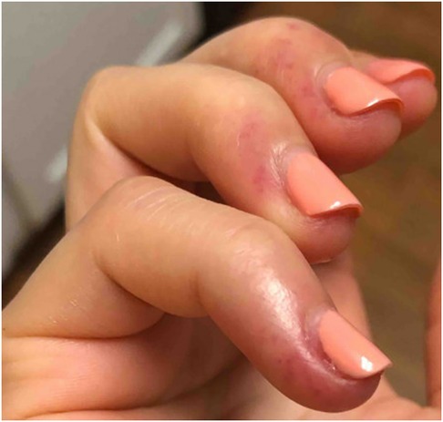
Figure 1. Distal fingers of the right hand showing features similar to chilblains.
Discussion
Interpretation of clinical features
This case highlights an unexpected trigger for a photosensitive rash with features similar to chilblains, a form of chronic cutaneous lupus. Our patient developed discrete violaceous macules, swelling, and a painful and itching quality that resolved without scarring the skin. Her cutaneous features developed over the summer and improved without using vasodilating agents, without similar features developing in the UV-protected areas (e.g., face, dorsum of the hands) and reported cold exposure. Cutaneous lupus erythematosus was considered the most likely diagnosis in the absence of an alternative explanation, and a biopsy was not performed, given the fleeting nature of the rash. Other diagnoses cannot thus be entirely excluded.
Final diagnosis
This UCTD patient had no history of photosensitivity and fastidiously adhered to the rheumatologist's recommendation to avoid UV light exposure. Her inadvertent UV exposure resulted in the development of multiple erythematous/violaceous macules, most consistent with a form of chronic cutaneous lupus. With her elevated anti-dsDNA antibody and her newly identified rash, she met the classification criteria for SLE (9).
Takeaway lessons
Clinicians often educate patients with SLE diagnosis on UV protective measures. This case highlights the need to ask about unknown photo-provocation as a potential diagnostic tool in those with UCTD. A thorough history may reveal an unintentional photo-provocation in those who are suspected of having or being at risk for developing SLE.
Patient perspective
During my first rheumatology appointment, we reviewed that symptoms, such as skin rashes and photosensitivity, may develop over time. I was advised to avoid direct sun exposure and to use sun protection. Between my initial appointment and my follow-up, I began doing at-home gel nail polish manicures, which involved using a nail lamp to cure the gel nail polish. After the first few uses, I noticed warmth and some redness in the skin around my nails. With continued use, this developed further discoloration, itching, warmth, and a swollen feeling in my fingers. This would appear a day or two after using the nail lamp and remain for about a week.
Having previously discussed that skin conditions and photosensitivity may be relevant to my condition, I brought up these symptoms at my follow-up rheumatology visit. Although sold as an LED light, I confirmed with the manufacturer that the lamp emits a UV light, and I was advised to discontinue using the UV-LED nail lamp. The symptoms did not return after I discontinued using the lamp.
Data availability statement
The original contributions presented in the study are included in the article/Supplementary Material; further inquiries can be directed to the corresponding author.
Ethics statement
Written informed consent was obtained from the participants/patient(s) for the publication of this case report. Written informed consent was obtained from the individual(s) for the publication of any potentially identifiable images or data included in this article.
Author contributions
DR generated the case report with supervision from JK. All authors contributed to the article and approved the submitted version.
Funding
JK received support from the National Institutes of Health NIAMS through K24AR076975.
Conflict of interest
The authors declare that the research was conducted in the absence of any commercial or financial relationships that could be construed as a potential conflict of interest.
Publisher's note
All claims expressed in this article are solely those of the authors and do not necessarily represent those of their affiliated organizations, or those of the publisher, the editors and the reviewers. Any product that may be evaluated in this article, or claim that may be made by its manufacturer, is not guaranteed or endorsed by the publisher.
References
1. Millard TP, Hawk JL, McGregor JM. Photosensitivity in lupus. Lupus. (2000) 9(1):3–10. doi: 10.1177/096120330000900103
2. Sim JH, Ambler WG, Sollohub IF, Howlader MJ, Li TM, Lee HJ, et al. Immune cell–stromal circuitry in lupus photosensitivity. J Immunol. (2021) 206(2):302–9. doi: 10.4049/jimmunol.2000905
3. Estadt SN, Maz MP, Musai J, Kahlenberg JM. Mechanisms of photosensitivity in autoimmunity. J Invest Dermatol. (2022) 142(3):849–56. doi: 10.1016/j.jid.2021.05.007
4. Kuhn A, Sonntag M, Richter-Hintz D, Oslislo C, Megahed M, Ruzicka T, et al. Phototesting in lupus erythematosus: a 15-year experience. J Am Acad Dermatol. (2001) 45(1):86–95. doi: 10.1067/mjd.2001.114589
5. Kuhn A, Beissert S. Photosensitivity in lupus erythematosus. Autoimmunity. (2005) 38(7):519–29. doi: 10.1080/08916930500285626
6. Kuhn A, Wozniacka A, Szepietowski JC, Gläser R, Lehmann P, Haust M, et al. Photoprovocation in cutaneous lupus erythematosus: a multicenter study evaluating a standardized protocol. J Invest Dermatol. (2011) 131(8):1622–30. doi: 10.1038/jid.2011.101
7. Hasan T, Pertovaara M, Yli-Kerttula U, Luukkaala T, Korpela M. Seasonal variation of disease activity of systemic lupus erythematosus in Finland: a 1 year follow up study. Ann Rheum Dis. (2004) 63(11):1498–500. doi: 10.1136/ard.2003.012740
8. Keyes E, Grinnell M, Vazquez T, Diaz DA, Werth VP. Ultraviolet light exposure from manicures in cutaneous lupus erythematosus. Rheumatology. (2022) 61(2):E38–9. doi: 10.1093/rheumatology/keab720
Keywords: UV light, photo-provocation, photosensitivity, cutaneous lupus erythematosus, systemic lupus erythematosus, undifferentiated connective tissue disease
Citation: Roofeh D and Kahlenberg JM (2023) Undifferentiated connective tissue disease with an incidental photo-provocation: a case report. Front. Lupus 1:1223001. doi: 10.3389/flupu.2023.1223001
Received: 15 May 2023; Accepted: 8 August 2023;
Published: 12 September 2023.
Edited by:
R. Hal Scofield, Oklahoma Medical Research Foundation, United StatesReviewed by:
Jillian M. Richmond, University of Massachusetts Medical School, United StatesWilliam D. Shipman, Yale University, United States
© 2023 Roofeh and Kahlenberg. This is an open-access article distributed under the terms of the Creative Commons Attribution License (CC BY). The use, distribution or reproduction in other forums is permitted, provided the original author(s) and the copyright owner(s) are credited and that the original publication in this journal is cited, in accordance with accepted academic practice. No use, distribution or reproduction is permitted which does not comply with these terms.
*Correspondence: David Roofeh RGF2aWQuUm9vZmVoQFJ1dGdlcnMuZWR1