 Marnie L. Freckelton
 Lone Høj
 Bruce F. Bowden1,3
- 1College of Science and Engineering, James Cook University, Townsville, QLD, Australia
- 2Australian Institute of Marine Science, Townsville, QLD, Australia
- 3AIMS@JCU, Division for Research and Innovation, James Cook University, Townsville, QLD, Australia
Bacterial Quorum Sensing (QS), the indirect regulation of gene expression through production and detection of small diffusible molecules, has emerged as a point of interaction between eukaryotic host organisms and their associated microbial communities. The extracellular nature of QS molecules enables interference in QS systems, in many cases via mimicry. This study targeted QS induction and inhibition in soft coral holobionts, as many soft coral species commonly contain compounds with structural similarities to the well-studied bacterial QS molecules acyl homoserine lactones. Screening with two bacterial biosensors, Agrobacterium tumefaciens A136 and Chromobacterium violaceum CV026, demonstrated that QS interference differed between the two biosensor strains and extended across the soft coral families Alcyoniidae, Clavulariidae, Nephtheidae, and Xeniidae. Bioassay-guided fractionation revealed chemical activity patterns, particularly in the induction of QS. Cembranoid diterpenes from active fractions were purified and tested for QS interference activity. Interestingly, the type of QS activity (induction or inhibition) in A. tumefaciens A136 correlated with structural variability of the secondary oxygen ring; cembranoid diterpenes with a furan ring or five-membered lactone induced QS, while compounds with larger (six or seven membered) lactone rings inhibited QS. Addition of the dominant cembranoid diterpene in the soft coral Lobophytum compactum, isolobophytolide, to bacterial culture media increased the number and morphological diversity of bacteria recovered from the mucosal layer of this soft coral, demonstrating a selective effect on certain members of the soft coral bacterial community. The identity and QS activity of recovered isolates differed between the mucosal layers of L. compactum and Sinularia flexibilis. In conclusion, this study provides information on the complexity of the interaction between soft corals and their associated bacteria, as well as, a structural understanding of how QS mimic compounds are able to interfere with a bacterial communication system.
Introduction
Quorum Sensing (QS) is one form of cell to cell signaling employed by bacteria to coordinate gene expression across entire populations through release and detection of extracellular signal molecules (Miller and Bassler, 2001). The aspects of multicellularity gained through QS, enable bacteria to perform many important ecological functions such as the ability to interact with their physical and biological environment (Miller and Bassler, 2001), form biofilms (Rice et al., 2005), and secrete virulence factors (Zhu and Mekalanos, 2003). The extracellular nature of QS signaling molecules facilitates their disruption and mimicry (Chhabra et al., 2005). Consequently, many bacteria possess the ability to detect and respond to QS signals of other species (Joint et al., 2007). Indeed, QS systems are more prevalent amongst bacteria associated with mixed bacterial biofilms and macro-organisms, suggesting that possession of QS systems confers an advantage in these habitats (Dudler and Eberl, 2006).
QS mimics are extrinsic signals that can interfere directly with QS gene expression (Bauer and Robinson, 2002). To be effective, QS mimic compounds must specifically interfere with the target QS system. Multiple QS systems have been discovered; in Gram-negative bacteria (Papenfort and Bassler, 2016) the most well studied system is the Auto Inducer One system (AI-1), which utilizes acyl homoserine lactones (AHLs) as signal molecules. Studies into the structure and functions of AHLs suggest that the ɤ-lactone ring is required for QS activity and that the length and functionality of the acyl side chain provides specificity (Parsek and Greenberg, 2000; Watson et al., 2002; Geske et al., 2008). For this reason, it has been hypothesized that AI-1 QS mimics would also contain a ɤ lactone ring or homologous functionality such as the furanones of the red alga Delisea pulchra. The furanones are one of the few QS inhibitors to have been structurally elucidated and the presence of an oxygenated ring was demonstrated to be essential to their activity (Manefield et al., 1999).
Disruption and mimicry of QS signals are increasingly recognized as mechanisms that are commonly employed by macro-organisms to regulate and manipulate their associated microbial communities (Bauer and Robinson, 2002; González and Keshavan, 2006). QS interference by host organisms can confer the ability to respond to the presence of certain pathogenic or mutualistic bacteria quickly and reliably (Kjelleberg et al., 1997; Mathesius et al., 2003), render a pathogenic species of bacteria non-pathogenic (Dong et al., 2007; Swem et al., 2009), and enable manipulation of the abundance and composition of its associated bacterial assemblies (Givskov et al., 1996). A host's microbiota can be a first line of defense against pathogen invasion (McFall-Ngai et al., 2013), therefore manipulation of QS could strengthen the resilience of the holobiont (González and Keshavan, 2006; Teplitski and Ritchie, 2009).
The ability of some bacterial species to detect non-native QS molecules has allowed the development of bacterial biosensor strains. QS bacterial biosensor strains are genetically modified bacterial isolates that require external addition of QS signal molecules. Their expression of QS-regulated genes is linked to reporter genes, which typically produce a pigment or bioluminescence (Steindler and Venturi, 2007). Two of the most commonly used AI-1 bacterial biosensor strains are based on the species Chromobacterium violaceum and Agrobacterium tumefaciens. In C. violaceum, QS regulates the production of the secondary metabolite violacein, which is purple in color (McClean et al., 1997). Bacterial biosensors based on this species utilize the LuxI/LuxR homolog genes CivI/CivR and are sensitive to AHLs with C4–C8 carbon chains as well as 3-oxo-C6 and -C8 carbon chains (McClean et al., 1997; Steindler and Venturi, 2007). A. tumefaciens QS biosensors have a genetically modified QS plasmid such that QS by these strains results in an enzymatic breakdown of X-gal and the formation of an indigo colored product (Zhu et al., 1998; Farrand et al., 2002; Zhu and Mekalanos, 2003). A. tumefaciens QS biosensors often utilize the TraI/TraR genes that provide sensitivity to AHLs with C6–C14 acyl side chains as well their equivalent 3-oxo-acyl side chains and C6–C10 hydroxy acyl side chains (Zhu et al., 1998; Farrand et al., 2002; Zhu and Mekalanos, 2003; Steindler and Venturi, 2007). Despite the partly overlapping acyl chain lengths detected by these two sensors, previous studies have observed differences in their responses during screening of isolates (Chong et al., 2012).
Widespread QS inhibitory activity has been observed to occur in the marine benthos, particularly in sponges and soft corals (Taylor et al., 2004; Skindersoe et al., 2008; Hunt et al., 2012). Soft corals contain a number of secondary metabolites with the structural potential to mimic QS, including furanocembrenes (cembranoid diterpenes with fused 5-membered ether rings) and cembranolides (cembranoids that possess a fused second ring in the form of a lactone). Cembranoid diterpenes are most commonly, but not exclusively, found within the family Alcyoniidae. Furthermore, cembranoid diterpenes are inherently variable in the presence, position and size of oxygenated ring systems (Wahlberg and Eklund, 1992). Variation of substituents, direction of cyclisation and the corresponding position of the isoprenoid double bonds of different diterpenes from different species of soft coral are also encountered (Wahlberg and Eklund, 1992) making cembranoid diterpenes a natural pool of compounds to investigate QS mimic structure-activity relationships.
Soft corals have a high incidence of QS interference; however, so far all screened soft corals have been from a single family, Alcyoniidae, and the presence of QS induction has not yet been investigated. While QS and QS interference have been implicated in the regulation of mixed bacterial communities including those found in the surface mucosal layer (SML) of hard corals (Tait et al., 2010; Golberg et al., 2011, 2013), it is unknown whether these communities may be regulated by QS mimics. Currently very little known about bacterial communities that associate with soft corals. In hard corals, bacteria in the SML, the first and largest point of interaction between a coral and the environment, are considered important to the health and resilience of the holobiont (Reshef et al., 2006; Rosenberg et al., 2007; Zilber-Rosenberg and Rosenberg, 2008; Shnit-Orland and Kushmaro, 2009).
This study aims to assess the potential of soft corals to interact with their associated microbes via QS, with a focus on their associated secondary metabolites such as cembranolides and furanocembrenes. Crude extracts of 15 soft coral species, representing 4 families of soft corals, were tested for their ability to induce or inhibit QS in 2 bacterial biosensors, A. tumefaciens A136 and C. violaceum CV026. Cembranolides and furanocembrenes were isolated from soft corals and the effect of their structural variability on QS interference was assessed. To gain a better understanding of the role that QS can play in regulating eukaryote associated bacterial communities, bacterial strains isolated from the surface mucosal layer (SML) of two soft corals, Sinularia flexibilis and Lobophytum compactum were similarly evaluated for QS activity. The possible role that isolobophytolide, the major secondary metabolite of L. compactum, might play in bacterial selection within the soft corals mucus was also assessed by supplementing culture media with this secondary metabolite.
Materials and Methods
Soft Coral Collection
Twenty four specimens of soft coral, representing 15 species (Supplementary Tables 1–3), were collected at a depth of 1–3 m near Orpheus Island (Great Barrier Reef, Australia; latitude 18°36.878′S; longitude 146°29.990′E). All specimens except Cespitularia sp. were photographed (Supplementary Figure 1) and sampled underwater, with samples placed directly into plastic bags filled with seawater. Samples were frozen (−80°C) within 1 h of collection and stored until freeze drying. Additional colonies of L. compactum, S. flexibilis, and Pachyclavularia violacea were collected for fraction analysis and isolation of pure compounds (Supplementary Tables 2,3). 1H NMR spectra of the crude extracts of all soft coral samples were compared between collections and with reference spectra from the Bowden laboratory to ensure consistency in the metabolites and species tested.
Soft Coral Extract Preparation
Dried soft coral tissue was weighed and homogenized before extraction. Three extracts of different polarity were generated for each soft coral sample. Solvents used for extraction were, in order, dichloromethane (DCM), methanol (MeOH), and water (H2O). Each extract was the result of three successive applications of solvent. Extracts were concentrated by rotary evaporation before being dried under a stream of nitrogen (N2) and stored at −20°C until analysis. The DCM and MeOH extracts were dissolved in ethanol and the aqueous extracts in H2O to a concentration of 20 mg/ml. All soft coral extracts were tested at two concentrations (4 μg/well and 40 μg/well) three times for the presence of QS induction and QS inhibition activity in A. tumefaciens A136 and C. violaceum CV026 in at least two independent experiments.
Soft Coral Extract Fractionation
To further investigate the patterns of QS activity observed in crude extracts, nine soft coral species from five genera (three families) were chosen for further fractionation (Supplementary Table 2). The chosen species displayed five patterns of crude extract activity (Supplementary Table 4). In addition to four species with strong crude extract activities, five species were selected to investigate observed within genera variation in activity patterns or because they represent common genera on the Great Barrier Reef (GBR). Ten fractions of decreasing polarity were generated from the dichloromethane extracts of the chosen soft corals. Extracts were fractionated using flash column chromatography on RP-C18 silica cartridges (Phenomenex Strata C18-E 55 μm 70 Å, 1,000 mg) eluted with a stepwise 20–100% MeOH: H2O gradient followed by a 1:1 DCM: MeOH wash. The resulting fractions were concentrated to dryness under a stream of N2 gas and re-dissolved in ethanol for QS screening as described above. 1H-NMR spectra (600 MHz) of active fractions were recorded with a Bruker 600 Avance spectrometer in deuterated chloroform (CDCl3).
Isolation of Purified Metabolites From the Corals
Ten cembranoid diterpenes with variable secondary ring structures were assessed in this study (Figure 1, Table 1). Isoneocembrene A (1) represents the base cembrene backbone, without an additional ring (Figure 1, Table 1). Compounds 2-4 are ɤ-lactones (5-membered rings). Two δ-lactones (6-membered rings) and one ε-lactone (7-membered ring) are also included in compounds 6-8 respectively (Figure 1, Table 1). The final two compounds are furanocembrenes (Figure 1, Table 1). Pure samples of isoneocembrene A (1), lobolide (4) and sarcophine (5) were acquired from the Bowden Laboratory, Townsville Australia. Isolobophytolide (2), and isolobophytolide monoacetates (3) were isolated from freshly collected and extracted L. compactum (see below). Flexibilide (6), dihydroflexibilide (7) and sinulariolide (8) were isolated from freshly collected and extracted S. flexibilis (see below). The furanocembrenes, pachyclavulariadiol (9) and pachyclavulariadiol diacetate (10), were isolated from freshly collected and extracted P. violacea. Extracts of each species were generated as described previously (section Soft Coral Extract Preparation).
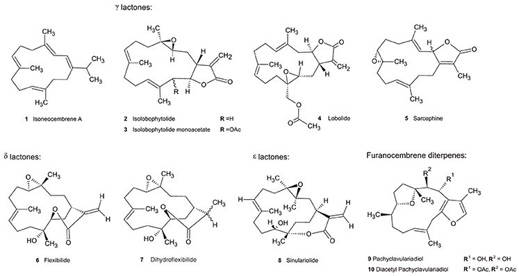
Figure 1. Soft coral cembranoid diterpenes tested for QS interference capability.
Table 1. Functional group analysis of tested soft coral metabolites.
For the L. compactum extract, vacuum liquid chromatography of active crude DCM extract (2 g) was performed over reverse phase C18 silica gel (Phenomenex Luna 10 mm C18 silica gel) and 10 fractions (200 ml) were collected using MeOH: H2O 0–100% stepwise gradient for each extract. Activity was identified in the 80% MeOH fraction and this fraction was subjected to RP-HPLC 60–100% MeOH gradient over 30 min (Phenomenex Gemini 3 μm NX-C18 110 Å, LC Column 30 × 4.6 mm). Isolobophytolide (2) eluted at 15 min and the two isomers of isolobophytolide monoacetate (3) eluted at 17 min.
For the S. flexibilis extract, flexibilide (6) and dihydroflexibilide (7) were isolated as reported by Kazlauskas et al. (1978). In brief, the DCM extract was subjected to normal phase flash column chromatography with combinations of hexane, DCM and EtOAc. Flexibilide was eluted by 6:1 DCM: EtOAc, 4:1 DCM: EtOAc yielded a mixture of 6 and 7 and 3:1 DCM: EtOAc afforded pure dihydroflexibilide. Sinulariolide (8) was isolated separately by a method adapted from Tursch et al. (1975) by direct crystallization of the DCM extract after dissolution in diethyl ether. Purification of compounds was performed by HPLC as described above.
Due to instability of the furanocembrenes from P. violacea, the pure compounds were generated semi-synthetically as per Bowden et al. (1979). In brief, the DCM extract was prepared at 4°C then partitioned between hexane and 10% aqueous MeOH. After removal of the solvent, the aqueous MeOH fraction was subjected to normal phase flash chromatography with a hexane: EtOAc gradient. All fractions containing (by TLC and 1H NMR) pachyclavulariadiol, diacetyl pachyclavulariadiol and the two monoacetyl pachyclavulariadiols were combined and hydrolyzed to pachyclavulariadiol (9) by incubation for 24 h at room temperature with MeOH containing 1% (w/v) potassium hydroxide. Methanol was removed under vacuum and the residue was partitioned between diethyl ether and water. The ether fraction was evaporated and the residue was dissolved in hexane. Diacetyl pachyclavulariadiol (10) was acquired by acetylation of half of the obtained pachyclavuriadiol (9). Acetylation was affected by incubation for 24 h with 1:1 acetic anhydride in pyridine before evaporation of the solvent and retrieval via liquid a partition between hexane and water. The semi-synthesis of monoacetyl pachyclavulariadiols was not performed as both 9 and 10 exhibited similar activity, so it was considered unlikely that the activities of monoacetyl pachyclavulariadiols would be different.
Structure and purity of each extracted compound was confirmed by 1D and 2D NMR and comparison with literature values. 1H-NMR (600 MHz) and 13C -NMR (150 MHz) spectra were recorded with a Bruker 600 Avance spectrometer in CDCl3, with tetramethylsilane (TMS) as internal standard. High resolution mass spectra were collected using an unmodified Bruker BioAPEX 47e mass spectrometer equipped with an Analytica model 103426 (Branford, CT) electrospray ionization (ESI) source. Analytical thin layer chromatography (TLC) was performed on Merck Kieselgel 60. Spots were visualized by UV light or by spraying with a 1% vanillin in acidified ethanol solution. Pure compounds were re-solubilized in ethanol and serially diluted to generate five different concentration solutions (1 × 10² mM to 1 × 10⁻² mM) for each compound.
Collection, Culture and Extraction of Bacterial Isolates From S. flexibilis and L. compactum Surface Mucosal Layer
Surface mucosal layer samples (SML) were collected from three healthy replicate colonies of S. flexibilis and L. compactum from the same depth and location. SML samples were collected underwater from the mid-capitulum region of the coral colony using 50 ml needleless sterile syringes. Samples were maintained at ambient temperatures and processed within 3 h of collection. At the same time as SML samples were retrieved, tissue samples of each replicate were collected and their metabolite 1H NMR spectral profiles were compared with samples from S. flexibilis and L. compactum collected previously to ensure correct species identification.
SML samples were serially diluted (10−2, 10−3, 10−4) using autoclaved artificial seawater (Instant Ocean; Spectrum Brands, Madison, WI, USA). One hundred microliters of each dilution were spread plated in triplicate on two types of media commonly used for studies of marine bacteria: 50% Marine Agar (50MA; BD) and Glycerol Artificial Seawater (GASW) agar (Smith and Hayasaka, 1982). Additionally, Thiosulfate Citrate Bile Salts (TCBS; BD) agar, which specifically selects for members of the family Vibrionaceae, was included. L. compactum SML dilutions were additionally plated onto 50MA and GASW agar supplemented with L. compactum's major secondary metabolite, isolobophytolide. All plates were incubated at 28°C and sampled after 48 h, 72 h, 1, and 2 weeks. Representatives of each colony morphotype from each plate were sub-cultured to purity for identification.
Where possible, two representatives of each morphotype were selected for QS activity screening. Where bacteria had initially been isolated using media embedded with isolobophytolide, growth was attempted on the equivalent medium without isolobophytolide. Strains that could not be cultured without isolobophytolide were not included in the screening. Screening was performed on acidified ethyl acetate (EtOAc) extracts of the cell free supernatant of soft coral isolates. These were acquired by transferring single colonies from 50MA plates to liquid culture (10 ml 50% Marine broth culture, 28°C at 170 rpm) and grown to late exponential phase. Cultures were centrifuged for 10 min at 4°C at 10,000 g to obtain the cell free supernatant (CFS). Each CFS was subjected to exhaustive extraction with acidified EtOAc (1% acetic acid) and concentrated to dryness under a stream of N2 gas. Extracts were then dissolved and diluted to a concentration of 20 mg/l with ethanol.
Bacterial QS Biosensor Strains and Culture Medium
The biosensor strains A. tumefaciens A136 (Fuqua and Winans, 1996) and C. violaceum CV026 (McClean et al., 1997) were used for detection of QS induction and inhibition in soft coral extracts. A. tumefaciens A136 was grown on ABt media (Clarrk and Maaloe, 1967) and C. violaceum CV026 was grown on Luria Bertani (LB) media (Bertani, 1951). In order to ensure that the QS plasmid was intact and functional, QS biosensor strains were grown in the presence of the appropriate antibiotic (Ravn et al., 2001). A. tumefaciens A136 was grown on media supplemented with 4.5 μg/ml of tetracycline and 50 μg/ml of spectinomycin, whereas, C. violaceum CV026 was grown on media supplemented with 20 μg/ml of kanamycin (Ravn et al., 2001).
QS Screening Assays
The presence of AHL type QS induction activity in soft coral extracts, fractions and pure compounds was detected by performing an agar diffusion assay as described in detail by Ravn et al. (2001). The QS biosensor strain, either A. tumefaciens A136 or C. violaceum CV026, was embedded within the agar and the sample being tested was added to a well cut or formed in the agar. For induction of QS, N-hexanoyl homoserine lactone was used as a positive control and extraction solvents were used as negative controls. Positive results were read as a blue coloration surrounding the wells of A. tumefaciens A136 and a purple coloration surrounding the wells of C. violaceum CV026 (see above and Supplementary Figure 2). The intensity of the response was measured as the diameter of the colored zone and normalized to the response of the positive control.
The agar diffusion assays described above were modified in order to detect QS inhibition. Briefly, as A. tumefaciens A136 and C. violaceum CV026 are not able to QS without the exogenous addition of AHLs, 8.5 μmol n-hexanoyl homoserine lactone was added into the agar embedded with the biosensor strain in order to test for QS inhibition. Two positive controls were chosen based on their previously reported ability to inhibit QS: n-dodecanoyl-DL-homoserine lactone (McClean et al., 1997) and vanillin (Choo et al., 2006). These controls proved effective for both biosensors. The extraction solvents were once again used as negative controls. Positive results in the inhibition assay were read as inhibition of blue or purple coloration of the plates containing A. tumefaciens A136 and C. violaceum CV026, respectively (Supplementary Figure 2). The intensity of the response was measured as the width of the inhibition zone surrounding the well and normalized to the positive control.
To generate dose response curves for pure compounds, agar containing the respective biosensor was poured into custom built molds with 28 preformed wells 4 mm in diameter. After solidification of the agar, 20 μl of sample was added to each well. Pure compounds were only tested with the A. tumefaciens A136 strain as it was the only strain to have both QS induction and inhibition activity.
Bacterial DNA Extraction, PCR and Sequencing
Genomic DNA of bacterial isolates was extracted using the Promega Wizard Genomic DNA Isolation kit (Promega, Madison WI USA) according to the manufacturer's directions. PCR amplification of 16S rRNA gene fragments was performed using the primers 27F and 1492R (Lane, 1991). The PCR reactions contained the following reagents: 0.4 mM of each primer, 1x MyTAQ buffer (Bioline, Australia), 1.25 U MyTAQ (Bioline, Australia), 1 μL DNA extract (final volume of 50 μL). Cycling conditions were an initial denaturing step of 94°C for 5 min, followed by 30 cycles at 95°C for 1 min, 56°C for 45 s, 72°C for 60 s, and a final elongation step at 72°C for 10 min. PCR products were verified by agarose gel electrophoresis and purified for sequencing using the Qiaquick PCR purification Kit (Qiagen, Valencia, CA) according to company supplied directions. Sanger sequencing was carried out at Macrogen (Seoul, South Korea) using both 27f and 1492R as sequencing primers.
Phylogenetic Analysis of Bacterial Isolates
Sequence fragments were assembled using Sequencher (Version 5, Gene Codes, Ann Arbour, USA). For each isolate, the 16S rRNA gene sequence was aligned with sequences in the nr and Ref_Seq database at the NCBI using the megablast tool (RRID:SCR_001598; Altschul et al., 1990) to identify closely related database sequences. Sequences of isolates and database matches were imported into MEGA6 (MEGA Software, RRID:SCR_000667) and aligned using ClustalW (Larkin et al., 2007). A Maximum Likelihood-based phylogenetic tree was constructed using the Maximum Parsimony algorithm for the starting tree, the Tamura-Nei model for nucleotide substitution, and 500 bootstrap replicates (Supplementary Figure 3). The 16S rRNA gene sequences for the 72 bacterial isolates were deposited into the NCBI Genbank database, under accession numbers KM360403-KM360473. Quantification and statistical analysis of CFUs and isolate morphotypes.
Colony forming unit (CFU) concentrations were estimated based on dilutions yielding between 30 and 300 colonies per plate. Differences in the number of CFUs between samples were determined based on three replicates for the corresponding dilution and media type. Statistical differences were determined using the non-parametric Kruskal-Wallis statistic, as the data violated both normality and homogeneity of variances required for ANOVA. Colony morphotype profile analysis was conducted on the variables color, size and texture and were compared using a nonmetric Multidimensional Scaling (nMDS) analysis in the vegan package (version 2.5-1; Oksanen et al., 2018) in R. The nMDS was chosen based on its suitability for spatial representation of complex data sets containing multiple variables, large numbers of zeroes and non-normal distributions (Rabinowitz, 1975). The statistical analyses were performed using Graphpad PRISM (GraphPad Prism, RRID:SCR_015807).
Results
Quorum Sensing Activities of Soft Coral Crude Extracts
Crude soft coral extracts demonstrated the ability to both induce and inhibit QS in the biosensors tested. While both induction and inhibition of QS were observed for the biosensor A. tumefaciens A136, none of the extracts, regardless of source coral species, polarity, or concentration, were able to induce QS in C. violaceum CV026 (Figure 2A). Inhibition of QS was more prevalent than induction across all polarity extracts (Figure 2A). For both biosensors, the number of active soft coral extracts was reduced at lower extract concentration, with none of the aqueous extracts retaining their activity at the lower dosage (Figure 2B).
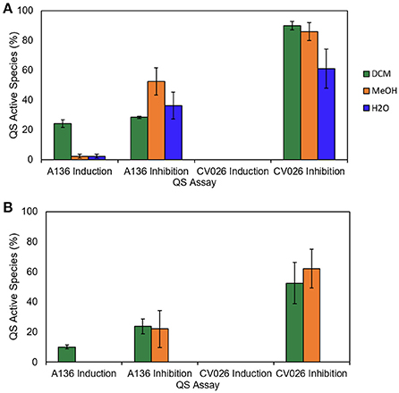
Figure 2. Percentage of soft coral species displaying QS induction or inhibition against A. tumefaciens A136 and C. violaceum CV026 after addition of (A) 40 μg and (B) 4 μg extract. Error bars represent the standard error from three screening efforts.
Crude extracts of five species induced QS in A. tumefaciens A136; two species from the family Alcyoniidae (L. compactum, Lobophytum sarcophytoides), one from the family Nephtheidae (Nephthea chabroli), one from the family Clavulariidae (P. violacea), and one from the family Xeniidae (Cespitularia sp.) (Figure 3). The highest incidence and strength of induction activity was seen for DCM crude extracts, with the largest haloes of coloration produced by DCM extracts of L. compactum and P. violacea (Figure 3).

Figure 3. Results of the A. tumefaciens A136 QS induction assay for the soft coral extracts from all polarity solvent extracts (dichloromethane, methanol and water). The bars represent positive responses, normalized to the response of the positive control (8.5 μmol N-hexanoyl-DL-homoserine lactone). Error bars represent the standard error from three screening efforts.
Low level QS inhibition of A. tumefaciens A136 was demonstrated for DCM crude extracts of most species, with the only exceptions being L. microlobulatum, Sinularia polydactyla, P. violacea, Clavularia sp., and N. chabroli (Figure 4A). The same trend was seen for MeOH crude extracts, with the additional exception of Sarcophyton sp. 2 (Figure 4A). Inhibition of QS in C. violaceum CV026 was present in DCM crude extracts of all species except P. violacea and Clavularia sp. (Figure 4B). Of the five species that induced QS in A. tumefaciens A136, three (L. compactum, L. sarcophytoides and Cespitularia sp.) also inhibited QS in both biosensors (Figure 4). This contrasted with the other two species capable of QS induction; N. chabroli extracts inhibited QS only in C. violaceum CV026 (Figure 4) while P. violacea demonstrated no QS inhibitory activity with either biosensor (Figure 4).
Figure 4. Results of the QS inhibition assay for the soft coral extracts from all polarity solvent extracts (dichloromethane, methanol and water) (A) A. tumefaciens A136 and (B) C. violaceum CV026 QS inhibition assay for the soft coral extracts from all polarity solvent extracts (dichloromethane, methanol and water). The bars represent positive responses, normalized to the response of the positive control (vanillin). Error bars represent the standard error from three screening efforts.
Quorum Sensing Activities of Soft Coral Fractions
For all fractionated species, at least two fractions induced QS in A. tumefaciens A136 (Figure 5), regardless of whether their corresponding crude extracts were active (Figure 4). All species produced fractions that induced QS and inhibited QS in at least one biosensor strain. The only exception was P. violacea, which did not inhibit QS in either strain (Figure 5) consistent with crude extract results (Figure 4). The two largest inductive haloes were observed for N. chabroli and L. compactum, which also retained their activity at the higher (1:10) dilution level (Figure 4 The two major bands of induction activity seen in the A. tumefaciens A136 bioassay occurred for fractions eluted at 60% MeOH and at 80–90% MeOH (Figure 5). Many of the latter fractions (80–90% MeOH) also showed activity in the corresponding QS inhibition bioassay (Figure 5). The distinct patterns of QS activity observed for A. tumefaciens A136 contrasted strongly with the broad C. violaceum CV026 inhibition activity (Figure 5). The presence of cembrene diterpenes correlated well with the QS active fractions (data not shown).
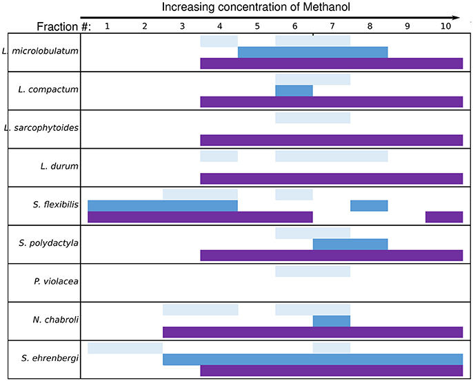
Figure 5. Active soft coral fractions by QS biosensor assay. Fractions were generated using a C18 flash column with a stepwise MeOH:H2O gradient. Fraction numbers reflect elution order with increasing MeOH percentage. Light blue bars represent the A. tumefaciens A136 induction assay, dark blue bars represent the A. tumefaciens A136 inhibition assay and the purple bars represent the C. violaceum CV026 inhibition assay.
Quorum Sensing Activities of Pure Compounds
The response to pure compounds also depended on the biosensor strain utilized. None of the tested cembranoid diterpenes induced QS in C. violaceum CV026. In contrast, both QS induction and inhibition activity was observed against A. tumefaciens A136 over three to four orders of magnitude, with a loss of activity at higher concentrations (Figure 6). No QS interference was observed for isoneocembrene A (Compound 1; Figure 6). The strongest induction of QS in A. tumefaciens A136 was observed for pachyclavulariadiol and diacetyl pachyclavulariadiol (Compounds 9 and 10; Figure 6). Induction was also observed in isolobophytolide (Compounds 2 and 3), lobolide (Compound 4), and sarcophine (Compound 5). QS inhibition was strongest in the ε-lactone ring of sinulariolide (Figure 6). Peak QS interference for all compounds occurred at approximately 1 × 10⁻⁵ mM (or 3 ppm; Figure 6).
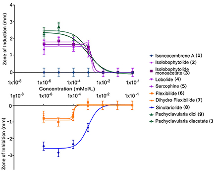
Figure 6. Dose response patterns of QS in A. tumefaciens A136 in cembranoid diterpene compounds isolated from soft corals. Although all zones of activity had positive values, for clarity, the zone sizes depicted as positive represent induction of QS and those represented as negative represent inhibition of QS. A zone is defined as the size in mm of either pigment production or pigment inhibition. Concentration refers to the concentration of the compound that was present in the agar wells.
Culturable Bacteria
A significantly higher number of colony forming units (CFUs) were estimated for mucus of S. flexibilis as compared to L. compactum (Kruskal-Wallis, H = 7.200, 2 d.f., P = 0.0036; Supplementary Figure 4). Interestingly, when growth media for L. compactum were amended with isolobophytolide, the estimated number of CFUs increased for this species but remained lower than the estimates for S. flexibilis (Supplementary Figure 4). The number and type of colony morphotypes also differed between L. compactum and S. flexibilis (Figure 7). S. flexibilis showed little variation in the morphotype profiles of GASW or 50MA media, forming a tight cluster on the nMDS biplot (Figure 7). In comparison, the morphotype profiles generated from L. compactum showed higher variation in the same culture media. This trend was also consistent when isolobophytolide was added as a selection agent (Figure 7).
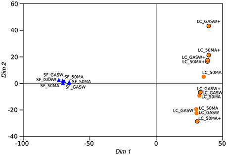
Figure 7. nMDS plot of bacterial isolate morphotype profiles generated from S. flexibilis and L. compactum. Profiles generated from S. flexibilis are indicated by the prefix SF whereas profiles from L. compactum are indicated by the prefix LC. 50MA indicates a profile from a 50% marine agar plate, GASW indicates a profile from a Glycerol Artificial Seawater plate. The plus symbol indicates the presence of isolobophytolide in the isolation media.
Sinularia flexibilis Bacterial Isolates
In total, 20 bacterial isolates from S. flexibilis were identified through 16S rRNA gene sequencing followed by BLAST searches and construction of phylogenetic trees (Supplementary Table 5, Figures 8, 9). The isolates were dominated by Gammaproteobacteria belonging to the family Vibrionaceae both for the non-Vibrionaceae targeted media (GASW and 50MA) as well as the Vibrionaceae targeted medium (TCBS). Other Gammaproteobacteria included two isolates whose closest relative was “Spongiobacter nickelotolerans” (hereafter referred to as Endozoicomonas, see below); and three Alteromonas-related strains. Finally, one isolate was identified with 99% sequence identity to Bacillus megaterium and Bacillus aryabhattai (phylum Firmicutes).
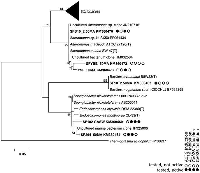
Figure 8. A phylogenetic tree based on partial 16S rRNA gene sequences retrieved from bacterial isolates from the mucus of the soft coral S. flexibilis. Details of the Vibrionaceae are shown in Figure 9. The tree is based on maximum-likelihood analysis, using a 50% conservation filter. The scale bar indicates 5% estimated sequence divergence. Thermoplasma acidophilum was used as the outgroup for analysis. Isolated sequences and their accession numbers are indicated in bold type. The nearest matches from the NCBI databases are included. T indicates that the sequence originates in the species type strain.
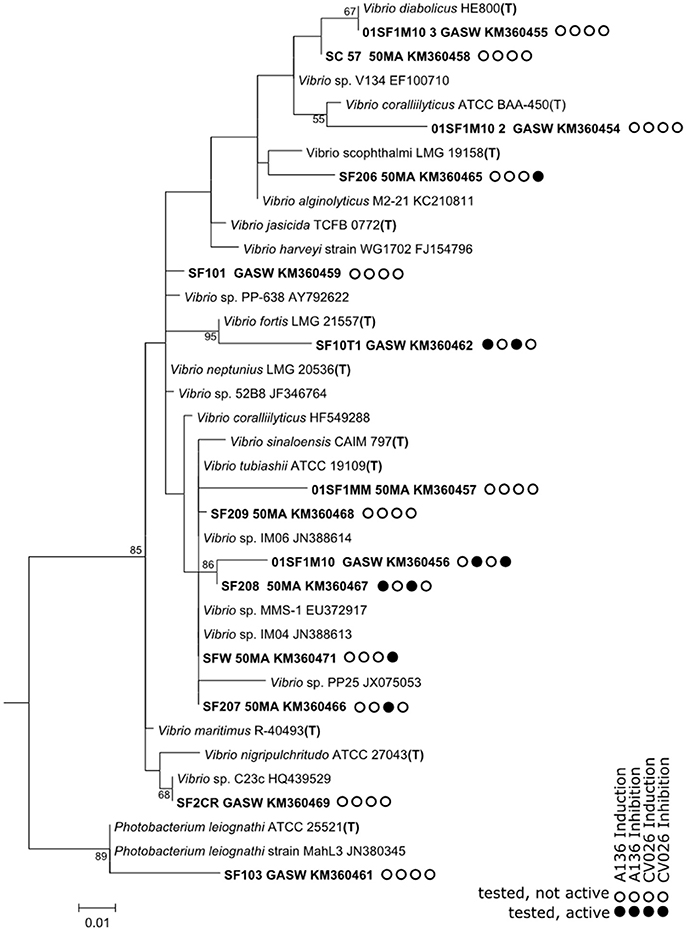
Figure 9. Vibrionaceae sub-tree based on 16S rRNA gene sequences retrieved from an analysis of bacterial isolates from the mucus of the soft coral S. flexibilis. The tree is based on maximum-likelihood analysis, using a 50% conservation filter. The scale bar indicates 1% estimated sequence divergence. Thermoplasma acidophilum was used as the outgroup for analysis. Isolated sequences and their accession numbers are indicated in bold type. The nearest matches from the NCBI databases are included. T indicates that the sequence originates in the species type strain.
The potential of soft coral isolates from S. flexibilis to participate in AHL-type QS communication systems was investigated using the same reporter bioassays as used for coral extracts. Quorum sensing activity under the used test conditions was demonstrated for 52.4% of the tested S. flexibilis isolates. Both tested Alteromonas strains exhibited QS induction activity. The Alteromonas SFB10_2 strain triggered QS induction in both sensors strains, whereas, the Alteromonas YSF strain only triggered QS induction in C. violaceum CV026 (Figure 8). Both Endozoicomonas-related strains triggered QS induction in C. violaceum CV026 only. In addition, both strains triggered QS inhibition in A. tumefaciens A136, while QS inhibition in C. violaceum CV026 was only triggered by strain SF102 (Figure 8). We note that out of the 62 bacterial strains screened in this study, the two Endozoicomonas-related strains from S. flexibilis were the only strains with both induction and inhibition QS activity. Of the tested Vibrionaceae strains, 6 of 14 strains showed QS activity and this was evenly split between induction (3 strains) and inhibition (3 strains) activity.
Lobophytum compactum Bacterial Isolates
The isolates cultured from L. compactum demonstrated a number of similarities to the bacteria isolated from S. flexibilis (Supplementary Table 5, Figures 10, 11). Firstly, the majority of L. compactum isolates were gammaproteobacteria of the genus Vibrionaceae (30/51). Secondly, strains related to the genera Endozoicomonas and Bacillus, and the order Alteromonadales, were isolated also from this soft coral species. In this instance, however, the diversity of Alteromonadales-related strains was higher with strains related not only to the genus Alteromonas (seven strains) but also to the genera Pseudoalteromonas (six strains), Paramoritella, Ferrimonas, and Shewanella. In contrast to the S. flexibilis isolates, the L. compactum isolates also included strains belonging to the genera Psychrobacter (class Gammaproteobacteria), Erythrobacter (class Alphaprotebacteria), and Micrococcus (class Actinobacteria).
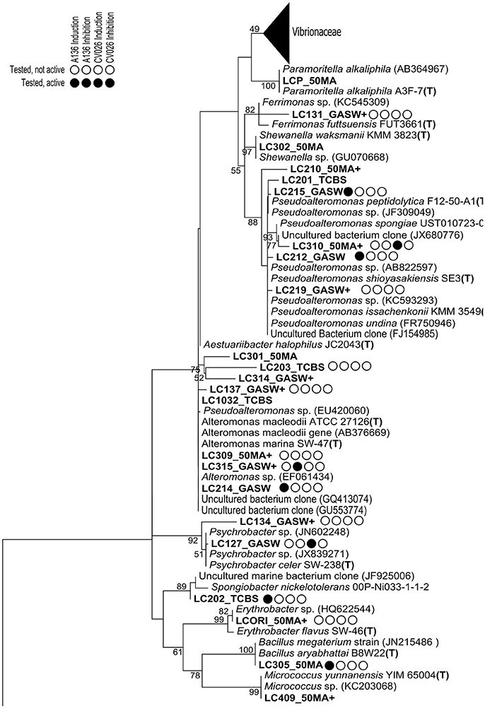
Figure 10. A phylogenetic tree based on 16S rRNA gene sequences retrieved from an analysis of bacterial isolates from the mucus of the soft coral L. compactum. Details of the Vibrionaceae are shown in Figure 11. The tree is based on maximum-likelihood analysis, using a 50% conservation filter. The scale bar indicates 10% estimated sequence divergence. Thermoplasma acidophilum was used as the outgroup for analysis. Isolated sequences and their accession numbers are indicated in bold type. The nearest matches from the NCBI databases are included. T indicates that the sequence originates in the species type strain.
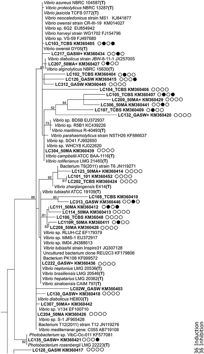
Figure 11. Vibrionaceae sub-tree tree (part of tree presented in Figure 10) based on 16S rRNA gene sequences retrieved from an analysis of bacterial isolates from the mucus of the soft coral L. compactum. The tree is based on maximum-likelihood analysis, using a 50% conservation filter. The scale bar indicates 10% estimated sequence divergence. Thermoplasma acidophilum was used as the outgroup for analysis. Isolated sequences and their accession numbers are indicated in bold type. The nearest matches from the NCBI databases are included. T indicates that the sequence originates in the species type strain.
The potential of soft coral isolates from L. compactum to participate in AHL-type QS communication systems was also investigated (Figure 10). Of the tested isolates from L. compactum, 47.5% demonstrated QS activity at the growth conditions tested and activity was mixed between induction and inhibition. Three of the tested Vibrio strains (LC111, LC103, and LC105) showed inhibitory activity against both biosensors. Of the strains that were initially isolated with media containing isolobophytolide, 41% were unable to be cultured in the absence of this compound and consequently were not tested for QS activity.
Discussion
Soft Coral Extracts
This study has demonstrated that QS interference extends across at least four soft coral families (Alcyoniidae, Clavulariidae, Nephtheidae, and Xeniidae). Further, it was shown that both induction and inhibition QS activity extends across both polar and non-polar fractions, indicating that QS interference capability is widespread in soft corals from the central Great Barrier Reef, Australia.
Widespread activity, across not only species that are known to contain different metabolite types but also across a range polarities (indicated by the activities of extracts obtained by use of solvents with different polarities), is probably indicative of active compounds of more than one structural type. This is further supported by the finding that most (except P. violacea) of the soft coral species that induced QS in A. tumefaciens A136, also inhibited both biosensors. The widespread prevalence of QS inhibition as well as the presence of QS induction in the soft corals screened here is consistent with QS activity found across a range of marine invertebrates (Taylor et al., 2004; Skindersoe et al., 2008; Hunt et al., 2012). The dual presence of induction and inhibition of QS is similar to that found previously in gorgonian coral extracts (Hunt et al., 2012) but contrasts with the sole QS inhibition activity that was identified in D. pulchra (Kjelleberg et al., 1997). QS induction was also established in extracts of marine sponges and sponge associated bacteria (Taylor et al., 2004). Results from this study highlight the need to examine both induction and inhibition of QS to generate a realistic understanding of the complexity of ecological interactions between a host organism and its associated bacteria.
Soft Coral Fractions
In contrast with the initial crude extract testing, all the fractionated soft corals displayed at least one active fraction in the A. tumefaciens A136 QS induction assay. This may reflect an inherent increase in concentration of the active components or a decrease in complexity of the samples being tested. Soft coral extracts and fractions may be highly complex mixtures of compounds with contrasting QS regulatory activities. The potential for activity masking within extracts, a phenomenon previously observed in the QS screening of marine sponge extracts (Taylor et al., 2004), is high. This is particularly true if only one concentration or level of complexity is tested.
The QS induction pattern of the soft coral fractions was generally limited to one or two active fractions (80 and 90% methanol elution) for each species suggests that the inductive capability may be due to the presence of structurally similar compounds. Cembranoid diterpenes are well documented in eleven of the soft coral species tested (MarinLit Database, 2013) and correlated well to QS induction therefore could be responsible for the observed QS activity in these species. The same diterpene scaffolds, however, are not commonly known in the genera Nephthea (Amir et al., 2012a,b) Cespitularia (Elshamy et al., 2016) or in gorgonian corals (Changyun et al., 2008), so cembranoid diterpenes cannot fully explain the observed QS interference in these species. Isolation of cembranoid diterpenes was therefore required to understand the relative importance of these secondary metabolites to act as QS mimics.
A similarly discrete pattern was not reflected in the QS inhibition profile for these fractions. The broad QS inhibition profile of these fractions, might be due to multiple compounds, or, the compound(s) responsible may not be suited to the method of fractionation used and the same compounds could be spread across several fractions. The presence of multiple QS compounds within a single holobiont would potentially enable a larger number of interactions with different bacterial strains and / or trigger different QS responses. This complexity may also reflect the capability of some bacteria to possess multiple QS systems (Reshef et al., 2006), with each system regulating a different process or interaction.
Pure Compounds
The strength and type of QS interference by cembranoid diterpenes was observed to correlate with the size of the oxygenated ring. Those cembranoid diterpenes that contained either a five membered furan or lactone ring were capable of inducing QS in A. tumefaciens A136, whereas the cembranoid diterpenes with larger lactone rings (six or seven membered) were seen to inhibit QS in A. tumefaciens A136. The type of oxygenated functional group also appears to impact the strength these QS mimics, with the furans tested (Compounds 9 and 10) having higher activities than the ɤ lactones (Compounds 2-5). In keeping with our current understanding of the QS mechanism, QS interference was only observed for cembranoid diterpenes possessing secondary oxygen rings (Fuqua et al., 2001; Watson et al., 2002; Geske et al., 2008). The presence of other minor functional groups (epoxides, acetates or level of saturation) had minimal discernible impact on the strength of QS interference observed and no obvious effect on the type of activity observed with respect to A. tumefaciens A136. QS mimics have previously been isolated that possess a ɤ lactone, however, these mimics (such as the furanones of D. pulchra) are often associated with QS inhibition rather than the inductive activity demonstrated here (Givskov et al., 1996; Defoirdt et al., 2013). In the case of the furanones from D. pulchra, bromine substituents are also present and may be influencing the type of activity. The presence of these metabolites in soft corals is strongly correlated to their taxonomy and may represent different strategies of interaction between species.
A common feature of QS mimic compounds previously identified from eukaryotic extracts is multiple forms of biological activity (Davies, 2006; Yim et al., 2007; Defoirdt et al., 2013). Cembranoid diterpenes appear to be no different with a number, including those identified in the current study, having previously been reported to demonstrate antibiotic (Aceret et al., 1995), cytotoxic (Maida et al., 1993) and algacidal properties. The antimicrobial activity identified in flexibilide (7) by Aceret et al. (1995), however, was exhibited at concentrations at least one order of magnitude higher than those that produced QS interference in this study. The peak QS active concentration occurred 1 × 10⁻⁵ mM (or 3 ppm), reflecting the concentrations of flexibilide and sarcophytoxide in the mucous and water column surrounding S. flexibilis and S. crassocaule detected by Coll et al. (1982). Rather than being incompatible, the contrasting activities could be evidence of a hormetic response. Hormetic relationships have been previously observed in the QS mimics from garlic (Persson et al., 2005) and some antibiotic compounds, whereby growth stimulation or cell signaling properties are exhibited at concentrations below their minimum growth inhibitory concentration (Davies, 2006; Yim et al., 2007). A hormetic response could be relevant in soft corals with loosely packed sclerites, where uptake or release of water from the tissue can lead to large changes in volume over a matter of hours (Freckelton, 2015). As a result, associated metabolites will show a correspondingly dramatic change in concentration in the tissues on a volumetric basis over the same time period. More research is required to understand the potential of hormetic relationships in QS mimics and how such concentration changes could be manipulated in the defense of the coral.
Strong evidence for the ecological role of cembranolides and furanocembranoid diterpenes as QS mimics is further exhibited in the strong differences in the ability of the two biosensor strains to respond to QS mimics in the soft corals. The QS inductive compounds present within the soft corals were more readily detected by the A. tumefaciens A136 strain. In contrast, QS inhibition was observed more frequently for C. violaceum CV026. This could suggest that C. violaceum CV026 can be inhibited by a broader range of compounds or that it is more sensitive to a broader range of compound concentrations. This difference in sensitivity is despite an overlap of AHL acyl chain length detection by the two biosensor strains (Steindler and Venturi, 2007). A. tumefaciens A136 utilizes the TraR QS response regulator system and responds to a broad range of acyl chain lengths in AHL molecules (Steindler and Venturi, 2007). C. violaceum responds to a shorter range of acyl chain lengths in AHL molecules and utilizes the CivR QS response regulator (Steindler and Venturi, 2007). The differential responses of these two biosensors highlights the advantage of using multiple biosensors when screening for QS mimics where chain length sensitivities may have little applicability.
Isolated Bacteria
This study strongly suggests that isolobopytolide, the major secondary metabolite in L. compactum, is an important selection factor regulating the microbial community of this soft coral. Firstly, we demonstrated that isolobophytolide can interfere with the QS activity of sensor strains, and secondly we demonstrated that addition of isolobophytolide to culture media increased the number and morphological variation of colonies produced from L. compactum. Moreover, the latter result suggests that the inclusion of secondary metabolites in growth media can improve the success of culturing soft coral associated bacterial isolates.
Most isolates generated in this study had high sequence identity with bacterial sequences sourced from the marine environment, including marine invertebrate hosts (Supplementary Table 5). Many of the recovered genera have also previously been isolated from coral mucus samples including Alteromonas, Bacillus, Endozoicomonas, Erythrobacter, Micrococcus, Pseudoalteromonas, Shewanella, and Vibrio (Lampert et al., 2006; Nithyanand and Pandian, 2009; Pootakham et al., 2017). The isolates were dominated by gammaproteobacteria belonging to the family Vibrionaceae, a result that is consistent with previous observations in scleractinian corals (Kvennefors et al., 2010, 2012). For both coral species, several Vibrio strains were isolated whose sequences clustered together and separately from the most closely related database sequences and hence may represent novel species. Scleractinian corals have previously been recognized as harboring a number of novel bacterial taxa (Rohwer et al., 2002; Sunagawa et al., 2010). This situation still remains, with amplicon-based studies of coral microbiomes returning many unassigned OTUs (Blackall et al., 2015). The bacteria of alcyonacean corals are less well studied and it is reasonable to assume that a similar situation could exist.
Multiple strains capable of inducing and/or disrupting QS in the bacterial biosensors were isolated from L. compactum and S. flexibilis (57.5 and 57.8% of tested strains, respectively). The genera of all the bacteria isolated, regardless of activity detected in this study, have previously been reported to possess or interact with QS systems (Ansaldi et al., 2002; Long et al., 2003; Waters and Bassler, 2005; Case et al., 2008; Tait et al., 2009, 2010; Nithya et al., 2010; Albuquerque and Casadevall, 2012; Lade et al., 2014), providing support for the hypothesis that QS is one of the mechanisms regulating coral associated microbial communities.
There was no clear taxonomic pattern of QS activity within the Vibrionaceae, which is consistent with a previous study that assessed QS activity in 29 Vibrionaceae strains (Tait et al., 2010). It is well recognized that QS in Vibrio spp. is tightly regulated by environmental conditions including host-released cues and nutritional status (Waters and Bassler, 2005). Vibrio spp. are ubiquitous in the marine environment (Urakawa and Rivera, 2006), however many Vibrio strains have been implicated in disease either as primary or opportunistic pathogens (Urakawa and Rivera, 2006). Given that QS is involved in the regulation of a number of the genes involved in pathogenicity (de Kievit and Iglewski, 2000; LaSarre and Federle, 2013), the presence of a wide range of Vibrio spp. with QS capabilities in otherwise healthy corals warrants further investigation to elucidate which genes are under QS control in these species.
“Spongiobacter,” now recognized as belonging to the genus Endozoicomonas (Neave et al., 2016), was originally recovered from a marine sponge (Pike et al., 2013) but is also present in many gorgonian (Sunagawa et al., 2010; La Rivière et al., 2013) and scleractinian corals (Raina et al., 2009; Blackall et al., 2015; Bourne et al., 2016). “Spongiobacter” strains have been attributed a number of ecological roles; “Spongiobacter” strains from A. millepora demonstrated a dependence on DMSP and consequently a role in the biogeochemical sulfur cycle was postulated (Raina et al., 2009), whereas, “Spongiobacter” strains from the sponge Suberites carnosus demonstrated antibacterial activity (Flemer et al., 2012). Of greatest interest to this study is the QS activity detected in “Spongiobacter” strains from the sponges Mycale laxissima and Ircinia strobilina (Mohamed et al., 2008). The Endozoiocomonas-related strains SF102 and SF204 from S. flexibilis that were tested in this study induced QS activity in C. violaceum CV026 and not in A. tumefaciens A136, whereas Mohamed and coworkers found the opposite response (positive in A. tumefaciens and negative in C. violaceum).
In this study, QS activities were assessed for the culturable fraction of bacteria associated with the mucus of two soft coral species. In future, functional gene analysis and gene expression analysis may allow a more complete assessment of the genes that are responsible for and regulated by QS in these bacteria. Moreover, new –omics techniques will allow investigations of quorum sensing genes and their expression also in bacteria that cannot easily be cultured with standard methods. A combination of culture-independent studies and manipulative experiments using isolates holds great promise for further elucidation of QS mechanisms in soft coral holobiomes.
Conclusion
This research establishes a framework for the importance of QS and the identity of potential QS mimics within the soft coral holobiont, highlighting the potential value of soft corals as a model system for both structural and ecological investigations of QS mimics. The results presented here clearly show that cembranolides and furanocembrenes are partially responsible for previously observed QS interference in soft coral extracts. Their QS interference translates to a potentially new structural backbone for QS mimic compounds. The size of the oxygenated ring had more bearing on the activity expressed than the presence or position of epoxides, double bonds or acetate groups, an observation which extends the structural understanding of QS mimics. QS interference extended however also to soft coral species not known to contain cembranolides and furanocembrenes, suggesting that new structural backbones with QS activity remain to be elucidated. The presence of both QS metabolites and QS bacteria within soft corals supports the role of QS as a way of mediating soft coral associated microbial communities. If the active compounds in these extracts are indeed produced by the soft coral, this interaction with QS could be important to the health and resilience of the host organism and may reflect a more widespread strategy of sessile marine invertebrates.
Ethics Statement
Soft coral samples were legally collected between 2009 and 2015 under Great Barrier Reef Marine Park Authority (GBRMPA) permits (G09/30327.1, G12/35236.1).
Data Availability Statement
The raw data supporting the conclusions of this manuscript will be made available by the authors, without undue reservation, to any qualified researcher.
Author Contributions
MF experimental design, sample collection, lab work, data analysis and write-up. LH experimental design, data analysis and write-up. BB experimental design, sample collection, data analysis and write-up.
Conflict of Interest Statement
The authors declare that the research was conducted in the absence of any commercial or financial relationships that could be construed as a potential conflict of interest.
Acknowledgments
The authors would like to thank Dr. Cherie Motti at the Biomolecular Analysis Facility, Australian Institute of Marine Science, for her invaluable experience and technical expertise. We would also like to thank Dr. Linda Blackall (now University of Melbourne) for preliminary discussions on Quorum Sensing. Dr. Tilmann Harder and Dr. Nete Bernbom at the Centre for Marine Biofouling and Bio-Innovation, University of New South Wales, are thanked for giving access to and providing invaluable training in the use of quorum sensing assays. This research was funded by the Australian Institute of Marine Science Futures project and by the AIMS@JCU Joint Venture as part of MF's Ph.D. project. MF was supported by an Australian government research training program scholarship. The presented data have previously been published online as part of MF's doctoral dissertation (Freckelton, 2015).
Supplementary Material
The Supplementary Material for this article can be found online at: https://www.frontiersin.org/articles/10.3389/fmars.2018.00198/full#supplementary-material
References
Aceret, T. L., Sammarco, P. W., and Coll, J. C. (1995). Toxic effects of alcyonacean diterpenes on scleractinian corals. J. Expt. Mar. Biol. Ecol. 188, 63–78. doi: 10.1016/0022-0981(94)00186-H
Albuquerque, P., and Casadevall, A. (2012). Quorum sensing in fungi-a review. Med. Mycol. 50, 337–345. doi: 10.3109/13693786.2011.652201
Altschul, S. F., Gish, W., Miller, W., Myers, E. W., and Lipman, D. J. (1990). Basic local alignment search tool. J. Mol. Biol. 215, 403–410. doi: 10.1016/S0022-2836(05)80360-2
Amir, F., Koay, Y. C., and Yam, W. S. (2012a). Chemical constituents and biological properties of the marine soft coral Nephthea: a review (Part 1). Trop. J. Pharm. Res. 11, 485–498.
Amir, F., Koay, Y. C., and Yam, W. S. (2012b). Chemical constituents and biological properties of the marine soft coral Nephthea: a review (Part 2). Trop. J. Pharm. Res. 11, 499–517.
Ansaldi, M., Marolt, D., Stebe, T., Mandic-Mulec, I., and Dubnau, D. (2002). Specific activation of the Bacillus quorum-sensing systems by isoprenylated pheromone variants. Mol. Microbiol. 44, 1561–1573. doi: 10.1046/j.1365-2958.2002.02977.x
Bauer, W. D., and Robinson, J. B. (2002). Disruption of bacterial quorum sensing by other organisms. Curr Opin Biotechnol. 13, 234–237 doi: 10.1016/S0958-1669(02)00310-5
Blackall, L. L., Wilson, B., and Van Oppen, M. J. H. (2015). Coral—the world's most diverse symbiotic ecosystem. Molec. Ecol. 24, 5330–5347. doi: 10.1111/mec.13400
Bourne, D. G., Morrow, K. M., and Webster, N. S. (2016). Insights into the Coral Microbiome: Underpinning the Health and Resilience of Reef Ecosystems. Annu. Rev. Microbiol. 70:317–340 doi: 10.1146/annurev-micro-102215-095440
Bowden, B. F., Coll, J. C., Mitchell, S. J., Raston, C. L., Stokie, G. J., and White, A. H. (1979). Studies of Australian Soft Corals. XV. The structure of pachyclavulariadiol, a novel furano-diterpene from Pachyclavularia violacea. Aust. J. Chem. 32, 2265–2274. doi: 10.1071/CH9792265
Case, R. J., Labbate, M., and Kjelleberg, S. (2008). AHL-driven quorum-sensing circuits: their frequency and function among the Proteobacteria. ISME J. 2, 345–349. doi: 10.1038/ismej.2008.13
Changyun, W., Haiyan, L., Changlun, S., Yanan, W., Liang, L., and Huashi, G. (2008). Chemical defensive substances of soft corals and gorgonians. Acta Ecol. Sin. 28, 2320–2328.
Chhabra, S. R., Philipp, B., Eberl, L., Givskov, M., Williams, P., and Cámara, M. (2005). “Extracellular communication in bacteria,” in The Chemistry of Pheromones and Other Semiochemicals II, ed S. Schultz (Berlin: Springer), 279–315.
Chong, G., Kimyon, O., Rice, S. A., Kjelleberg, S., and Manefield, M. (2012). The presence and role of bacterial quorum sensing in activated sludge Microb. Biotechnol. 5, 621–633
Choo, J. H., Rukayadi, Y., and Hwang, J.-K. (2006). Inhibition of bacterial quorum sensing by vanilla extract. Lett. Appl. Microbiol. 42, 637–641. doi: 10.1111/j.1472-765X.2006.01928.x
Clarrk, D., and Maaloe, O. (1967). DNA replication and division cycle in Escherichia coli J Mol Biol. 23, 99–112.
Coll, J. C., Bowden, B. F., Tapiolas, D. M., and Dunlap, W. C. (1982). In situ isolation of allelochemicals released from soft corals (Coelenterata: Octocorallia): A totally submersible sampling apparatus. J. Exp. Mar. Biol. Ecol. 60, 293–299. doi: 10.1016/0022-0981(82)90166-6
Davies, J. (2006). Are antibiotics naturally antibiotics? J Ind. Microbiol. Biotechnol. 33, 496–499. doi: 10.1007/s10295-006-0112-5
Defoirdt, T., Brackman, G., and Coenye, T. (2013). Quorum sensing inhibitors: how strong is the evidence? Trends Microbiol. 21, 619–624.
de Kievit, T., and Iglewski, B. H. (2000). Bacterial quorum sensing in pathogenic relationships. Infect. Immun. 68, 4839–4849. doi: 10.1128/IAI.68.9.4839-4849.2000
Dong, Y. H., Wang, L. Y., and Zhang, L. H. (2007). Quorum-quenching microbial infections: mechanisms and implications. Philos. Trans. R. Soc. Lond. B. Biol. Sci. 362, 1201–1211. doi: 10.1098/rstb.2007.2045
Dudler, R., and Eberl, L. (2006). Interactions between bacteria and eukaryotes via small molecules. Curr. Opin. Biotechnol. 17, 268–273. doi: 10.1016/j.copbio.2006.04.004
Elshamy, A. I., Nassar, M. I., Mohamed, T. A., and Hegazy, M.-E. F. (2016). Chemical and biological profile of Cespitularia species: a mini review. J. Adv. Res. 7, 209–224. doi: 10.1016/j.jare.2015.07.003
Farrand, S., Qin, Y., and Oger, P. (2002). Quorum-sensing system of Agrobacterium plasmids: analysis and utility. Meth. Enzymol. 358, 452–484. doi: 10.1016/S0076-6879(02)58108-8
Flemer, B., Kennedy, J., Margassery, L., Morrissey, J., O'Gara, F., and Dobson, A. (2012). Diversity and antimicrobial activities of microbes from two Irish marine sponges, Suberites carnosus and Leucosolenia sp. J. Appl. Microbiol. 112, 289–301. doi: 10.1111/j.1365-2672.2011.05211.x
Freckelton, M. L. (2015). Quorum Sensing in Australian Soft Corals. Ph.D. thesis. Townsville, QLD: James Cook University. Available online at: http://researchonline.jcu.edu.au/41083
Fuqua, C., Parsek, M. R., and Greenberg, E. P. (2001). Regulation of gene expression by cell-to-cell communication: acyl-homoserine lactone quorum sensing. Annu. Rev. Genet. 35, 439–468. doi: 10.1146/annurev.genet.35.102401.090913
Fuqua, C., and Winans, S. C. (1996). Conserved cis-acting promoter elements are required for density-dependent transcription of Agrobacterium tumefaciens conjugal transfer genes. J. Bacteriol. 178, 435–440. doi: 10.1128/jb.178.2.435-440.1996
Geske, G. D., O'Neill, J. C., Miller, D. M., Wezeman, R. J., Mattmann, M. E., Lin, Q., et al. (2008). Comparative analyses of N-acylated homoserine lactones reveal unique structural features that dictate their ability to activate or inhibit quorum sensing. Chembiochem. 9, 389–400. doi: 10.1002/cbic.200700551
Givskov, M., de Nys, R., Manefield, M., Gram, L., Maximilien, R. I., Eberl, L., et al. (1996). Eukaryotic interference with homoserine lactone-mediated prokaryotic signalling. J. Bacteriol. 178, 6618–6622. doi: 10.1128/jb.178.22.6618-6622.1996
Golberg, K., Eltzov, E., Shnit-Orland, M., Marks, R. S., and Kushmaro, A. (2011). Characterization of quorum sensing signals in coral-associated bacteria. Microb. Ecol. 61, 783–792. doi: 10.1007/s00248-011-9848-1
Golberg, K., Pavlov, V., Marks, R. S., and Kushmaro, A. (2013). Coral-associated bacteria, quorum sensing disrupters, and the regulation of biofouling. Biofouling. 29, 669–682. doi: 10.1080/08927014.2013.796939
González, J. E., and Keshavan, N. D. (2006). Messing with bacterial quorum sensing. Microbiol. Mol. Biol. Rev. 70, 859–875. doi: 10.1128/MMBR.00002-06
Hunt, L. R., Smith, S. M., and Downum, K. R. (2012). Microbial regulation in gorgonian corals. Mar. Drugs. 10, 1225–1243. doi: 10.3390/md10061225
Joint, I., Downie, A. J., and Williams, P. (2007). Bacterial conversations: talking, listening and eavesdropping. An introduction. Philos. Trans. R. Soc. Lond. B. Biol. Sci. 362, 1115–1117. doi: 10.1098/rstb.2007.2038
Kazlauskas, R., Murphy, P. T., Wells, R. J., Schonholzer, P., and Coll, J. C. (1978). Cembranoid constituents from an Australian collection of the soft coral Sinularia flexibilis. Aust. J. Chem. 31, 1817–1824. doi: 10.1071/CH9781817
Kjelleberg, S., Steinberg, P., Givskov, M., Gram, L., Manefield, M., and de Nys, R. (1997). Do marine natural products interfere with prokaryotic AHL regulatory systems? Aquat. Microb. Ecol. 13, 85–93. doi: 10.3354/ame013085
Kvennefors, E. C. E., Sampayo, E., Kerr, C., Vieira, G., Ro,-, G., and Barnes, A. C. (2012). Regulation of bacterial communities through antimicrobial activity by the coral holobiont. Microb. Ecol. 63, 605–618. doi: 10.1007/s00248-011-9946-0
Kvennefors, E. C. E., Sampayo, E., Ridgway, T., Barnes, A. C., and Hoegh- Guldberg, O. (2010). Bacterial communities of two ubiquitous Great Barrier Reef corals reveals both site-and species-specificity of common bacterial associates. PLoS ONE 5:e10401. doi: 10.1371/journal.pone.0010401
Lade, H., Paul, D., and Kweon, J. H. (2014). Isolation and molecular characterization of biofouling bacteria and profiling of quorum sensing signal molecules from membrane bioreactor activated sludge. Int. J Mol. Sci. 15, 2255–2273. doi: 10.3390/ijms15022255
Lampert, Y., Kelman, D., Dubinsky, Z., Nitzan, Y., and Hill, R. T. (2006). Diversity of culturable bacteria in the mucus of the Red Sea coral Fungia scutaria. FEMS. Microbiol. Ecol. 58, 99–108. doi: 10.1111/j.1574-6941.2006.00136.x
Lane, D. (1991). “16S/23S rRNA sequencing”, in Nucleic Acid Techniques in Bacterial Systematic, eds E. Stackebrandt, and M. Goodfellow (New York, NY: John Wiley and Sons), 115–175.
La Rivière, M., Roumagnac, M., Garrabou, J., and Bally, M. (2013). Transient shifts in bacterial communities associated with the temperate gorgonian Paramuricea clavata in the northwestern Mediterranean Sea. PLoS ONE 8:e57385. doi: 10.1371/journal.pone.0057385
Larkin, M. A., Blackshields, G., Brown, N., Chenna, R., McGettigan, P. A., McWilliam, H., et al. (2007). Clustal W and Clustal X version 2.0. Bioinformatics. 23, 2947–2948. doi: 10.1093/bioinformatics/btm404
LaSarre, B., and Federle, M. J. (2013). Exploiting quorum sensing to confuse bacterial pathogens. Microbiol. Mol. Biol. Rev. 77, 73–111. doi: 10.1128/MMBR.00046-12
Long, R. A., Qureshi, A., Faulkner, D. J., and Azam, F. (2003). 2-n-Pentyl-4- quinolinol produced by a marine Alteromonas sp. and its potential ecological and biogeochemical roles. Appl. Environ. Microbiol. 69, 568–576. doi: 10.1128/AEM.69.1.568-576.2003
Maida, M., Carroll, A. R., and Coll, J. C. (1993). Variability of terpene content in the soft coral Sinularia flexibilis (Coelenterata: Octocorallia), and its ecological implications. J. Chem. Ecol. 19, 2285–2296. doi: 10.1007/BF00979664
Manefield, M., de Nys, R., Kumar, N., Read, R., Givskov, M., Steinberg, P., et al. (1999). Evidence that halogenated furanones from Delisea pulchra inhibit acylated homoserine lactone (AHL)-mediated gene expression by displacing the AHL signal from its receptor protein. Microbiology. 145, 283–291
Mathesius, U., Mulders, S., Gao, M., Teplitski, M., Caetano-Anolles, G., Rolfe, B. G., et al. (2003). Extensive and specific responses of a eukaryote to bacterial quorum-sensing signals. Proc. Natl. Acad. Sci. U.S.A. 100, 1444–1449. doi: 10.1073/pnas.262672599
MarinLit Database (2013). MarinLit Database. Canterbury: Department of Chemistry, University of Canterbury.
McClean, K. H., Winson, M. K., Fish, L., Taylor, A., Chhabra, S. R., Camara, M., et al. (1997). Quorum sensing and Chromobacterium violaceum: exploitation of violacein production and inhibition for the detection of N-acyl homoserine lactones. Microbiology. 143, 3703–3711. doi: 10.1099/00221287-143-12-3703
McFall-Ngai, M., Hadfield, M. G., Bosch, T. C., Carey, H. V., Domazet-Lošo, T., Douglas, A. E., et al. (2013). Animals in a bacterial world, a new imperative for the life sciences. Proc. Nat. Acad. Sci. 110, 3229–3236. doi: 10.1073/pnas.1218525110
Miller, M. B., and Bassler, B. L. (2001). Quorum sensing in bacteria. Ann Rev. Microbiol. 55, 165–199. doi: 10.1146/annurev.micro.55.1.165
Mohamed, N. M., Cicirelli, E. M., Kan, J., Chen, F., Fuqua, C., and Hill, R. T. (2008). Diversity and quorum-sensing signal production of Proteobacteria associated with marine sponges. Environ. Microbiol. 10, 75–86. doi: 10.1111/j.1462-2920.2007.01431.x
Neave, M. J., Apprill, A., Ferrier-Pagès, C., and Voolstra, C. R. (2016). Diversity and function of prevalent symbiotic marine bacteria in the genus Endozoicomonas. Appl. Microbiol. Biotechnol. 100, 8315–8324 doi: 10.1007/s00253-016-7777-0
Nithya, C., Begum, M. F., and Pandian, S. K. (2010). Marine bacterial isolates inhibit biofilm formation and disrupt mature biofilms of Pseudomonas aeruginosa PAO1. Appl. Microbiol. Biotechnol. 88, 341–358. doi: 10.1007/s00253-010-2777-y
Nithyanand, P., and Pandian, S. K. (2009). Phylogenetic characterization of culturable bacterial diversity associated with the mucus and tissue of the coral Acropora digitifera from the Gulf of Mannar. FEMS. Microbiol. Ecol. 69, 384–394. doi: 10.1111/j.1574-6941.2009.00723.x
Oksanen, J., Blanchet, F. G., Friendly, M., Kindt, R., Legendre, P., McGlinn, D., et al. (2018). vegan: Community Ecology Package. R package version 2.5–1.
Papenfort, K., and Bassler, B. L. (2016). Quorum sensing signal–response systems in Gram-negative bacteria. Nat. Rev. Microbiol. 14, 576. doi: 10.1038/nrmicro.2016.89
Parsek, M. R., and Greenberg, E. P. (2000). Acyl-homoserine lactone quorum sensing in Gram-negative bacteria: a signaling mechanism involved in associations with higher organisms. Proc. Natl. Acad. Sci. U.S.A. 97, 8789–8793. doi: 10.1073/pnas.97.16.8789
Persson, T., Givskov, M., and Nielsen, J. (2005). Quorum sensing inhibition: targeting chemical communication in Gram-negative bacteria. Curr. Med. Chem. 12, 3103–3115.
Pike, R. E., Haltli, B., and Kerr, R. G. (2013). Endozoicomonas euniceicola sp. nov. and Endozoicomonas gorgoniicola sp. nov., bacteria isolated from the octocorals, Eunicea fusca and Plexaura sp. Int. J. Syst. Evol. Microbiol. 63, 4294–4302. doi: 10.1099/ijs.0.051490-0
Pootakham, W., Mhuantong, W., Yoocha, T., Putchim, L., Sonthirod, C., Naktang, C., et al. (2017). High resolution profiling of coral associated bacterial communities using full-length 16S rRNA sequence data from PacBio SMRT sequencing system. Sci. Rep. 7:2774. doi: 10.1038/s41598-017-03139-4
Rabinowitz, G. B. (1975). An introduction to nonmetric multidimensional scaling. Am J of Polit. Sci. 19, 343–390. doi: 10.2307/2110441
Raina, J.-B., Tapiolas, D., Willis, B. L., and Bourne, D. G. (2009). Coral-associated bacteria and their role in the biogeochemical cycling of sulfur. Appl. Environ. Microbiol. 75, 3492–3501. doi: 10.1128/AEM.02567-08
Ravn, L., Christensen, A. B., Molin, S., Givskov, M., and Gram, L. (2001). Methods for detecting acylated homoserine lactones produced by Gram-negative bacteria and their application in studies of AHL-production kinetics. J. Microbiol. Methods. 44, 239–251. doi: 10.1016/S0167-7012(01)00217-2
Reshef, L., Koren, O., Loya, Y., Zilber-Rosenberg, I., and Rosenberg, E. (2006). The coral probiotic hypothesis. Environ. Microbiol. 8, 2068–2073. doi: 10.1111/j.1462-2920.2006.01148.x
Rice, S., Koh, K., Queck, S., Labbate, M., Lam, K., and Kjelleberg, S. (2005). Biofilm formation and sloughing in Serratia marcescens are controlled by quorum sensing and nutrient cues. J. Bacteriol. 187, 3477–3485. doi: 10.1128/JB.187.10.3477-3485.2005
Rohwer, F., Seguritan, V., Azam, F., and Knowlton, N. (2002). Diversity and distribution of coral-associated bacteria. Mar. Ecol. Prog. Ser. 243, 1–10. doi: 10.3354/meps243001
Rosenberg, E., Koren, O., Reshef, L., Efrony, R., and Zilber-Rosenberg, I. (2007). The role of microorganisms in coral health, disease and evolution. Nat. Rev. Microbiol. 5, 355–362. doi: 10.1038/nrmicro1635
Shnit-Orland, M., and Kushmaro, A. (2009). Coral mucus-associated bacteria: a possible first line of defense. FEMS Microbiol. Ecol. 67, 371–380. doi: 10.1111/j.1574-6941.2008.00644.x
Skindersoe, M. E., Ettinger-Epstein, P., Rasmussen, T. B., Bjarnsholt, T., de Nys, R., and Givskov, M. (2008). Quorum sensing antagonism from marine organisms. Mar. Biotechnol. (N.Y.). 10, 56–63. doi: 10.1007/s10126-007-9036-y
Smith, G. W., and Hayasaka, S. S. (1982). Nitrogenase activity associated with Halodule wrightii Roots. Appl. Environ. Microbiol. 143, 1244–1248.
Steindler, L., and Venturi, V. (2007). Detection of quorum-sensing N-acyl homoserine lactone signal molecules by bacterial biosensors. FEMS Microbiol. Lett. 266, 1–9. doi: 10.1111/j.1574-6968.2006.00501.x
Sunagawa, S., Woodley, C. M., and Medina, M. (2010). Threatened corals provide underexplored microbial habitats. PLoS ONE 5:e9554. doi: 10.1371/journal.pone.0009554
Swem, L. R., Swem, D. L., O'Loughlin, C. T., Gatmaitan, R., Zhao, B., Ulrich, S. M., et al. (2009). A quorum-sensing antagonist targets both membrane-bound and cytoplasmic receptors and controls bacterial pathogenicity. Mol. Cell. 35, 143–153. doi: 10.1016/j.molcel.2009.05.029
Tait, K., Hutchison, Z., Thompson, F. L., and Munn, C. B. (2010). Quorum sensing signal production and inhibition by coral-associated Vibrios. Environ. Microbiol. Rep. 2, 145–150. doi: 10.1111/j.1758-2229.2009.00122.x
Tait, K., Williamson, H., Atkinson, S., Williams, P., Cámara, M., and Joint, I. (2009). Turnover of quorum sensing signal molecules modulates cross-kingdom signalling. Environ. Microbiol. 11, 1792–1802. doi: 10.1111/j.1462-2920.2009.01904.x
Taylor, M. W., Schupp, P. J., Baillie, H. J., Charlton, T. S., de Nys, R., Kjelleberg, S., et al. (2004). Evidence for acyl homoserine lactone signal production in bacteria associated with marine sponges. Appl. Environ. Microbiol. 70, 4387–4389. doi: 10.1128/AEM.70.7.4387-4389.2004
Teplitski, M., and Ritchie, K. (2009). How feasible is the biological control of coral diseases? Trends Ecol. Evol. 24, 378–385. doi: 10.1016/j.tree.2009.02.008
Tursch, B., Braekman, J., Daloze, D., Herin, M., Karlsson, R., and Losman, D. (1975). Chemical studies of marine invertebrates XI. Tetrahedron. 31, 129–133. doi: 10.1016/0040-4020(75)85006-X
Urakawa, H., and Rivera, I. M. G. (2006). “Aquatic environment,” in The Biology of Vibrios, eds F. L. Thompson and A. B. Swings (Washington DC: ASM Press), 175–189.
Wahlberg, I., and Eklund, A. M. (1992). “Cyclised cembranoids of natural occurrence,” in Progress in the Chemistry of Organic Natural Products, Vol. 60, eds W. Herz, G. W. Kirby, R. E. Moore, W. Steglich, and C. Tamm (New York, NY: Springer), 1–141.
Waters, C. M., and Bassler, B. L. (2005). Quorum sensing: cell-to-cell communication in bacteria. Annu. Rev. Cell Dev. Biol. 21, 319–346. doi: 10.1146/annurev.cellbio.21.012704.131001
Watson, W. T., Minogue, T. D., Val, D. L., Beck von Bodman, S., and Churchill, M. E. A. (2002). Structural basis and specificity of acyl-homoserine lactone signal production in bacterial quorum sensing. Mol. Cell. 9, 685–694. doi: 10.1016/S1097-2765(02)00480-X
Yim, G., Wang, H. H., and Davies, J. (2007). Antibiotics as signalling molecules. Philos. Trans. R. Soc. Lond. B. Biol. Sci. 362, 1195–1200. doi: 10.1098/rstb.2007.2044
Zhu, J., Beaber, J., More, M., Fuqua, C., Eberhard, A., and Winans, S. (1998). Analogs of the autoinducer 3-oxooctanoyl-homoserine lactone strongly inhibit activity of the TraR protein of Agrobacterium tumefaciens. J. Bacteriol. 180, 5398–5405.
Zhu, J., and Mekalanos, J. J. (2003). Quorum sensing-dependent biofilms enhance colonization in Vibrio cholerae. Dev. Cell. 5, 647–656. doi: 10.1016/S1534-5807(03)00295-8
Keywords: quorum sensing, soft coral, bacterial isolates, cembrenolide, quorum sensing mimic
Citation: Freckelton ML, Høj L and Bowden BF (2018) Quorum Sensing Interference and Structural Variation of Quorum Sensing Mimics in Australian Soft Coral. Front. Mar. Sci. 5:198. doi: 10.3389/fmars.2018.00198
Received: 01 February 2018; Accepted: 17 May 2018;
Published: 06 June 2018.
Edited by:
Stanley Chun Kwan Lau, Hong Kong University of Science and Technology, Hong KongReviewed by:
Hauke Fabian Kegler, Leibniz Centre for Tropical Marine Research (LG), GermanyFernando Reyes, Fundación Centro de Excelencia en Investigación de Medicamentos Innovadores en Andalucía, Spain
Copyright © 2018 Freckelton, Høj and Bowden. This is an open-access article distributed under the terms of the Creative Commons Attribution License (CC BY). The use, distribution or reproduction in other forums is permitted, provided the original author(s) and the copyright owner are credited and that the original publication in this journal is cited, in accordance with accepted academic practice. No use, distribution or reproduction is permitted which does not comply with these terms.
*Correspondence: Lone Høj, bC5ob2pAYWltcy5nb3YuYXU=
†Present Address: Marnie L. Freckelton, Pacific Biosciences Research Center, University of Hawaii at Mānoa, Honolulu, HI, United States