- 1Applied Technology for Neuro-Psychology Lab, IRCCS Istituto Auxologico Italiano, Milan, Italy
- 2Department of Psycholgy, Università Cattolica del Sacro Cuore, Milan, Italy
After experiencing a stroke in the right hemisphere, almost 50% of patients showed Unilateral Spatial Neglect (USN). In recent decades, Virtual Reality (VR) has been used as an effective tool both for the assessment and rehabilitation of USN. Indeed, this advanced technology allows post-stroke patients to interact with ecological and engaging environments similar to real ones, but in a safe and controlled way. To provide an overview of the most recent VR applications for the assessment and rehabilitation of USN, a systematic review has been carried out. Since 2010, 13 studies have proposed and tested innovative VR tools for USN. After a wide description of the selected studies, we discuss the main features of these VR tools in order to provide crucial indications for future studies, neurorehabilitation interventions, and clinical practice.
Introduction
Each year about 500,000 people suffer a stroke. Strokes are the third leading cause of death in Western countries (after cardiovascular and neoplastic diseases) and one of the leading causes of long-term severe disability (Sudlow and Warlow, 1997; Pendlebury et al., 2009; Zhang et al., 2012). Indeed, it is a catastrophic and often unexpected event with a wide range of physical and psychological consequences that involve both patients and their relatives (Wolfe, 2000; Di Carlo, 2009). Due to the debilitating initial symptoms and long-term impairment in daily life activities like locomotion and speech, the consequences of a stroke depend on type, severity, and location of the occlusion. After a stroke, it is commonly possible to identify two basic categories of impairment or disability: motor disability (including the inability to walk, problems with coordination and balance, hemiparesis, or hemiplegia) and cognitive impairments (including aphasia, memory, and visuo-spatial and executive functions impairments) (Hendricks et al., 2002; Pohjasvaara et al., 2002; Hackett et al., 2005; Langhorne et al., 2009; Lloyd-Jones et al., 2009; Sundar and Adwani, 2010). The most common cognitive impairment after a stroke, which appears in approximately 50% of patients, is Unilateral Spatial Neglect (USN) (Bowen et al., 1999; Appelros et al., 2002; Nijboer et al., 2013a). USN commonly (in 90% of cases) occurs after lesions in the right hemisphere, particularly in the parietal (inferior), temporal (superior), and/or frontal (ventral) cortex and sometimes in subcortical nuclei (Buxbaum et al., 2004). This complex syndrome can be defined as “a failure to report, respond, or orient to contralateral stimuli that is not caused by an elemental sensorimotor deficit” (Heilman et al., 1985). Patients with USN may show several symptoms in everyday life, such as eating food only on the right side of the plate, putting make-up only on the right side of their face and, forgetting to look left before crossing the street (Nijboer et al., 2013b, 2014b). For these reasons, USN is a poor prognostic sign for both motor and cognitive rehabilitation outcomes (Buxbaum et al., 2004; Jehkonen et al., 2006; Mutai et al., 2012; Nijboer et al., 2014a).
An increasing number of theories have been proposed to explain the behaviors characteristic to USN; to date, the most interesting theories are attentional-based (Bartolomeo and Chokron, 2002; Corbetta et al., 2005; Corbetta and Shulman, 2011). Specifically, Bartolomeo said that “left neglect does not reflect an attentional deficit but an attentional bias consisting of enhanced attention to the right” (Bartolomeo and Chokron, 2002, p. 221). Indeed, Bartolomeo and Chokron argue that USN may be caused by an impairment in the exogenous (i.e., stimulus-related) orienting of attention because the endogenous (i.e., strategy-driven) way is relatively well-preserved, although it operates slowly (Bartolomeo and Chokron, 2002). In the same direction, Corbetta and colleagues (Corbetta et al., 2005; Corbetta and Shulman, 2011) argued that the attentional deficits in UNS may be mediated by a dysfunction, both functional and structural, of the two frontoparietal attention networks, in addition to damages resulting from the lesion (Corbetta et al., 2005; Corbetta and Shulman, 2011).
Paper-and-pencil tests are traditionally used to assess the presence of USN symptoms in a clinical setting. In “cancelation tasks,” patients are required to find a target symbol mixed with several other distractors. The most common tests are cancelation of line (Albert, 1973), letter (Diller and Weinberg, 1976), circle (Vallar and Perani, 1986), and star (Wilson et al., 1987). However, as noted by Rengachary et al. (2009), these paper-and-pencil tests may be particularly poor at detecting USN symptoms, especially in the chronic stage (Halligan et al., 1989). Driven by attentional-based theories, it is crucial to acknowledge that patients may be able to learn a compensatory attentional strategy and, consequently, to pass a test in which they have unlimited time to identify static targets. In clinical practice, two major methods for USN rehabilitation are visual searching and stimulation techniques: the first one is meant to improve voluntary exploration of the contralesional space (Pierce and Buxbaum, 2002; Paci et al., 2010), while the second one implicitly forces the patients to explore contralesional space (i.e., prismatic adaptation or caloric, galvanic, and optokinetic stimulation) (Kerkhoff and Schenk, 2012).
None of these approaches alone is the gold standard for rehabilitation of UNS (Pierce and Buxbaum, 2002; Bowen et al., 2013); it is strictly recommended that a combination of multiple approaches be used to develop a personalized rehabilitation process (Kerkhoff and Schenk, 2012).
Computerized methods offer a promising alternative approach for USN assessment and rehabilitation (Gontkovsky et al., 2002; Pflugshaupt et al., 2004; Deouell et al., 2005; Yong Joo et al., 2010; Bonato, 2012; Rabuffetti et al., 2012; Bonato and Deouell, 2013; Smit et al., 2013; Dalmaijer et al., 2014; Vaes et al., 2015). Computerized tests are able to identify subtle deficits that a static paper-and-pencil test might miss. Moreover, the traditional methods may lack ecological validity (which is crucial for rehabilitation) (Perez-Garcia et al., 1998; Levick, 2010), and there is often no correspondence between performance at the task and performance in real life (Eslinger et al., 1992, 2004; Vriezen et al., 2001). Finally, these protocols are time-consuming and tedious both for therapists and patients because people suffering from UNS also often experience anosognosia, meaning that they are unaware of their disability.
One of the most promising solutions to improve the quality of neuropsychological assessment and rehabilitation is the use of Virtual Reality (VR). VR can make more neuropsychological practice more involving, generalizable, and ecological thanks to its ability to measure behavior in valid, safe, and controlled environments objectively and automatically; dynamic learning also may increase engagement of the patients (Rizzo et al., 2002; Brooks and Rose, 2003; Riva, 2009; Sugarman et al., 2011). First, a systematic review about the potentiality of VR for USN assessment and rehabilitation was carried out by Tsirlin et al. (2009). They underlined that VR provides an advanced human-computer interface that allows the patients to interact with, and become immersed in, a computer-generated environment similar to the real-life experience. Thanks to this advanced technology, patients can be evaluated and trained through simulations that are relevant for everyday life, eliminating the necessity to use real environments that are not always available inside a hospital. VR can also improve traditional assessment methods by providing information about head and eye movements, postural deviations, and limb kinematics, which can be useful in detecting subtle deficits. Finally, Tsirlin et al. (2009) argued that VR assessment and rehabilitation of USN could be more engaging and consequently more effective than traditional methods. Despite the incredible potential of VR for assessment and rehabilitation of USN, Tsirlin et al. (2009) noted that there are several challenges that may limit future applications in this field: the ergonomic aspects of VR systems (considering the reduced mobility of post-stroke patients), the necessary collaboration between clinicians and technicians to set up VR systems, and the costs related to the design, maintenance and use of a VR system.
Thanks to the dramatic development of VR technology, several researchers have exploited the potential of VR both for the cognitive evaluation and rehabilitation of USN. On this basis, the main goal of this systematic review is to provide an overview of the latest applications in the field of assessment and rehabilitation of USN with VR applications since 2010 to provide crucial indications for future studies and neurorehabilitation interventions. Below, we analyze the articles and describe the methodology and technology used in the articles in order to understand the developments and new perspectives.
Methods
We followed the Preferred Reporting Items for Systematic Reviews and Meta-Analysis (PRISMA) guidelines (Moher et al., 2009).
Search Strategy
To achieve this, a computer-based search in several databases was performed for relevant publications. Databases used for the search were: PsycINFO, Web of Science (Web of Knowledge), PubMed and Medline.
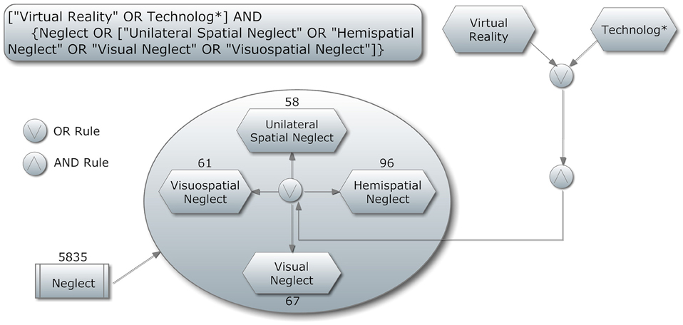
The search string was: (“Virtual Reality” OR Technolog*) AND [“Neglect” OR (“Unilateral Spatial Neglect” OR “Hemispatial Neglect” OR “Visual Neglect” OR “Visuospatial Neglect”)]. A graphical representation of the search string can been seen in Figure 1.
Our choice to search for both “virtual reality” and “technolog*” was to avoid missing papers due to the misleading terminologies that are often used in some studies. Acting within this strategy, we can be confident that this review is both replicable and inclusive of all possible records.
The articles were individually scanned to elaborate whether they fulfill the following inclusion criteria: (a) research article; (b) providing information about the used sample; (c) providing information about measures, and (d) published in English. These inclusion criteria were used for several reasons. As noted above, information about the sample and measures are a prerequisite.
The second search strategy (with the term Technolog*) had as a further exclusion criterion being present in the first list (already screened).
Systematic Review Flow
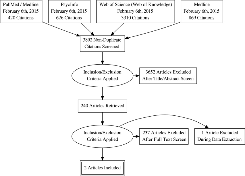
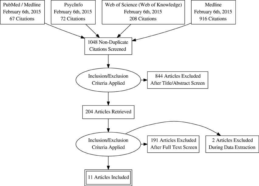
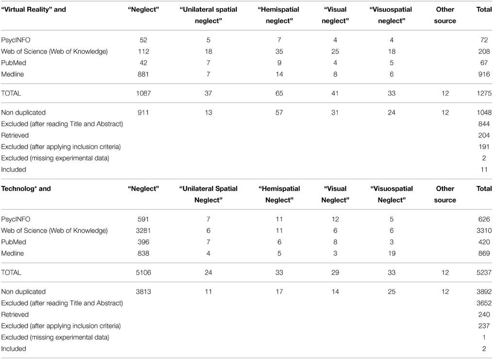
The flow chart of the systematic review is shown in Figure 2 for the term “Virtual Reality” and in Figure 3 for the term Technolog*. By searching in PsycINFO, PubMed, Medline and Web of Science (Web of Knowledge: WoK), our initial search yielded 1048 non-duplicate citations screened with “Virtual Reality” and 3892 with “Technolog*.” More details are available in the Search Strategy Table (Table 1). After the application of the inclusion criteria, papers were reduced to 204 and 240 articles, respectively. A deeper investigation of the full papers resulted in the exclusion of 191 and 237 articles, respectively. During the data extraction procedure, three additional full papers were excluded. In the end, 13 studies met the full criteria and were included in this review (Table 1). A flow diagram showing the procedure is detailed in Figure 2 for “Virtual Reality” search strategy and in Figure 3 for “Technolog*.”
Expert colleagues in the field were contacted for suggestions on further studies to consider in our search. Four new studies arose and have been included in the analyzed studies. To assess a risk of bias, PRISMA recommendations for systematic literature analysis have been strictly followed. Three authors (E.P., S.S., and P.C.) independently selected paper abstracts and titles and analyzed the full papers that met the inclusion criteria, resolving disagreements through consensus.
Results
In the current systematic review, we aim to provide a review of state-of-the-art experimental studies (from 2010 to 2014) focused on the use of VR for the assessment and rehabilitation of USN. In total, 12 studies met the inclusion criteria, were critically reviewed, and are summarized in Table 2.
In the following paragraphs, we critically reviewed the selected studies by dividing them according to the main purposes of the virtual tools proposed: (1) neuropsychological assessment of USN symptoms; (2) neuropsychological rehabilitation of USN symptoms; and (3) comprehensive platform for both assessment and rehabilitation of USN symptoms.
The Application of VR in the Assessment of USN
As it was described in the introduction, USN is typically evaluated by paper-and-pencil tests despite the aforementioned limitations of these tools. In this section, to deeply review the potential of VR for improving and/or integrating the traditional evaluations of USN, we analyzed the selected articles to provide an overview of the most recent virtual diagnostic tasks.
The first article analyzed was written by Kim et al. (2010), who used a 3D immersive VR program for street-crossing to assess USN in post-stroke patients. They assessed 32 patients, 16 with USN and 16 without USN. USN was assess by physiatrists and occupational therapists. They observe patients in the real life situations in order to find evidence of USN.
Patients was assesses during one session both with virtual and paper-and-pencil test. The test used are the Line Bisection Test (Schenkenberg et al., 1980) and the Line Cancellation Test (Albert, 1973).
At the beginning of virtual task, the patient see an avatar in front of a traffic light, the mission is cross the street without accident. If a car approaching to the avatar, patient have to push a stop button in order to avoid an accident. If patients failed to recognize the car approaching, they had visual and auditory cues to stop the avatar before failing their mission.
The results demonstrated that the two groups (patients with USN vs. patients without USN) showed differences in several variables analyzed during the task: deviation angle, left-to-right reaction time ratio, left visual, auditory cue rates, and left failure rate. Kim et al. (2010) showed that USN can be detected and measured easily and safely using their VR test. The authors also compared these virtual tools to the paper-and-pencil tests and found one correlation: the Line Bisection Test (Schenkenberg et al., 1980) correlated significantly with the deviation angle in the USN group.
In a similar test developed by Mesa-Gresa et al. (2011), they used a conventional LCD monitor, a surround system, a navigation and interaction joystick, and an optical tracking system (TRACKIR). Head movements were detected thanks to a cap with three reflecting markers and a USB infrared camera. A sample of 25 patients was analyzed, divided into neglect patients (n = 5) and non-neglect patients (n = 20) according to results obtained at the following tests: Behavioral Inattention Test (BIT), Color Trail Making Test (CTT), and Conners' Continuous Performance Test-II (CPT-II) (Peña-Casanova et al., 2006).
They planned a training session before the task that consisted of crossing a two-way road twice to arrive at a supermarket and then return. The task ended when patients went to and came back from the supermarket twice, making a maximum of four collisions with a car. They evaluated the following: how many times the participants looked to the left and to the right, the total time needed, the total number of accidents, whether the task was successfully accomplished, and a neuropsychological battery. During the VRSCT, negligent subjects showed a higher number of collision with a car than the other group, indeed indicating that the tool was able to discriminate between the two groups in clinical practice.
Peskine et al. (2011) developed a task that took place in a virtual city: patients have to count the number of bus stops they see. The sample included nine patients with a history of right cerebrovascular accidents (five of whom had visuospatial USN) and matched controls both for age and sex. USN was assessed using the Bells Cancellation Test (Gauthier et al., 1989) and the Catherine Bergego Scale (CBS; Azouvi et al., 2006). Patients used an HMD with an electromagnetic sensor system able to detect movements and sat on a swivel chair to turn on their own vertical axis. They had to move in the city, locate the swings in a park, and count all the bus stops; the examiner noted the patient's progress. The virtual assessment was done in just one session. Results showed that patients omitted more targets than controls and, most importantly, four patients without USN during the cancelation test showed USN in the virtual task.
Another navigation task was developed by Buxbaum et al. (2012). They created a “Virtual Reality Lateralized Attention Test” (VRLAT), a computerized measure of USN. They compared 71 USN patients with 10 control subjects. For the clinical assessment Buxbaum et al. (2012) used: a modified version of Bell Cancellation Test (Gauthier et al., 1989), the Letter Cancellation and Line Bisection Tests (Wilson et al., 1987), a modified version of the “fluff” test (Cocchini et al., 2001), a laser line-bisection task (Buxbaum et al., 2004), and a modified version of the Moss Real World Navigation (RWN) test (Buxbaum et al., 2008). During the VRLAT patient had to name all stationary objects in the scene while following a virtual winding path (i.e., navigation can be executed sometimes by the participants and sometimes by the experimenter). The program included three array conditions (i.e., simple, complex, and enhanced), and all patients completed all levels twice, once “coming” and once “going.” The software ran on a personal computer with a flat-screen video display; patients used a Logitech Attack 3 joystick.
This test seems to be better than traditional tests at predicting performance in real world. For this reason the VRLAT is a good tool for the assessment of USN. It's quick and easy to use, doesn't require specialized equipment, and could be useful both in clinical settings and in rehabilitation.
Aravind and colleagues (Aravind and Lamontagne, 2014; Aravind et al., 2015) developed a navigation task in a virtual room divided into three sub-tasks and analyzed the performance of 12 patients. A diagnosis of USN was based on the motor free visual perceptual test (MVPT; Colarusso and Hammill, 1972), and/or the Star Cancellation Test (Wilson et al., 1987). Clinical assessment included: Bells Cancellation Test (Gauthier et al., 1989), Line Bisection Tests (Wilson et al., 1987), the Montreal Cognitive Assessment (MOCA; Nasreddine et al., 2005), and the Trail Making Test-B (Army Individual Test Battery, 1944). Two of these tasks (“obstacle detection task” and the “joystick-driven obstacle avoidance task”) were analyzed in the first selected paper (Aravind et al., 2015); the other task, “locomotor obstacle avoidance task,” was described in another publication (Aravind and Lamontagne, 2014). Patients wearing a Visor SX60 head-mounted display (HMD) (NVIS, USA) and had a joystick (Attack3, Logitech, USA) to interact with the environment.
In the “locomotor obstacle avoidance task” (Aravind and Lamontagne, 2014) patient had to walking toward a target and avoid a collision with an moving object. The moving obstacle may approaching from center, right, or left.
During the “obstacle detection task” (Aravind et al., 2015) the patient was seated at a table with a joystick in the non-paretic hand. One of the three objects placed in center, right or left in the other side of the virtual room may approach toward the patient. When patient perceived the object had to push the button.
During the “joystick-driven obstacle avoidance task” (Aravind et al., 2015) the patient is passively moved toward a target and must avoid objects that move at him. The patient may avoid the object moving to the right or left or up or slow down the speed of movement with the joystick.
In the first task, patients detected contralesional obstacles at closer proximities compared to ipsilesional ones. For the “joystick-driven obstacle avoidance task,” participants begin to avoid objects at the last moment before the collision. Instead, they found that the performances on these paper-and-pencil tests were negatively associated with distances at detection, but the association lost significance with the exclusion of one patient (an outlier). For the “locomotor obstacle avoidance task,” Aravind and colleagues (Aravind and Lamontagne, 2014; Aravind et al., 2015) showed that 8 out of 12 subjects collided with either contralesional or head-on obstacles or both. Delay in detection and execution of avoidance strategies and smaller distances from obstacles were observed for colliders subjects compared to non-colliders one. After analyzing all three tasks, Aravind and colleagues (Aravind and Lamontagne, 2014; Aravind et al., 2015) argued that their system showed a typical pattern for USN patients and thus can be used for assessment.
The last article in this section is that of Fordell et al. (2011). They designed a VR Diagnostic Test Battery (VR-DiSTRO). The battery included the virtual version of four classical sub-tests: Star Cancellation and Line Bisection Test (Wilson et al., 1987), Visual Extinction Test (Geeraerts et al., 2005), and Baking Tray Task (Tham, 1996). During the experiment, patients have to do both virtual and classic versions of the test. The patients used a robotic pen (Phantom Omni haptic device) and shutter glasses for stereoscopic vision. All virtual tests took 15 min. The sample was composed of 31 post-stroke patients: 12 had a left-sided lesion and 19 had a right-sided one. VR-DiSTRO correctly identified the USN patients in the group, showing a 100% sensitivity and 82% specificity to correctly identify USN in the sample. Additionally, 77% of the sample said that the system was easy to use. The agreement with paper-and-pencil tests was moderate to almost perfect, indicating that this virtual battery was able to detect USN at least as well as the classic tests.
The Application of VR in the Rehabilitation of USN
In order to investigate the potential of VR in USN rehabilitation, we provided an overview of the most recent studies showing different and alternative solutions compared with the traditional methods of rehabilitation. First of all, neuropsychological rehabilitation of USN must take into account the specific needs of each patient. For this reason, a more customizable neuropsychological application is essential.
The traditional rehabilitation methods are often characterized by repetitive exercises, non-consideration of the individual patients' differences and needs, and the inability to generalize the performance and outcomes as not measured and quantified. For instance, the prisms technique, one of the most effective techniques in the neuropsychological rehabilitation of USN, induces an optical shift of the visual field to the right; the patients have an adaptation to this visual distortion that reduces neglect symptoms (Rossetti et al., 1998; Jacquin-Courtois et al., 2013; Leigh et al., 2015). Between the various techniques it is the most effective one, but not yet to be widely used in clinical practice. For this reason there is a need for innovative rehabilitations methods able to decrease USN behavior for long-term.
Kim et al. (2011) examined 24 stroke patients with USN divided into two groups. The VR group (n = 12) received a VR training with a system equipped with a monitor, a video camera, and computer-recognizing gloves. Patients had to complete three tasks: “Bird and Ball” (i.e., they had to touch a flying ball to turn it into a bird), “Coconut” (i.e., they had to catch coconuts falling from a tree), and “Container” (i.e., they had to move a box from one side to another). The control group (n = 12) received conventional USN therapy such as reading, visual tracking, writing, drawing and copying, and puzzles. Both groups had daily sessions of 30 min day, five sessions per week for 3 weeks. Both groups were assessed with conventional USN tests such as: the Star Cancellation Test and the Line Bisection Test (Wilson et al., 1987), the CBS (Azouvi et al., 2003) and the Korean version of the Modified Bartel Index (K-MBI; Jung et al., 2007). Results showed that only the VR group improved in the Star Cancellation Test (Wilson et al., 1987) and in the CBS (Azouvi et al., 2003) after the rehabilitation period.
Navarro et al. (2013) assessed the clinical validation, usability, and convergent validity of the “Virtual Street Crossing System” (Mesa-Gresa et al., 2011) to find out if it could be used for rehabilitation of USN. Their sample was composed of 17 USN patients, 15 non-USN patients and 15 control subjects. The rehabilitation task was the same used by Mesa-Gresa and colleagues in their study (Mesa-Gresa et al., 2011) and described previously. After the virtual task patients were administered a modified version of the Short Feedback Questionnaire (SFQm; Witmer and Singer, 1998). Patients were also assessed with some neuropsychological tests like: BIT, CPT-II, Stroop Test, Color Trail Test, BADS—Zoo Map Test, and Key Search Test (Peña-Casanova et al., 2006). The assessment was administered 3 days before or after the VR session. Patients with USN showed a lack of efficacy in the task, for example, they made more accidents than other groups. The results of their study showed the clinical effectiveness of the street-crossing system as confirmed by the VR outcomes, and the correlation with the scores of the neuropsychological tests.
The “Duckneglect” platform was developed by Mainetti et al. (2013). They analyzed a single case in order to check the improvement in USN using their system for rehabilitation. The patient, IB, was a 65-year-old male with a right fronto-temporal intraparenchymal hemorrhagic lesion that occurred in 2009; he's right-handed and has had 18 years of education. This system included specially-designed games requiring patients to reach some targets through different levels of difficulty using visual and auditory cues. A webcam, connected to the host PC, was positioned frontally to the patient's face, and two loud speakers were positioned near the patient to create a spatialized sound. Video of the patient was acquired from the camera and real-time processed to extract his silhouette from the background. The silhouette was then pasted onto the virtual scene of the rehabilitation task. In the end, the final scene was displayed on a screen in front of the patient. Before and after rehabilitation training a fully neuropsychological battery was administered: Line Cancellation Test (Albert, 1973), Letter Cancellation Test (Diller and Weinberg, 1976), Line Bisection Test (Schenkenberg et al., 1980), the Mini Mental State Examination (MMSE; Folstein et al., 1983), the Attentional Matrices (Spinnler and Tognoni, 1987), and the Token Test (DE RENZI and Vignolo, 1962). The rehabilitation lasted for half an hour every day, 5 days a week for 1 month. 5 months later, the patient came back for a follow-up and exhibited a significant improvement both on a classic paper-and-pencil test and other neuropsychological tasks. The improvement was also present for activities of daily living.
van Kessel et al. (2013) analyzed the performance of 29 post-stroke (right hemisphere) patients during their rehabilitation with a new computerized training method based on the “Visual Scanning Training” (TSVS) of Pizzamiglio (Pizzamiglio, 1990). Patients were divided into two groups: the experimental group (n = 14) received the computerized training while control group (n = 15) received traditional training. All patients received 30 training sessions 5 days a week for 6 weeks, 1 h per day. They used several tests for pre- and post-training assessment: paper-and-pencil tests, observation scales and the Driving Simulator Tasks. The paper-and-pencil tests are: Line Cancellation Test (Albert, 1973), Letter Cancellation Test (Diller and Weinberg, 1976), Bells Cancellation Test (Gauthier et al., 1989), Line Bisection Test (Schenkenberg et al., 1980), Word Reading Task (Làdavas et al., 1997), Gray Scales (Tant et al., 2002), and Baking Tray Task (Tham and Tegnér, 1996). The observation scales include: Semi-structured scale for the evaluation of personal and extrapersonal neglect (Zoccolotti et al., 1992), and Subjective Neglect Questionnaire (Towle and Lincoln, 1991). In the Driving Simulator Tasks, patients had to perform three tasks: Line Tracking Task, Single Detection Task (CVRT), and a combination of the previous two tasks. During the training sessions, the TSVS was composed of the following exercises: Large Screen Digit Detection, copying lines drawn on a dot matrix, reading and copying training and figure description. During the first and third weeks, both groups received the same treatment: on Monday and on Wednesday they did the TSVS tasks and on Thursday and Friday they did the TSVS and the lane tracking. During the second and fourth weeks, patients worked for just 2 days: the experimental group did the TSVS and the dual task while the control group did the TSVS and the lane tracking. van Kessel et al. (2013) didn't find any significant group or interaction effects that might underline additional positive effects of the dual task training; they weren't the result of other factors like spontaneous recovery or learning effects.
The Application of Integrated Platform for USN
Two of the selected studies proposed integrated VR platforms that are useful both for assessment and rehabilitation of USN.
An interesting example was given by Tanaka et al. (2010), who developed an HMD for the assessment and rehabilitation of USN. They tested two post-stroke patients with USN using a combined system (Charge-Coupled Device camera, HMD, and a computer) programmed to show in the display a modified version of the classic Line Cancellation Test (Albert, 1973). They administered the standard paper-and-pencil task and six modified versions task created by manipulating the zoom (in or out), the coordinates of visual field (object-centered and egocentric), and the presence of cue (arrows). These manipulations have been made in order to find and identify the left neglect area. The study confirmed that, thanks to the special assessment through HDM, it was easier to identify the neglected area of the patients. These results might provide a more precise assessment and a more focused rehabilitation. Tanaka et al. (2010) showed that, with a reduced image condition and the arrows condition, performance at the cancelation task improved.
The other article was a feasibility study by Sugarman et al. (2011) proposing new tools that could be used both for assessment and rehabilitation: SeeMe. The system was tested on a single USN patient (66 years old) who had a right hemisphere stroke 15 months previously. The woman was invited to use the tool for 1 h each day for 8 weeks. The patient stood in a specific area in front of a large monitor that displayed the virtual scenes, seeing himself on the screen in real time and being able to use trunk and limb movements to interact with the virtual environment. A single screen-mounted camera and a vision-based tracking system captured and converted the user's movements. Three tasks were used for the rehabilitation and four for the assessment (i.e., React task, the patient have to touch the virtual balls that appear randomly on both sides of the screen). The patient was assessed on the first and on the last day of the treatment with SeeMe and with the standard paper-and-pencil tests. Also the SFQ (Witmer and Singer, 1998) an open ended interview was administered on the last day of treatment. To the SFQ (Witmer and Singer, 1998) patient assigns 5 points out of 5 in almost every question except the one that assesses whether the virtual environment looks real. To this question the patient assigns a score of 2 out of 5. For the assessment task results indicated a difference between movement times (defined as “the time elapsed between the appearance of the target and the subject's virtual contact with the target”) in the right and the left space. Moreover, after training there was an improvement in movement times for the neglected space and in the paper-and-pencil test for USN.
Conclusions
The aim of this review is to describe and to critically analyze the most recent virtual tools developed and tested for the assessment and rehabilitation of USN in order to provide crucial indications for future studies, neurorehabilitation interventions, and clinical practice.
To date, traditional paper-and-pencil methods are still the most widely used technique in the clinical practice, despite several concerns both for assessment and rehabilitation of USN symptoms.
Regarding the assessment, the traditional paper-and-pencil tests may be deficient in detecting USN symptoms in the chronic stage of the disease (Rengachary et al., 2009), and their sensitivity and specificity varies between 38 and 52% (Agrell et al., 1997; Lindell et al., 2007; Fordell et al., 2011). On the other hand, regarding rehabilitative interventions, there is the prisms' technique, which is one of the most effective, but not the most used, techniques in neuropsychological rehabilitation of USN. It typically consists of sessions of repetitive exercises that have to be done several times a week but, unfortunately, have a limited effect in time (Rossetti et al., 1998; Newport and Schenk, 2012).
It is possible to note that paper-and-pencil tools use static, two-dimensional, and geometrical targets, which are far from those of a real, or virtual, environment. These tasks generally require a simple visual search in the near space, allowing only the diagnosis of peripersonal USN (Robertson and Halligan, 1999; Deouell et al., 2005; Kim et al., 2010; Aravind and Lamontagne, 2014). Otherwise, a real environment requires dynamic responses to the relevant stimuli that, in personal and extrapersonal space, change every time (Deouell et al., 2005; Buxbaum et al., 2008; Kim et al., 2010). This is a crucial feature of virtual environments since personal and extrapersonal USN are two subtypes of this syndrome that can be dissociated (Robertson and Halligan, 1999; Halligan et al., 2003). Specifically for rehabilitation, the use of moving stimuli may be crucial to modulate patients' visual attention; these kinds of objects can capture and drive attention to the left side of the space. Indeed, some recent evidence has reported that a moving cue in the left side of a task's space improved target detection in that area (Butter et al., 1990; Mattingley et al., 1994; Tanaka et al., 2010).
Moreover, both for static and moving stimuli there were different gradients of increasing reaction times, with a progression from the ipsilesional field toward the midline and into the contralesional field (Smania et al., 1998; Deouell et al., 2005; Dvorkin et al., 2007). Because of this feature, the computer version of reaction time tasks was generally more sensitive than paper-and-pencil tests (Rengachary et al., 2009; Bonato et al., 2012). One of the reasons for this behavioral pattern could be the predisposition of the patient with USN to initiating visual scanning of the environment from the ipsilesional side (Smania et al., 1998; Dvorkin et al., 2007; Aravind and Lamontagne, 2014; Aravind et al., 2015).
VR technologies offer impressive opportunities both for the rehabilitation and assessment of different cognitive deficits, including USN (Schultheis and Rizzo, 2001; Riva et al., 2004; Bohil et al., 2011).
According to the results of this systematic review, VR seems a promising instruments both for the assessment and rehabilitation of USN.
However, the trade-off between the incredible progress of VR and the need of methodological rigor and the possibility to the apply experimental protocols in the clinical practice has still to cope with different challenges.
First, as mentioned previously, Tsirlin and colleagues in their review (Tsirlin et al., 2009) underlined some characteristics of VR technologies that should be taken into consideration for future VR applications in this field.
The most important one is the ergonomic aspect of VR tools. Patients have specific needs to be considered, especially post-stroke patients who typically have to use a wheelchair for locomotion (Tsirlin et al., 2009). Our analysis showed that most of the selected studies have proposed VR assessment tools with greater attention paid to the ergonomic aspect in order to meet the needs of patients. In particular, it emerged that most of the recent VR systems could possibly be used with a chair or a wheelchair. Moreover, three selected studies have proposed some VR systems that can be easily controlled with one hand (Fordell et al., 2011; Kim et al., 2011), this is a great advantage for USN patients since hemiparesis is extremely common. Given this disability, it is very important to analyze usability aspects of the setting as Kim et al. (2011), Mainetti et al. (2013), Navarro et al. (2013), and van Kessel et al. (2013) did for their tools.
A second critical challenge for the clinician is the technical usability of the VR system/software since the clinical staff often has no programming skills. For this reason, cooperation with software developers is necessary for the use and customization of the technology. By designing intuitive VR applications and providing adequate training, developers may also help medical personnel in using these tools independently. First of all, Mainetti et al. (2013) emphasized the necessity of close collaboration between technical and clinical staff to tailor virtual environments to the specific requirements of patients. Moreover, three selected studies specifically addressed these issues, emphasizing the need for an easy-to-use application (Fordell et al., 2011; Sugarman et al., 2011; Sedda et al., 2013). Sugarman et al. (2011) have commented on their special attention to the usability aspects of their system, specifying that “SeeMe does not require any equipment beyond a webcam camera and a standard computer with a good video card” (p. 1). Indeed, there is a growing diffusion of VR-based telerehabilitation systems for post-stroke patients (for a review, see Brochard et al., 2010), which has allowed new directions for the design of ecological scenarios supporting multimodal interaction (Perez-Marcos et al., 2012).
The third important challenge that may limit the use of VR in the assessment and rehabilitation of USN is the high costs often required for designing and testing a technological system. Our analysis showed that only two selected studies have tried to pay particular attention to the costs (Kim et al., 2011; Mainetti et al., 2013), while the others tried to use cutting-edge technology in order to maximize the performance of the system.
Specifically for the neuropsychological rehabilitation of USN, it is essential to take into account the specific needs of the different patients. For this reason, a more customizable neuropsychological rehabilitation would be essential. A platform that allows the clinician to customize the tasks might also make a difference.
Finally, all the articles analyzed suggest several methods for the assessment and rehabilitation of USN, but there are some “methodological weaknesses.” Few studies compared VR methods with conventional ones (Kim et al., 2010, 2011; Mesa-Gresa et al., 2011; van Kessel et al., 2013; Aravind and Lamontagne, 2014; Aravind et al., 2015), only few studies compared the results with a control group (Kim et al., 2010; Peskine et al., 2011; Buxbaum et al., 2012; Navarro et al., 2013) and often the samples were too small to allow a generalization of the result (Peskine et al., 2011; Sugarman et al., 2011; Mainetti et al., 2013; Aravind and Lamontagne, 2014; Aravind et al., 2015), while controlled randomized trials testing the VR training in comparison with traditional protocol should be important. For further research, we also recommend adequate follow-up to maximize the benefits and monitor the persistence of the effect of neglect rehabilitation interventions. More, to enhance the potentiality of a multi-sensory and engaging VR stimulation, it is reasonable that USN patients should start a VR rehabilitation program in the acute stage.
However, the results obtained from the reviewed studies are promising and showed that VR systems stimulate interest and participation of patients (Kim et al., 2011). Indeed, VR simulations can be highly engaging by supporting a process known as “transformation of flow” (Riva et al., 2006), defined as an individual's ability to use and identify an optimal experience (i.e., flow) to promote new and unexpected psychological resources. This process may be particularly important since rehabilitation programs can be particularly demanding for patients. However, it is crucial to take into account potential transient side effects of immersive VR, such as cyber-sickness which occurs as a result of conflicts between visual, vestibular and proprioceptive signals. In addition to technological advancements, reducing the VR sessions (i.e., between 20 and 30 min) and giving precise explanations may alleviate any symptoms of discomfort.
Despite the great improvements in technology over the last 6 years, very few articles use new tools for assessment or rehabilitation in neuropsychology. The new technology systems for VR and the devices for “communication” with the virtual world could be very useful for neuropsychology; the possibility of acting in a virtual environment like in the real one is an important goal. Many efforts are aimed at improving the immersive virtual reality system, and there are two particularly important tools: VR wearable visors and the Cave Automatic Virtual Environment (CAVE). The VR head-mounted display is developed for virtual reality systems and video games. This tool uses custom tracking technology and creates a stereoscopic 3D view with excellent depth, scale and parallax by presenting unique and parallel images for each eye and using a 3-axis gyroscope, accelerometer and magnetometer to process data. The CAVE is a room with projection screens on the walls, floors and, in some cases, ceiling. The stereoscopic projectors are used for a 3D effect. These characteristics, together with the high-resolution of the graphics, allow an increase in the sense of presence. Users in the CAVE use head-trackers and hand-trackers in order to allow natural movements to interact with the virtual environment. CAVE is used mostly for design and fashion applications, but recently there have been some clinical applications, principally for the treatment of phobias and emotional disorders (Meyerbröker et al., 2010; Bouchard et al., 2013).
In terms of input devices, the classics are controllers for game consoles like Wii or Xbox. Wired gloves could be a way to improve the usability and comfort of the interaction and allow more fluid and natural movement in the environment. To remove the intermediation of tools, a solution could be using cameras to recognize models and identify motion, like Kinect or Vicon.
One input and output device is the Haptic device that allows people to feel the physical characteristics of the environment like gravity and viscosity.
The critical aspects of these devices are the high price and the complexity of both software and hardware components. To implement this device, support from technicians and developers is necessary in order to create the environments. Despite this limitation, this device has great potential to improve clinical practice. This new device allows for a completely different interaction with the virtual world and offers endless opportunities to analyze subject behavior in multiple ecological and controlled situations.
The aim of future studies could be to explore these possibilities in order to better understand the characteristics of each patient and his disorder and to create customized rehabilitation programs.
Additionally, the development of portable devices with good performance and reasonable prices may improve research concerning telemedicine, which would open the door to patients being treated at home without sacrificing medical supervision. Patients and doctors would be linked by a virtual platform that would allow monitoring of the patient's progress. The medical data could be mixed with the cognitive to make a complete picture of the healthy state of the patient. Medical data can be recorded by a wearable device capable of acquiring physiological signals.
Conflict of Interest Statement
The authors declare that the research was conducted in the absence of any commercial or financial relationships that could be construed as a potential conflict of interest.
Acknowledgments
This work was supported by the Italian funded project VRehab. Virtual Reality in the Assessment and TeleRehabilitation of Parkinson's Disease and “Post-Stroke Disabilities” - RF-2009-1472190.
References
Agrell, B. M., Dehlin, O. I., and Dahlgren, C. J. (1997). Neglect in elderly stroke patients: a comparison of five tests. Psychiatry Clin. Neurosci. 51, 295–300. doi: 10.1111/j.1440-1819.1997.tb03201.x
Albert, M. L. (1973). A simple test of visual neglect. Neurology 23, 658–664. doi: 10.1212/WNL.23.6.658
Appelros, P., Karlsson, G. M., Seiger, A., and Nydevik, I. (2002). Neglect and anosognosia after first-ever stroke: incidence and relationship to disability. J. Rehabil. Med. 34, 215–220. doi: 10.1080/165019702760279206
Aravind, G., Darekar, A., Fung, J., and Lamontagne, A. (2015). Virtual reality-based navigation task to reveal obstacle avoidance performance in individuals with visuospatial neglect. IEEE Trans. Neural Syst. Rehabil. Eng. 23, 179–188. doi: 10.1109/TNSRE.2014.2369812
Aravind, G., and Lamontagne, A. (2014). Perceptual and locomotor factors affect obstacle avoidance in persons with visuospatial neglect. J. Neuroeng. Rehabil. 11:38. doi: 10.1186/1743-0003-11-38
Army Individual Test Battery. (1944). Manual of Directions and Scoring. Washington, DC: War Department, Adjutant General's Office.
Azouvi, P., Bartolomeo, P., Beis, J. M., Perennou, D., Pradat-Diehl, P., and Rousseaux, M. (2006). A battery of tests for the quantitative assessment of unilateral neglect. Restor. Neurol. Neurosci. 24, 273–285.
Azouvi, P., Olivier, S., de Montety, G., Samuel, C., Louis-Dreyfus, A., and Tesio, L. (2003). Behavioral assessment of unilateral neglect: study of the psychometric properties of the Catherine Bergego Scale. Arch. Phys. Med. Rehabil. 84, 51–57. doi: 10.1053/apmr.2003.50062
Bartolomeo, P., and Chokron, S. (2002). Orienting of attention in left unilateral neglect. Neurosci. Biobehav. Rev. 26, 217–234. doi: 10.1016/S0149-7634(01)00065-3
Bohil, C. J., Alicea, B., and Biocca, F. A. (2011). Virtual reality in neuroscience research and therapy. Nat. Rev. Neurosci. 12, 752–762. doi: 10.1038/nrn3122
Bonato, M. (2012). Neglect and extinction depend greatly on task demands: a review. Front. Hum. Neurosci. 6:195. doi: 10.3389/fnhum.2012.00195
Bonato, M., and Deouell, L. Y. (2013). Hemispatial neglect: computer-based testing allows more sensitive quantification of attentional disorders and recovery and might lead to better evaluation of rehabilitation. Front. Hum. Neurosci. 7:162. doi: 10.3389/fnhum.2013.00162
Bonato, M., Priftis, K., Marenzi, R., Umiltà, C., and Zorzi, M. (2012). Deficits of contralesional awareness: a case study on what paper-and-pencil tests neglect. Neuropsychology 26, 20. doi: 10.1037/a0025306
Bouchard, S., Bernier, F., Boivin, É., Dumoulin, S., Laforest, M., Guitard, T., et al. (2013). Empathy toward virtual humans depicting a known or unknown person expressing pain. Cyberpsychol. Behav. Soc. Netw. 16, 61–71. doi: 10.1089/cyber.2012.1571
Bowen, A., Hazelton, C., Pollock, A., and Lincoln, N. B. (2013). Cognitive rehabilitation for spatial neglect following stroke. Cochrane Database Syst. Rev. 7:CD0035860 doi: 10.1002/14651858.CD003586.pub3
Bowen, A., McKenna, K., and Tallis, R. C. (1999). Reasons for variability in the reported rate of occurrence of unilateral spatial neglect after stroke. Stroke 30, 1196–1202. doi: 10.1161/01.STR.30.6.1196
Brochard, S., Robertson, J., Médée, B., and Rémy-Néris, O. (2010). What's new in new technologies for upper extremity rehabilitation? Curr. Opin. Neurol. 23, 683–687. doi: 10.1097/WCO.0b013e32833f61ce
Brooks, B. M., and Rose, F. D. (2003). The use of virtual reality in memory rehabilitation: Current findings and future directions. NeuroRehabilitation 18, 147–157.
Butter, C. M., Kirsch, N. L., and Reeves, G. (1990). The effect of lateralized dynamic stimuli on unilateral spatial neglect following right hemisphere lesions. Restor. Neurol. Neurosci. 2, 39–46.
Buxbaum, L. J., Dawson, A. M., and Linsley, D. (2012). Reliability and validity of the Virtual Reality Lateralized Attention Test in assessing hemispatial neglect in right-hemisphere stroke. Neuropsychology 26, 430. doi: 10.1037/a0028674
Buxbaum, L. J., Ferraro, M. K., Veramonti, T., Farne, A., Whyte, J., Ladavas, E., et al. (2004). Hemispatial neglect: subtypes, neuroanatomy, and disability. Neurology 62, 749–756. doi: 10.1212/01.WNL.0000113730.73031.F4
Buxbaum, L. J., Palermo, M. A., Mastrogiovanni, D., Read, M. S., Rosenberg-Pitonyak, E., Rizzo, A. A., et al. (2008). Assessment of spatial attention and neglect with a virtual wheelchair navigation task. J. Clin. Exp. Neuropsychol. 30, 650–660. doi: 10.1080/13803390701625821
Cocchini, G., Beschin, N., and Jehkonen, M. (2001). The fluff test: a simple task to assess body representation neglect. Neuropsychol. Rehabil. 11, 17–31. doi: 10.1080/09602010042000132
Colarusso, R. P., and Hammill, D. D. (1972). Motor-free Visual Perception Test. Novato, CA: Academic Therapy Pub.
Corbetta, M., and Shulman, G. L. (2011). Spatial neglect and attention networks. Annu. Rev. Neurosci. 34, 569. doi: 10.1146/annurev-neuro-061010-113731
Corbetta, M., Kincade, M. J., Lewis, C., Snyder, A. Z., and Sapir, A. (2005). Neural basis and recovery of spatial attention deficits in spatial neglect. Nat. Neurosci. 8, 1603–1610. doi: 10.1038/nn1574
Dalmaijer, E. S., Van der Stigchel, S., Nijboer, T. C., Cornelissen, T. H., and Husain, M. (2014). CancellationTools: All-in-one software for administration and analysis of cancellation tasks. Behav. Res. Methods. doi: 10.3758/s13428-014-0522-7. [Epub ahead of print].
DE RENZI, E., and VIGNOLO, L. A. (1962). The token test: a sensitive test to detect receptive disturbances in aphasics. Brain 85, 665. doi: 10.1093/brain/85.4.665
Deouell, L. Y., Sacher, Y., and Soroker, N. (2005). Assessment of spatial attention after brain damage with a dynamic reaction time test. J. Int. Neuropsychol. Soc. 11, 697–707. doi: 10.1017/s1355617705050824
Di Carlo, A. (2009). Human and economic burden of stroke. Age Ageing 38, 4–5. doi: 10.1093/ageing/afn282
Diller, L., and Weinberg, J. (1976). Hemi-inattention in rehabilitation: the evolution of a rational remediation program. Adv. Neurol. 18, 63–82.
Dvorkin, A. Y., Rymer, W. Z., Settle, K., and Patton, J. L. (2007). “Perceptual assessment of spatial neglect within a virtual environment,” in Paper Presented at the Virtual Rehabilitation (Venice).
Eslinger, P. J., Flaherty-Craig, C. V., and Benton, A. L. (2004). Developmental outcomes after early prefrontal cortex damage. Brain Cogn. 55, 84–103. doi: 10.1016/S0278-2626(03)00281-1
Eslinger, P. J., Grattan, L. M., Damasio, H., and Damasio, A. R. (1992). Developmental consequences of childhood frontal lobe damage. Arch. Neurol. 49, 764–769. doi: 10.1001/archneur.1992.00530310112021
Folstein, M. F., Robins, L. N., and Helzer, J. E. (1983). The Mini-mental state examination. Arch. Gen. Psychiatry 40, 812. doi: 10.1001/archpsyc.1983.01790060110016
Fordell, H., Bodin, K., Bucht, G., and Malm, J. (2011). A virtual reality test battery for assessment and screening of spatial neglect. Acta Neurol. Scand. 123, 167–174. doi: 10.1111/j.1600-0404.2010.01390.x
Gauthier, L., Dehaut, F., and Joanette, Y. (1989). The bells test: a quantitative and qualitative test for visual neglect. Int. J. Clin. Neuropsychol. 11, 49–54.
Geeraerts, S., Lafosse, C., Vandenbussche, E., and Verfaillie, K. (2005). A psychophysical study of visual extinction: ipsilesional distractor interference with contralesional orientation thresholds in visual hemineglect patients. Neuropsychologia 43, 530–541. doi: 10.1016/j.neuropsychologia.2004.07.012
Gontkovsky, S. T., McDonald, N. B., Clark, P. G., and Ruwe, W. D. (2002). Current directions in computer-assisted cognitive rehabilitation. NeuroRehabilitation 17, 195–199.
Hackett, M. L., Yapa, C., Parag, V., and Anderson, C. S. (2005). Frequency of depression after stroke a systematic review of observational studies. Stroke 36, 1330–1340. doi: 10.1161/01.STR.0000165928.19135.35
Halligan, P. W., Fink, G. R., Marshall, J. C., and Vallar, G. (2003). Spatial cognition: evidence from visual neglect. Trends Cogn. Sci. 7, 125–133. doi: 10.1016/S1364-6613(03)00032-9
Halligan, P. W., Marshall, J. C., and Wade, D. T. (1989). Visuospatial neglect: underlying factors and test sensitivity. Lancet 334, 908–911. doi: 10.1016/S0140-6736(89)91561-4
Heilman, K. M., Watson, R. T., and Valenstein, E. (1985). Neglect and Related Disorders. New York, NY: Oxford University Press.
Hendricks, H. T., van Limbeek, J., Geurts, A. C., and Zwarts, M. J. (2002). Motor recovery after stroke: a systematic review of the literature. Arch. Phys. Med. Rehabil. 83, 1629–1637. doi: 10.1053/apmr.2002.35473
Jacquin-Courtois, S., O'shea, J., Luauté, J., Pisella, L., Revol, P., Mizuno, K., et al. (2013). Rehabilitation of spatial neglect by prism adaptation: a peculiar expansion of sensorimotor after-effects to spatial cognition. Neurosci. Biobehav. Rev. 37, 594–609. doi: 10.1016/j.neubiorev.2013.02.007
Jehkonen, M., Laihosalo, M., and Kettunen, J. (2006). Impact of neglect on functional outcome after stroke-a review of methodological issues and recent research findings. Restor. Neurol. Neurosci. 24, 209–215.
Jung, H. Y., Park, B. K., Shin, H. S., Kang, Y. K., Pyun, S. B., Paik, N. J., et al. (2007). Development of the Korean version of Modified Barthel Index (K-MBI): multi-center study for subjects with stroke. J. Korean Acad. Rehabil. Med. 31, 283–297.
Kerkhoff, G., and Schenk, T. (2012). Rehabilitation of neglect: an update. Neuropsychologia 50, 1072–1079. doi: 10.1016/j.neuropsychologia.2012.01.024
Kim, D. Y., Ku, J., Chang, W. H., Park, T. H., Lim, J. Y., Han, K., et al. (2010). Assessment of post-stroke extrapersonal neglect using a three-dimensional immersive virtual street crossing program. Acta Neurol. Scand. 121, 171–177. doi: 10.1111/j.1600-0404.2009.01194.x
Kim, Y. M., Chun, M. H., Yun, G. J., Song, Y. J., and Young, H. E. (2011). The effect of virtual reality training on unilateral spatial neglect in stroke patients. Ann. Rehabil. Med. 35, 309–315. doi: 10.5535/arm.2011.35.3.309
Làdavas, E., Shallice, T., and Zanella, M. T. (1997). Preserved semantic access in neglect dyslexia. Neuropsychologia 35, 257–270. doi: 10.1016/S0028-3932(96)00066-8
Langhorne, P., Coupar, F., and Pollock, A. (2009). Motor recovery after stroke: a systematic review. Lancet Neurol. 8, 741–754. doi: 10.1016/S1474-4422(09)70150-4
Leigh, S., Danckert, J., and Eliasmith, C. (2015). Modelling the differential effects of prisms on perception and action in neglect. Exp. Brain Res. 233, 751–766. doi: 10.1007/s00221-014-4150-3
Levick, W. R. (2010). Observer rating of memory in children: a review. Brain Impair. 11, 144–151. doi: 10.1375/brim.11.2.144
Lindell, A. B., Jalas, M. J., Tenovuo, O., Brunila, T., Voeten, M. J., and Hämäläinen, H. (2007). Clinical assessment of hemispatial neglect: evaluation of different measures and dimensions. Clin. Neuropsychol. 21, 479–497. doi: 10.1080/13854040600630061
Lloyd-Jones, D., Adams, R., Carnethon, M., De Simone, G., Ferguson, T. B., Flegal, K., et al. (2009). Heart disease and stroke statistics–2009 update: a report from the American Heart Association Statistics Committee and Stroke Statistics Subcommittee. Circulation 119, 21–181. doi: 10.1161/CIRCULATIONAHA.108.191261
Mainetti, R., Sedda, A., Ronchetti, M., Bottini, G., and Borghese, N. A. (2013). Duckneglect: video-games based neglect rehabilitation. Technol. Health Care 21, 97–111. doi: 10.3233/THC-120712
Mattingley, J. B., Bradshaw, J. L., and Bradshaw, J. A. (1994). Horizontal visual motion modulates focal attention in left unilateral spatial neglect. J. Neurol. Neurosurg. Psychiatry 57, 1228–1235. doi: 10.1136/jnnp.57.10.1228
Mesa-Gresa, P., Lozano, J. A., Llórens, R., Alcañiz, M., Navarro, M. D., and Noé, E. (2011). “Clinical validation of a virtual environment test for safe street crossing in the assessment of acquired brain injury patients with and without neglect,” in Paper Presented at the Proceedings of the 13th IFIP TC 13 International Conference on Human-computer Interaction - Volume Part II (Lisbon).
Meyerbröker, K., Morina, N., Kerkhof, G., and Emmelkamp, P. M. (2010). Virtual reality exposure treatment of agoraphobia: a comparison of computer automatic virtual environment and head-mounted display. Stud. Health Technol. Inform. 167, 51–56. doi: 10.3233/978-1-60750-766-6-51
Moher, D., Liberati, A., Tetzlaff, J., Altman, D. G., and PRISMA Group. (2009). Preferred reporting items for systematic reviews and meta-analyses: the PRISMA statement. PLoS Med. 6:e1000097. doi: 10.1371/journal.pmed.1000097
Mutai, H., Furukawa, T., Araki, K., Misawa, K., and Hanihara, T. (2012). Factors associated with functional recovery and home discharge in stroke patients admitted to a convalescent rehabilitation ward. Geriatr. Gerontol. Int. 12, 215–222. doi: 10.1111/j.1447-0594.2011.00747.x
Nasreddine, Z. S., Phillips, N. A., Bédirian, V., Charbonneau, S., Whitehead, V., Collin, I., et al. (2005). The Montreal Cognitive Assessment, MoCA: a brief screening tool for mild cognitive impairment. J. Am. Geriatr. Soc. 53, 695–699. doi: 10.1111/j.1532-5415.2005.53221.x
Navarro, M. D., Lloréns, R., Noé, E., Ferri, J., and Alcañiz, M. (2013). Validation of a low-cost virtual reality system for training street-crossing. A comparative study in healthy, neglected and non-neglected stroke individuals. Neuropsychol. Rehabil. 23, 597–618. doi: 10.1080/09602011.2013.806269
Newport, R., and Schenk, T. (2012). Prisms and neglect: what have we learned? Neuropsychologia 50, 1080–1091. doi: 10.1016/j.neuropsychologia.2012.01.023
Nijboer, T. C., Kollen, B. J., and Kwakkel, G. (2013a). Time course of visuospatial neglect early after stroke: a longitudinal cohort study. Cortex 49, 2021–2027. doi: 10.1016/j.cortex.2012.11.006
Nijboer, T. C. W., Kollen, B. J., and Kwakkel, G. (2014a). The impact of recovery of visuo-spatial neglect on motor recovery of the upper paretic limb after stroke. PLoS ONE 9:e100584. doi: 10.1371/journal.pone.0100584
Nijboer, T. C. W., Ten Brink, A. F., Kouwenhoven, M., and Visser-Meily, J. M. A. (2014b). Functional assessment of region-specific neglect: are there differential behavioural consequences of peripersonal versus extrapersonal neglect? Behav. Neurol. 2014:526407. doi: 10.1155/2014/526407
Nijboer, T. C., van de Port, I., Schepers, V., Post, M., and Visser-Meily, A. (2013b). Predicting functional outcome after stroke: the influence of neglect on basic activities in daily living. Front. Hum. Neurosci. 7:182. doi: 10.3389/fnhum.2013.00182
Paci, M., Matulli, G., Baccini, M., Rinaldi, L. A., and Baldassi, S. (2010). Reported quality of randomized controlled trials in neglect rehabilitation. Neurol. Sci. 31, 159–163. doi: 10.1007/s10072-009-0198-4
Peña-Casanova, J., Gramunt Fombuena, N., and Gich Fullà, J. (2006). Neurophychological Tests. Barcelona: Elsevier.
Pendlebury, S. T., Giles, M. F., and Rothwell, P. M. (2009). Transient Ischemic Attack and Stroke: Diagnosis, Investigation and Management. Cambridge: Cambridge University Press.
Perez-Garcia, M., Godoy-Garcia, J. F., Vera-Guerrero, N., Laserna-Triguero, J. A., and Ouente, A. E. (1998). Neuropsychological evaluation of everyday memory. Neuropsychol. Rev. 8, 203–227. doi: 10.1023/A:1021622319851
Perez-Marcos, D., Solazzi, M., Steptoe, W., Oyekoya, O., Frisoli, A., Weyrich, T., et al. (2012). A fully-immersive set-up for remote interaction and neurorehabilitation based on virtual body ownership. Front. Neurol. 3:110. doi: 10.3389/fneur.2012.00110
Peskine, A., Rosso, C., Box, N., Galland, A., Caron, E., Rautureau, G., et al. (2011). Virtual reality assessment for visuospatial neglect: importance of a dynamic task. J. Neurol. Neurosurg. Psychiatr. 82, 1407–1409. doi: 10.1136/jnnp.2010.217513
Pflugshaupt, T., Bopp, S. A., Heinemann, D., Mosimann, U. P., von Wartburg, R., Nyffeler, T., et al. (2004). Residual oculomotor and exploratory deficits in patients with recovered hemineglect. Neuropsychologia 42, 1203–1211. doi: 10.1016/j.neuropsychologia.2004.02.002
Pierce, S. R., and Buxbaum, L. J. (2002). Treatments of unilateral neglect: a review. Arch. Phys. Med. Rehabil. 83, 256–268. doi: 10.1053/apmr.2002.27333
Pohjasvaara, T., Leskelä, M., Vataja, R., Kalska, H., Ylikoski, R., Hietanen, M., et al. (2002). Post−stroke depression, executive dysfunction and functional outcome. Eur. J. Neurol. 9, 269–275. doi: 10.1046/j.1468-1331.2002.00396.x
Rabuffetti, M., Farina, E., Alberoni, M., Pellegatta, D., Appollonio, I., Affanni, P., et al. (2012). Spatio-temporal features of visual exploration in unilaterally brain-damaged subjects with or without neglect: results from a touchscreen test. PLoS ONE 7:e31511. doi: 10.1371/annotation/bf311a56-bc48-44b6-9b0f-7ccc97fc290f
Rengachary, J., d'Avossa, G., Sapir, A., Shulman, G. L., and Corbetta, M. (2009). Is the posner reaction time test more accurate than clinical tests in detecting left neglect in acute and chronic stroke? Arch. Phys. Med. Rehabil. 90, 2081–2088. doi: 10.1016/j.apmr.2009.07.014
Riva, G. (2009). Virtual reality: an experiential tool for clinical psychology. Br. J. Guid. Couns. 37, 337–345. doi: 10.1080/03069880902957056
Riva, G., Castelnuovo, G., and Mantovani, F. (2006). Transformation of flow in rehabilitation: the role of advanced communication technologies. Behav. Res. Methods 38, 237–244. doi: 10.3758/BF03192775
Riva, G., Mantovani, F., and Gaggioli, A. (2004). Presence and rehabilitation: toward second-generation virtual reality applications in neuropsychology. J. Neuroeng. Rehabil. 1, 9. doi: 10.1186/1743-0003-1-9
Rizzo, A. A., Buckwalter, J. G., and van der Zaag, C. (2002). “Applications for neuropsychological assessment and rehabilitation,” in Handbook of Virtual Environments, ed K. Stanney (New York, NY: L. A. Earlbaum), 1027–1064.
Robertson, I. H., and Halligan, P. W. (1999). Spatial Neglect: A Clinical Handbook for Diagnosis and Treatment. Hove: Psychology Press.
Rossetti, Y., Rode, G., Pisella, L., Farné, A., Li, L., Boisson, D., et al. (1998). Prism adaptation to a rightward optical deviation rehabilitates left hemispatial neglect. Nature 395, 166–169. doi: 10.1038/25988
Schenkenberg, T., Bradford, D. C., and Ajax, E. T. (1980). Line bisection and unilateral visual neglect in patients with neurologic impairment. Neurology 30, 509–509. doi: 10.1212/WNL.30.5.509
Schultheis, M. T., and Rizzo, A. A. (2001). The application of virtual reality technology in rehabilitation. Rehabil. Psychol. 46, 296. doi: 10.1037/0090-5550.46.3.296
Sedda, A., Borghese, N. A., Ronchetti, M., Mainetti, R., Pasotti, F., Beretta, G., et al. (2013). Using virtual reality to rehabilitate neglect. Behav. Neurol. 26, 183–185. doi: 10.1155/2013/810279
Smania, N., Martini, M., Gambina, G., Tomelleri, G., Palamara, A., Natale, E., et al. (1998). The spatial distribution of visual attention in hemineglect and extinction patients. Brain 121, 1759–1770. doi: 10.1093/brain/121.9.1759
Smit, M., Van der Stigchel, S., Visser-Meily, J. M., Kouwenhoven, M., Eijsackers, A. L., and Nijboer, T. C. (2013). The feasibility of computer-based prism adaptation to ameliorate neglect in sub-acute stroke patients admitted to a rehabilitation center. Front. Hum. Neurosci. 7:353. doi: 10.3389/fnhum.2013.00353
Spinnler, H., and Tognoni, G. (1987). Standardizzazione e Taratura Italiana di Test Neuropsicologici. Milano: Masson Italia Periodici.
Sudlow, C. L. M., and Warlow, C. P. (1997). Comparable studies of the incidence of stroke and its pathological types - Results from am international collaboration. Stroke 28, 491–499. doi: 10.1161/01.STR.28.3.491
Sugarman, H., Weisel-Eichler, A., Burstin, A., and Brown, R. (2011). “Use of novel virtual reality system for the assessment and treatment of unilateral spatial neglect: A feasibility study,” in Paper Presented 2011 International Conference on the Virtual Rehabilitation (ICVR) (Zurich). doi: 10.1109/ICVR.2011.5971859
Sundar, U., and Adwani, S. (2010). Post-stroke cognitive impairment at 3 months. Ann. Indian Acad. Neurol. 13, 42. doi: 10.4103/0972-2327.61276
Tanaka, T., Ifukube, T., Sugihara, S., and Izumi, T. (2010). A case study of new assessment and training of unilateral spatial neglect in stroke patients: effect of visual image transformation and visual stimulation by using a head mounted display system (HMD). J. Neuroeng. Rehabil. 7:20. doi: 10.1186/1743-0003-7-20
Tant, M. L. M., Kuks, J. M. B., Kooijman, A. C., Cornelissen, F. W., and Brouwer, W. H. (2002). Grey scales uncover similar attentional effects in homonymous hemianopia and visual neglect. Neuropsychologia 40, 1474–1481. doi: 10.1016/S0028-3932(01)00197-X
Tham, K. (1996). The baking tray task: a test of spatial neglect. Neuropsychol. Rehabil. 6, 19–26. doi: 10.1080/713755496
Tham, K., and Tegnér, R. (1996). The baking tray task: a test of spatial neglect. Neuropsychol. Rehabil. 6, 19–25. doi: 10.1080/713755496
Towle, D., and Lincoln, N. B. (1991). Development of a questionnaire for detecting everyday problems in stroke patients with unilateral neglect. Clin. Rehabil. 5, 135–140. doi: 10.1177/026921559100500208
Tsirlin, I., Dupierrix, E., Chokron, S., Coquillart, S., and Ohlmann, T. (2009). Uses of virtual reality for diagnosis, rehabilitation and study of unilateral spatial neglect: review and analysis. Cyberpsychol. Behav. 12, 175–181. doi: 10.1089/cpb.2008.0208
Vaes, N., Lafosse, C., Nys, G., Schevernels, H., Dereymaeker, L., Oostra, K., et al. (2015). Capturing peripersonal spatial neglect: An electronic method to quantify visuospatial processes. Behav. Res. Methods 47, 27–44. doi: 10.3758/s13428-014-0448-0
Vallar, G., and Perani, D. (1986). The anatomy of unilateral neglect after right-hemisphere stroke lesions. A clinical/CT-scan correlation study in man. Neuropsychologia 24, 609–622. doi: 10.1016/0028-3932(86)90001-1
van Kessel, M. E., Geurts, A. C., Brouwer, W. H., and Fasotti, L. (2013). Visual scanning training for neglect after stroke with and without a computerized lane tracking dual task. Front. Hum. Neurosci. 7:358. doi: 10.3389/fnhum.2013.00358
Vriezen, E. R., Pigott, S. D., and Pelletier, P. M. (2001). Developmental implications of early frontal-lobe damage: a case study. Brain Cogn. 47, 222–225.
Wilson, B., Cockburn, J., and Halligan, P. (1987). Development of a behavioral test of visuospatial neglect. Arch. Phys. Med. Rehabil. 68, 98–102.
Witmer, B., and Singer, M. (1998). Measuring presence in virtual environments: a presence questionnaire. Presence 7, 225–240. doi: 10.1162/105474698565686
Wolfe, C. D. A. (2000). The impact of stroke. Br. Med. Bull. 56, 275–286. doi: 10.1258/0007142001903120
Yong Joo, L., Soon Yin, T., Xu, D., Thia, E., Pei Fen, C., Kuah, C. W., et al. (2010). A feasibility study using interactive commercial off-the-shelf computer gaming in upper limb rehabilitation in patients after stroke. J. Rehabil. Med. 42, 437–441. doi: 10.2340/16501977-0528
Zhang, Y., Chapman, A. M., Plested, M., Jackson, D., and Purroy, F. (2012). The incidence, prevalence, and mortality of stroke in France, Germany, Italy, Spain, the UK, and the US: a literature review. Stroke Res. Treat. 2012:436125. doi: 10.1155/2012/436125
Keywords: virtual reality, USN, assessment, rehabilitation, PRISMA
Citation: Pedroli E, Serino S, Cipresso P, Pallavicini F and Riva G (2015) Assessment and rehabilitation of neglect using virtual reality: a systematic review. Front. Behav. Neurosci. 9:226. doi: 10.3389/fnbeh.2015.00226
Received: 19 May 2015; Accepted: 10 August 2015;
Published: 25 August 2015.
Edited by:
Katiuscia Sacco, University of Turin, ItalyReviewed by:
Seth Davin Norrholm, Emory University School of Medicine, USATanja Nijboer, Utrecht University, Netherlands
Copyright © 2015 Pedroli, Serino, Cipresso, Pallavicini and Riva. This is an open-access article distributed under the terms of the Creative Commons Attribution License (CC BY). The use, distribution or reproduction in other forums is permitted, provided the original author(s) or licensor are credited and that the original publication in this journal is cited, in accordance with accepted academic practice. No use, distribution or reproduction is permitted which does not comply with these terms.
*Correspondence: Elisa Pedroli, Applied Technology for Neuro-Psychology Lab, IRCCS Istituto Auxologico Italiano, Via Magnasco 2, 20149 Milan, Italy,ZS5wZWRyb2xpQGF1eG9sb2dpY28uaXQ=
 Elisa Pedroli
 Silvia Serino
 Pietro Cipresso
 Federica Pallavicini
 Giuseppe Riva


