 Runze Wang1†
 Yang Gao
 TianYang Yu
 Liyuan Wang
- 1Heilongjiang University of Chinese Medicine, Harbin, China
- 2Endocrinology Department, Heilongjiang Academy of Sciences of Traditional Chinese Medicine, Harbin, China
- 3Acupuncture Department, Second Affiliated Hospital of Heilongjiang University of Chinese Medicine, Harbin, China
- 4Ophthalmology Department, First Affiliated Hospital of Heilongjiang University of Chinese Medicine, Harbin, China
Background: Oculomotor nerve palsy (ONP) is a clinically occurring neurological disorder. Due to the complex anatomy and long travel distances of the oculomotor nerve, the causes of ONP vary and manifest in various ways. With continued interest in this area, it has become necessary to conduct a bibliometric study in ONP. This work aims to synthesize and visually identify current research themes and future trends in ONP through a literature-based analysis.
Method: Articles and reviews on ONP published from 2002 to 2021 were derived from the Web of Science Core Collection (WoSCC) database. We generated visual images and performed quantitative and qualitative analysis through an online bibliometric tool, Citespace and VOSviewer software.
Results: A total of 1,205 published articles were included in this analysis. The annual number of this area's publications is showing an overall upward trend, with the number of citations increasing every year, reaching 2,698 by 2021. The United States (367) and Japan (116) dominated the list with the most numerous articles published. The University of California Los Angeles in the USA is the institution that published the highest number of articles (47). Engle EC (23) and the JOURNAL OF NEUROSURGERY (46) are the most influential authors and journals in this field. The co-occurrence network analysis divided the keywords into five main research themes, which mainly include clinical manifestations of ONP, aneurysms, cerebral neurological symptoms, diseases with ONP as a complication, and other neurological disorders.
Conclusion: This study is the first comprehensive and systematic bibliometric analysis of the current state of global ONP research over the past 20 years. We organized current hotspots and expected trends and provided key information for exploring potential research frontiers in ONP.
Introduction
The oculomotor nerve (ON) is the main nerve that innervates eye movements. Its damage may lead to typical manifestations such as ptosis, diplopia, unable to do eye movements, and diminished or absent pupillary light reflex (1–6). In an early study of 290 patients with oculomotor nerve palsy (ONP), results showed 23.1% of patients had unknown causes, 20.7% had vascular lesions, 16.2% had head trauma, 14.5% had other causes, 13.8% had aneurysms, and 11.7% had tumors (7). Singh's report indicated that the causes of ONP were congenital or acquired, congenital for 43%, trauma for 20%, inflammation for 13%, intracranial aneurysms for 7% in pediatric patients, while vascular disease, intracranial aneurysms and trauma were the most common causes among adult patients (8). At the same time, diabetes mellitus, painful ophthalmoplegia, and cerebral infarction are also the etiology of this disease (9–11). A 10-year cohort study of Koreans showed that the incidence of ONP increased annually from 2006 to 2015 and was more prevalent in older adults, with a significantly accelerated incidence after age 60, severely affecting patients' quality of life (12). Therefore, the number of studies on ONP has increased dramatically in the past few decades. However, studies on publication patterns, literature characteristics, and bibliometrics on ONP are scarce.
Bibliometrics is the science of qualitative and quantitative analysis of existing research fields through mathematics and statistics (13, 14). Bibliometrics can provide readers with a visual and intuitive image of research results, including the contributions and productivity of different countries, institutions, and journals, as well as the evolution of information about the creators, the priorities, and the dissemination channels of the field. It emerged as a solution to the dilemma of access to knowledge due to the enormous growth of information and offered the possibility of interdisciplinary collaboration, crossover and integration.
In this study, we conducted a bibliometric analysis of published ONP-related studies and reviews in the Web of Science Core Collection (WoSCC) database over the past 20 years. This study can assist researchers in obtaining a brief overview of current hotspots in this field to gain insight into future research trends, to improve the quality of ONP research.
Materials and methods
Data acquisition and search strategy
All data were screened from the Science Citation Index-expanded (SCI-E) of the WoSCC database on November 6, 2022. After repeated attempts in a limited range, we finally used the query as follows: TS = (Oculomotor nerve disease* OR Oculomotor nerve disorder OR Oculomotor neuropathy OR Oculomotor nerve paralysis OR Oculomotor nerve palsy) with a publication date from 2001-01-01 to 2021-12-31. We limited the document type as “articles” and “reviews” and the language as “English.” Finally, a total of 1,205 records were retrieved for bibliometric analysis.
Data extraction
To facilitate processing with the software, we exported all records as “full records and citation references” and stored them as plain text files. The basic information included authors' names, institution, country/region, corresponding authors, citations count, journal, and keywords. The H-index and the average citations per item (ACI) were extracted from the Web of Science (WoS) webpage by using the “Create Citation Report” function.
Data analysis
Microsoft Excel 2019 are mainly used to summarize basic information such as the number of annual publications and the number of citations. The different country publications over the years and country collaboration analysis were using the bibliometric platform (https://bibliometric.com/).
Citespace is the most widely used knowledge mapping tool for co-occurrence and burst analysis of institutions, authors, and keywords (15). In the mapping created by Citespace (Version 6.1.R4), the number or frequency of different item types (countries, institutions, and authors) is indicated by the size of the nodes. At the same time, the color represents the year in which the item appeared, with darker colors representing later appearances. The thicker the line, the more frequent the association.
VOSviewer, also a bibliometric software, is better at knowledge clustering and visual analysis (16). Therefore, in this study, we used this software (Version 1.6.18) to generate corresponding graphs to perform co-authorship and co-citation analyses of authors, journals, and references.
Result
Overall publication performance
The actual number of literatures in a field over a certain period can reveal the intensity of research in the discipline and give a general indication of its trends. As shown in Figure 1, the number of ongoing papers in the field has generally been upward over the last 20 years. The number of research publications was growing slowly from 2001 to 2009, with a significant increase in the annual production of publications in 2010, followed by a fluctuating upward trend over the next 11 years, which indicates that ONP has been a hot issue in the research field. Despite a small decline in the number of publications in 2021, it is still significant and considerable. According to our statistics, the total number of cited articles in this field was 15,001 (14,322 after removing self-citations), and the total number of citations was 22,625 (18,727 after removing self-citations), with an average of 17.95 citations per paper. The citation volume has been growing consistently and rapidly year over year, which indicates that ONP-related research has a certain influence.
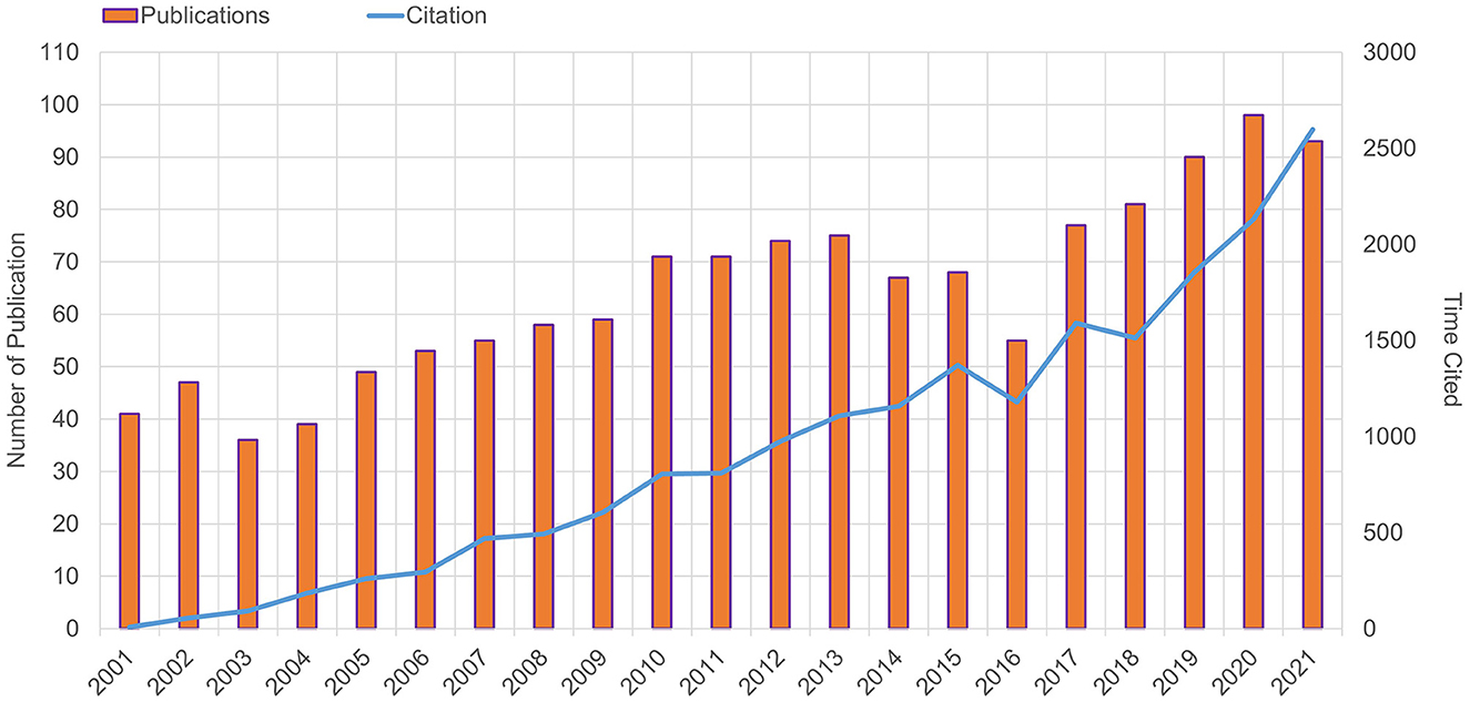
Figure 1. Publication and citation. Trends in publications and citations per year from 2001 to 2021.
Countries and regions
The statistics indicated that altogether 1,205 documents got published from 75 different countries/regions. USA (340, 28.2%), Japan (142, 11.8%), China (102, 8.5%), Germany (84, 7.0%), and South Korea (83, 6.9%) were the top five countries in terms of the number of papers published showed in Figure 2A. The volume of articles issued by all countries was visualized in Figure 2B, while the annual volume was shown in Figure 2C. Figure 2D shows the network diagram of cooperation between countries. We analyzed the cooperation between countries and found that the USA cooperates most closely with Germany.
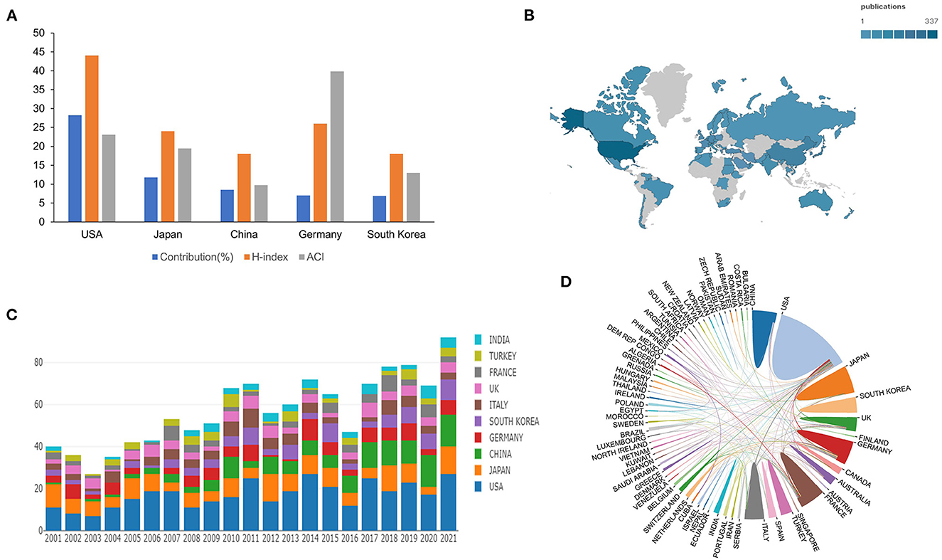
Figure 2. Countries and regions. (A) Top 5 most productive countries contribution. (B) Geographical distribution of countries in ONP. (C) The annual publication counts of the top 10 countries from 2001 to 2021. (D) Collaborative relationships among different countries.
Institutions
The top 15 most productive institutions are listed in Table 1. It shows that the University of California Los Angeles, Harvard University, Boston Children's Hospital, and Harvard Medical School have published more publications in the last two decades. Figure 3A visualizes the collaborative relationships among the institutions. From this figure, the University of California Los Angles collaborates more closely with Harvard University and Boston Children's Hospital. At the same time, Seoul National University has more publications but few collaborations with other institutions, which shows that the collaboration between different national institutions needs to be enhanced. Figure 3B reflects the timing of ONP-related studies published by each institution, which shows that Capital Medical School, Boston Children's Hospital, and Harvard Medical School have been very interested in this disease in the recent past.
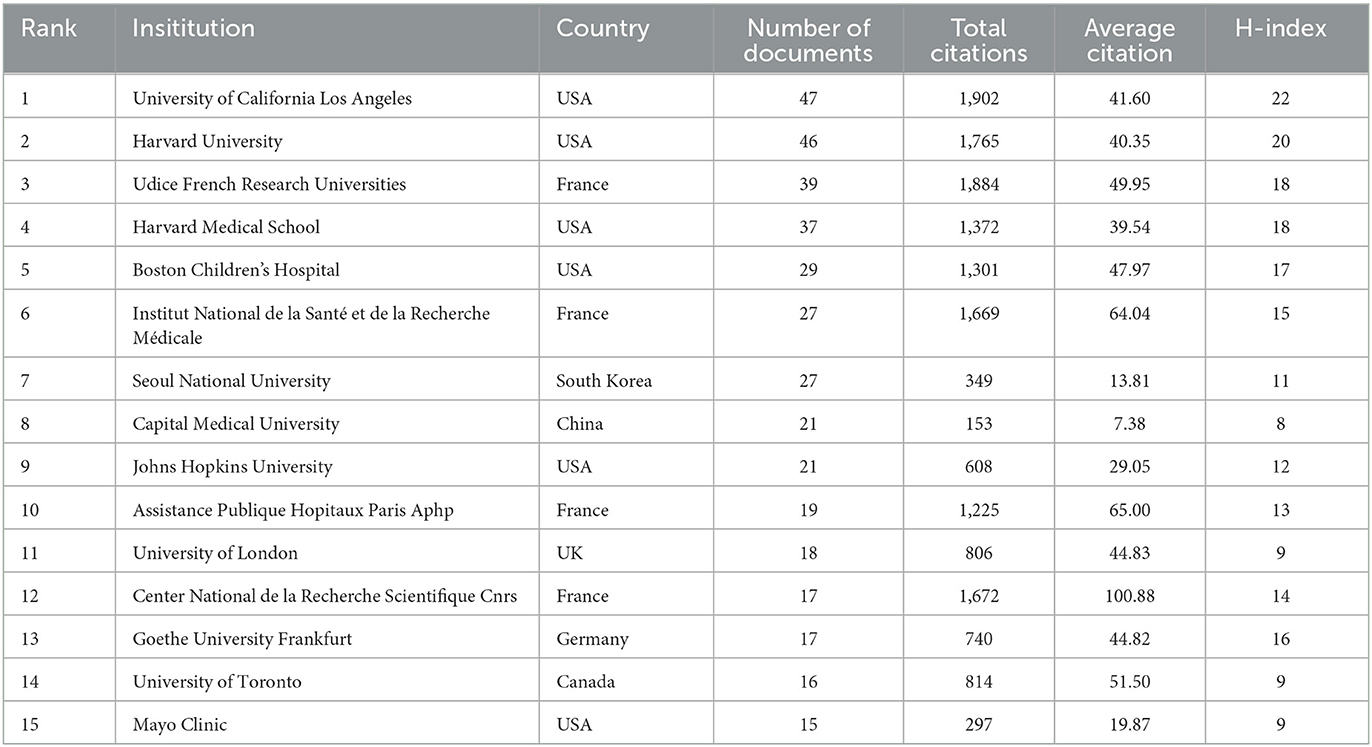
Table 1. Top 15 most productive institutions between 2001 to 2021 on ONP.
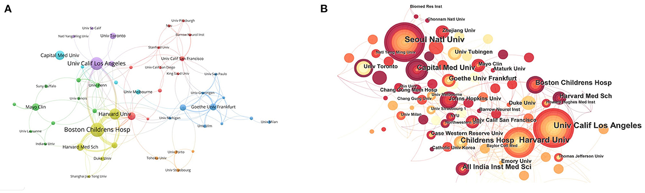
Figure 3. Institutions. (A) Co-occurrence analysis of the main institutions. The thicker the line between the institutions, the more frequently the two cooperate. (B) A Citespace visualization map of the cooperated institutions involved in ONP. The color of the node represents the time of the most recent ONP field paper published by this institution.
Author and co-cited author
A total of 5,609 authors participated in the ONP study, with an average of 4.65 authors per article. Table 2 lists the top 15 authors regarding the number of publications, with Engle EC being the author with the most articles (n = 23), followed closely by Demer JL (n = 20). In Figure 4A, we visualized the collaborative relationships between authors and revealed that authors from the USA, France, and Germany collaborated more closely, but collaborative links between other countries remained low.
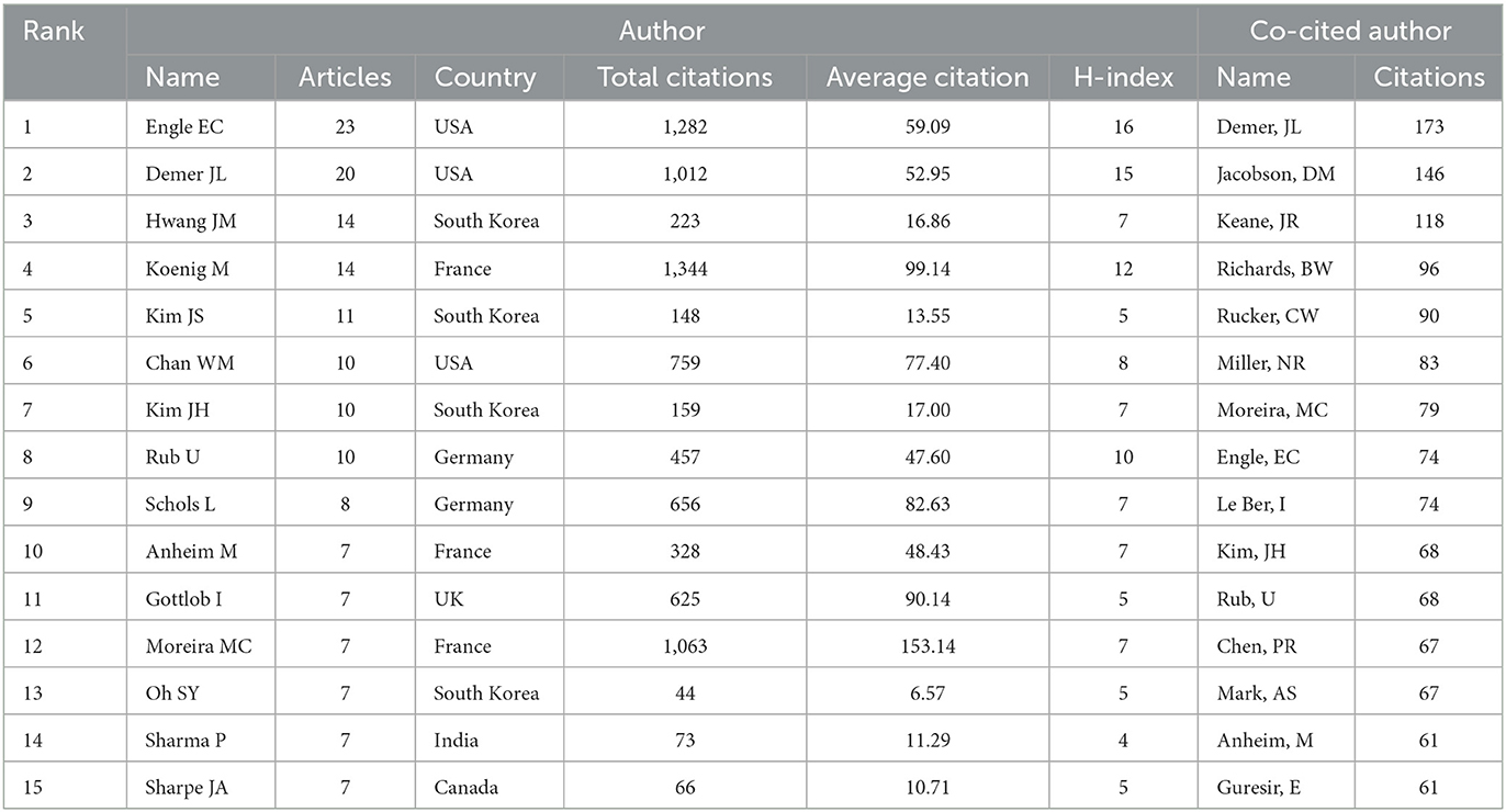
Table 2. The top 15 productive authors and co-cited authors on ONP research from 2001 to 2021.
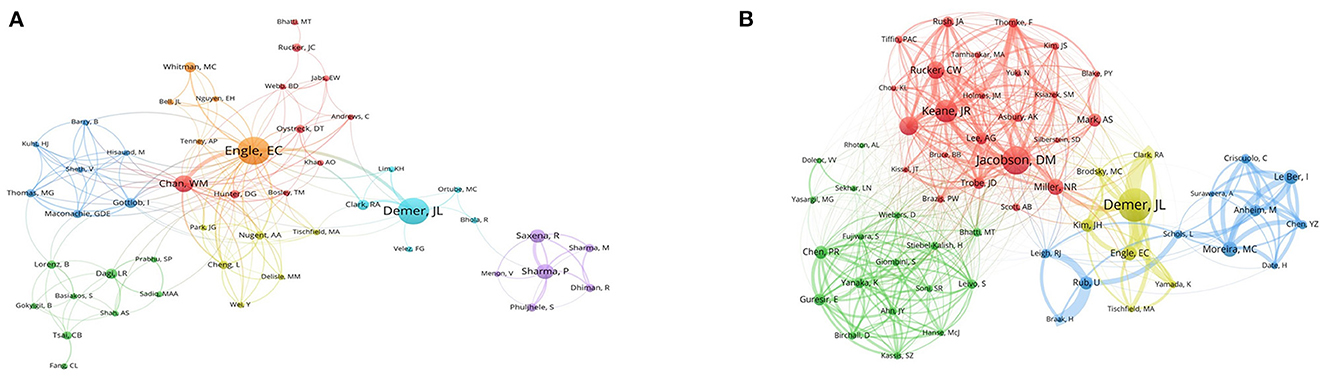
Figure 4. Author and co-cited author. (A) Network analysis of authors involved in ONP. (B) Network analysis of co-cited authors involved in ONP. The thicker the line between the institutions, the more frequently the two cooperate.
A total of 18,416 authors were cited in 1,205 publications, and we have compiled the information of the top 15 cited authors in Table 2. As can be seen from this table, three of the authors with more than 100 citations are Demer JL (n = 173), Jacobson DM (n = 146), and Keane JR (n = 118). After filtering out the authors with more than 30 citations, we used VOSviewer to visualize the contribution of these authors to the field in Figure 4B. As shown in this figure, Miller NR collaborated with the largest number of authors, while Demer JL had the most comprehensive impact.
Journals
The top 15 journals with the most publications are listed in Table 3, which shows that Journal of Neurosurgery has published the highest number of documents (n = 46), followed by World Neurosurgery (n = 42). The highest impact factor is Brain, which also has the highest number of citations and average citations, indicating its considerable impact in this field. According to the Journal Citation Report 2021, only 46.7% are located in Q1 and Q2 among the top 15 involved journals. Figure 5 visualizes the journal co-citation, revealing that Journal of Neuro-Ophthalmology's publications are cited by more journals, while Neurosurgery is the one with the highest overall weight.
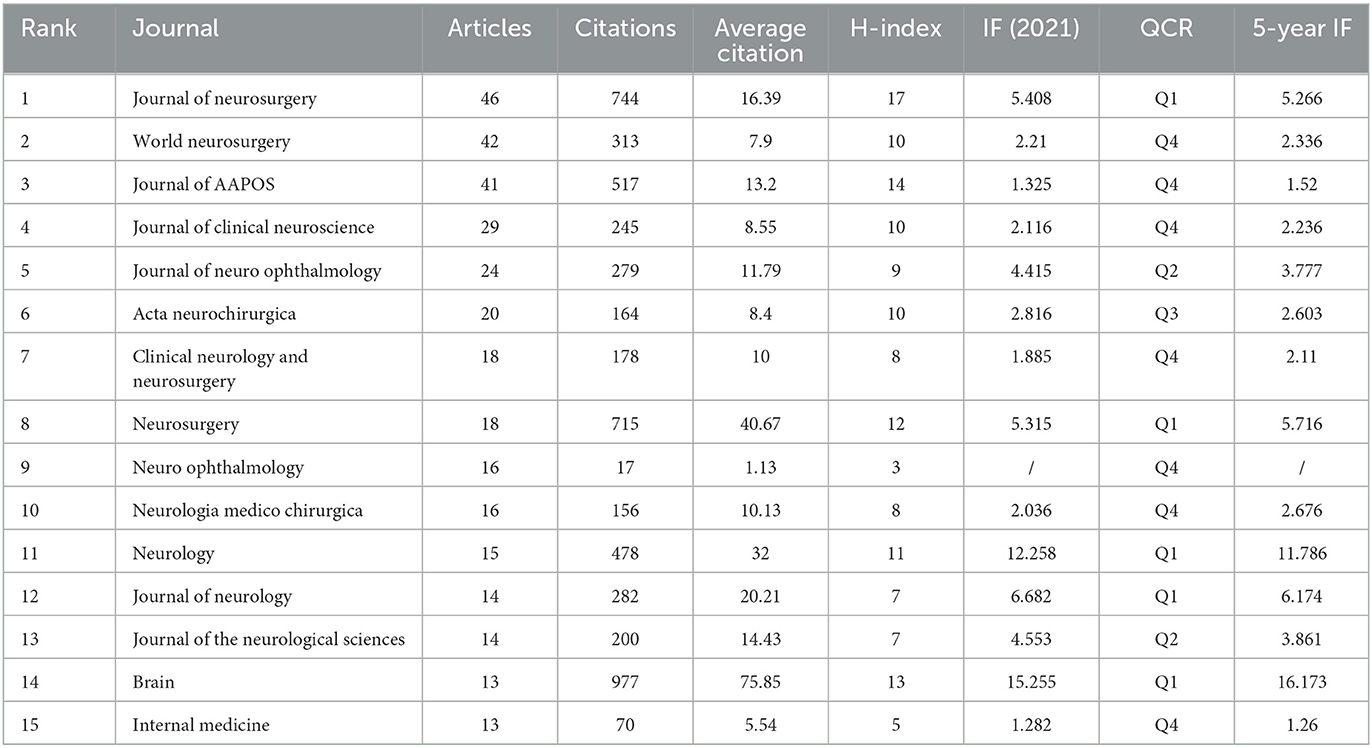
Table 3. The top 15 journals regarding the number of publications on ONP.

Figure 5. Journals. Visualized analysis of co-occurrence of journals built with VOSviewer.
Co-cited references
According to our statistics, 1,205 articles were cited by 24,720 pieces of literature. Table 4 lists the information of the top 10 cited articles. We used publications cited more than 20 times to form a co-citation network to show the citation relationships among articles. As shown in Figure 6A, the article published by Richards BW in 1992 in the American Journal of Ophthalmology is the most co-cited literature. This study has a positive relationship with other studies, which became the basis for many subsequent studies.
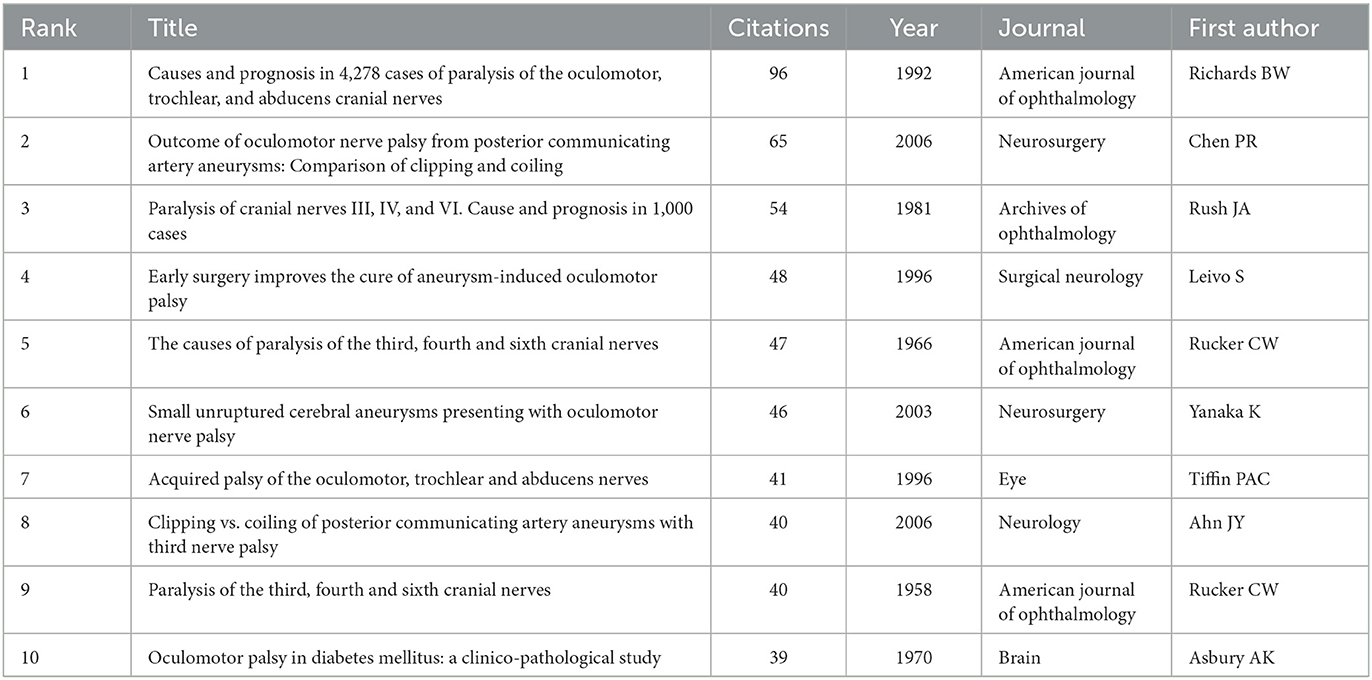
Table 4. Top 10 most cited references.
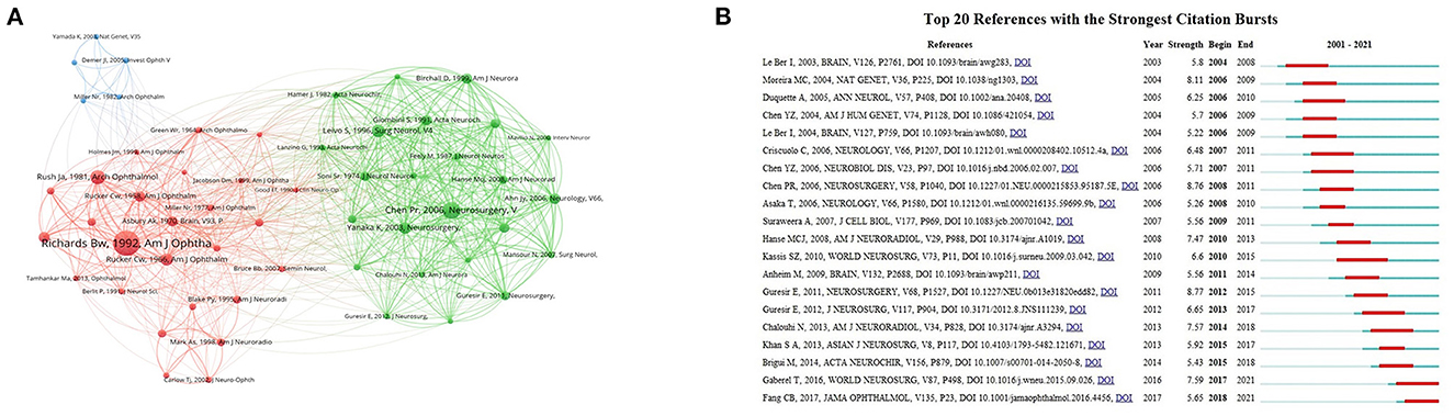
Figure 6. Co-cited references. (A) Network map of co-cited references on ONP. (B) The top 20 references with highest burst values on ONP from 2001 to 2021. The solid blue line represents the length of time the keyword appeared, and the red portion in the middle indicates the time of the reference burst.
Citation bursts can show the references that researchers follow in a certain time. As shown in Figure 6B, the top 20 references with the strongest citation bursts have burst values ranging from 5.22 to 8.77. Kassis (17), World Neurosurg has the longest burst, while Guresir (18), Neurosurgery has the highest burst value. The two most recent citation bursts are Gaberel (19), World Neurosurg and Fang et al. (20), JAMA Ophthalmol, with burst values of 7.59 and 5.65.
Keywords and hotspots
We extracted 4,401 keywords from 1,205 articles. Among these keywords, the top 10 most frequent keywords were oculomotor nerve (n = 301), oculomotor nerve palsy (n = 275), paralysis (n = 223), management (n = 96), MRI (n = 94), cranial nerve (n = 88), ophthalmoplegia (n = 71), surgery (n = 68), cavernous sinus (n = 61), and diagnosis (n = 58). After limiting the number of occurrences of keywords to more than 20, we obtained 57 keywords and plotted the co-occurrence network using VOSviewer in Figure 7A. The keywords were divided into 5 clusters. Cluster 1 (red) is mainly about clinical manifestations of ONP, such as ophthalmoplegia, diplopia, and ptosis. Cluster 2 (blue) is mainly related to aneurysms, such as intracranial and cerebral aneurysms. Cluster 3 (yellow) is mainly connected with other cerebral neurological symptoms and prognosis, such as trochlear, headache, and migraine. Cluster 4 (purple) is related to diseases with ONP as a complication, such as tumors, pituitary apoplexy, and cavernous sinus. Cluster 5 (green) is associated with other neurological disorders which may cause ONP, such as spinocerebellar ataxia, peripheral neuropathy, and gene mutations.
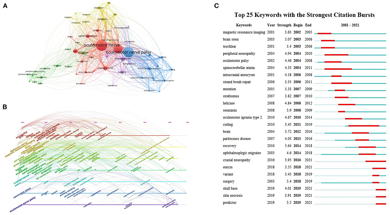
Figure 7. Keywords and hotspots. (A) Network map of ONP-related keywords co-citation. (B) Timeline map of ONP-related keywords appearance. (C) The top 25 keywords with the highest values of bursts in the ONP studies.
Using Citespace is possible to plot a timeline of keyword occurrences, and the timeline allows visualization of the stage hotspots and direction of ONP research. As shown in Figure 7B, researchers focused mainly on exploring the types of diseases associated with ONP symptoms in the first 10 years (2001–2010), with keywords including cavernous sinus, cerebral aneurysm, subarachnoid hemorrhage, pituitary adenoma, spinocerebellar ataxia, and amyotrophic lateral sclerosis. The research focus of 2011–2020 is tilted toward the treatment, prognosis, and management of ONP. The main keywords are as follows: clinical feature, predictor, risk factor, surgical technique, and complication.
Keyword bursts can further demonstrate the evolution of the field's hotspots and predict the next development trend. Figure 7C indicates that the burst value of ONP ranged from 3.35 to 5.64. “Recovery” is the keyword with the strongest emergent strength (5.64), and “coiling” has the longest outbreak time lasting a total of 8 years. Recent keywords include “skull base,” “skin necrosis,” and “predictor.”
Discussion
ONP may lead to serious morbidity or life-threatening causes, often viewed as a diagnostic concern in clinical practice (20, 21). Most of these lesions causing ONP occurred in the subarachnoid space and cavernous sinus (22, 23). There are also some untypically causes, such as schwannoma, meningioma, and radiation, may also lead to ONP (21). The ON's local anatomy is complex, its course covers a long distance, and the distribution of the oculomotor nuclei in the midbrain is more dispersed, creating a higher risk of injury to this nerve (24). The main symptoms of ONP, such as ptosis, diplopia, and difficulty with eye movements, seriously affect patients' quality of life, making it of high research value. To our knowledge, there are no patterns, literature characteristics, or bibliometric analyses of ONP. In order to better understand the research hotspots and predict future research trends on ONP, we have conducted this manuscript to provide researchers with quick access to this information.
In this study, we presented an overall review of the literature in the field of ONP from 2001 to 2021. We provided a qualitative and quantitative overview of the allocation of studies over countries, authors, and journals. Moreover, we reviewed keywords and citations involved in the literature to clarify how they have developed and evolved. In terms of the number of papers, the number of articles on ONP has been on the rise in the past 20 years and peaking in 2020, with citations increasing year by year, indicating that researchers have devoted considerable effort to ONP in recent years, and this trend will continue in the future. An analysis of institutions and countries shows that the United States publishes the most articles on ONP and has the most productive research institutions. A total of 6 of the 15 most published institutions are in the United States. These institutions are ranked at the top of the H-index and ACI, which shows the influence of the US in the ONP field. However, the node centrality representing the United States is still low (0.12). Coupled with the relatively scattered locations of institutional co-occurrence maps, research countries and institutions with absolute dominance have not yet emerged. At the same time, academic exchanges and cooperation among institutions need to be further reinforced. It is interesting to note that although Germany ranks 4th in publication numbers, its ACI is the highest, reflecting the high recognition of ONP-related research in Germany.
The institution analysis shows that the University of California Los Angeles collaborates more closely with Harvard University and Boston Children's Hospital. After further searching, we found that the primary area of collaboration between these institutions is in the molecular biology of congenital fibrosis of the extraocular muscles (CFEOM). CFEOM is a rare hereditary strabismus characterized by extraocular muscle restriction and congenital ptosis, pathologically characterized by partial or complete damage to the oculomotor nucleus and the extraocular muscles innervated by the motor nerve (25, 26). The two most cited articles in the collaborative publications of these institutions reported mutations in the KIF21A gene that causes the CFEOM1 and mutations in the TUBB3 gene that causes the CFEOM3 (27, 28). In addition, neuroimaging studies of CFEOM caused by different mutations are also the focus of collaborative studies at these institutions (26, 29). These findings have contributed significantly to the development of the molecular genetics and clinical imaging manifestations of CFEOM. Author analysis shows Professor Engle EC, based at Harvard University, was the most productive author studying the molecular genetics of specific types of strabismus that manifest as damage to the ON, abducens nerve, and talocrural nerve. Engle first performed genetic analysis and two-point linkage mapping on two autosomal dominant CFEOM families in 1994 and localized the CFEOM1 gene to the 8 centimorgans of chromosome 12 between the D12S87 and D12S85 region (30). In subsequent studies, Engle's research team defined three CFEOM phenotypes due to different gene mutations by finer genetic mapping (31–33). They investigated the structural basis of oculomotor abnormalities in different types of CFEOM by magnetic resonance imaging (MRI) (26, 29). In a 2017 published article, she revealed a role for the ephrin bidirectional signaling pathway upstream of mutant alpha2 chimeric proteins in Duane retraction syndrome (DRS), which may contribute to the selective vulnerability of abducens motor neurons (34).
The journal analysis helps researchers access information on core and refereed journals in their subject areas (35). Table 3 shows that the Journal of Neurosurgery published the most articles, but the journal with the highest number of citations and average citations was the Brain. The majority of journals shown in this table have impact factors below 5, and only 5 journals are from Q1, suggesting that the quality of articles on ONP needs to be improved. However, it may also be related to researchers focusing more on diagnostic issues of ONP rather than information such as pathological mechanisms. The most cited article analyzes the causes and prognosis in 4,278 cases of paralysis of the oculomotor, trochlear, and abducens cranial nerves written by Richards BW in 1992 (n = 96) (36) published on the American journal of ophthalmology. Rush and Younge (7), Rucker (37), and Tiffin et al. (38) have conducted clinical studies on the analysis of the causes and prognosis of ONP-related diseases. The rest of the top 10 citations focus more on the effect of surgical treatment on ONP from artery aneurysms. Surgery focuses on preventing aneurysm rupture, re-rupture risk, and mitigating the nerve damage caused by compression (39). Surgical clipping, which immediately reduces the occupancy effect, has long been the preferred treatment for aneurysm-induced ONP (18, 40), but other research shown that endovascular treatment is more likely to result in better recovery of neurological function in patients (41–43). The prevailing explanation for coiling's ability to reduce or eliminate aneurysmal pulsatility is more beneficial to nerve recovery than surgical clipping that directly separates the oculomotor nerve from the adjacent aneurysm (39, 44, 45). Citation analysis of articles is likely to be affected by length-time effects (46), so we performed a burst analysis of the co-cited literature to interpret the results. A systematic review and meta-analysis of endovascular coiling and surgical clipping for ONP, published by Gaberel et al. (19) in World Neurosurgery in 2016, has recently seen a citation explosion. Another recent outbreak article focused on the etiology of ONP, and the characteristics of developing populations were published by Fang et al. (20) in 2017. As can be seen, most of the highly cited literature types are retrospective clinical studies that provide a reliable basis for the diagnosis, treatment, and prognosis of ONP.
Cooccurrence and burst analysis of keywords can reveal the hotspot distribution and the evolution of research trends in a field (47). As shown in Figure 7A, the keyword clustering of ONP is closely related to the clinical manifestations of the disease as well as its etiology. The pathogenesis of ONP is both congenital and acquired, and its common identifiable etiologies include cerebrovascular disease, aneurysm, trauma, brain tumor, and demyelinating disease (6, 48). The timeline provides us with a more accurate picture of the changes in each cluster over time. As can be seen in Figure 7B, “infarction,” “clinical feature,” “risk factor,” “ophthalmic artery,” and “skull base” have remained hot topics of research in ONP in the last decade, suggesting that risk and diagnostic studies of ONP still promising. As vascular ischemia is the most common clinical cause and risk factor of ONP in adults, the diagnosis should be based on the exclusion of patient history, symptoms, and radiological and biological information (49). Studies have shown that MRI 3D-CISS techniques can exclude aneurysm or tumor compression, local inflammation, and infection along the oculomotor nerve, which can help clarify the clinical diagnosis and thus avoid further extensive laboratory and imaging examinations (50). Notably, the keyword “skin necrosis” appears in recent outbreaks, which may be relevant to the new etiology of ONP highlighted by recent case reports. Sung et al. (51) reported a case of progressive ONP and patchy skin necrosis after calcium hydroxylapatite filler injection for nose augmentation in 2010, while Kwon et al. (52) reported a case of sudden onset of ONP and gradually occurring skin necrosis after filling soft tissue with hyaluronic acid injections in 2013, Bae et al. (53), Kim et al. (54), and Lucaciu et al. (55) reported similar cases in the past 5 years, reminding plastic surgeons that they should be aware of these risks. In Figure 7C, the keyword bursts show the evolution of ONP research hotspots and trends. The earliest outbreak of keywords was magnetic resonance imaging (MRI), which indicates that the diagnosis of diseases related to ONP, such as aneurysms and cerebrovascular diseases in the early years were mainly relied on MRI and other traditional imaging methods. MRI can also evaluate some genetic diseases that exhibit ONP, such as CFEOM and DRS. However, in recent years, the emergence of keywords, such as “predictors,” suggests that advances in molecular biology and gene localization studies have helped us gradually understand the underlying mechanisms of these genetic diseases. Moreover, it helped us to find predictive and diagnostic methods for these genetic diseases leading to ONP. The recent increase of the keyword “cranial neuropathy” suggests that researchers are becoming more concerned about ONP caused by congenital abnormal innervation of cranial nerves.
Several limitations of our study still need to be pointed out, although this does not impact our conclusions. First, this work only selected literature in the WOSCC database where the language was restricted to English, which may have led to incomplete results. Secondly, to ensure the accuracy and completeness of data trends over time, we only analyzed papers from 2001 to 2021, which may have led to the omission of some major articles published recently.
Conclusion
In general, after analyzing 1,205 articles, we found that the U.S. institutions, authors, and the overall number of publications and citations are in the leading position, indicating the U.S.'s influence in the ONP field. Analysis of the co-cited literature shows that research on ONP in the last 20 years has focused on clinical diagnosis and the selection of surgical modalities. Clustering visualization of keywords revealed that the main causes of ONP are cerebrovascular disease, aneurysms, tumors, and demyelinating disease. However, burst analysis of keywords suggests we still need to focus on new causes of ONP and its complications during treatment.
This study provides a comprehensive and systematic overview of the current state of global ONP research over the past 20 years. The findings can provide researchers with key information in this field, such as current hotspots and expected trends, and guide them to explore potential research frontiers.
Data availability statement
The original contributions presented in the study are included in the article/supplementary material, further inquiries can be directed to the corresponding authors.
Author contributions
These articles were retrieved and downloaded by RW, YG, and SW. RW and YG were responsible for collecting data and visualizing the results. RW and XC wrote the first draft. TY and LW were provided guidance throughout the work, revising drafts, and approving the final manuscript. All authors contributed significantly to the final article.
Funding
This study was supported by the National Natural Science Foundation of China (No. 82004425), the Heilongjiang Applied Technology Research and Development Program (GA19C108), and the Heilongjiang Postdoctoral Foundation (LBH-Z21216 and LBH-Z22269).
Conflict of interest
The authors declare that the research was conducted in the absence of any commercial or financial relationships that could be construed as a potential conflict of interest.
Publisher's note
All claims expressed in this article are solely those of the authors and do not necessarily represent those of their affiliated organizations, or those of the publisher, the editors and the reviewers. Any product that may be evaluated in this article, or claim that may be made by its manufacturer, is not guaranteed or endorsed by the publisher.
References
1. Biousse V, Newman NJ. Third nerve palsies. Semin Neurol. (2000) 20:55–74. doi: 10.1055/s-2000-6833
2. Lv X, Jiang C, Li Y, Yang X, Wu Z. Isolated oculomotor nerve palsy in interventional neuroradiology. Eur J Radiol. (2010) 74:441–4. doi: 10.1016/j.ejrad.2009.04.012
3. Kung NH, Van Stavern GP. Isolated ocular motor nerve palsies. Semin Neurol. (2015) 35:539–48. doi: 10.1055/s-0035-1563568
4. Del-Rio-Vellosillo M, Garcia-Medina JJ, Pinazo-Duran MD, Abengochea-Cotaina A, Barbera-Alacreu M. Ocular motor palsy after spinal puncture. Reg Anesth Pain Med. (2017) 42:1–9. doi: 10.1097/AAP.0000000000000504
5. Newman NJ, Biousse V. Third nerve palsies-less frequent but just as concerning. JAMA Ophthalmol. (2017) 135:29–30. doi: 10.1001/jamaophthalmol.2016.4448
6. Kim T, Nam K, Kwon BS. Isolated oculomotor nerve palsy in mild traumatic brain injury. Am J Phys Med Rehabil. (2020) 99:430–5. doi: 10.1097/PHM.0000000000001316
7. Rush JA, Younge BR. Paralysis of cranial nerves III, IV, and VI. Cause and prognosis in 1,000 cases. Arch Ophthalmol. (1981) 99:76–9.
8. Singh A, Bahuguna C, Nagpal R, Kumar B. Surgical management of third nerve palsy. Oman J Ophthalmol. (2016) 9:80–6. doi: 10.4103/0974-620X.184509
9. Tokunaga M, Fukunaga K, Nakanishi R, Watanabe S, Yamanaga H. Midbrain infarction causing oculomotor nerve palsy and ipsilateral cerebellar ataxia. Intern Med. (2014) 53:2143–7. doi: 10.2169/internalmedicine.53.1613
10. Ilgen Uslu F, Özkan M. Painful ophthalmoplegia: a case report and literature review. Agriculture. (2015) 27:219–23. doi: 10.5505/agri.2015.67699
11. Raza HK, Chen H, Chansysouphanthong T, Cui G. The aetiologies of the unilateral oculomotor nerve palsy: a review of the literature. Somatosens Mot Res. (2018) 35:229–39. doi: 10.1080/08990220.2018.1547697
12. Jung EH, Kim SJ, Lee JY, Cho BJ. The incidence and etiologies of third cranial nerve palsy in koreans: a 10-year nationwide cohort study. Ophthalmic Epidemiol. (2020) 27:460–7. doi: 10.1080/09286586.2020.1773870
13. Gu J, Hu M, Gu Z, Yu J, Ji Y, Li L, et al. Bibliometric analysis reveals a 20-year research trend for chemotherapy-induced peripheral neuropathy. Front Neurol. (2021) 12:793663. doi: 10.3389/fneur.2021.793663
14. Fang LJ, Wang CY. Bibliometric analysis of studies on the treatment of hemifacial spasm. Front Neurol. (2022) 13:931551. doi: 10.3389/fneur.2022.931551
15. Chen C. Searching for intellectual turning points: progressive knowledge domain visualization. Proc Natl Acad Sci U S A. (2004) 101:5303–10. doi: 10.1073/pnas.0307513100
16. Van Eck NJ, Waltman L. Software survey: VOSviewer, a computer program for bibliometric mapping. Scientometrics. (2010) 84:523–38. doi: 10.1007/s11192-009-0146-3
17. Kassis SZ, Jouanneau E, Tahon FB, Salkine F, Perrin G, Turjman F. Recovery of third nerve palsy after endovascular treatment of posterior communicating artery aneurysms. World Neurosurg. (2010) 73:11–6; discussion e2. doi: 10.1016/j.surneu.2009.03.042
18. Güresir E, Schuss P, Setzer M, Platz J, Seifert V, Vatter H. Posterior communicating artery aneurysm-related oculomotor nerve palsy: influence of surgical and endovascular treatment on recovery: single-center series and systematic review. Neurosurgery. (2011) 68:1527–33; discussion 1533–1524. doi: 10.1227/NEU.0b013e31820edd82
19. Gaberel T, Borha A, Di Palma C, Emery E. Clipping vs. coiling in the management of posterior communicating artery aneurysms with third nerve palsy: a systematic review and meta-analysis. World Neurosurg. (2016) 87:498–506.e494. doi: 10.1016/j.wneu.2015.09.026
20. Fang C, Leavitt JA, Hodge DO, Holmes JM, Mohney BG, Chen JJ. Incidence and etiologies of acquired third nerve palsy using a population-based method. JAMA Ophthalmol. (2017) 135:23–8. doi: 10.1001/jamaophthalmol.2016.4456
21. Tian G, Kardon R, Feng C, Hong R, Sha Y, Sun X, et al. Oculomotor nerve palsy due to unusual causes. J Neuroophthalmol. (2021) 41:e244–50. doi: 10.1097/WNO.0000000000001032
22. Keane JR. Third nerve palsy: analysis of 1,400 personally-examined inpatients. Can J Neurol Sci. (2010) 37:662–70. doi: 10.1017/S0317167100010866
23. Margolin E, Freund P. Third nerve palsies: review. Int Ophthalmol Clin. (2019) 59:99–112. doi: 10.1097/IIO.0000000000000279
24. Iaconetta G, De Notaris M, Cavallo LM, Benet A, Enseñat J, Samii M, et al. The oculomotor nerve: microanatomical and endoscopic study. Neurosurgery. (2010) 66:593–601; discussion 601. doi: 10.1227/01.NEU.0000365422.36441.C8
25. Nakano M, Yamada K, Fain J, Sener EC, Selleck CJ, Awad AH, et al. Homozygous mutations in ARIX(PHOX2A) result in congenital fibrosis of the extraocular muscles type 2. Nat Genet. (2001) 29:315–20. doi: 10.1038/ng744
26. Demer JL, Clark RA, Tischfield MA, Engle EC. Evidence of an asymmetrical endophenotype in congenital fibrosis of extraocular muscles type 3 resulting from TUBB3 mutations. Invest Ophthalmol Vis Sci. (2010) 51:4600–11. doi: 10.1167/iovs.10-5438
27. Yamada K, Andrews C, Chan WM, Mckeown CA, Magli A, De Berardinis T, et al. Heterozygous mutations of the kinesin KIF21A in congenital fibrosis of the extraocular muscles type 1 (CFEOM1). Nat Genet. (2003) 35:318–21. doi: 10.1038/ng1261
28. Tischfield MA, Baris HN, Wu C, Rudolph G, Van Maldergem L, He W, et al. Human TUBB3 mutations perturb microtubule dynamics, kinesin interactions, and axon guidance. Cell. (2010) 140:74–87. doi: 10.1016/j.cell.2009.12.011
29. Demer JL, Clark RA, Engle EC. Magnetic resonance imaging evidence for widespread orbital dysinnervation in congenital fibrosis of extraocular muscles due to mutations in KIF21A. Invest Ophthalmol Vis Sci. (2005) 46:530–9. doi: 10.1167/iovs.04-1125
30. Engle EC, Kunkel LM, Specht LA, Beggs AH. Mapping a gene for congenital fibrosis of the extraocular muscles to the centromeric region of chromosome 12. Nat Genet. (1994) 7:69–73.
31. Wang SM, Zwaan J, Mullaney PB, Jabak MH, Al-Awad A, Beggs AH, et al. Congenital fibrosis of the extraocular muscles type 2, an inherited exotropic strabismus fixus, maps to distal 11q13. Am J Hum Genet. (1998)63:517–25.
32. Doherty EJ, Macy ME, Wang SM, Dykeman CP, Melanson MT, Engle EC. CFEOM3: a new extraocular congenital fibrosis syndrome that maps to 16q24.2-q24.3. Invest Ophthalmol Vis Sci. (1999) 40:1687–94.
33. Engle EC, Mcintosh N, Yamada K, Lee BA, Johnson R, O'keefe M, et al. CFEOM1, the classic familial form of congenital fibrosis of the extraocular muscles, is genetically heterogeneous but does not result from mutations in ARIX. BMC Genet. (2002) 3:3. doi: 10.1186/1471-2156-3-3
34. Nugent AA, Park JG, Wei Y, Tenney AP, Gilette NM, Delisle MM, et al. Mutant α2-chimaerin signals via bidirectional ephrin pathways in Duane retraction syndrome. J Clin Invest. (2017) 127:1664–82. doi: 10.1172/JCI88502
35. Chen D, Zhang G, Wang J, Chen S, Wang J, Nie H, et al. Mapping trends in moyamoya angiopathy research: a 10-year bibliometric and visualization-based analyses of the web of science core collection (WoSCC). Front Neurol. (2021) 12:637310. doi: 10.3389/fneur.2021.637310
36. Richards BW, Jones FR, Younge BR. Causes and prognosis in 4,278 cases of paralysis of the oculomotor, trochlear, and abducens cranial nerves. Am J Ophthalmol. (1992) 113:489–96.
37. Rucker CW. The causes of paralysis of the third, fourth and sixth cranial nerves. Am J Ophthalmol. (1966) 61:1293–8.
38. Tiffin PA, Macewen CJ, Craig EA, Clayton G. Acquired palsy of the oculomotor, trochlear and abducens nerves. Eye. (1996) 10(Pt 3):377–84.
39. Chalouhi N, Theofanis T, Jabbour P, Dumont AS, Gonzalez LF, Starke RM, et al. Endovascular treatment of posterior communicating artery aneurysms with oculomotor nerve palsy: clinical outcomes and predictors of nerve recovery. AJNR Am J Neuroradiol. (2013) 34:828–32. doi: 10.3174/ajnr.A3294
40. Chen PR, Amin-Hanjani S, Albuquerque FC, Mcdougall C, Zabramski JM, Spetzler RF. Outcome of oculomotor nerve palsy from posterior communicating artery aneurysms: comparison of clipping and coiling. Neurosurgery. (2006) 58:1040–6; discussion 1040–1046. doi: 10.1227/01.NEU.0000215853.95187.5E
41. Mansour N, Kamel MH, Kelleher M, Aquilina K, Thornton J, Brennan P, et al. Resolution of cranial nerve paresis after endovascular management of cerebral aneurysms. Surg. Neurol. (2007) 68:500–4; discussion 504. doi: 10.1016/j.surneu.2006.12.061
42. Zhang SH, Pei W, Cai XS, Cheng G. Endovascular management and recovery from oculomotor nerve palsy associated with aneurysms of the posterior communicating artery. World Neurosurg. (2010) 74:316–9. doi: 10.1016/j.wneu.2010.05.036
43. Ko JH, Kim YJ. Oculomotor nerve palsy caused by posterior communicating artery aneurysm: evaluation of symptoms after endovascular treatment. Interv Neuroradiol. (2011) 17:415–9. doi: 10.1177/159101991101700403
44. Ahn JY, Han IB, Yoon PH, Kim SH, Kim NK, Kim S, et al. Clipping vs. coiling of posterior communicating artery aneurysms with third nerve palsy. Neurology. (2006) 66:121–3. doi: 10.1212/01.wnl.0000191398.76450.c4
45. Albuquerque FC. Coiling vs. clipping for posterior communicating artery aneurysms associated with oculomotor nerve palsy: only time will tell. World Neurosurg. (2010) 74:250–1. doi: 10.1016/j.wneu.2010.06.024
46. Ismail I, Saqr M. A quantitative synthesis of eight decades of global multiple sclerosis research using bibliometrics. Front Neurol. (2022) 13:845539. doi: 10.3389/fneur.2022.845539
47. Zhu G, Fu Z, Su S, Tang Y, Liu F, Yu W. Global trends and hotspots in trigeminal neuralgia research from 2001 to 2021: a bibliometric analysis. Front Neurol. (2022) 13:894006. doi: 10.3389/fneur.2022.894006
48. Wei H, Yin H, Huang M, Guo Z. The 2019 novel cornoavirus pneumonia with onset of oculomotor nerve palsy: a case study. J Neurol. (2020) 267:1550–3. doi: 10.1007/s00415-020-09773-9
49. Hung CH, Chang KH, Chu CC, Liao MF, Chang HS, Lyu RK, et al. Painful ophthalmoplegia with normal cranial imaging. BMC Neurol. (2014) 14:7. doi: 10.1186/1471-2377-14-7
50. Roques M, Biotti D, Darcourt J, Adam G, Varenne F, Bonneville F. Microvascular ischemic isolated oculomotor nerve palsy revealed by contrast-enhanced 3D-CISS imaging. J Neuroradiol. (2021) 48:466–7. doi: 10.1016/j.neurad.2021.01.001
51. Sung MS, Kim HG, Woo KI, Kim YD. Ocular ischemia and ischemic oculomotor nerve palsy after vascular embolization of injectable calcium hydroxylapatite filler. Ophthalmic Plast Reconstr Surg. (2010) 26:289–91. doi: 10.1097/IOP.0b013e3181bd4341
52. Kwon SG, Hong JW, Roh TS, Kim YS, Rah DK, Kim SS. Ischemic oculomotor nerve palsy and skin necrosis caused by vascular embolization after hyaluronic acid filler injection: a case report. Ann Plast Surg. (2013) 71:333–4. doi: 10.1097/SAP.0b013e31824f21da
53. Bae IH, Kim MS, Choi H, Na CH, Shin BS. Ischemic oculomotor nerve palsy due to hyaluronic acid filler injection. J Cosmet Dermatol. (2018) 17:1016–8. doi: 10.1111/jocd.12498
54. Kim BJ, You HJ, Jung I, Kim DW. Ophthalmoplegia with skin necrosis after a hyaluronic acid filler injection. J Cosmet Dermatol. (2020) 19:1307–10. doi: 10.1111/jocd.13403
Keywords: oculomotor nerve paralysis, bibliometric analysis, Citespace, VOSviewer, web of science
Citation: Wang R, Gao Y, Wu S, Cai X, Yu T and Wang L (2023) Research hotspots and trends of oculomotor nerve palsy from 2001 to 2021 based on web of science: A bibliometric analysis. Front. Neurol. 14:1112070. doi: 10.3389/fneur.2023.1112070
Received: 30 November 2022; Accepted: 02 February 2023;
Published: 22 February 2023.
Edited by:
Catherine Vignal Clermont, Fondation Ophtalmologique Adolphe de Rothschild, FranceReviewed by:
Shan-Qiang Zhang, Shantou University, ChinaTong Liu, Guangdong Second Hospital of Traditional Chinese Medicine, China
Copyright © 2023 Wang, Gao, Wu, Cai, Yu and Wang. This is an open-access article distributed under the terms of the Creative Commons Attribution License (CC BY). The use, distribution or reproduction in other forums is permitted, provided the original author(s) and the copyright owner(s) are credited and that the original publication in this journal is cited, in accordance with accepted academic practice. No use, distribution or reproduction is permitted which does not comply with these terms.
*Correspondence: TianYang Yu,  dHl5dV90Y21AMTYzLmNvbQ==; Liyuan Wang, d2FuZ2xpeXVhbjE5ODkxMDI5QDE2My5jb20=
†These authors have contributed equally to this work and share first authorship