 Yu Wang1,2,3
 Sinan Wang
 Xuan Zhou
- 1Department of Maxillofacial and Otorhinolaryngological Oncology, Tianjin Medical University Cancer Institute and Hospital, Tianjin, China
- 2Key Laboratory of Cancer Prevention and Therapy, Tianjin Cancer Institute, Tianjin, China
- 3National Clinical Research Center of Cancer, Tianjin, China
- 4Department of Gastroenterology and Hepatology, Tianjin Medical University General Hospital, Tianjin, China
- 5Tianjin Gastroenterology and Hepatology Institute, Tianjin Medical University, Tianjin, China
- 6Tianjin Research Center of Basic Medical Science, Tianjin Medical University, Tianjin, China
Head and neck squamous cell carcinoma (HNSCC) is the sixth most common type of human malignancy. For decades, research into HNSCC invasion and metastasis has been dedicated to the study of protein-coding genes. Along with whole-genome and transcriptome sequencing development, long non-coding RNA (lncRNA) has attracted greater attention. Compelling evidence has proven the critical role of lncRNAs in the occurrence and development of HNSCC by means of epigenetic modifications, regulation of gene transcription, and post-transcription level. More importantly, crosstalk between lncRNAs and microRNAs was recently proven to regulate HNSCC metastasis through EMT modification. Based on these, this review summarizes the critical roles of lncRNAs in HNSCC metastasis and the crosstalk between lncRNAs and microRNAs as well as the detailed regulatory mechanism of the interaction. Thus, a deeper understanding of the lncRNA network in cancer metastasis is finally uncovered in order to provide a rationale and innovative concepts toward new therapeutic strategies for the highly metastatic HNSCC.
Introduction
Head and neck squamous cell carcinoma (HNSCC) is the sixth most common type of human malignancy and involves carcinoma of several anatomic sites, such as lip, oral cavity, pharynx (nasopharynx, oropharynx, hypopharynx), and larynx, with an annual incidence of ~500,000 (1). Even through systemic therapeutic strategies have developed, the 5-years overall survival (OS) of HNSCC patients is hardly satisfying (2). Evidence has shown that the relatively poor prognosis and high recurrence of HNSCC are mainly due to the high rate of local invasion and distant metastasis (3). Consequently, it is essential to explore the detailed molecular mechanisms involved in cancer metastatic cascade so as to promote the development of target therapy and improve the overall survival of HNSCC.
Experimental and clinical studies have attempted to establish the biological basis of this metastasis cascade. Mountains of evidence highlight the irreplaceable role of long non-coding RNA (lncRNA) in cancer metastasis, including HNSCC (4, 5). Such transcripts are widely validated not to produce functional proteins, but regulate gene expression at multiple levels and participate in cancer evolution and development (6). More importantly, unique cross-regulation between lncRNA and miRNA was recently mentioned, and emerging evidence shows that such crosstalk has a great effect on human cancer metastasis, partially through EMT regulation (7). In this review, we summarize the correlation between lncRNA and EMT mediation and highlight the leading role of lncRNA/miRNA crosstalk in the metastasis of HNSCC.
LncRNAs Involved in HNSCC Invasion and Metastasis
LncRNAs are a heterogenous group of RNAs containing more than 200 nucleotides and recently involved in many biological processes (8). Their number is significantly larger than that of protein-coding genes and can act in a cis and/or in trans manner during the development of human cancers (9). They have well-defined subcellular sites, mainly concentrated in the nucleus and involved in the regulation of chromatin and chromosomal conformation (10).
In the past few years, there has been a paradigm shift in the understanding of non-coding RNAs (ncRNAs) and their role in cancer biology (11, 12). Due to the discovery of alternative splicing in 1970s, a major focus in various pathological and physiological processes shifted to the role of proteins and protein-coding RNAs and mutations as prominent mechanisms in disease etiology and pathophysiology. However, in 1977, the discovery of introns and ribozymes suggested the role of ncRNA as a regulatory molecule (13). Since then, more and more research focuses on lncRNAs, which are identified as playing critical roles in cancer metastasis regulation. LncRNAs are not only responsible for tumor proliferation, cell death regulation, and angiogenesis (14–16), but also for the invasion and metastasis of HNSCC (17–19). These lncRNAs described as potential transfer regulators are shown in Table 1.
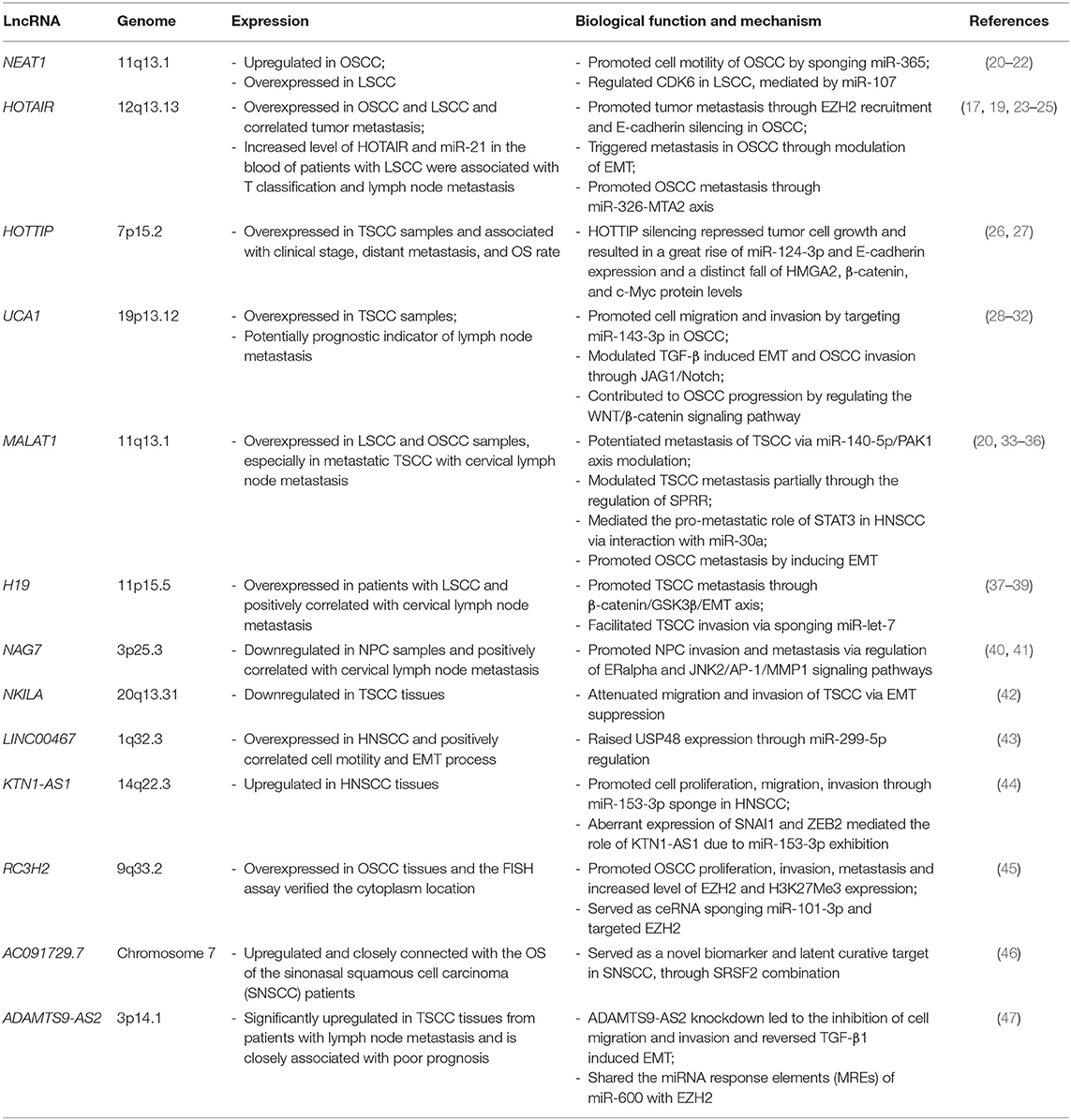
Table 1. lncRNAs in HNSCC metastasis.
LncRNA HOTAIR
LncRNA HOX transcript antisense RNA (HOTAIR) is one of the most well-studied oncogenic lncRNAs, originally characterized as a regulator of the HOX gene family, helping to control cellular identity (48). The 5′ terminal of HOTAIR can be combined with chromatin modified complex, and the 3′ terminal can bind to histone demethylase I complex. Therefore, HOTAIR regulates methylation or demethylation of H3K4me2 at the H3K27 site, which is involved in proliferation (49), apoptosis (50), and metastasis (51) of tumor cells. It was found that the expression of HOTAIR is increased in many subtypes of HNSCC. Compared with the normal oral epithelial cell lines, HOTAIR in OSCC Cal-27 and UM-1 cell lines increased significantly (52). In addition, in vitro experiments demonstrate that, compared with low invasiveness, HOTAIR in the invasive oral squamous cell carcinoma cell lines is upregulated. Moreover, knocking down HOTAIR expression levels globally inhibits cell proliferation, migration, and invasion (53). Furthermore, HOTAIR is confirmed to promote HNSCC invasion and metastasis (23) and triggers the EMT process through EZH2/H3K27me3 recruitment, which is proven to be negatively associated with clinical outcomes in HNSCC patients (54).
LncRNA UCA1
In tongue squamous cell carcinoma (TSCC), the expression level of lncRNA urothelial carcinoma antigen 1 (UCA1) is significantly increased and correlated with lymph node metastasis. In addition, the expression of UCA1 in lymph node metastasis is higher than that in the primary tumor. In a cell culture of TSCC, the overexpression of UCA1 promotes cell migration but has little effect on cell proliferation (32). Furthermore, UCA1 is also revealed to attenuate cell growth and metastasis of OSCC cell lines in vitro and in vivo, through WNT/β-catenin activation (28). Consequently, it is suggested that UCA1 may promote the metastasis of cancer cells and may be a prognostic indicator of lymph node metastasis in HNSCC.
LncRNA MALAT1
Abnormal expression of lncRNA metastasis–associated lung adenocarcinoma transcript 1 (MALAT1) is reported in multiple human cancers, including prostate cancer (55), colorectal cancer (56), hepatocellular carcinoma (57), and HNSCC (33, 34). Fang et al. reported globally increased MALAT1 expression levels in TSCC (32), especially in those with lymph node metastasis (35). DNA microarray analysis shows that MALAT1 significantly increases TSCC cell motility through regulating LAYN, CCT4, CTHRC1, and FHL1 expression levels, which are small proline-rich protein (SPRR) members (35). In parallel, Zhou and colleagues reveal that MALAT1 is significantly associated with poor prognosis in patients with OSCC and could promote invasion and metastasis of OSCC by means of EMT activation (34). In addition, by means of ChIP-PCR and RIP-PCR analysis, we also reveal that STAT3 may accelerate EMT progression and cancer metastasis through interaction with the MALAT1/miR-30a axis (33).
LncRNA H19
LncRNA H19 is described as participating in the metastasis of various cancers. In TSCC, H19 is demonstrated to be upregulated in the tumor tissue compared with adjacent samples. Furthermore, the expression level of H19 in metastatic tumor is significantly higher than in non-metastatic tumor. Subsequently, H19 is demonstrated to function as ceRNA to sponge let-7a, resulting in HMGA2 enhancement, and finally, increasing the capacity of TSCC invasion and metastasis (37). H19 is also found to be overexpressed in nasopharyngeal carcinoma (NPC) and to promote NPC cell invasion capacity via E-cadherin silencing and miR-630/EZH2 regulation (58). Mechanistically, Wu T. and colleagues report that H19 is overexpressed in laryngeal squamous cell carcinoma (LSCC) and accelerates LSCC tumor progression through miR-148a-3p attenuation and DNMT1 enhancement (39).
LncRNAs Regulate HNSCC Cell Motility via EMT Mediation
The main regulating factors of EMT include the EMT effect factor, EMT core regulating factor, and EMT induction factor (59). EMT effect factors are usually proteins that define epithelial or mesenchymal properties, such as E-cadherin, α- Catenin, γ-Catenin, Vim, and Fibronectin, which promote cell migration and invasion during EMT. Among them, E-cadherin is considered to be the leading force (60). The core regulatory factors of EMT are composed of transcription factors, including Snail-1, Snail-2, ZEB1, ZEB2, Twist-1, and Twist-2 as well as the newly discovered pair-related homeobox transcription factor 1 (Prrx1), which regulates the EMT process through E-cadherin mediation (60). Moreover, EMT inducers consist of several signaling pathways, including TGF-β/Smad, Wnt/β-catenin, Notch, and GF receptor signaling cascade. Most importantly, the TGF-β/Smad signaling pathway appears to be the major activator of EMT. In addition, tumor inflammation, and the hypoxia microenvironment also serve basic roles in EMT promotion.
Recently, it is confirmed that EMT is also regulated by post transcription factor lncRNA, which plays its role through regulatory effectors, transcription factors, and signal transduction pathways (61). Unlike microRNAs, which repress target gene expression levels post-transcriptionally, functional lncRNAs may influence the EMT process during cancer metastasis by regulating gene expression at different levels, including chromatin modification, transcription, and post-transcriptional processing (Figure 1). MALAT1 is widely confirmed as one of the most well-studied oncogenic lncRNAs that is confirmed to be involved in the EMT process. Fan Y. and colleagues illustrate that TGF-β overexpression in the tumor microenvironment could induce cancer metastasis through EMT regulation and validate MALAT1 as an important mediator of TGF-β related EMT (62). Mechanistically, MALAT1 is then proven to promote EMT through Suz12 recruitment, which acts as an H3K27 methyltransferase binding E-cadherin promoter and inhibiting its expression in a PRC2-dependent manner. Moreover, subsequent ChIP-PCR and luciferase reporter assays show that STAT3 might bind to the MALAT1 promoter region and transcriptionally activate its expression in order to induce EMT and accelerate HNSCC metastasis (33). In OSCC, MALAT1 is also reported to play oncogenic roles in EMT-related cancer metastasis. By means of siRNA, MALAT1 is validated to be required for maintaining EMT-mediated cell migration and invasion. MALAT1 knockdown significantly suppressed the expression levels of N-cadherin and Vimentin, but raised E-cadherin in vitro. Meanwhile, both cytoplasm and the nucleus NF-κB/β-catenin axis is significantly triggered after MALAT1 elevation. It is noteworthy that targeting MALAT1 globally inhibits the proliferation capacity of TSCCA-induced xenograft tumor, suggesting MALAT1 as an important prognostic factor of OSCC and a satisfactory target with therapeutic potential. Furthermore, MALAT1 also acts as a transcriptional regulator within the regulation of activating the Wnt/β-catenin signaling pathway (63). In addition to MALAT1, there are other lncRNAs proven to participate in EMT regulation, such as HOTAIR and H19. In OSCC, a significant negative correlation between HOTAIR and E-cadherin expression levels is found in both tumor tissues and cell lines. Meanwhile, HOTAIR is validated to trigger E-cadherin silencing through the recruitment of EZH2 and H3K27me3 in the promoter region of E-cadherin (23), indicating that HOTAIR might regulate OSCC metastasis in an epigenetic manner. On the other hand, compared with matched normal tissues, the expression of H19 is upregulated in TSCC specimens and significantly correlated with lymph node metastasis. Subsequently, H19 attenuation significantly suppresses cell motility in vitro through activation of β-Catenin/GSK3β/E-cadherin signaling. In addition, animal models show that H19 inhibition significantly impairs tumor progression and lung metastasis (38). Apart from those prometastatic lncRNAs, lncRNA NKILA is validated to inhibit the migration and invasion of OSCC (42). Mechanistic study shows that NKILA inhibits the phosphorylation of IκBα and NF-κB activation as well as the induction of the EMT process. An in vivo experimental metastasis model also demonstrates that NKILA inhibits lung metastasis of NOD/SCID mice with TSCC tumors, suggesting NKILA as a potential predictor for OS and distant metastasis in patients with TSCC.
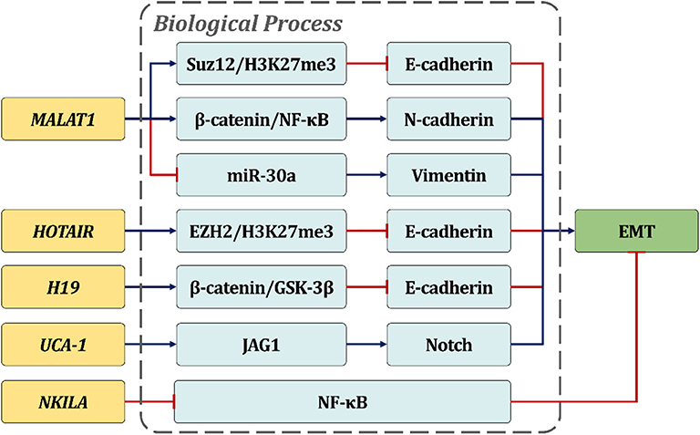
Figure 1. LncRNAs regulate HNSCC cell motility via EMT mediation. The text in the boxes depicts the potential pro/anti-EMT mechanisms of lncRNAs in HNSCC.
LncRNA/Microrna Interaction in HNSCC Metastasis
During ncRNA crosstalk, on the one hand, the stability of lncRNA can be affected due to coaction with specific miRNA. On the other hand, lncRNA, also known as competitive endogenous RNA, could bind certain miRNAs to isolate the miRNA from its target mRNA, thereby antagonizing miRNA's function (Figure 2). The tumor suppressor miR-217 is reported to inhibit MALAT1 through the Ago2-mediated pathway in order to inhibit EMT-related metastasis through upregulating E-cadherin and N-cadherin suppression (64). Similarly, the recruitment of miR-30a also reduces the stability of MALAT1 in HNSCC in order to inhibit the invasion capacity of tumor cells (33). Additionally, MALAT1 knockdown is also seen to completely suppress tumor progression through miR-140-5p elevation and PAK1 inhibition, both in vitro and in TSCC-induced xenograft tumors (36). Another lncRNA H19 is also widely mentioned in lncRNA/miRNA interaction. Kou N. and colleagues illustrate that H19 can act as ce-RNA to sponge let-7a, leading to the accumulation of metastasis regulator HMGA2, which is enriched in TSCC tissues and cell lines. Intriguingly, let-7a suppression significantly rescues the weakened tumor cell motility induced by sh-H19. These findings demonstrate that the H19/let-7a crosstalk plays a critical role in TSCC migration and invasion (37). Meanwhile, H19 is validated to regulate EZH2 by miR-630 silencing, which is a repressor of EZH2 and interacts with H19 in a sequence-specific manner, to inhibit the expression level of E-cadherin and eventually accelerate the invasion and metastasis of NPC (58). Other examples involving lncRNA ZFAS1 (65) and lncRNA UCA1 (29, 30) can also function as ceRNA during EMT-related cancer metastasis.
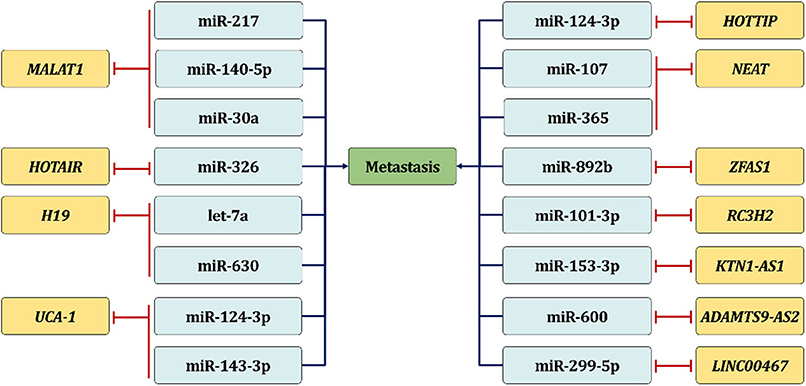
Figure 2. LncRNA/microRNA interaction in HNSCC metastasis. LncRNA can serve as ceRNA in order to bind certain miRNA to isolate the miRNA from its target mRNA, thereby promoting HNSCC metastasis.
Perspectives
LncRNA is constitutively deregulated during the progression and development of human cancers and globally suggested as a critical regulator in tumor cell motility. At present, the understanding of lncRNA in HNSCC metastasis remains confused and ambiguous, and there is little information about the function of lncRNA in HNSCC, which needs further research. This review provides a comprehensive study of the expression profile of lncRNA in HNSCC and summarizes the control of ncRNA crosstalk on the EMT process, emphasizing the leading influence of lncRNA crosstalk in the metastasis of HNSCC. Even so, a great deal of work is still urgently required to characterize the complex ncRNA networks that contribute to HNSCC metastasis, and it is necessary to carry out further research to clarify the relationship between lncRNA and miRNA in order to seek better treatment strategies.
Consent for Publication
All authors agreed to the publication of this review.
Author Contributions
YW and SW contributed to conception, drafting, interpretation, and manuscript revision. XZ and YR contributed to interpretation and manuscript revision. All authors provided final approval and agreed to be accountable for all aspects of the study.
Funding
This work was supported by the China National Natural Scientific Fund No. 81872206 (XZ); the Tianjin Municipal Education Commission Support Grant No. 2019KJ188 (YW); and the China National Natural Scientific Fund No. 81872495 (YR).
Conflict of Interest
The authors declare that the research was conducted in the absence of any commercial or financial relationships that could be construed as a potential conflict of interest.
References
1. Egger G, Liang G, Aparicio A, Jones PA. Epigenetics in human disease and prospects for epigenetic therapy. Nature. (2004) 429:457–63. doi: 10.1038/nature02625
2. Lam L, Logan RM, Luke C. Epidemiological analysis of tongue cancer in South Australia for the 24-year period, 1977-2001. Austr Dental J. (2006) 51:16–22. doi: 10.1111/j.1834-7819.2006.tb00395.x
3. Bhave SL, Teknos TN, Pan Q. Molecular parameters of head and neck cancer metastasis. Crit Rev Eukaryot Gene Expr. (2011) 21:143–53. doi: 10.1615/CritRevEukarGeneExpr.v21.i2.40
4. Guglas K, Bogaczynska M, Kolenda T, Rys M, Teresiak A, Blizniak R, et al. lncRNA in HNSCC: challenges and potential. Contemp Oncol. (2017) 21:259–66. doi: 10.5114/wo.2017.72382
5. Cao W, Liu JN, Liu Z, Wang X, Han ZG, Ji T, et al. A three-lncRNA signature derived from the Atlas of ncRNA in cancer (TANRIC) database predicts the survival of patients with head and neck squamous cell carcinoma. Oral Oncol. (2017) 65:94–101. doi: 10.1016/j.oraloncology.2016.12.017
6. Sanchez Calle A, Kawamura Y, Yamamoto Y, Takeshita F, Ochiya T. Emerging roles of long non-coding RNA in cancer. Cancer Sci. (2018) 109:2093–100. doi: 10.1111/cas.13642
7. Liz J, Esteller M. lncRNAs and microRNAs with a role in cancer development. Biochim Biophys Acta. (2016) 1859:169–76. doi: 10.1016/j.bbagrm.2015.06.015
8. Djebali S, Davis CA, Merkel A, Dobin A, Lassmann T, Mortazavi A, et al. Landscape of transcription in human cells. Nature. (2012) 489:101–8. doi: 10.1038/nature11233
9. Zhang R, Xia LQ, Lu WW, Zhang J, Zhu JS. LncRNAs and cancer. Oncol Lett. (2016) 12:1233–9. doi: 10.3892/ol.2016.4770
10. Khalil AM, Guttman M, Huarte M, Garber M, Raj A, Rivea Morales D, et al. Many human large intergenic non-coding RNAs associate with chromatin-modifying complexes and affect gene expression. Proc Natl Acad Sci USA. (2009) 106:11667–72. doi: 10.1073/pnas.0904715106
11. Salyakina D, Tsinoremas NF. Non-coding RNAs profiling in head and neck cancers. NPJ Genomic Med. (2016) 1:15004. doi: 10.1038/npjgenmed.2015.4
12. Manikandan M, Deva Magendhra Rao AK, Arunkumar G, Manickavasagam M, Rajkumar KS, Rajaraman R, et al. Oral squamous cell carcinoma: microRNA expression profiling and integrative analyses for elucidation of tumourigenesis mechanism. Mol Cancer. (2016) 15:28. doi: 10.1186/s12943-016-0512-8
13. Morris KV, Mattick JS. The rise of regulatory RNA. Nat Rev Genet. (2014) 15:423–37. doi: 10.1038/nrg3722
14. Ding L, Ren J, Zhang D, Li Y, Huang X, Hu Q, et al. A novel stromal lncRNA signature reprograms fibroblasts to promote the growth of oral squamous cell carcinoma via LncRNA-CAF/interleukin-33. Carcinogenesis. (2018) 39:397–406. doi: 10.1093/carcin/bgy006
15. Ma WQ, Chen J, Fang W, Yang XQ, Zhu A, Zhang D, et al. LncRNA INHBA-AS1 promotes cell growth, migration, and invasion of oral squamous cell carcinoma by sponging miR-143-3p. Eur Rev Med Pharm Sci. (2020) 24:1821–8. doi: 10.26355/eurrev_202002_20360
16. Zhang P, Liu Y, Li C, Zhang L, Liu Q, Jiang T. LncRNA PAPAS promotes oral squamous cell carcinoma by upregulating transforming growth factor-beta1. J Cell Biochem. (2019) 120:16120–7. doi: 10.1002/jcb.28893
17. Tao D, Zhang Z, Liu X, Zhang Z, Fu Y, Zhang P, et al. LncRNA HOTAIR promotes the invasion and metastasis of oral squamous cell carcinoma through metastasis-associated gene 2. Mol Carcinogen. (2020) 59:353–64. doi: 10.1002/mc.23159
18. Wang Y, Zhang X, Wang Z, Hu Q, Wu J, Li Y, et al. LncRNA-p23154 promotes the invasion-metastasis potential of oral squamous cell carcinoma by regulating Glut1-mediated glycolysis. Cancer Lett. (2018) 434:172–83. doi: 10.1016/j.canlet.2018.07.016
19. Lu MY, Liao YW, Chen PY, Hsieh PL, Fang CY, Wu CY, et al. Targeting LncRNA HOTAIR suppresses cancer stemness and metastasis in oral carcinomas stem cells through modulation of EMT. Oncotarget. (2017) 8:98542–52. doi: 10.18632/oncotarget.21614
20. Tang H, Wu Z, Zhang J, Su B. Salivary lncRNA as a potential marker for oral squamous cell carcinoma diagnosis. Mol Med Rep. (2013) 7:761–6. doi: 10.3892/mmr.2012.1254
21. Zou AE, Ku J, Honda TK, Yu V, Kuo SZ, Zheng H, et al. Transcriptome sequencing uncovers novel long non-coding and small nucleolar RNAs dysregulated in head and neck squamous cell carcinoma. RNA. (2015) 21:1122–34. doi: 10.1261/rna.049262.114
22. Wang P, Wu T, Zhou H, Jin Q, He G, Yu H, et al. Long non-coding RNA NEAT1 promotes laryngeal squamous cell cancer through regulating miR-107/CDK6 pathway. J Exp Clin Cancer Res. (2016) 35:22. doi: 10.1186/s13046-016-0297-z
23. Wu Y, Zhang L, Zhang L, Wang Y, Li H, Ren X, et al. Long non-coding RNA HOTAIR promotes tumor cell invasion and metastasis by recruiting EZH2 and repressing E-cadherin in oral squamous cell carcinoma. Int J ogy. (2015) 46:2586–94. doi: 10.3892/ijo.2015.2976
24. Wang J, Zhou Y, Lu J, Sun Y, Xiao H, Liu M, et al. Combined detection of serum exosomal miR-21 and HOTAIR as diagnostic and prognostic biomarkers for laryngeal squamous cell carcinoma. Med Oncol. (2014) 31:148. doi: 10.1007/s12032-014-0148-8
25. Nie Y, Liu X, Qu S, Song E, Zou H, Gong C. Long non-coding RNA HOTAIR is an independent prognostic marker for nasopharyngeal carcinoma progression and survival. Cancer Sci. (2013) 104:458–64. doi: 10.1111/cas.12092
26. Zhang H, Zhao L, Wang YX, Xi M, Liu SL, Luo LL. Long non-coding RNA HOTTIP is correlated with progression and prognosis in tongue squamous cell carcinoma. Tumour Biol. (2015) 36:8805–9. doi: 10.1007/s13277-015-3645-2
27. Xiong L, Tang Y, Tang J, Liu Z, Wang X. Downregulation of lncRNA HOTTIP suppresses the proliferation, migration, and invasion of oral tongue squamous cell carcinoma by regulation of HMGA2-mediated Wnt/beta-catenin pathway. Cancer Biotherap Radiopharmac. (2020). doi: 10.1089/cbr.2019.3017. [Epub ahead of print].
28. Yang YT, Wang YF, Lai JY, Shen SY, Wang F, Kong J, et al. Long non-coding RNA UCA1 contributes to the progression of oral squamous cell carcinoma by regulating the WNT/beta-catenin signaling pathway. Cancer Sci. (2016) 107:1581–9. doi: 10.1111/cas.13058
29. Zhang TH, Liang LZ, Liu XL, Wu JN, Su K, Chen JY, et al. LncRNA UCA1/miR-124 axis modulates TGFbeta1-induced epithelial-mesenchymal transition and invasion of tongue cancer cells through JAG1/Notch signaling. J Cell Biochem. (2019) 120:10495–504. doi: 10.1002/jcb.28334
30. Duan Q, Xu M, Wu M, Zhang X, Gan M, Jiang H. Long non-coding RNA UCA1 promotes cell growth, migration, and invasion by targeting miR-143-3p in oral squamous cell carcinoma. Cancer Med. (2020) 9:3115–29. doi: 10.1002/cam4.2808
31. Han R, Chen S, Wang J, Zhao Y, Li G. LncRNA UCA1 affects epithelial-mesenchymal transition, invasion, migration and apoptosis of nasopharyngeal carcinoma cells. Cell Cycle. (2019) 18:3044–53. doi: 10.1080/15384101.2019.1667707
32. Fang Z, Wu L, Wang L, Yang Y, Meng Y, Yang H. Increased expression of the long non-coding RNA UCA1 in tongue squamous cell carcinomas: a possible correlation with cancer metastasis. Oral Surg Oral Med Oral Pathol Oral Radiol. (2014) 117:89–95. doi: 10.1016/j.oooo.2013.09.007
33. Wang Y, Wu C, Zhang C, Li Z, Zhu T, Chen J, et al. TGF-beta-induced STAT3 overexpression promotes human head and neck squamous cell carcinoma invasion and metastasis through malat1/miR-30a interactions. Cancer Lett. (2018) 436:52–62. doi: 10.1016/j.canlet.2018.08.009
34. Zhou X, Liu S, Cai G, Kong L, Zhang T, Ren Y, et al. Long non coding RNA MALAT1 promotes tumor growth and metastasis by inducing epithelial-mesenchymal transition in oral squamous cell carcinoma. Sci Rep. (2015) 5:15972. doi: 10.1038/srep15972
35. Fang Z, Zhang S, Wang Y, Shen S, Wang F, Hao Y, et al. Long non-coding RNA MALAT-1 modulates metastatic potential of tongue squamous cell carcinomas partially through the regulation of small proline rich proteins. BMC Cancer. (2016) 16:706. doi: 10.1186/s12885-016-2735-x
36. Zhu M, Zhang C, Chen D, Chen S, Zheng H. lncRNA MALAT1 potentiates the progression of tongue squamous cell carcinoma through regulating miR-140-5p-PAK1 pathway. OncoTargets Ther. (2019) 12:1365–77. doi: 10.2147/OTT.S192069
37. Kou N, Liu S, Li X, Li W, Zhong W, Gui L, et al. H19 facilitates tongue squamous cell carcinoma migration and invasion via sponging miR-let-7. Oncol Res. (2019) 27:173–82. doi: 10.3727/096504018X15202945197589
38. Zhang DM, Lin ZY, Yang ZH, Wang YY, Wan D, Zhong JL, et al. IncRNA H19 promotes tongue squamous cell carcinoma progression through beta-catenin/GSK3beta/EMT signaling via association with EZH2. Am J Transl Res. (2017) 9:3474–86.
39. Wu T, Qu L, He G, Tian L, Li L, Zhou H, et al. Regulation of laryngeal squamous cell cancer progression by the lncRNA H19/miR-148a-3p/DNMT1 axis. Oncotarget. (2016) 7:11553–66. doi: 10.18632/oncotarget.7270
40. Zhang W, Huang C, Gong Z, Zhao Y, Tang K, Li X, et al. Expression of LINC00312, a long intergenic non-coding RNA, is negatively correlated with tumor size but positively correlated with lymph node metastasis in nasopharyngeal carcinoma. J Mol Histol. (2013) 44:545–54. doi: 10.1007/s10735-013-9503-x
41. Huang C, Wu M, Tang Y, Li X, Ouyang J, Xiao L, et al. NAG7 promotes human nasopharyngeal carcinoma invasion through inhibition of estrogen receptor alpha and up-regulation of JNK2/AP-1/MMP1 pathways. J Cell Physiol. (2009) 221:394–401. doi: 10.1002/jcp.21867
42. Huang W, Cui X, Chen J, Feng Y, Song E, Li J, et al. Long non-coding RNA NKILA inhibits migration and invasion of tongue squamous cell carcinoma cells via suppressing epithelial-mesenchymal transition. Oncotarget. (2016) 7:62520–32. doi: 10.18632/oncotarget.11528
43. Chen Y, Ding Y. LINC00467 enhances head and neck squamous cell carcinoma progression and the epithelial-mesenchymal transition process via miR-299-5p/ubiquitin specific protease-48 axis. J Gene Med. (2020) 22:e3184. doi: 10.1002/jgm.3184
44. Jiang Y, Wu K, Cao W, Xu Q, Wang X, Qin X, et al. Long non-coding RNA KTN1-AS1 promotes head and neck squamous cell carcinoma cell epithelial-mesenchymal transition by targeting miR-153-3p. Epigenomics. (2020) 12:487–505. doi: 10.2217/epi-2019-0173
45. Wu K, Jiang Y, Zhou W, Zhang B, Li Y, Xie F, et al. Long non-coding RNA RC3H2 facilitates cell proliferation and invasion by targeting MicroRNA-101-3p/EZH2 axis in OSCC. Mol Therapy Nucleic Acids. (2020) 20:97–110. doi: 10.1016/j.omtn.2020.02.006
46. Yu B, Qu L, Wu T, Yan B, Kan X, Zhao X, et al. A novel LncRNA, AC091729.7 promotes sinonasal squamous cell carcinomas proliferation and invasion through binding SRSF2. Front Oncol. (2019) 9:1575. doi: 10.3389/fonc.2019.01575
47. Li Y, Wan Q, Wang W, Mai L, Sha L, Mashrah M, et al. LncRNA ADAMTS9-AS2 promotes tongue squamous cell carcinoma proliferation, migration and EMT via the miR-600/EZH2 axis. Biomed Pharmacotherapy. (2019) 112:108719. doi: 10.1016/j.biopha.2019.108719
48. Rinn JL, Kertesz M, Wang JK, Squazzo SL, Xu X, Brugmann SA, et al. Functional demarcation of active and silent chromatin domains in human HOX loci by non-coding RNAs. Cell. (2007) 129:1311–23. doi: 10.1016/j.cell.2007.05.022
49. Bao X, Ren T, Huang Y, Sun K, Wang S, Liu K, et al. Knockdown of long non-coding RNA HOTAIR increases miR-454-3p by targeting Stat3 and Atg12 to inhibit chondrosarcoma growth. Cell Death Dis. (2017) 8:e2605. doi: 10.1038/cddis.2017.31
50. Li X, Wu Z, Mei Q, Li X, Guo M, Fu X, et al. Long non-coding RNA HOTAIR, a driver of malignancy, predicts negative prognosis and exhibits oncogenic activity in oesophageal squamous cell carcinoma. Br J Cancer. (2013) 109:2266–78. doi: 10.1038/bjc.2013.548
51. Gupta RA, Shah N, Wang KC, Kim J, Horlings HM, Wong DJ, et al. Long non-coding RNA HOTAIR reprograms chromatin state to promote cancer metastasis. Nature. (2010) 464:1071–6. doi: 10.1038/nature08975
52. Sun S, Wu Y, Guo W, Yu F, Kong L, Ren Y, et al. STAT3/HOTAIR signaling axis regulates HNSCC growth in an EZH2-dependent manner. Clin Cancer Res. (2018) 24:2665–77. doi: 10.1158/1078-0432.CCR-16-2248
53. Su SC, Hsieh MJ, Lin CW, Chuang CY, Liu YF, Yeh CM, et al. Impact of HOTAIR gene polymorphism and environmental risk on oral cancer. J Dental Res. (2018) 97:717–24. doi: 10.1177/0022034517749451
54. Cao W, Feng Z, Cui Z, Zhang C, Sun Z, Mao L, et al. Up-regulation of enhancer of zeste homolog 2 is associated positively with cyclin D1 overexpression and poor clinical outcome in head and neck squamous cell carcinoma. Cancer. (2012) 118:2858–71. doi: 10.1002/cncr.26575
55. Stone L. Prostate cancer: escaping enzalutamide: Malat1 contributes to resistance. Nat Rev Urol. (2017) 14:450. doi: 10.1038/nrurol.2017.91
56. Li P, Zhang X, Wang H, Wang L, Liu T, Du L, et al. MALAT1 Is associated with poor response to oxaliplatin-based chemotherapy in colorectal cancer patients and promotes chemoresistance through EZH2. Mol Cancer Therap. (2017) 16:739–51. doi: 10.1158/1535-7163.MCT-16-0591
57. Wang J, Wang H, Zhang Y, Zhen N, Zhang L, Qiao Y, et al. Mutual inhibition between YAP and SRSF1 maintains long non-coding RNA, Malat1-induced tumourigenesis in liver cancer. Cell Signal. (2014) 26:1048–59. doi: 10.1016/j.cellsig.2014.01.022
58. Li X, Lin Y, Yang X, Wu X, He X. Long non-coding RNA H19 regulates EZH2 expression by interacting with miR-630 and promotes cell invasion in nasopharyngeal carcinoma. Biochem Biophys Res Commun. (2016) 473:913–9. doi: 10.1016/j.bbrc.2016.03.150
59. Tsai JH, Yang J. Epithelial-mesenchymal plasticity in carcinoma metastasis. Genes Dev. (2013) 27:2192–206. doi: 10.1101/gad.225334.113
60. Nantajit D, Lin D, Li JJ. The network of epithelial-mesenchymal transition: potential new targets for tumor resistance. J Cancer Res Clin Oncol. (2015) 141:1697–713. doi: 10.1007/s00432-014-1840-y
61. Guo F, Parker Kerrigan BC, Yang D, Hu L, Shmulevich I, Sood AK, et al. Post-transcriptional regulatory network of epithelial-to-mesenchymal and mesenchymal-to-epithelial transitions. J Hematol Oncol. (2014) 7:19. doi: 10.1186/1756-8722-7-19
62. Fan Y, Shen B, Tan M, Mu X, Qin Y, Zhang F, et al. TGF-beta-induced upregulation of malat1 promotes bladder cancer metastasis by associating with suz12. Clini Cancer Res. (2014) 20:1531–41. doi: 10.1158/1078-0432.CCR-13-1455
63. Liang J, Liang L, Ouyang K, Li Z, Yi X. MALAT1 induces tongue cancer cells' EMT and inhibits apoptosis through Wnt/beta-catenin signaling pathway. J Oral Pathol Med. (2017) 46:98–105. doi: 10.1111/jop.12466
64. Lu L, Luo F, Liu Y, Liu X, Shi L, Lu X, et al. Posttranscriptional silencing of the lncRNA MALAT1 by miR-217 inhibits the epithelial-mesenchymal transition via enhancer of zeste homolog 2 in the malignant transformation of HBE cells induced by cigarette smoke extract. Toxicol Appl Pharmacol. (2015) 289:276–85. doi: 10.1016/j.taap.2015.09.016
Keywords: head and neck squamous cell carcinoma, long non-coding RNA, metastasis, EMT, invasion
Citation: Wang Y, Wang S, Ren Y and Zhou X (2020) The Role of lncRNA Crosstalk in Leading Cancer Metastasis of Head and Neck Squamous Cell Carcinoma. Front. Oncol. 10:561833. doi: 10.3389/fonc.2020.561833
Received: 13 May 2020; Accepted: 27 August 2020;
Published: 02 October 2020.
Edited by:
Wei Cao, Shanghai Jiao Tong University, ChinaReviewed by:
Cheng Wang, Sun Yat-sen University, ChinaYanjie Zhang, Shanghai Jiao Tong University, China
Madhu Khullar, Post Graduate Institute of Medical Education and Research (PGIMER), India
Copyright © 2020 Wang, Wang, Ren and Zhou. This is an open-access article distributed under the terms of the Creative Commons Attribution License (CC BY). The use, distribution or reproduction in other forums is permitted, provided the original author(s) and the copyright owner(s) are credited and that the original publication in this journal is cited, in accordance with accepted academic practice. No use, distribution or reproduction is permitted which does not comply with these terms.
*Correspondence: Xuan Zhou, byron2000zhou@sina.com