 María A. Rodríguez-Soto1
 Alejandra Riveros-Cortés1
 Ian C. Orjuela-Garzón1
 Inés María Fernández-Calderón1
 Cristian F. Rodríguez1
 Natalia Suárez Vargas1Carlos Ostos2
 Carolina Muñoz Camargo1
 Juan C. Cruz1
 Seungil Kim3Antonio D'Amore3William R. Wagner3Juan C. Briceño1,4*
- 1Department of Biomedical Engineering, Universidad de los Andes, Bogotá, Colombia
- 2Instituto de Química, Facultad de Ciencias Exactas y Naturales, Universidad de Antioquia, Medellín, Colombia
- 3McGowan Institute for Regenerative Medicine and Department of Bioengineering, University of Pittsburgh, Pittsburgh, PA, United States
- 4Department of Congenital Heart Disease and Cardiovascular Surgery, Fundación CardioInfantil Instituto de Cardiología, Bogotá, Colombia
by Rodríguez-Soto MA, Riveros-Cortés A, Orjuela-Garzón IC, Fernández- Calderón IM, Rodríguez CF, Vargas NS, Ostos C, Camargo CM, Cruz JC, Kim S, D’Amore A, Wagner WR and Briceño JC (2024). Front. Bioeng. Biotechnol. 12:1410863. doi: 10.3389/fbioe.2024.1410863
In the published article, there was an error in Figure 9 as published. The image labeled as “2D control Day 1 panel of Phalloidin (AF 488)” was mistakenly similar to the image labeled as “ML + P + P Lumen Day 7 panel of Phalloidin (AF 488) due to a mislabeling during file organization.” The corrected Figure 9 and its caption “Endothelialization potential of ML + P + P with HUVECs seeded on the luminal surface. (A) Phalloidin staining at days 1 and 7 compared with a 2D control on a glass slide. (B) SEM images of Endothelial cell lining. Black arrows highlight cells and cell nuclei, yellow arrows indicate cell boundaries, and red arrows correspond to cracks in the fixed cell monolayer resulting from sample processing; beneath this layer, electrospun fibers are visible. (C) Percentage of covered surface area by HUVECs, data normalized from with 2D control. (D) DNA quantification at 7 days. (E) RNA expression profile. (E) VEGF and NO release. (F) Intracellular ROS production. (Mean ± SD) where, ns = no significant *p ≤ 0.05, **p ≤ 0.01, ***p ≤ 0.001, ****p ≤ 0.0001.” appear below.
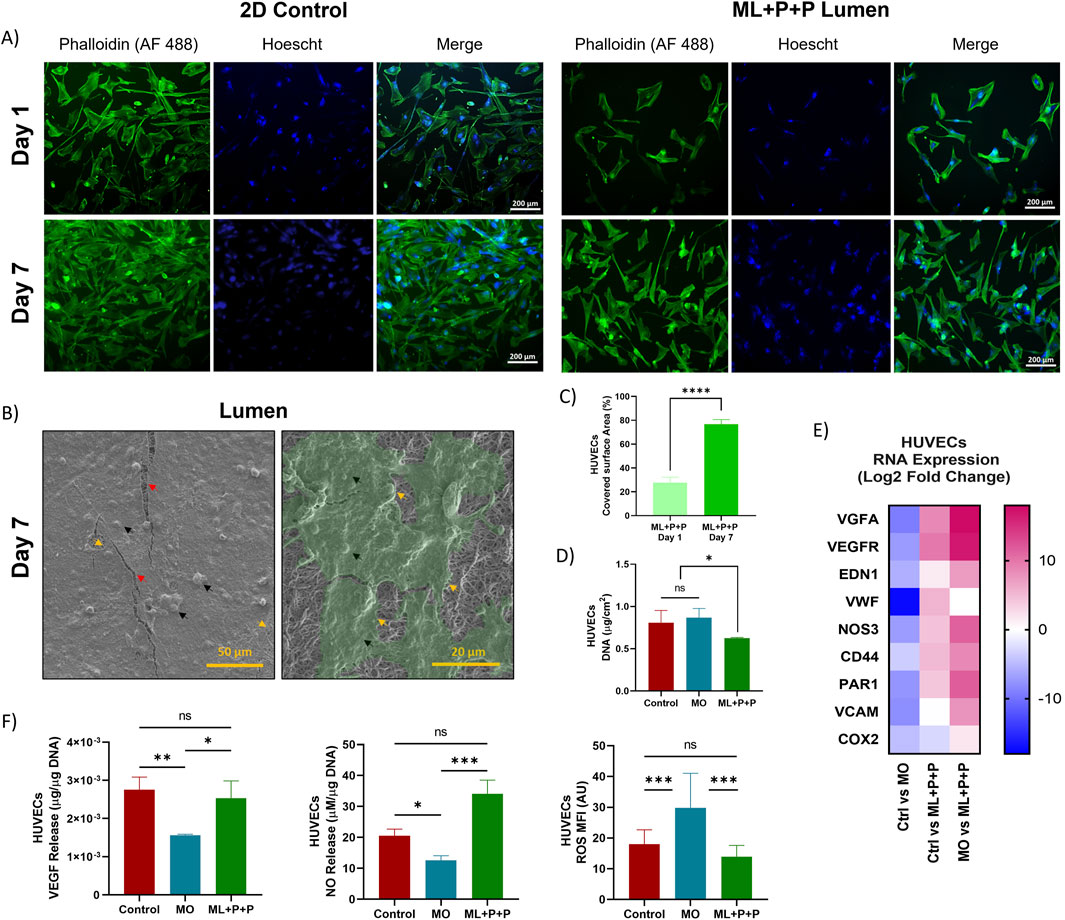
Figure 9. Endothelialization potential of ML + P + P with HUVECs seeded on the luminal surface. (A) Phalloidin staining at days 1 and 7 compared with a 2D control on a glass slide. (B) SEM images of Endothelial cell lining. Black arrows highlight cells and cell nuclei, yellow arrows indicate cell boundaries, and red arrows correspond to cracks in the fixed cell monolayer resulting from sample processing; beneath this layer, electrospun fibers are visible. (C) Percentage of covered surface area by HUVECs, data normalized from with 2D control. (D) DNA quantification at 7 days. (E) RNA expression profile. (E) VEGF and NO release. (F) Intracellular ROS production. (Mean ± SD) where, ns = no significant *p ≤ 0.05, **p ≤ 0.01, ***p ≤ 0.001, ****p ≤ 0.0001.
The authors apologize for this error and state that this does not change the scientific conclusions of the article in any way. The original article has been updated.
Publisher’s note
All claims expressed in this article are solely those of the authors and do not necessarily represent those of their affiliated organizations, or those of the publisher, the editors and the reviewers. Any product that may be evaluated in this article, or claim that may be made by its manufacturer, is not guaranteed or endorsed by the publisher.
Keywords: tissue engineered vascular grafts, regenerative medicine, biomaterials, inflammatory response, immunomodulation, M1/M2 macrophage polarization, endothelialization, cell signaling
Citation: Rodríguez-Soto MA, Riveros-Cortés A, Orjuela-Garzón IC, Fernández-Calderón IM, Rodríguez CF, Suárez Vargas N, Ostos C, Muñoz Camargo C, Cruz JC, Kim S, D'Amore A, Wagner WR and Briceño JC (2025) Corrigendum: Redefining vascular repair: revealing cellular responses on PEUU—gelatin electrospun vascular grafts for endothelialization and immune responses on in vitro models. Front. Bioeng. Biotechnol. 13:1607125. doi: 10.3389/fbioe.2025.1607125
Received: 07 April 2025; Accepted: 09 April 2025;
Published: 24 April 2025.
Edited and reviewed by:
Wolfgang Holnthoner, AUVA Research Centre, AustriaCopyright © 2025 Rodríguez-Soto, Riveros-Cortés, Orjuela-Garzón, Fernández-Calderón, Rodríguez, Suárez Vargas, Ostos, Muñoz Camargo, Cruz, Kim, D'Amore, Wagner and Briceño. This is an open-access article distributed under the terms of the Creative Commons Attribution License (CC BY). The use, distribution or reproduction in other forums is permitted, provided the original author(s) and the copyright owner(s) are credited and that the original publication in this journal is cited, in accordance with accepted academic practice. No use, distribution or reproduction is permitted which does not comply with these terms.
*Correspondence: Juan C. Briceño, amJyaWNlbm9AdW5pYW5kZXMuZWR1LmNv