 Ruth Pulido1
 Nelson Naveas2,3*
 Francisco Javier Fernández-Alonso2,3,4
 Miguel Manso-Silván2,3,4Leonardo Soriano2
 Carlos Torres-Ulloa5Karel Mena-Ulecia6Gonzalo Recio-Sánchez7
 Juan Paulo Garcia-Sandoval8
 Jacobo Hernández-Montelongo5,9*
- 1Departamento de Química, Universidad de Antofagasta, Antofagasta, Chile
- 2Departamento de Física Aplicada, Universidad Autónoma de Madrid, Campus de Cantoblanco, Madrid, Spain
- 3Instituto Universitario de Ciencia de Materiales “Nicolás Cabrera” (INC), Universidad Autónoma de Madrid, Campus de Cantoblanco, Madrid, Spain
- 4Centro de Microanálisis de Materiales, Universidad Autónoma de Madrid, Campus de Cantoblanco, Madrid, Spain
- 5Departmento de Ciencias Matemáticas y Físicas, Universidad Católica de Temuco, Temuco, Chile
- 6Departmento de Ciencias Biológicas y Químicas, Universidad Católica de Temuco, Temuco, Chile
- 7Facultad de Ingeniería, Arquitectura y Diseño, Universidad San Sebastián, Concepción, Chile
- 8Departmento de Ingeniería Química, Universidad de Guadalajara, Guadalajara, Mexico
- 9Departamento de Bioingeniería Traslacional, Universidad de Guadalajara, Guadalajara, Mexico
Urinary catheters (UCs) are critical in biomedical applications, but prolonged use increases the risk of catheter-associated urinary tract infections (CAUTIs), a leading cause of healthcare-associated infections (HAIs). The present study presents a dual strategy to create an antibacterial surface on commercial Foley silicone UCs by combining a contact-killing effect with the controlled release of antimicrobial compounds. We designed a drug delivery system using a layer-by-layer (LbL) antibacterial coating of carboxymethylcellulose (CMC) and chitosan-silver (CHI-Ag) complexes, with ciprofloxacin (CFX) as the model drug. The resulting LbL coating, about 1
1 Introduction
Urethral catheters (UCs) are pliable plastic tubes carefully inserted into the urinary bladder through the urethra, serving the purpose of collecting urine in a drainage bag (Chuang and Tambyah, 2021; Newman et al., 2018). As indispensable devices in patient care, catheters rank among the most extensively utilized medical tools in hospital settings. They find application in patients with urinary obstruction, both preceding and following specific surgical procedures. Additionally, catheters play a crucial role in aiding various treatments, such as ensuring precise measurement of urine output, managing incontinence, or facilitating the direct administration of particular medications into the bladder.
Despite their valuable utility, prolonged use of these devices comes with the risk of serious complications, notably the occurrence of catheter-associated urinary tract infections (CAUTIs), recognized as the most prevalent cause of healthcare-associated infections (HAIs) (Teixeira-Santos et al., 2022; Tenke et al., 2017). Alarmingly, approximately 15%–25% of hospitalized patients undergo catheterization, underlining the widespread usage and the associated risks. In both Europe and the United States, the annual incidence of CAUTIs surpasses one million cases, contributing to escalated morbidity and mortality rates (Cai et al., 2023; Öztürk and Murt, 2020).
In this context, rates of CAUTIs are significantly higher in Latin American hospitals than in industrialized countries (Yin et al., 2023). For example, CAUTIs in Chile have shown a notable and consistent increase. Prevalence studies conducted by the Ministry of Health (Unidad de Infecciones Intrahospitalarias, 2020) highlight CAUTIs as the most frequently reported infections across hospitals of varying complexity levels nationwide. Noteworthy is the significant prevalence of CAUTIs observed from 2014 to 2019, primarily attributed to the extensive use of UCs within a substantial patient population. Among the six medical devices examined in the HAIs surveillance report, UCs emerged as the most widely employed, boasting the highest utilization percentage compared to oral parenteral nutrition, other probes, and mechanical ventilation (Unidad de Infecciones Intrahospitalarias, 2020).
In that sense, catheterization could be considered a contraindication as it is closely associated with a heightened probability of CAUTIs (Chuang and Tambyah, 2021). These infections result from the formation of biofilms on UCs, consisting of bacteria that exhibit reduced sensitivity to antibiotics. Additionally, CAUTIs are associated with increased mortality rates, prolonged hospitalization times, and higher hospital expenses (Gray et al., 2023; Hollenbeak and Schilling, 2018). In some cases, this CAUTIs incidence was worsened by the impact of the COVID-19 pandemic due to the large volume of patients requiring advanced medical care and subsequent depleted resources (Hyte et al., 2023; Mitra et al., 2021).
Gram-negative bacteria are the most common causative agents of urinary tract infections (UTIs), including Escherichia coli, Proteus mirabilis, Klebsiella pneumoniae, and Pseudomonas aeruginosa (Majumder et al., 2018). Although Gram-positive bacteria are generally less prevalent (Alshomrani et al., 2023), recent studies have reported a growing incidence of S. aureus in cases of asymptomatic bacteriuria and complicated UTIs, particularly among elderly individuals, recently hospitalized patients, and those with indwelling urinary catheters (Paudel et al., 2023). Furthermore, Candida species can thrive and proliferate in the urinary tract environment (Yao et al., 2022). All these CAUTI-associated pathogens exhibit multiple virulence factors including fimbrial adhesins, urease activity, biofilm formation, and immune evasion proteins. These traits facilitate catheter colonization and persistence. Ciprofloxacin (CFX) targets bacterial DNA gyrase but may be ineffective against sessile, biofilm-embedded cells. Conversely, silver (Ag) exerts membrane-disruptive and metabolic effects independent of active replication, offering complementary protection. Understanding the interplay between drug action and pathogen virulence is key to designing effective coatings (Govindarajan and Kandaswamy, 2022; Sivaramalingam et al., 2024).
In that sense, different strategies have been used to develop antimicrobial surfaces for UCs, which can be grouped into five strategies (Teixeira-Santos et al., 2022): 1) release of antimicrobial compounds (Srisang et al., 2020; Lin et al., 2021; Miao et al., 2023), 2) contact-killing (Gao et al., 2020; Tailly et al., 2021; Bai et al., 2023), 3) anti-adhesive (Yu et al., 2017; Zhang et al., 2019; Prateeksha et al., 2021), 4) disruption of biofilm architecture (Durgadevi et al., 2020; Swidan et al., 2022; da Silva et al., 2024), and 5) benign biofilms to inhibit pathogen colonization (Zhu et al., 2015; Carvalho et al., 2021).
Studies have reported antimicrobial coatings using silver nanoparticles (AgNPs) combined with antibiotics. However, many of these strategies face challenges such as nanoparticle aggregation, burst drug release, and limited control over release kinetics under physiological conditions (Kadirvelu et al., 2024). Our study advances this field by introducing a drug delivery system based on an antibacterial layer-by-layer (LbL) coating applied to commercial silicone urinary catheters (UCs) of two sizes (14Fr and 20Fr). The LbL architecture, fabricated by sequentially assembling carboxymethylcellulose (CMC) with the antibacterial chitosan-silver complex (CHI-Ag), allows modular control of coating thickness, drug loading, and silver integration. CFX was incorporated as a model drug to investigate controlled release performance. This dual-action system combines contact-killing properties with the sustained release of antimicrobial compounds. The coated UCs were subjected to morphological and physicochemical characterization, and their antibacterial efficacy was evaluated against both gram-negative E. coli and gram-positive S. aureus strains. Biological in vitro assays were performed using pristine, coated, and CFX-loaded coated samples. To elucidate the mechanisms of CFX loading and release, we implemented a mathematical model based on unidirectional diffusion governed by Fick’s second law. Additionally, molecular dynamics (MD) simulations were employed to investigate the interactions between CFX and the coating matrix, using catheter-relevant geometries (14Fr/20Fr) to better represent real-world performance.
2 Materials and methods
2.1 Materials
14Fr and 20Fr Foley silicone UCs were acquired from Chen Kang® (China); their nominal diameters were 4.7 mm and 6.7 mm, respectively. The following reagents were procured from Sigma–Aldrich (United States): polyethylenimine (PEI), a 50 wt% solution in water with an average molecular weight (Mw)
Additionally, the following chemicals and substances were obtained from Merck (Germany): silver nitrate (
2.2 Layer-by-layer coating
UCs samples were cut to a length of 2.5 cm using a scalpel and sealed at both ends using a silicone gun. Subsequently, the samples underwent oxidation in an oxygen plasma equipment (Harrick Plasma, United States) under low pressure (0.2 mmHg) for 15 min. This process enhances surface hydrophilicity and activates silanol groups on the PDMS surface, which improves the subsequent adsorption of PEI and the adhesion of the LbL assembly (Hernandez-Montelongo et al., 2017). Following this, a preliminary layer of PEI solution (1 mg/mL, pH = 4) was applied for 15 min, followed by rinsing with Milli-Q water, pre-adjusted to pH 4.
The LbL coating incorporating silver was then executed using the method proposed by (Naveas et al., 2023). Briefly, the samples were immersed for 10 min in a CMC solution (0.1% m/v, pH = 4) and rinsed with Milli-Q water adjusted to pH 4. Subsequently, the samples were immersed for 10 min in a chitosan-silver (CHI-Ag) solution (0.1% m/v CHI solution with 0.1 mM
2.3 Physicochemical characterization
The surface wettability of the samples was assessed using a water contact angle measuring system (Dropletlab, Canada) in static sessile drop mode. A 10
For the evaluation of carboxylic and amino groups in UCs with LbL coating, samples underwent immersion in MB (0.001 M, pH = 7) and RB (0.001 M, pH = 5) solutions. This was followed by rinses with Milli-Q water. Subsequently, stains from the samples were extracted for UV–vis spectroscopy measurements in absorbance mode. Samples treated with MB were immersed in a 5% (v/v) solution of glacial acetic acid, while those with RB were immersed in a 0.1 M NaOH solution. The absorbance values for MB and RB were measured at 663 nm (Eltaweil et al., 2020) and 545 nm (Bayraktutan, 2020), respectively, using a UV-Vis spectrophotometer (Evolution 220 model, Thermo Scientific, United States).
Chemical analysis of the samples was conducted using Attenuated Total Reflectance Fourier-Transform Infrared Spectroscopy (ATR-FTIR). An FTIR spectrometer coupled with an ATR accessory using a zinc selenide crystal (CARY 630 FTIR Agilent Technologies, United States) was employed within the range of 4,000 to 600
The morphology of the samples was explored using a variable pressure scanning electron microscope (VP-SEM, SU-3500 Hitachi, Japan) with an acceleration voltage of 10 kV. The acquired images underwent processing using the freely available ImageJ software, version 1.52k. For elemental mapping and atomic percentage determination, energy-dispersive X-ray analysis (EDX) was employed with an INCA X-sight system from Oxford Instruments integrated into the VP-SEM equipment.
X-ray Photoelectron Spectroscopy (XPS) was employed to analyze the surface chemical composition of the samples. XPS spectra were obtained using the Surface Analysis Station 150 XPS RQ300/2 (TAIB Instruments) equipped with a hemispherical electron analyzer and a Mg-anode X-ray source. The pass energy was set at 20 eV, providing an overall resolution of 0.9 eV. All XPS binding energies were referenced to the adventitious C 1s carbon peak at a binding energy of 284.6 eV to account for surface charging effects. The XPS spectra were fitted using the CasaXPS software.
In-depth profiling of the silver into LbL coating was studied through Rutherford Backscattering Spectroscopy (RBS). RBS experiments were conducted at the standard beamline of the Center of Micro-analysis of Materials (CMAM, Spain) (Redondo-Cubero et al., 2021), which houses a 5 MV Cockroft-Walton tandetron accelerator. In these experiments, 4 MeV He
2.4 Drug loading and release profiles
Following the assembly of the LbL coating, each sample underwent a 12-h immersion in a ciprofloxacin (CFX) solution with a concentration of 1.33 mg/mL. Subsequently, the samples were thoroughly rinsed with Milli-Q water and allowed to dry. To generate the release profiles of the CFX-loaded samples, they were placed in a phosphate-buffered saline (PBS) solution within a horizontal shaker set at 37 °C, pH of 7.6, and 50 rpm. The concentration of CFX in PBS was determined at various release times over a 15-day period using a UV-Vis spectrophotometer (Evolution 220 model, Thermo Scientific, United States). CFX was detected at a wavelength of 275 nm (Uddin et al., 2022). All experiments were conducted in triplicate, with non-functionalized samples serving as controls in the kinetic release experiments.
2.5 Antibacterial assays
The antibacterial activity of the samples was assessed using the agar diffusion method against two bacterial strains: Escherichia coli ATCC 25922 (gram-negative) and Staphylococcus aureus ATCC 25923 (gram-positive). Mueller-Hilton agar served as the medium for the diffusion method test. Bacterial strains were adjusted to a 0.5 McFarland standard concentration and inoculated onto solid media under aseptic conditions. Samples were then applied vertically onto the surface of the solid media and allowed to dry for 1 h. The inoculated plates were incubated for 24 h at 35 °C, after which the inhibition zones around the samples were measured from photographs of the plates using ImageJ software. Data were analyzed statistically by analysis of variance (ANOVA) with a subsequent Tukey post-hoc test using OriginLab v. 2022b software; p-values of 0.05 or less were considered statistically significant. All assays were performed in triplicate.
2.6 Mathematical model
The mathematical model utilized in this study is built around three consecutive processes centered on unidirectional diffusion governed by Fick’s second law (Hernandez-Montelongo et al., 2022). These processes include drug loading, where samples are loaded with a highly concentrated drug solution; a resting stage, during which the drug continues to diffuse into the matrix samples; and drug delivery, where the drug is released from the samples into a PBS solution at 37 °C, simulating bodily fluid conditions.
The diffusion equation for the delivery system is shown in (Equation 1):
where
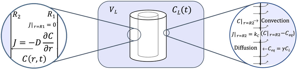
Figure 1. Scheme of the drug delivery system.
At the beginning, the UC does not contain CFX, therefore the initial condition must be Equation 2:
The inner region of the UC only contains air because it is closed before the experiment starts, thus we assume that CFX does not diffuse to this region and the boundary condition at
On the other hand, the CFX’s flux on the external surface of the UC varies depending on the stage of the process. At the CFX load stage, from
Here in (Equation 4),
Finally, at the delivery stage, the UC is in contact with the PBS solution that initially has zero CFX concentration, but as the CFX diffuses from the UC surface to the PBS solution, the external CFX concentration in the PBS solution,
while the external CFX concentration in the PSB solution varies according to (Equation 7):
where
In particular, if we consider a well mixed convection during the loading and delivery stages, then
while the dynamical behavior of the external CFX concentration in the PBS solution becomes (Equation 10):
In summary, the model is described by PDE (Equation 1), initial condition (Equation 2), and boundary condition (Equation 3) for all the domain of time. The boundary conditions (Equation 8) and (Equation 5) are used at load and rest stages, respectively, while boundary condition (Equation 9) and ODE (Equation 10) are used at delivery stage.
2.7 Molecular dynamics (MD) simulations
To gain deeper insights into the mechanisms governing the experimental behavior of controlled CFX release, quantum mechanical tools were employed in this study (Aazam and Thomas, 2024). The chemical structure of CFX was retrieved from the DrugBank database, identified by the code DB00537 (Knox et al., 2024). PDMS, CMC and the CHI-Ag complex were sketched using the freely available online tool Marvin JS (https://marvinjs-demo.chemaxon.com/latest/demo.html). These structures were obtained in XYZ format and individually optimized to their minimum energy states using the valence triple-zeta with two sets of polarization functions (def2-TZVPP) basis set for all atoms and the Becke, 3-parameter, Lee–Yang–Parr (B3LYP) hybrid functional (Weigend and Ahlrichs, 2005; Becke, 1992; AL-Khaykanee, 2013; Orio et al., 2009), implemented in the Orca 5.0.1 software package (Neese, 2012; Neese, 2022). The full optimized geometry of all molecules was verified by examining their imaginary frequencies.
After confirming that all molecules had reached their energy minima, the systems under investigation were assembled. These systems comprised two complexes: complex-1 consisted of three layers, with the inner and outer layers composed of PDMS molecules and a layer of three CFX molecules sandwiched between them at a distance of 3Å. Complex-2 consisted of six layers: the outermost layer contained two CFX molecules, followed by a layer of CHI-Ag at a distance of 3Å, a layer of two CFX molecules, a layer of CMC, another layer of two CFX molecules, a layer of one CHI-Ag molecule, and an innermost layer of three CFX molecules. Both complexes were constructed using the Chemcraft version 1.8 program for Linux (Andrienko, 2010) and simulated using the ab initio molecular dynamics (AIMD) method (Bocharov et al., 2020; Tse, 2002; Marx and Hutter, 2000). Each system underwent a simulation period of 100 fs (2000 steps) with a time step of 0.5 fs at a temperature of 298.15 K. All ab initio MD simulations were conducted using the Orca 5.0.1 program package (Neese, 2012; Neese, 2022; Snyder and Kucukkal, 2021).
3 Results and discussion
In this work, UCs of varying diameters (14Fr and 20Fr) underwent a coating process involving a thin film of CMC/CHI-Ag, assembled through a layer-by-layer technique (LbL). The synthesis process is outlined schematically in Figure 2. Given that polydimethylsiloxane (PDMS) serves as the basis for the silicone UCs (Zare et al., 2021), the surface of these devices is inherently hydrophobic. Thus, prior to coat the samples, a surface pretreatment involving
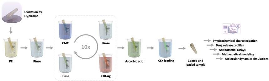
Figure 2. Schematic representation illustrating the assembly process of the coating composed of carboxymethylcellulose (CMC) and chitosan-silver (CHI-Ag) leading to a the Layer-by-Layer technique Ag loaded coating (LbL): The sample is immersed alternately in the respective polysaccharide electrolyte, followed by rinsing steps and
Figure 3A depicts UCs before and after the LbL coating, evident from the light brown appearance. Following the LbL treatment, the wettability of UC samples transitioned to a hydrophilic behavior, registering a change from 100
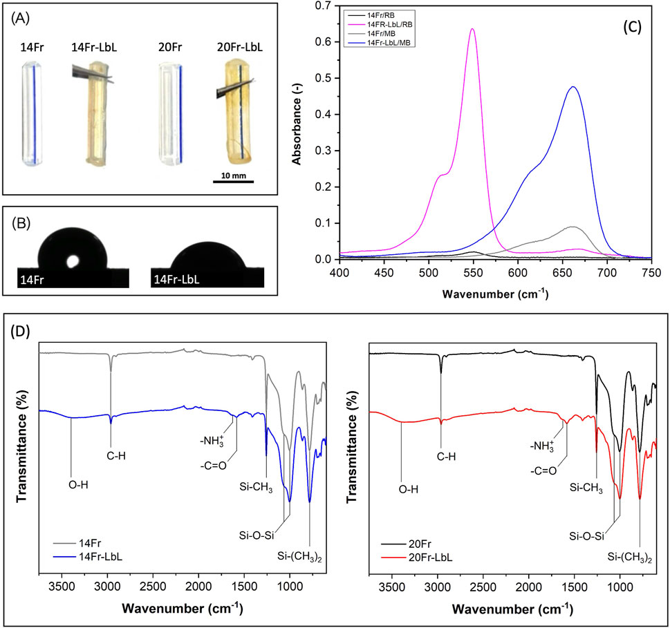
Figure 3. (A) Urinary catheters before and after the layer-by-layer coating. (B) Water contact angle of pristine and coated sample (14Fr). (C) Absorbance of Rose Bengal and Methylene Blue obtained from pristine and coated samples (14Fr). (D) ATR-FTIR spectra of urinary catheters before and after the layer-by-layer coating.
UV–Vis spectroscopy served as a tool to indirectly identify the carboxylic groups of CMC (negatively charged) by utilizing the oppositely charged stain methylene blue (MB) (positively charged) (Eltaweil et al., 2020). Likewise, to indirectly detect the ammonium groups of CHI (positively charged), the stain Rose Bengal (RB) (negatively charged) was applied (Bayraktutan, 2020). Figure 3C illustrates the absorbance peaks of MB and RB for both the 14Fr and 14Fr-LbL samples. The pristine 14Fr sample displayed low absorbance values: 0.089 for MB and 0.020 for RB. In contrast, the coated 14Fr-LbL sample exhibited significantly higher absorbance peaks, measuring 0.476 for MB and 0.635 for RB. A similar trend was observed in the 20Fr and 20Fr-LbL samples.
The chemical analysis of the samples was conducted using ATR-FTIR to directly identify the surface modifications (Figure 3D). The bending vibrations of the fingerprint functional groups of PDMS were discerned, including Si-(
Figure 4A displays cross-sectional SEM images of both UCs. The 14Fr sample exhibited external and internal diameters of 4.7 mm and 2.7 mm, respectively. In the case of the 20Fr sample, its external and internal diameters were 6.7 mm and 4.7 mm, respectively. Both UCs presented a wall thickness of 1 mm. Moreover, the typical inflation cuff orifice of Foley catheters was also observed in both samples. UCs submitted to the layer-by-layer treatment showed a thin coating of 1.3
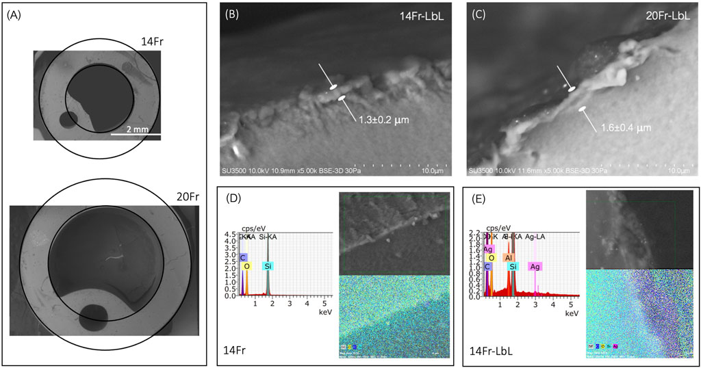
Figure 4. (A) Cross-sectional SEM images of 14Fr and 20Fr catheters. (B) Thickness of the 14Fr-LbL film. (C) Thickness of the 20Fr-LbL film. (D) EDX analysis and mapping of the 14Fr sample. (E) EDX analysis and mapping of the 14Fr-LbL sample.
Rutherford Backscattering Spectrometry (RBS) was performed to analyze the diffusion of silver (Ag) in the UCs. Figure 5A shows the RBS spectrum obtained for the 14Fr-LbL sample. Five element-related signals can be observed, corresponding to carbon (C), nitrogen (N), oxygen (O), silicon (Si), and silver (Ag). The Ag signal shows a peak at a backscattering energy of 3,359 keV, whereas, for Ag atoms on the surface, a backscattering energy of 3,447 keV would be expected. This indicates that silver has infiltrated towards the bulk material of the LbL coating.
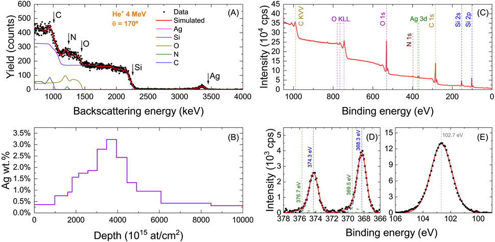
Figure 5. (A) RBS experimental and simulated spectra and (B) derived in-depth concentration profile of Ag obtained via RBS fitting. XPS analysis on 14Fr-(LbL-Ag) sample: (C) survey spectra of 14Fr-(LbL-Ag) sample and, XPS spectra of (D) Ag 3d and (E) Si 2p core levels.
The in-depth compositional profile of silver was estimated by fitting the RBS spectrum using the Simnra 7.03 software, as shown in Figure 5B. For this analysis, a structure composed of up to 20 layers of material was considered, each with varying thickness and concentrations of C, N, O, Si, and Ag. The results confirm that the concentration of Ag on the coating surface is only 0.4%, while deeper within the material, the weight percentage increases up to 3.2%.
The surface chemical composition of the UCs was studied via X-ray Photoelectron Spectroscopy (XPS). Figure 5C shows the XPS spectrum of the 14Fr-LbL samples. The survey spectrum reveals the presence of carbon (C), nitrogen (N), oxygen (O), silicon (Si), and silver (Ag). A quantitative estimation of the surface composition was performed based on the intensity of the signals associated to each element, using the sensitivity factors compiled by (Wagner et al., 1981). In this way, the surface concentration of silver was estimated to be 0.4%, consistent with the amount of silver observed in the outermost layer as estimated by RBS.
Figure 5D shows the spectral region corresponding to Ag 3d. Two peaks are observed at 368.3 eV and 374.3 eV, corresponding to the photoemission peaks associated with the Ag
Figure 5E shows the spectral region corresponding to Si 2p. The region is accurately fitted with a single Gaussian-Lorentzian peak centered at 102.7 eV, which is consistent with the formation of Si-O bond (Kaur et al., 2016; Kong et al., 2020).
To evaluate the ciprofloxacin (CFX) delivery performance of the coated samples, experiments were conducted in PBS at 37 °C and pH 7.6 under stirring conditions. A pH of 7.6 is close to physiological conditions and falls within the typical range of urine (pH 4.5–8.0), which can vary depending on hydration levels and overall health status. Both control and coated samples were loaded with highly concentrated solutions of the antibiotic for 12 h and then stored for 24 h (resting stage) prior to use in the experiments. The drug release profiles obtained are presented in Figures 6A,B. The results indicate that pristine samples (14Fr and 20Fr) exhibited significant release of CFX, demonstrating the chemical compatibility between the antibiotic’s structure and the silicone substrates (Hui et al., 2008). In that sense, the released amount of CFX for both control samples was similar, approximately 30
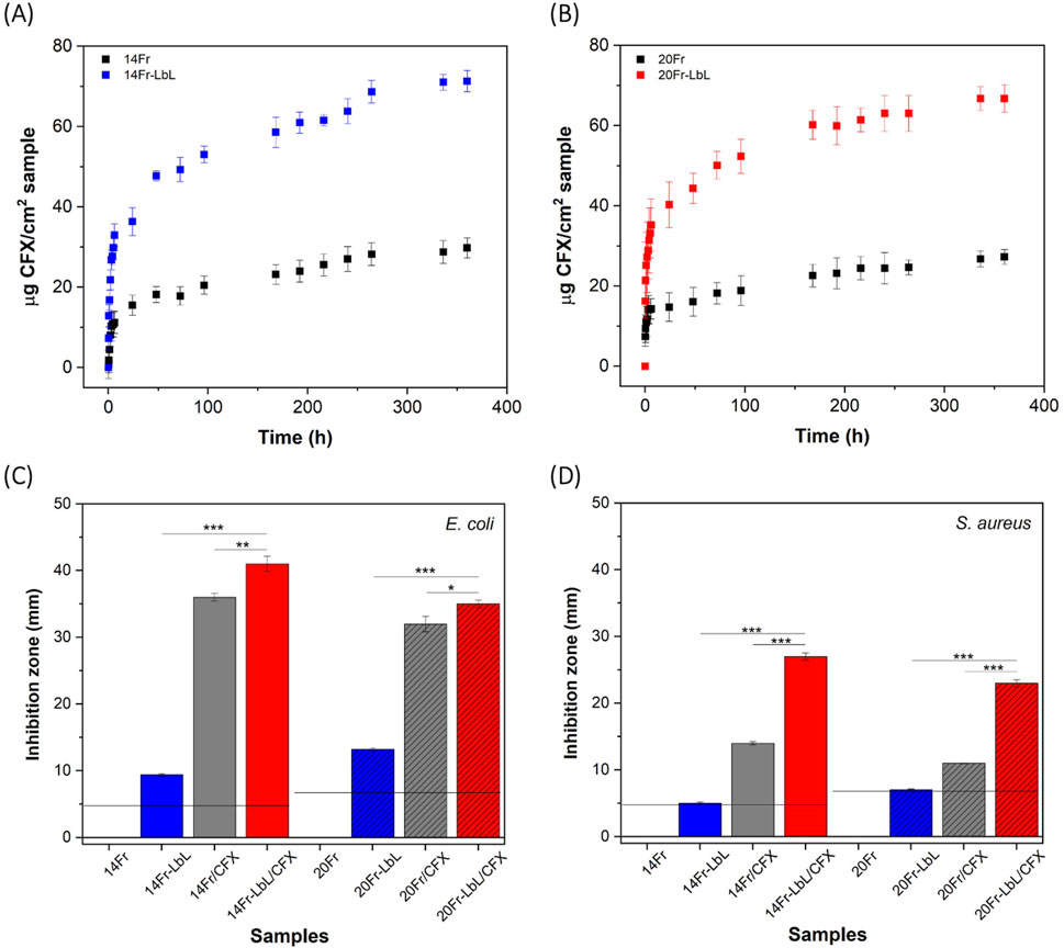
Figure 6. Drug release profiles of CFX from: (A) 14Fr and 14Fr-LbL, and (B) 20Fr and 20Fr-LbL. Antibacterial tests of samples against: (C) Escherichia coli, and (D) Staphylococcus aureus. Labels mean 14Fr and 20Fr are uncoated, 14Fr-LbL and 20Fr-LbL are LbL coated, 14Fr/CFX and 20Fr/CFX are uncoated but CFX-loaded, 14Fr-LbL/CFX and 20Fr-LbL/CFX are LbL-coated and CFX-loaded. Results represent mean
Furthermore, Figures 6C,D illustrate the antibacterial efficacy of the samples against E. coli and S. aureus. For the E. coli strain (Figure 6C), the pristine samples (14Fr and 20Fr) exhibited no antibacterial activity. However, the coated samples 14Fr-LbL and 20Fr-LbL displayed inhibition areas of 10 and 13 mm, respectively. These inhibition zones noted for the LbL-only samples might indicate limited diffusion of silver ions caused by surface-anchored
Also, it is important to note that an increase in the surface roughness and the transition from a hydrophobic to a hydrophilic surface (as shown in Figures 3A,B) after the LBL procedure may enhance bacterial adhesion. However, this effect could be counteracted by the strong antibacterial properties of silver and CFX, which help prevent early-stage colonization. Nonetheless, further studies are needed to evaluate long-term biofilm formation under dynamic conditions in the urinary tract.
In the case of the S. aureus strain (Figure 6D), pristine samples also showed no antibacterial effect, while coated samples only exhibited an inhibition zone corresponding to the diameter of the UCs: 4.7 mm for 14Fr-LbL and 6.7 mm for 20Fr-LbL. This indicates that the LbL coating solely functioned as an antibacterial surface. However, samples loaded with CFX demonstrated increased antibacterial efficacy: 14Fr/CFX and 20Fr/CFX exhibited inhibition zones of 15 and 10 mm, respectively.Notably, the combined antibacterial activity resulting from the combination of the LbL coating and the CFX antibiotic was significantly enhanced for this strain. The 14Fr-LbL/CFX and 20Fr-LbL/CFX samples demonstrated inhibition zones of 27 mm and 23 mm, respectively. Both values exceed the CLSI threshold of
To gain deeper insights into the mechanisms governing the release of CFX from both uncoated and coated samples, we developed a mathematical model accounting for the drug loading step and the resting stage (Section 2.6). The kinetic parameters obtained from this model are presented in Table 1. Given that the wall thickness of the UCs is 1 mm and the LbL coating is a micron-thick film, we assumed that the diffusivity (
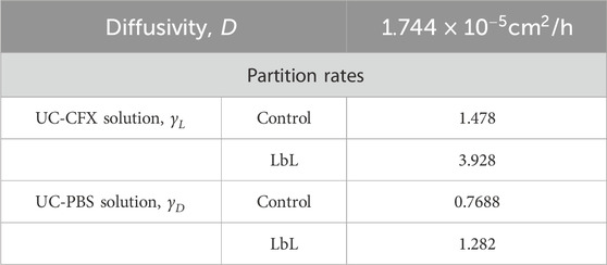
Table 1. Kinetic parameters.
Using the kinetic parameters from Table 1, several simulations were conducted. The Supplementary Figure S1A illustrates the CFX concentration inside the UCs immediately after the 12-h loading period. In all cases, both pristine (14Fr and 20Fr) and coated samples (14Fr-LbL and 20Fr-LbL) exhibited CFX accumulation at the border of the external wall of the UCs. However, after the 24-h rest stage preceding the drug delivery experiments (Supplementary Figure S1B), simulations showed how CFX continues to diffuse through the samples. With these insights, finally, we were able to simulate the experimental data of CFX delivery over the 15-day release period (Figure 7A). Additionally, owing to the characteristics of this mathematical model, we can predict how drug release profiles will differ based on varying drug loading and rest stage times (Supplementary Figure S1C; Figure 7B). Simulations indicated that extending the loading time may not necessarily result in higher CFX loading, as equilibrium is reached at the border of the external UCs wall. However, longer rest stage times enable more controlled release, as CFX becomes more homogeneously distributed throughout the samples.
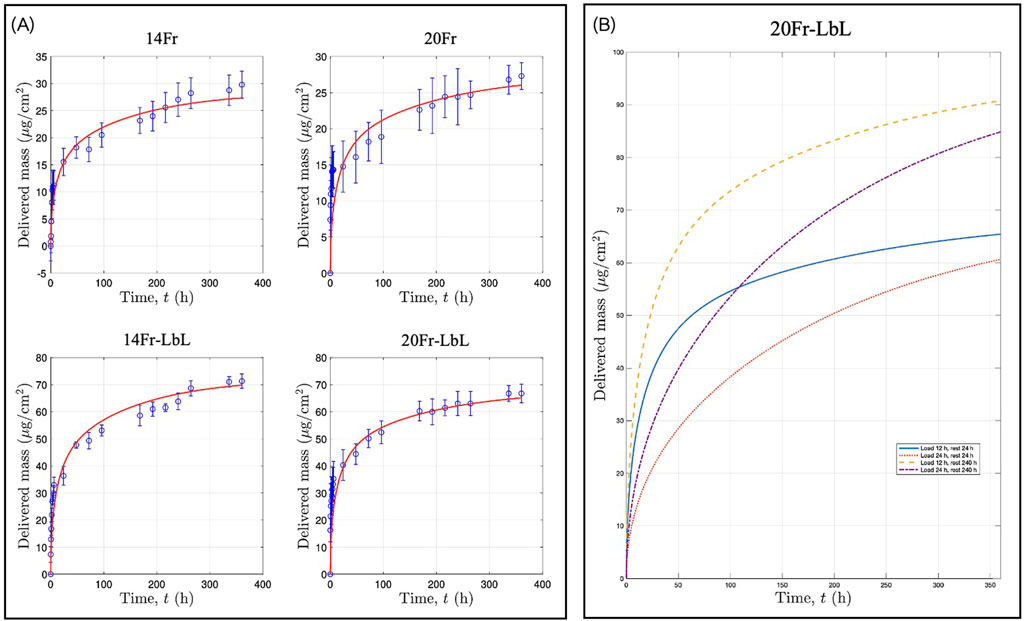
Figure 7. (A) CFX release profiles: experimental data and simulations. (B) Simulations of drug release profiles considering different drug loading and rest stage times.
Figure 8 presents the molecular dynamics simulations. Complex-1 (Figure 8A) shows a stable capacity for CFX loading within PDMS, as reflected by its consistent negative potential energy, approximately −5
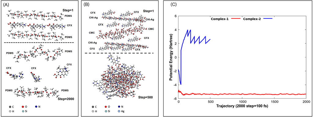
Figure 8. Molecular dynamics simulations: (A) Complex-1 [PDMS-CFX-PDMS] and (B) complex-2 [CFX-CHI-Ag-CFX-CMC-CFX-CHI-Ag-CFX]. 1 step = 0.05 fs. (C) Potential energy of simulated complexes.
4 Conclusion
This study successfully developed a dual strategy to create an antibacterial surface on commercial Foley silicone urinary catheters (UCs) by combining the contact-killing effect with the controlled release of antimicrobial compounds. Specifically, UCs were coated with an antibacterial drug delivery system using a Layer-by-Layer (LbL) approach that incorporated carboxymethylcellulose (CMC) and chitosan-silver (CHI-Ag) complexes, loaded with ciprofloxacin (CFX) as the model drug. The very thin LbL coating significantly enhanced drug loading and release, achieving over twice the amount of CFX release compared to uncoated catheters. The antibacterial efficacy was markedly improved in the coated samples, particularly against S. aureus than E. coli. Additionally, a mathematical model based on Fick’s second law was proposed to simulate the experimental CFX release profiles, effectively predicting drug release kinetics by incorporating both drug loading and resting stages. Molecular dynamics simulations further confirmed strong compatibility between CFX and the LbL layers, although with relatively low stability. Despite the promising results, the study has certain limitations that should be addressed in future research. First, the antibacterial performance was evaluated only against planktonic forms of E. coli and S. aureus, without directly assessing biofilm formation or eradication on the catheter surface. Given the clinical importance of biofilms in CAUTIs, future studies should focus on quantifying biofilm inhibition and evaluating antimicrobial durability under physiologically dynamic conditions. Second, the release kinetics of silver from the layer-by-layer (LbL) coating were not investigated. Although silver was reduced to its elemental form (
Data availability statement
The raw data supporting the conclusions of this article will be made available by the authors, without undue reservation.
Author contributions
RP: Writing – original draft, Writing – review and editing, Methodology, Conceptualization, Formal Analysis. NN: Formal Analysis, Methodology, Conceptualization, Writing – original draft, Writing – review and editing. FF-A: Writing – review and editing, Data curation, Methodology, Formal Analysis. MM-S: Formal Analysis, Data curation, Resources, Methodology, Writing – review and editing, Funding acquisition. LS: Methodology, Formal Analysis, Writing – review and editing. CT-U: Writing – review and editing, Methodology, Formal Analysis. KM-U: Writing – review and editing, Funding acquisition, Formal Analysis, Methodology, Resources. GR-S: Writing – review and editing, Methodology, Formal Analysis. JG-S: Formal Analysis, Methodology, Writing – review and editing. JH-M: Project administration, Formal Analysis, Methodology, Funding acquisition, Conceptualization, Writing – review and editing, Writing – original draft, Resources.
Funding
The author(s) declare that financial support was received for the research and/or publication of this article. This work received financial funding from FONDECYT–Chile (grant number 1230553), CONICYT PFCHA/DOCTORADO/2017–21172001, and was partially supported by the supercomputing infrastructure of the NLHPC (ECM-02) at the Universidad de Chile and the Centro de Computación Científica-Universidad Autónoma de Madrid (CCC-UAM); thanks to CPU time and other resources granted by the institutions. Miguel Manso acknowledges support from the Spanish funding agency MCIN/AEI/10.13039/50110001 1033 (PID2020-112770RB-C22) for assistance with characterization. F.J.F-A acknowledges funding from the Formaci’ on de Profesorado Universitario programme, ref. FPU22/04365, and grants PID2022-141080OB-C22 and CNS2024-154729, funded by MCIN/AEI.
Conflict of interest
The authors declare that the research was conducted in the absence of any commercial or financial relationships that could be construed as a potential conflict of interest.
Generative AI statement
The author(s) declare that Generative AI was used in the creation of this manuscript. Generative AI was used solely to enhance English grammar. All content and interpretations are the sole responsibility of the authors.
Any alternative text (alt text) provided alongside figures in this article has been generated by Frontiers with the support of artificial intelligence and reasonable efforts have been made to ensure accuracy, including review by the authors wherever possible. If you identify any issues, please contact us.
Publisher’s note
All claims expressed in this article are solely those of the authors and do not necessarily represent those of their affiliated organizations, or those of the publisher, the editors and the reviewers. Any product that may be evaluated in this article, or claim that may be made by its manufacturer, is not guaranteed or endorsed by the publisher.
Supplementary material
The Supplementary Material for this article can be found online at: https://www.frontiersin.org/articles/10.3389/fbioe.2025.1614509/full#supplementary-material
References
Aazam, E. S., and Thomas, R. (2024). Understanding the behavior of a potential anticancer lamotrigine in explicit solvent (water and dmso) using quantum mechanical tools and abinitio molecular dynamics. Chem. Phys. Impact 8, 100404. doi:10.1016/j.chphi.2023.100404
Al-Khaykanee, M. K. (2013). Lee-yang-parr (b3lyp) density functional theory calculations of di-cyano naphthalene molecules group. Chem. Mater. Res. 3.
Aldakheel, F. M., Mohsen, D., El Sayed, M. M., Alawam, K. A., Binshaya, A. S., and Alduraywish, S. A. (2023). Silver nanoparticles loaded on chitosan-g-pva hydrogel for the wound-healing applications. Molecules 28, 3241. doi:10.3390/molecules28073241
Alshomrani, M. K., Alharbi, A. A., Alshehri, A. A., Arshad, M., Dolgum, S., Alshomrani, M., et al. (2023). Isolation of staphylococcus aureus urinary tract infections at a community-based healthcare center in riyadh. Cureus 15, e35140. doi:10.7759/cureus.35140
Andrienko, G. (2010). Chemcraft-graphical software for visualization of quantum chemistry computations. Available online at: https://www.chemcraftprog.com.
Bai, Y., Li, K., Ma, L., Wu, D., Xiang, J., Hu, Q., et al. (2023). Mussel-inspired surface modification of urinary catheters with both zwitterionic and bactericidal properties for effectively preventing catheter-associated infection. Chem. Eng. J. 455, 140766. doi:10.1016/j.cej.2022.140766
Bayraktutan, T. (2020). Investigation of photophysical and binding properties of rose bengal dye on graphene oxide and polyethyleneimine-functionalized graphene oxide nanocomposites. Chem. Pap. 74, 3017–3024. doi:10.1007/s11696-020-01130-4
Becke, A. D. (1992). Density-functional thermochemistry. i. the effect of the exchange-only gradient correction. J. Chem. Phys. 96, 2155–2160. doi:10.1063/1.462066
Bocharov, D., Krack, M., Rafalskij, Y., Kuzmin, A., and Purans, J. (2020). Ab initio molecular dynamics simulations of negative thermal expansion in scf3: the effect of the supercell size. Comput. Mater. Sci. 171, 109198. doi:10.1016/j.commatsci.2019.109198
Bodas, D., and Khan-Malek, C. (2006). Formation of more stable hydrophilic surfaces of pdms by plasma and chemical treatments. Microelectron. Eng. 83, 1277–1279. doi:10.1016/j.mee.2006.01.195
Bozoğlan, B. K., Duman, O., and Tunç, S. (2020). Preparation and characterization of thermosensitive chitosan/carboxymethylcellulose/scleroglucan nanocomposite hydrogels. Int. J. Biol. Macromol. 162, 781–797. doi:10.1016/j.ijbiomac.2020.06.087
Cai, Y., Yang, H., Li, J., Gu, R., Dong, Y., Zhao, Q., et al. (2023). Antibacterial agnps-paam-cs-pvp nanocomposite hydrogel coating for urinary catheters. Eur. Polym. J. 196, 112260. doi:10.1016/j.eurpolymj.2023.112260
Calderon, V. S., Galindo, R. E., Benito, N., Palacio, C., Cavaleiro, A., and Carvalho, S. (2013). Ag+ release inhibition from zrcn–ag coatings by surface agglomeration mechanism: structural characterization. J. Phys. D Appl. Phys. 46, 325303. doi:10.1088/0022-3727/46/32/325303
Carvalho, F. M., Teixeira-Santos, R., Mergulhao, F. J., and Gomes, L. C. (2021). Effect of lactobacillus plantarum biofilms on the adhesion of escherichia coli to urinary tract devices. Antibiotics 10, 966. doi:10.3390/antibiotics10080966
Chuang, L., and Tambyah, P. A. (2021). Catheter-associated urinary tract infection. J. Infect. Chemother. 27, 1400–1406. doi:10.1016/j.jiac.2021.07.022
da Silva, C. R., de Farias Cabral, V. P., Rodrigues, D. S., Ferreira, T. L., Barbosa, A. D., de Andrade Neto, J. B., et al. (2024). Antibiofilm activity of promethazine against esbl-producing strains of escherichia coli in urinary catheters. Microb. Pathog. 193, 106769. doi:10.1016/j.micpath.2024.106769
Din, M. S. U., Gohar, U. F., Hameed, U., Mukhtar, H., Morar, A., Herman, V., et al. (2022). Piper nigrum fruit extract as an antibiotic resistance reversal agent in mdr bacteria. Appl. Sci. 12, 12542. doi:10.3390/app122412542
Durgadevi, R., Kaleeshwari, R., Swetha, T. K., Alexpandi, R., Pandian, S. K., and Ravi, A. V. (2020). Attenuation of proteus mirabilis colonization and swarming motility on indwelling urinary catheter by antibiofilm impregnation: an in vitro study. Colloids Surfaces B Biointerfaces 194, 111207. doi:10.1016/j.colsurfb.2020.111207
Eltaweil, A. S., Elgarhy, G. S., El-Subruiti, G. M., and Omer, A. M. (2020). Carboxymethyl cellulose/carboxylated graphene oxide composite microbeads for efficient adsorption of cationic methylene blue dye. Int. J. Biol. Macromol. 154, 307–318. doi:10.1016/j.ijbiomac.2020.03.122
Ferraria, A. M., Carapeto, A. P., and Botelho do Rego, A. M. (2012). X-ray photoelectron spectroscopy: silver salts revisited. Vacuum 86, 1988–1991. doi:10.1016/j.vacuum.2012.05.031
Firet, N. J., Blommaert, M. A., Burdyny, T., Venugopal, A., Bohra, D., Longo, A., et al. (2019). Operando exafs study reveals presence of oxygen in oxide-derived silver catalysts for electrochemical co2 reduction. J. Mater. Chem. A 7, 2597–2607. doi:10.1039/C8TA10412C
Gao, L., Wang, Y., Li, Y., Xu, M., Sun, G., Zou, T., et al. (2020). Biomimetic biodegradable ag@ au nanoparticle-embedded ureteral stent with a constantly renewable contact-killing antimicrobial surface and antibiofilm and extraction-free properties. Acta Biomater. 114, 117–132. doi:10.1016/j.actbio.2020.07.025
Govindarajan, D. K., and Kandaswamy, K. (2022). Virulence factors of uropathogens and their role in host pathogen interactions. Cell Surf. 8, 100075. doi:10.1016/j.tcsw.2022.100075
Gray, J., Rachakonda, A., and Karnon, J. (2023). Pragmatic review of interventions to prevent catheter-associated urinary tract infections (CAUTIs) in adult inpatients. J. Hosp. Infect. 136, 55–74. doi:10.1016/j.jhin.2023.03.020
Gugala, N., Vu, D., Parkins, M. D., and Turner, R. J. (2019). Specificity in the susceptibilities of escherichia coli, pseudomonas aeruginosa and staphylococcus aureus clinical isolates to six metal antimicrobials. Antibiotics 8, 51. doi:10.3390/antibiotics8020051
Hernandez-Montelongo, J., Lucchesi, E., Nascimento, V., França, C., Gonzalez, I., Macedo, W., et al. (2017). Antibacterial and non-cytotoxic ultra-thin polyethylenimine film. Mater. Sci. Eng. C 71, 718–724. doi:10.1016/j.msec.2016.10.064
Hernandez-Montelongo, R., Salazar-Araya, J., Hernandez-Montelongo, J., and Garcia-Sandoval, J. P. (2022). Mathematical modeling of recursive drug delivery with diffusion, equilibrium, and convection coupling. Mathematics 10, 2171. doi:10.3390/math10132171
Hochvaldová, L., Panáček, D., Válková, L., Večeřová, R., Kolář, M., Prucek, R., et al. (2024). E. coli and s. aureus resist silver nanoparticles via an identical mechanism, but through different pathways. Commun. Biol. 7, 1552. doi:10.1038/s42003-024-07266-3
Hollenbeak, C. S., and Schilling, A. L. (2018). The attributable cost of catheter-associated urinary tract infections in the United States: a systematic review. Am. J. Infect. control 46, 751–757. doi:10.1016/j.ajic.2018.01.015
Hui, A., Boone, A., and Jones, L. (2008). Uptake and release of ciprofloxacin-hcl from conventional and silicone hydrogel contact lens materials. Eye and contact lens 34, 266–271. doi:10.1097/icl.0b013e3181812ba2
Humphries, R. M., Hindler, J. A., Shaffer, K., and Campeau, S. A. (2019). Evaluation of ciprofloxacin and levofloxacin disk diffusion and etest using the 2019 enterobacteriaceae clsi breakpoints. J. Clin. Microbiol. 57, e01797-18–1128. doi:10.1128/jcm.01797-18
Hyte, M., Clark, C., Pandey, R., Redden, D., Roderick, M., and Brock, K. (2023). How covid-19 impacted cauti and clabsi rates in Alabama. Am. J. Infect. Control 52, 147–151. doi:10.1016/j.ajic.2023.05.014
Jung, W. K., Koo, H. C., Kim, K. W., Shin, S., Kim, S. H., and Park, Y. H. (2008). Antibacterial activity and mechanism of action of the silver ion in staphylococcus aureus and escherichia coli. Appl. Environ. Microbiol. 74, 2171–2178. doi:10.1128/aem.02001-07
Kadirvelu, L., Sivaramalingam, S. S., Jothivel, D., Chithiraiselvan, D. D., Govindarajan, D. K., and Kandaswamy, K. (2024). A review on antimicrobial strategies in mitigating biofilm-associated infections on medical implants. Curr. Res. Microb. Sci. 6, 100231. doi:10.1016/j.crmicr.2024.100231
Kaur, A., Chahal, P., and Hogan, T. (2016). Selective fabrication of sic/si diodes by excimer laser under ambient conditions. IEEE Electron Device Lett. 37, 142–145. doi:10.1109/LED.2015.2508479
Knox, C., Wilson, M., Klinger, C. M., Franklin, M., Oler, E., Wilson, A., et al. (2024). Drugbank 6.0: the drugbank knowledgebase for 2024. Nucleic Acids Res. 52, D1265–D1275. doi:10.1093/nar/gkad976
Kong, K., Xu, G., Lan, Y., Jin, C., Yue, Z., Li, X., et al. (2020). Effect of siox-coating crystallinity on electrochemical performance of si@siox anode materials in lithium-ion batteries. Appl. Surf. Sci. 515, 146026. doi:10.1016/j.apsusc.2020.146026
Lin, E. M. J., Lay, C. L., Subramanian, G. S., Tan, W. S., Leong, S. S. J., Moh, L. C. H., et al. (2021). Control release coating for urinary catheters with enhanced released profile for sustained antimicrobial protection. ACS Appl. Mater. and Interfaces 13, 59263–59274. doi:10.1021/acsami.1c17697
Majumder, M. M. I., Ahmed, T., Ahmed, S., and Khan, A. R. (2018). Microbiology of catheter associated urinary tract infection. Microbiol. Urin. tract infections-microbial agents predisposing factors. doi:10.5772/intechopen.80080
Marx, D., and Hutter, J. (2000). Ab initio molecular dynamics: theory and implementation. Mod. methods algorithms quantum Chem. 1, 141.
Mayer, M. (2014). Improved physics in simnra 7. Nucl. Instrum. Methods Phys. Res. 332, 176–180. doi:10.1016/j.nimb.2014.02.056
Miao, J., Wu, X., Fang, Y., Zeng, M., Huang, Z., Ouyang, M., et al. (2023). Multifunctional hydrogel coatings with high antimicrobial loading efficiency and ph-responsive properties for urinary catheter applications. J. Mater. Chem. B 11, 3373–3386. doi:10.1039/d3tb00148b
Mitra, M., Ghosh, A., Pal, R., and Basu, M. (2021). Prevention of hospital-acquired infections: a construct during covid-19 pandemic. J. Fam. Med. Prim. Care 10, 3348–3354. doi:10.4103/jfmpc.jfmpc_742_21
Naveas, N., Pulido, R., Torres-Costa, V., Agulló-Rueda, F., Santibáñez, M., Malano, F., et al. (2023). Antibacterial films of silver nanoparticles embedded into carboxymethylcellulose/chitosan multilayers on nanoporous silicon: a layer-by-layer assembly approach comparing dip and spin coating. Int. J. Mol. Sci. 24, 10595. doi:10.3390/ijms241310595
Neese, F. (2012). The orca program system. Wiley Interdiscip. Rev. Comput. Mol. Sci. 2, 73–78. doi:10.1002/wcms.81
Neese, F. (2022). Software update: the orca program system—version 5.0. Wiley Interdiscip. Rev. Comput. Mol. Sci. 12, e1606. doi:10.1002/wcms.1606
Newman, D. K., Rovner, E. S., Wein, A. J., and Mucksavage, P. (2018). Ureteral stents, nephrostomy tubes, and urethral dilators. Clin. Appl. Urologic Catheters, Devices Prod., 105–132. doi:10.1007/978-3-319-14821-2_4
Orio, M., Pantazis, D. A., and Neese, F. (2009). Density functional theory. Photosynth. Res. 102, 443–453. doi:10.1007/s11120-009-9404-8
Öztürk, R., and Murt, A. (2020). Epidemiology of urological infections: a global burden. World J. urology 38, 2669–2679. doi:10.1007/s00345-019-03071-4
Paudel, S., Guedry, S., Obernuefemann, C. L., Hultgren, S. J., Walker, J. N., and Kulkarni, R. (2023). Defining the roles of pyruvate oxidation, tca cycle, and mannitol metabolism in methicillin-resistant staphylococcus aureus catheter-associated urinary tract infection. Microbiol. Spectr. 11, e05365–22. doi:10.1128/spectrum.05365-22
Prateeksha, P., Bajpai, R., Rao, C. V., Upreti, D. K., Barik, S. K., and Singh, B. N. (2021). Chrysophanol-functionalized silver nanoparticles for anti-adhesive and anti-biofouling coatings to prevent urinary catheter-associated infections. ACS Appl. Nano Mater. 4, 1512–1528. doi:10.1021/acsanm.0c03029
Redondo-Cubero, A., Borge, M., Gordillo, N., Gutiérrez Neira, C., Olivares, J., Casero, R., et al. (2021). Current status and future developments of the ion beam facility at the centre of micro-analysis of materials in madrid. Eur. Phys. J. Plus 136, 175. doi:10.1140/epjp/s13360-021-01085-9
Sivaramalingam, S. S., Jothivel, D., Govindarajan, D. K., Kadirvelu, L., Sivaramakrishnan, M., Chithiraiselvan, D. D., et al. (2024). Structural and functional insights of sortases and their interactions with antivirulence compounds. Curr. Res. Struct. Biol. 8, 100152. doi:10.1016/j.crstbi.2024.100152
Snyder, H. D., and Kucukkal, T. G. (2021). Computational chemistry activities with avogadro and orca. J. Chem. Educ. 98, 1335–1341. doi:10.1021/acs.jchemed.0c00959
Srisang, S., Wongsuwan, N., Boongird, A., Ungsurungsie, M., Wanasawas, P., and Nasongkla, N. (2020). Multilayer nanocoating of foley urinary catheter by chlorhexidine-loaded nanoparticles for prolonged release and anti-infection of urinary tract. Int. J. Polym. Mater. Polym. Biomaterials 69, 1081–1089. doi:10.1080/00914037.2019.1655752
Swidan, N. S., Hashem, Y. A., Elkhatib, W. F., and Yassien, M. A. (2022). Antibiofilm activity of green synthesized silver nanoparticles against biofilm associated enterococcal urinary pathogens. Sci. Rep. 12, 3869. doi:10.1038/s41598-022-07831-y
Tailly, T., MacPhee, R. A., Cadieux, P., Burton, J. P., Dalsin, J., Wattengel, C., et al. (2021). Evaluation of polyethylene glycol-based antimicrobial coatings on urinary catheters in the prevention of escherichia coli infections in a rabbit model. J. Endourology 35, 116–121. doi:10.1089/end.2020.0186
Teixeira-Santos, R., Gomes, L. C., and Mergulhao, F. J. (2022). Recent advances in antimicrobial surfaces for urinary catheters. Curr. Opin. Biomed. Eng. 22, 100394. doi:10.1016/j.cobme.2022.100394
Tenke, P., Mezei, T., Bőde, I., and Köves, B. (2017). Catheter-associated urinary tract infections. Eur. Urol. Suppl. 16, 138–143. doi:10.1016/j.eursup.2016.10.001
Tse, J. S. (2002). Ab initio molecular dynamics with density functional theory. Annu. Rev. Phys. Chem. 53, 249–290. doi:10.1146/annurev.physchem.53.090401.105737
Uddin, M. A., Sutonu, B. H., Rub, M. A., Mahbub, S., Alotaibi, M. M., Asiri, A. M., et al. (2022). Uv-visible spectroscopic and dft studies of the binding of ciprofloxacin hydrochloride antibiotic drug with metal ions at numerous temperatures. Korean J. Chem. Eng. 39, 664–673. doi:10.1007/s11814-021-0924-z
Unidad de Infecciones Intrahospitalarias, M. d. S. (2020). Informe de vigilancia de infecciones asociadas a la atención en salud 2020. Minist. Salud, Dep. Calid. Form. programa control Infecc. intrahospitalarias.
Wagner, C. D., Davis, L. E., Zeller, M. V., Taylor, J. A., Raymond, R. H., and Gale, L. H. (1981). Empirical atomic sensitivity factors for quantitative analysis by electron spectroscopy for chemical analysis. Surf. Interface Analysis 3, 211–225. doi:10.1002/sia.740030506
Weigend, F., and Ahlrichs, R. (2005). Balanced basis sets of split valence, triple zeta valence and quadruple zeta valence quality for h to rn: design and assessment of accuracy. Phys. Chem. Chem. Phys. 7, 3297–3305. doi:10.1039/b508541a
Yao, Q., Wu, C., Yu, X., Chen, X., Pan, G., and Chen, B. (2022). Current material engineering strategies to prevent catheter encrustation in urinary tracts. Mater. Today Bio 16, 100413. doi:10.1016/j.mtbio.2022.100413
Yin, R., Jin, Z., Lee, B. H., Alvarez, G. A., Stagnaro, J. P., Valderrama-Beltran, S. L., et al. (2023). Prospective cohort study of incidence and risk factors for catheter-associated urinary tract infections in 145 intensive care units of 9 Latin american countries: Inicc findings. World J. Urology 41, 3599–3609. doi:10.1007/s00345-023-04645-z
Yu, K., Lo, J. C., Yan, M., Yang, X., Brooks, D. E., Hancock, R. E., et al. (2017). Anti-adhesive antimicrobial peptide coating prevents catheter associated infection in a mouse urinary infection model. Biomaterials 116, 69–81. doi:10.1016/j.biomaterials.2016.11.047
Zare, M., Ghomi, E. R., Venkatraman, P. D., and Ramakrishna, S. (2021). Silicone-based biomaterials for biomedical applications: antimicrobial strategies and 3d printing technologies. J. Appl. Polym. Sci. 138, 50969. doi:10.1002/app.50969
Zhang, S., Wang, L., Liang, X., Vorstius, J., Keatch, R., Corner, G., et al. (2019). Enhanced antibacterial and antiadhesive activities of silver-ptfe nanocomposite coating for urinary catheters. ACS Biomaterials Sci. and Eng. 5, 2804–2814. doi:10.1021/acsbiomaterials.9b00071
Keywords: drug delivery, layer-by-layer (LbL), antibacterial coating, urinary catheters, mathematical model, molecular dynamics
Citation: Pulido R, Naveas N, Fernández-Alonso FJ, Manso-Silván M, Soriano L, Torres-Ulloa C, Mena-Ulecia K, Recio-Sánchez G, Garcia-Sandoval JP and Hernández-Montelongo J (2025) Drug delivery system based on an antibacterial layer-by-layer coating on urinary catheters: an experimental and simulation approach. Front. Bioeng. Biotechnol. 13:1614509. doi: 10.3389/fbioe.2025.1614509
Received: 19 April 2025; Accepted: 08 August 2025;
Published: 29 August 2025.
Edited by:
Sangram Keshari Samal, Regional Medical Research Center (ICMR), IndiaReviewed by:
Hongmin Sun, University of Missouri, United StatesSujatha Dodoala, Sri Padmavati Mahila Visvavidyalayam, India
Kumaravel Kandaswamy, Kumaraguru College of Technology, India
Stephen Waller, University of Kansas Medical Center, United States
Guangshun Yi, King Faisal Specialist Hospital and Research Centre, Saudi Arabia
Copyright © 2025 Pulido, Naveas, Fernández-Alonso, Manso-Silván, Soriano, Torres-Ulloa, Mena-Ulecia, Recio-Sánchez, Garcia-Sandoval and Hernández-Montelongo. This is an open-access article distributed under the terms of the Creative Commons Attribution License (CC BY). The use, distribution or reproduction in other forums is permitted, provided the original author(s) and the copyright owner(s) are credited and that the original publication in this journal is cited, in accordance with accepted academic practice. No use, distribution or reproduction is permitted which does not comply with these terms.
*Correspondence: Jacobo Hernández-Montelongo, amFjb2JvLmhlcm5hbmRlekB1Y3QuY2w=; Nelson Naveas, bmVsc29uLm5hdmVhc0Blc3R1ZGlhbnRlLnVhbS5lcw==