 Vijay Kumar Garlapati1*
 Swati Sharma1
 Deepak Sharma1†
 S. P. Jeevan Kumar2
 Samuel Jacob3
 Arindam Kuila4
 Ashok Kumar Gupta5
 Abhishek Chaudhary1
- 1Department of Biotechnology and Bioinformatics, Jaypee University of Information Technology, Waknaghat, Himachal Pradesh, India
- 2ICAR-Directorate of Floricultural Research, Pune, Maharashtra, India
- 3Department of Biotechnology, School of Bioengineering, College of Engineering and Technology, SRM Institute of Science and Technology, Kattankulathur, Tamil Nadu, India
- 4Department of Bioscience and Biotechnology, Banasthali Vidyapith, Tonk, Rajasthan, India
- 5Department of Civil Engineering, Jaypee University of Information Technology, Waknaghat, Himachal Pradesh, India
Nanoparticle-based treatment is one of the rapidly growing research domains in cancer treatment due to its associated structural, targeted, and stability features. The conventional (chemical and physical) nanoparticle (NP) synthesis suffers from drawbacks such as toxicity, cost, and unsustainable process methodologies, which necessitate the urgent need for sustainable green approaches to nanoparticle synthesis for envisioned cancer treatment options. The green synthesis of microalgal NPs is a promising approach for obtaining NPs for cancer treatment. As a result, this review presents the synthesis mechanism of microalgal NPs and the factors affecting their green synthesis. The mechanism of action of microalgal NPs in cancer treatment has been discussed in relation to their cytotoxic effects. The sustainability features, probable quality control regime of green-synthesized microlgal NPs, along with the prospects of incorporating synthetic biology and utilizing genetically engineered microalgae, have been highlighted in the context of cancer treatment.
1 Introduction
Cancer is a vast concept that refers to a variety of disorders in which the body’s cells grow in an unrestrained manner. There are approximately 200 distinct forms of cancer, and specific cancerous cells can spread to other tissues, creating deadly metastases. Cancer is the most significant cause of mortality worldwide due to population expansion and aging. Because of this substantial degree of impact, cancer treatment has received much interest from the research community (Cooper et al., 2023). According to a report by the UN Health Department, cancer is the leading cause of death worldwide, contributing to nearly 10 million deaths in 2020 or nearly one in six deaths (Siegel et al., 2023). Due to the side effects of existing cancer treatments, it remains challenging to identify more effective therapies, and the development of novel anticancer drugs for anticancer therapy is critical for sustained advancement. It is well known that approximately 60% of hematology and oncology medications come from naturally driven sources, and 33.3% of the most often prescribed drugs are natural substances or derivative products. Because natural solid substances (e.g., polyketides, steroids, phenolic compounds, terpenoids, and antioxidants) have previously been identified in marine species, there has been enormous growth in the research on marine bioactive metabolites (Liu and Qin, 2023).
The method of monitoring and diagnosing diseases by applying nanotech for control is termed “nanomedicine” (Ma and Shi, 2021). Cancer medications that are pharmacologically active reach the tumor tissue with low specificity and dose-limiting toxicity during treatment. Intravenous (IV) and oral routes are two common drug delivery modalities. These approaches have several drawbacks. For example, oral administration of capsules and tablets may result in chaotic pharmacokinetics due to drug exposure to the body’s metabolic pathways. This can lead to the administration of higher-than-necessary doses, which can lead to increased toxicity. Traditional IV approaches are frequently even more difficult. Some traditional intravenous medicines have limited specificity, causing injury to healthy tissues. Nanoparticle-based conjugates are one of the best ways to deliver drugs to target tissues (Kandula et al., 2023).
The photosynthetic microorganisms of marine species, specifically microalgae, can be categorized as eukaryotic (green algae, diatoms) or prokaryotes (blue‒green algae), which are able to produce some of the important compounds of medical interest (Figure 1). Seven marine-derived drugs are available on the marketplace; out of the seven available drugs, four are anticancer drugs. There are nearly 26 marine natural compounds in medical testing, 23 of which exhibit anticancer properties. There are ongoing clinical trials of anticancer drugs produced by green algae (Al-Zahrani et al., 2021). Microalgae produce secondary metabolites, lipid derivatives, carbohydrates, and proteins with various structures. These compounds have been clinically tested, and the results indicate that these conventional treatments strengthen the immune system and assist in cancer elimination (Saadaoui et al., 2020). Halogenated compounds, fatty acids, peptides, steroids, carotenoids, etc., are produced by green algae; these molecules bind at various sites, suppress the mitotic cycle, and cause apoptosis due to cellular pathway activation. In addition to their anticancer effects, these compounds have antioxidant, antimicrobial and anti-inflammatory effects (Ruzik, 2023).

Figure 1. Microalgae as a source of different bioactive compounds (Created in https://BioRender.com).
There has been an increase in research in the context of microalgal technologybecause of their rich nutritional bioactive componentsand most importantly, they are renewable, have a high growth rate, require no land, are easily cultivated and harvested, and can grow in all seasons. Some marine species are known to be beneficial health supplements. Antioxidants containing algae are indispensable in cancer therapies. Drugs used in chemotherapy eliminate cancer cells but stimulate the formation of highly reactive partially oxidized compounds called reactive oxygen species (ROS), which have harmful effects. Therefore, antioxidants are given alone or in combination with chemotherapeutics (Ferdous and Yusof, 2021). These antioxidants activate the defense system, prevent genomic instability caused by ROS, and inhibit the proliferation of cells (Abd El-Hack et al., 2019).
The bioactive compounds from microalgae include carotenoids, phycobilin, polyunsaturated fatty acids, polysaccharides, sterols, vitamins, enzymes, and proteins, which have multiple applications in the pharma and cosmetic sectors (Silva et al., 2022). Microalgae produce antioxidant compounds, e.g., microalgal-derived tetraterpenoids, which are antioxidantsthat exhibit antitumor activity (Ferdous et al., 2021). β-Carotene from D. salina has been reported to haveimmunomodulatory and anti-inflammatory effects and is atherapeutic agent for colon, prostate, breast, and lung cancers (Lee et al., 2020). In the cosmetic sector, algal carotenoids are antioxidants, anti-aging agents, and natural pigments (Foo et al., 2021). Algal bioactive compounds, such as α-tocopherol, ascorbic acid, and β-carotene, have shown promising effects on prostate cancer (PC-3) cells through their ability to reduce cell viability and increase reactive oxygen species (ROS) levels and lipid peroxidation (LOP) (Ayna, 2020). Another microalgal bioactive compound, namely, astaxanthin, has shown better antioxidant properties than carotenoids and helps to avoid cell and tissue damage; it is a therapeutic candidate for different malignant cells and has also been reported to act as an anti-aging agent through enhanced aerobic metabolism by preventing protein oxidative decomposition (Faraone et al., 2020; Zhuang et al., 2022).
Microalgae are a potent alternative source for sustainable nanoparticle (NP) synthesis. Low metal concentrations are necessary for microalgal cellular functions such as photosynthetic electron transfer, N2 assimilation, and cofactors for enzymatic reactions, with the foreseen synthesis parameters affecting their morphology and functions (Hamida et al., 2022; Hamida et al., 2020a; Jacob et al., 2021). Microalgal cells secrete metal chelating agents to mitigate high metal concentration toxicity, which aids in converting metals to nanosized metal nuclei, which is the basis for NP synthesis (Hamida et al., 2020b; Bin-Meferij and Hamida, 2019). Algal metallic NPs of 1–100 nm in size, such as silver (Ag), gold (Au), and platinum (Pt), have been reported to have significant therapeutic efficacy in treating different health ailments, such as cancers (Abed et al., 2022; Xue et al., 2021)), infectious diseases (Lin et al., 2021; Baby and Reji, 2022), and diabetes (Koushki et al., 2021). Microalgal NPs serve as potent candidates in cancer treatment due to their physicochemical properties (small size, large surface area, and surface chemistry), which aid in quickly penetrating cell membranes and organelles toward programmed cell death (Ferdous and Nemmar, 2020). AgNPs have been reported to have promising effects on cancer cells (Nguyen et al., 2021; Haque et al., 2021), with high therapeutic efficacy against MCF-7, Caco-2, HepG2, and HCT-116 malignant cells (Hamida et al., 2020c; Acharya et al., 2022; El-Naggar et al., 2018; Rana and Prajapati, 2023; Sharma et al., 2023; Sharma et al., 2022).
The present review focused on the green synthesis of microalgae-based nanoparticles towards cancer treatment by emphasizing microalgae as a potential for.
2 Green synthesis of microalgae-based NPs for cancer treatment
NPs are beneficial because they have a large surface area due to their small size (in Nanos), and they can easily cross the cell-tissue barrier to reach their target site. Some natural anticancer drugs, such as camptothecin, may not be used due to their poor solubility, and nanotechnology offers novel solutions to address such difficulties (İnan et al., 2021; Sun et al., 2021). Hydrophobic drugs are encapsulated in nanoparticles, making them soluble; on administration, such drugs are released from the nanoparticle onto the target. Nanoformulations carrying anticancer compounds offer certain advantages, such as better solubility, proper drug accumulation at the target site, enhanced half-life, less toxicity and low cost and stable release of drugs (Heinemann et al., 2021). NPs help bioactive compounds escape the immune system and reach their target to eliminate diseased cells by bypassing biological barriers (Khan et al., 2019).
Several classes of nanoparticles are known, such as (a) carbon-based nanoparticles, which have applications in biomedical fields and biosensors; (b) metal-based NPs, which are used in drug and gene delivery, radiotherapy, and anticancer; (c) polymeric NPs, which are used in biosensors and have environmental and agricultural applications; (d) ceramic NPs, which are used in bone repair; (d) lipid-based NPs, which can overcome biological barriers in the case of cell transfection; and (f) semiconductor NPs, which may have diodes, solar cells, and laser technology applications. The different classes of nanoparticles are depicted in Figure 2.
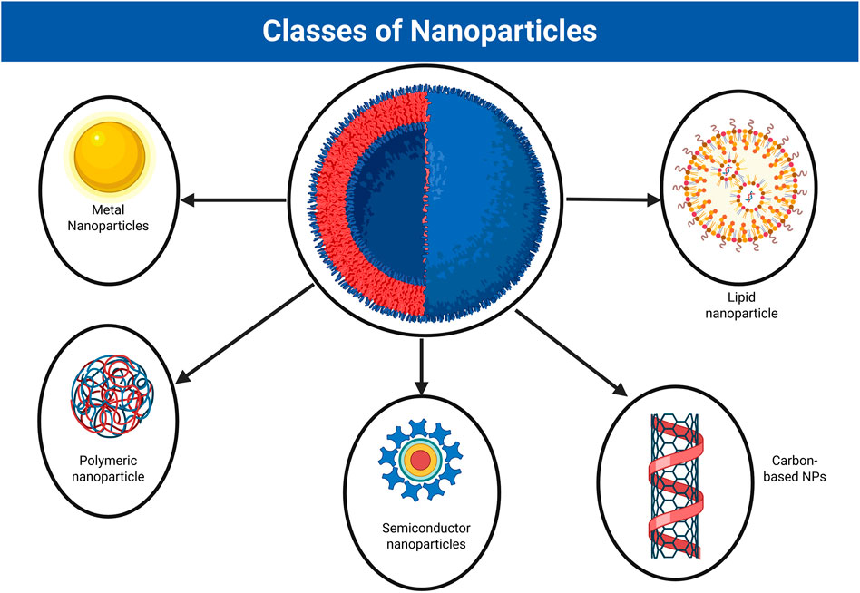
Figure 2. Different classes of nanoparticles (Created in https://BioRender.com).
Biosynthesized nanoparticles such as microalgae-based green synthesized NPs are preferred for cancer treatment becauseof their eco-friendly synthesis without the requirement of high pressures, high temperatures, and toxic chemicals (Khan et al., 2019; Alsammarraie et al., 2018). The microalgae processing for green nanoparticle synthesis consists of microalgal cell cultivation (upstream processing, USP) under stress conditions (Kaushik et al., 2023) and harvesting of microalgal biomass (downstream processing, DSP) (Katiyar and Arora, 2020; Khoo et al., 2020; Tang et al., 2020; Khanra et al., 2018; MatAron et al., 2021). The microalgae-based green synthesized gold nanoparticles (AuNPs) and silver nanoparticles (AgNPs) found to be effective as anticancer agentsthose findings were tabulated in Table1.

Table 1. Microalgal-based nanoparticles and their role in cancer therapy.
Various microalgal species, such as Chlorella, Spirulina, and Scendesmus, were utilized for green synthesis of AgNPs. The process involves the suspending of microalgal biomass in an aqueous silver nitrate solution (1 mM). The synthesized AgNPs were characterized by transmission electron microscopy (TEM), UV spectroscopy, energy dispersive X-ray energy dispersive spectroscopy (EDX) followed by the evaluating the cytotoxicity of the synthesized NPs for anti-cancerous activity (Tran et al., 2023). The green synthesized gold nanoparticles (AuNPs) of D. salina, includes the steps of cultivating the microalgae in MJM media followed by harvesting of microalgal biomass, suspending the aqueous microalgal biomass (20%) in aqueous silver chloride (1 M), and finally centrifuged to get the AuNPs. The green synthesized AuNPs were further characterized by TEM, FTIR, and XPS (Singh et al., 2019).
AgNPs may act against virus-, cancer-, bacteria-, and fungus-infected cells. Muneebaetal (İnan et al., 2021), used the DHM1, DHM3, and DHM3 strains for microalgalNP synthesis to assess their activity against a cancer cell line (MCF7). As a part of the procedure, silver nitrate solution (5mM, aqueous) was added to the microalgae-derived ethanolic extract (5:1). On overnight incubation, a color change was observed from green to yellow and AgNPs were obtained by centrifugation. Sathishkumar et al. (2019) prepared AgNPs from aqueous extract using Trichodesmium erythraeum via an environmentally friendly method that was found to have anti-proliferative and antioxidant properties. Algotiml et al. (2022) biosynthesized nanoparticles from Ulva rigida, Cystoseira myrica, and Gracilaria foliifera and tested them for antimicrobial and anticancer activity. The major algal extract components included fatty acids, amide proteins, terpenoids, flavonoids, polyphenols, fluoro aliphatic compounds, pyruvic acid, volatile compounds, and alkalines. AgNPs exhibited antimicrobial and antifungal activity against foodborne microbes and pathogenic fungi, respectively. These AgNPs were proven to be antidermophytes in the case of skin infections and anticancerous against breast cancer (MCF7) cell lines. Al-Zahrani et al. (2021) synthesized AgNPs by using the microalgae Chaetomorpha ligustica. These compounds were effective against HCT116 and HT29 (colon cancer cell lines) cells. Chaetomoroha ligustica extract and its nanoparticles proved to be cytotoxic, but the cytotoxicity depended on the dose. Several other biosynthesized nanoparticles prepared from Bifurcaria bifurcate, Chlorococcum humicola, Galaxuara elongata, Sargassum plagiophyllum, Amphora-46, Caulerpa racemose, Microcoleus sp., and Ulva fasciata are known for their antibacterial activity (Al-Radadi et al., 2022; Zaman et al., 2020).
2.1 Factors affecting the green synthesis of microalgae-based NPs
The optimal yield of microalgae-based NPs depends on the synthesis factors, namely, temperature, pH, reactant concentration, reaction time, capping agent, and choice of organism. These factors may affect the shape, size, and stability of nanoparticles, determining nanoparticle toxicity.
2.1.1 Temperature
Chemical methods such as electrochemical and solvothermal methods are highly influenced by temperature. Physical processes require a temperature of 350 °C, whereas chemical methods require a lower temperature. A temperature of 100 °C is needed for the synthesis of microalgal NPs. At high temperatures may increase the reduction rate (Shanmuganathan et al., 2023).
2.1.2 pH
pH affects the shape and size of NPs; a low pH causes the SPR peak to widen and shift toward a longer wavelength region, producing a variety of NPs (often triangular or circular, for example). In contrast, a high pH is ideal for making small NPs and promotes the formation of spherical NPs. NPs show additional stabilization in alkaline or acidic environments. Large pearl-sized NPs were produced in abundance under alkaline conditions and were far more stable than the clustered NPs made under acidic conditions (Zhang et al., 2022).
2.1.3 Reactant concentration
Varying effects on the generation of NPs can be caused by different reactant concentrations in an algal extract. The impact of reducing agent concentration on the size and quantity of selenium particles produced by Chlorella vulgaris extract was investigated. Transmission electron microscopy (TEM) analysis of the time-dependent creation of nanoparticles revealed a critical role for multiple twinned particles (MTPs) in this process. Additionally, it was discovered that the development of single-crystalline selenium nanotriles was caused by the sluggish nature of the reaction and the influence of the shape and direction of the extract (Dinc et al., 2022).
2.1.4 Reaction time
Reaction time is a critical factor in microalgal NPs synthesis. The same experiment can produce varied particle sizes if the reaction time is altered. The algal extract used to synthesize the NPs began to develop in 2 min and produced spherical NPs with a mean size of 12 nm after 5 min. Additionally, the progressive increase in contact duration and interaction between microalgae and silver ions (Ag+) at room temperature results in an increase in the SPR peak intensity and the rapid biosynthesis of non-agglomerated AgNPs (Moraes et al., 2021).
2.1.5 Choice of the microalgal strain
Cost-effective NPs synthesis depends not only on chemical-physical parameters but also on the chosen microalgae based on critical intrinsic properties such as biochemical pathways, growth rate, and enzyme activities; (ii) the size of the inoculum; and (iii) the selection of biocatalysts, which is essential for accelerating the rate of reaction (i.e., reduction). Whole cells and enzymes can all be employed as biocatalysts. Live whole cells are desirable because these coenzymes are expensive and may be recycled along the route, demonstrating their enormous efficacy (Moraes et al., 2021).
2.1.6 Capping agent
The stability of NPS should be improved to prevent aggregation and oxidation, particularly by utilizing organic protective ligands whose head group binds to metal NP surfaces with high affinity to stabilize highly reactive surface atoms. The alkyl spacer between the head and tail groups of the ligand is thought to act as a capping shell and regulate the interparticle spacing. Furthermore, the surface reactivity and solubility of NPs are greatly influenced by the functional tail groups of the ligand. Importantly, it was discovered that the kind of ligand (such as disulfide, ammonium, thiol, or citrate) and the level of ligand capping, along with the synthetic conditions used, could systematically change the size, shape, and ligand-to-metal ratio of the NPs and directly affect their chemical and physical (such as electronic and optical) properties (San and Shon, 2018).
3 Synthesis mechanism of microalgal NPs
Microalgae are used to prepare metallic nanoparticles that have applications in anticancer therapy. Phytochemicals in microalgae contain functional groups such as carboxyl, amino, and hydroxyl groups, which assist in reducing metals and serve as capping agents (providing coatings on nanoparticles). Nanoparticles of silver, cadmium, gold, lead, and silicon-germanium can be prepared using marine species (Restrepo and Villa, 2021). Green nanoparticle formation can be achieved via (a) intracellular synthesis, where compounds inside the cell carry out reduction; or by extracellular synthesis, where compounds outside the cell carry out reduction (Figure 3). In intracellular NPs synthesis, the metal ions taken by microalgal cells are reduced to NPs with the aid of microalgal metabolites, followed by NPs extraction from within the microalgal cells. In contrast, the extracellular NPs synthesis and stabilization proceed with the reduction of metal ions outside the microalgal cells (on the cell surface or algal extracts containing solutions) with the aid of secreted microalgal metabolites. In both approaches, microalgal metabolites/bioactive compounds act as reducing agents (by donating electrons, which reduces the metal ions to NPs synthesis) and capping agents (to stabilize and prevent clumping). The microalgal NPs synthesis also relies on the growth characteristics of microalgae and on the environmental factors (metal ion concentration, pH, temperature, etc.) which dictate the structural and stability features of synthesized NPs for tailored biomedical applications (Restrepo and Villa, 2021).

Figure 3. Extracellular and Intracellular synthesis mechanisms of microalgal NPs (Modified and adapted from (Chan et al., 2022)) (Created in https://BioRender.com).
The bioreduction process includes activation, growth, and termination. For instance, (a) during activation, metal ions are reduced, and reduced metal ions undergo nucleation; (b) during the growth phase, small nanoparticles aggregate into large particles marked with thermodynamic stability; and (c) termination is marked by biomineralization (inorganic (metal)-organic (microalgal assisted) composite) and nanoparticle stabilization (Figure 4). The formation of nanoparticles from Tetraselmisko chinensis and Sargassum muticum occurs via intracellular and extracellular synthesis (Chan et al., 2022). The general procedure for generating microalgal-based nanoparticles includes major steps, viz., (1) the addition of a microalgal extract to a metal ionic solution where the reduction of the metal ion is caused by the functional nature of the microalgal compound; (2) the incubation of the sample after centrifugation, where successful reduction is marked by a change in the color of the solution (e.g., yellow to purple); (3) characterization, where transmission electron microscopy (TEM) reveals the size of the nanoparticles, energy dispersive analysis (EDX) confirms the presence of an element (e.g., silver), Fourier transform infrared (FTIR) spectroscopy reveals the type of biomolecule responsible for the stabilization of the AgNPs, and UV-VIS spectra determine the structure and properties of the nanoparticles (Singh et al., 2019).

Figure 4. Schematic representation of microalgal NPs synthesis (Modified and adapted from (Chan et al., 2022)). (FTIR; Fourier Transform Infrared, SEM: Scanning Electron Microscopy, TEM: TransmissionElectroscopy Microscopy) (Created in https://BioRender.com).
Among different microalgae, the selection of microalgal species depends on the gamet of packed biomolecules posses by particular algal species, which dictates the metal ion stabilization towards NPs synthesis, and also on the type of expected metallic NPs with intended application domain. The selection of microalgae for NPs synthesis includes a systematic process of identifying the required metallic NPs for targeted application, with concrete proof of microalgal species for synthesizing particular metallic NPs. Once the type of NPs and microalgal species are identified, growth kinetics and particular metal uptake by selected microalgal species are determined, which follows the optimization of NPs synthesis reaction conditions (temperature, pH, and reaction time) towards better size, shape, and functionality. Finally, the synthesized microalgal NPs have to be characterized to determine the different structural and chemical properties for better suitability for targeted biomedical applications (Arteaga-Castrejón et al., 2024).
4 Characterization of the synthesized microalgal NPs
Characterization of the NPs was carried out to determine the microscopic structure and material properties through microscopy-, spectroscopy- and X-ray-based techniques (Figure 5). Table 2 highlights the characterization techniques, principles, and use of methods in nanoparticle synthesis and sample preparation.

Figure 5. Microscopy- and spectroscopy-based characterization techniques for Microalgal NPs (SEM: Scanning Electron Microscopy, TEM: Transmission ElectroscopyMicroscopy,AFM:Atomic Force Microscope, FTIS: Fourier Transform Infrared Spectroscopy) (Created in https://BioRender.com).
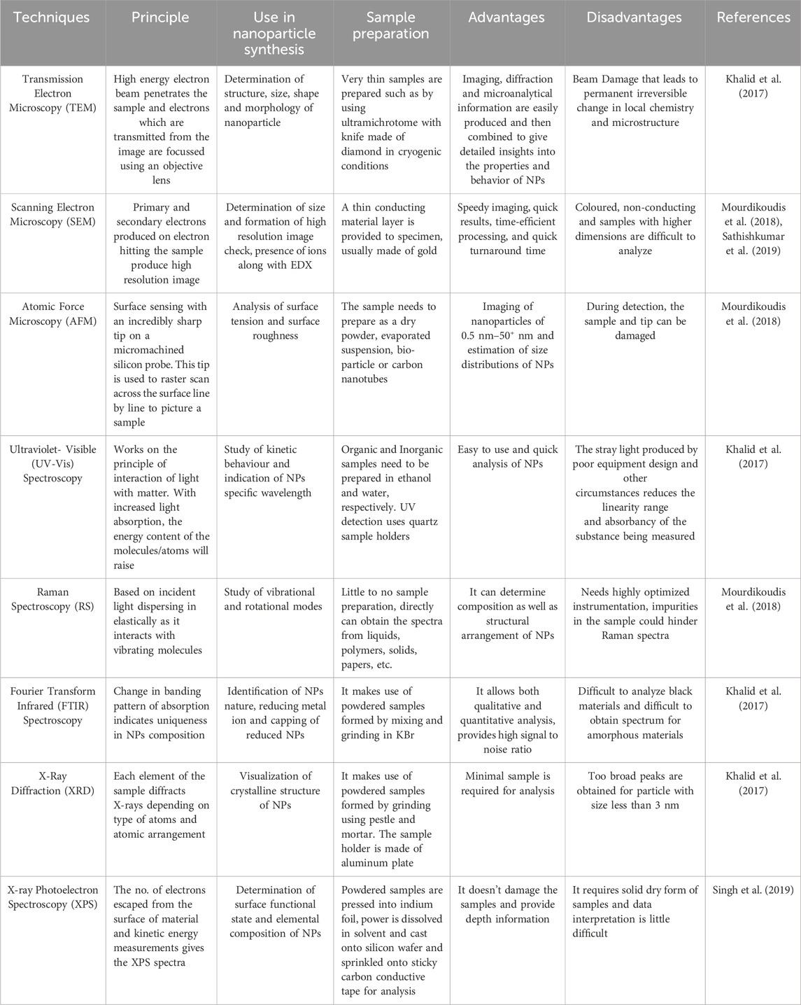
Table 2. Different characterization techniques of NPs and their features.
One of the advanced nanoparticle characterization techniques is nanoSIMS, which uses isotope labeling to analyze and study a complete biological system. It can distinguish between isotopes and combines fine spatial resolution with high atomic sensitivity. The zeta potential, also called the electrokinetic potential, is used to measure the electric charge on the nanoparticle surface. The zeta potential of the particles indicates nanoparticle stability (Fernandes et al., 2023). Other techniques include NMR, which is used to analyze NP size, atomic composition, electronic core structure, ligand density and ligand influence; dynamic light scattering (DLS), which is used to detect agglomerates and hydrodynamic size; SQUID-nanoSQUID, which is used to analyze magnetization remanence, blocking temperature and magnetization saturation; liquidTEM, which is used to study growth mechanisms, depict nanoparticle growth in realtime, superlattice formation and single particle motion; HRTEM, which is used to distinguish amorphous, poly- and monocrystallineNPs; electron diffraction, which is used to study lattice and long-range order parameters; cryo-TEM, which is used to explore aggregation pathways and complete growth mechanisms; electronomography, for 3D particle visualization, video, snapshots, and quantitative information at the atomic scale; electron backscatter diffraction (EBSD), which is used to examine microstructure, texture and defects in NPs; magnetic force microscopy (MFM), which provides information about the magnetic moment of NPs and differentiates them from nonmagnetic NPs (Mourdikoudis et al., 2018).
5 Microalgal NPs in cancer treatment
Microalgae are reservoirs of bioactive compounds such as sterols, polysaccharides, fatty acids, phycobiliproteins, phenolics, and vitamins that play major roles in preventing or curing cancer. Table 3 summarizes the different bioactive molecules produced by microalgae and their therapeutic potential.

Table 3. Microalgae derived bioactive molecules and their use in cancer.
5.1 Mechanism of action of microalgal NPs in cancer treatment
There are four major ways by which microalgae affect cancer cells. (1) Microalgae are known to decrease the binding capacity for tubulin polymerization, which inhibits the synthesis of microtubules (e.g., Cucarin A). (2) They alter the expression of COX-2, MMP-9, MMP-2, and ERK-2 (e.g., astaxanthin), which decreases invasion capacity. (3) They decrease VEGF, i.e., vascular endothelial growth factor (e.g., fucoidan), which results in antiangiogenic activity. (4) Increased fas, ICAM (intercellular adhesion molecule) and decreased bcl2 (e.g., C-phycocyanin), which cause the activation of caspase 2 3 4 6 8 9 10. Microalgae directly affect cancer in five ways. (1) PUFAs from microalgae, such as DHA, cause DNA fragmentation; (2) PUFAs, such as DHA, decrease the mitochondrial membrane potential. (3) PUFAs such as astaxanthin and DHA activate ERK, increasing p27. (4) Astaxanthin modulates NF-κB. (5) DHA increases cytochrome C, p53, and bax levels, leading to cell cycle arrest and an antiproliferative effect. All the above-mentioned factors lead to the apoptosis of cancer cells.The mechanism of action of microalgal AgNPs in cancer treatment is summarized in Figure 6 (Hamida et al., 2022).
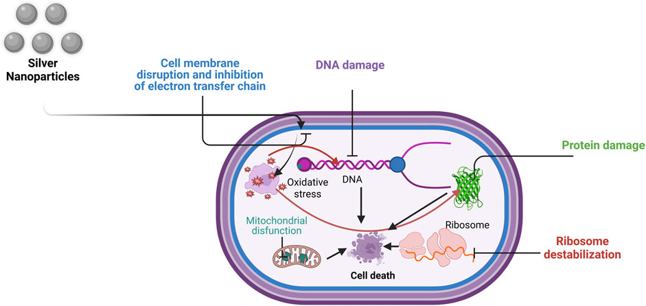
Figure 6. Mechanism of action of AgNPs in cancer treatment (Modified and adapted from (Hamida et al., 2022)) (Created in https://BioRender.com).
5.2 Cell line studies and toxicity studies associated with the microagal NPs
The cell toxicity, genotoxicity, and immunotoxicity of NPs have been studied. As proven by many studies, these compounds are toxic to biological systems. The toxicity depends on the structure, size, and material from which the nanoparticle is made. To date, evidence has confirmed that biologically synthesized NPs, especially microalgal NPs, are less toxic to normal cells. Gold nanoparticles synthesized by using the microalga D. salina were tested for their effect on cancer (MCF 7) and normal (MCF 10A) cells. These compounds were shown to be cytotoxic to cancer cells and had no negative impact on the normal cell line (Singh et al., 2019). AgNPs made from the microalgae T. erythraeum caused antiproliferation in MCF-7 and HeLa cell lines, and they were not harmful to normal cells (Sathishkumar et al., 2019). AgNPs made using U. rigida were lethal to MCF-7 cells, a breast cancer cell line, and were not cytotoxic to normal cells. Because the core of gold NPs is inert and nontoxic, they are believed to be relatively harmless. On the other hand, metallic NPs are highly cytotoxic to normal cells; for example, aluminum oxide NPs decrease the viability of cells, increase oxidative stress, alter mitochondrial function, and change protein expression in the blood‒brain barrier (Algotiml et al., 2022). Copper NPs cause impairment in the kidney, spleen, and liver. Metallic AgNPs accumulate in many organs and can cross the BBB and reach the brain. After exposing rats to silver-based NPs by inhalation or subcutaneous injection, AgNPs have been found in several organs, including the lungs, spleen, kidney, liver, and brain. Furthermore, these NPs have shown greater ROS formation and cell survival toxicity. The most prevalent harmful effects of zinc-based nanomaterials, including cell membrane damage, cytotoxicity, and enhanced oxidative stress, have been described in diverse mammalian cell lines. Exposure of human mesothelioma cells and rodent fibroblasts to high concentrations of zinc oxide nanoparticles caused cell death (Hamida et al., 2022).
6 Technical challenges in green synthesis of microalgal NPs
The green synthesis of microalgal NPs process starts with the culture of microalgal cells and harvesting. Batch and Fed-batchor continuous cultivation modes are the most common techniques. Biomass is collected and processed during the cultivation phase. The cost of biomass harvesting is estimated to be nearly 30% of the price of microalgal downstream processes. As a result, the high cost of harvesting is one of the most significant bottlenecks in commercializing microalgal processes. Due to the density of microalgal cells during growth, the medium is often low, and most microalgal cells have a negative charge, which causes them to be suspended; moreover, the microalgal harvesting procedure is costly and energy-intensive (Jacob et al., 2021).As a part of harvesting, several harvesting procedures were reported such as sedimentation, ultrasound, centrifugation, filtration, and flotation, to maximize algal biomass yield. Nonetheless, these approaches are not as practical as flocculation because of their high cost and lower efficiency. The flocculation harvesting process is far more comfortable and efficient than previous methods, yet there are still many hurdles to overcome. On the other hand, utilizing flocculants to collect microalgal biomass can pollute slurry concentrates, lowering the market value of algal biomass and making it difficult to isolate valuable microalgal compounds. As a result, the only method to reduce harvesting costs is to improve harvesting technology (Halder and Azad, 2019).
The application of NPs in biomedical applications is the most vulnerable circumstance. The interaction between nanotoxicity and its biomolecules has been the subject of various studies. Nonetheless, assessing and validating nanotoxicity in a living system is complex. The assessment of toxicity and the challenges in identifying the influence on living systems are historic. Researchers face challenges when evaluating nanomaterials in solution-based, powder form and dealing with biological systems using various methodologies for toxicity evaluation. Evaluation tools and characterization procedures are critical for overcoming these hurdles, whereas cytotoxic assays consider nanoparticle shape, size, and morphology (Gupta et al., 2019).
7 Trends, scope and sustainability aspects of green synthesis of microalgal NPs
Since the discovery of nanoparticles, physical and chemical production methods have been prominent. In 2009, a wave led to both increases in nanoparticle synthesis and the adoption of biological synthesis methods. There has been an increase in the green synthesis of NPs owing to their sustainability and advantages. By emphasizing the green synthesis of NPs by microalgae, numerous microalgae have been explored for their ability to produce nanoparticles (Bao and Lan, 2019).
Despite their production, what remains unexplored is the application part of synthesized NPs. Research has yet to be carried out to determine their specific role in diagnostics, specifically in diagnosis of cancer. Table 4 summarizes the different clinical application status of green synthesized microalgal NPs. The production of reported microalgal NPs has increased the scope of further research that can focus on exploring the potential uses of these NPs in diagnostics and cancer therapy.

Table 4. Microalgal NP’s and its clinical application status (Bao and Lan, 2019; Patil and Kim, 2018).
7.1 Sustainability features associated with the green synthesis of microalgal NPs
Adopting green synthesis of microalgal NPs route brings economic and environmental sustainability features to the process. In the context of the environment, sustainability involves making efficient use of resources and keeping them for use by future generations. Microalgae grow with the aid of sunlight, carbon dioxide and inorganic nutrients such as nitrogen and phosphate. Utilization of atmospheric CO2 and inorganic nutrients of wastewater by microalgae makes the process more sustainable in nature (Chan et al., 2022). This sustainability feature of green synthesized microalgal NPs outperforms the existing chemical- and physical-based NPs synthesis techniques (sol-gel technique, nonsputtering, reduction, and electrochemical methods) by avoiding the usage of toxic, expensive reagents, high energy, pressure requirements of process, and associated intricacy of the separation process (Chan et al., 2022). The microalgal biomolecules serves as the capping agents for stabilization of synthesized NPs in case of green synthesis of microalgal NPs which aligns with green chemistry fundamentals (Mondal et al., 2023). The envisaged conditions of microalgal NPs green synthesis contribute to a lower environmental impact, which also aids in the cost-effectiveness of the process. Moreover, choosing microalgal route for NPs synthesis contributes towards the CO2 sequestration, coupled with wastewater treatment aspects. Being a biological process, the microalgal route of NPs synthesis results in minimal hazardous waste generation with bioremediation potential and a scalable process (by employing photobioreactor systems) along with the better economics of value-added byproducts side chains. These economic and environmental aspects make green synthesis of microalgal NPs, a viable alternative for conventional chemical and physical approaches for NPs synthesis, offering the advantages of sustainability and industrial feasibility (Mondal et al., 2023).
8 Quality control aspects of green synthesized microalgal NPs
NP use is divided into two primary categories: pharmaceutical and medical. The components and procedures utilized in drug production are subject to quality control. It is crucial to accurately assess formulation excipients and active pharmaceutical ingredients (APIs) for optimizing and evaluating preformulations. To guarantee the safety and efficacy of medications throughout the regulatory timeframe, they must be of sufficient strength, purity, quality, and potency. Many nanostructure systems, such as liposomes, nanoemulsions, dendrimers, nanocrystals, and metal oxides (zinc oxide, superparamagnetic iron oxide, titanium dioxide), have been approved by the FDA. The European Medical Agency and European Commission designated doxorubicin polyisohexylcyanoacrylate nanoparticles for treating hepatocellular carcinoma as orphan drugs and awarded this classification to the Bio Alliance in October 2004. The first nanodrug to receive FDA approval was Doxil in 1995. Dosage forms contain pegylated liposomes that carry the chemotherapeutic drug doxorubicin. An injectable amphotericin liposome is known as an AmBisome® (Taghizadeh et al., 2021).
There are several requirements for using nanoparticles as drugs, such as size (primary particle size, volume, and surface area), agglomeration state, distribution in two or three dimensions, chemical composition (element identification and distribution, crystal shape, and particle size distribution), and surface composition (charge on the surface). These requirements are crucial for nanoparticles used in biomedical applications. However, specifics of nanoparticle size in bulk materials and intended pharmaceutical items are critical to comprehending a drug’s pharmacodynamics and pharmacokinetic characteristics. There is a clear association between the engineering of nanoparticles, including manufacturing, and their impact on cell surface composition, morphology (size and shape), surface composition, and aggregation. Consequently, for employing green synthesized microalgal NPs for medical usage, more research is needed to ensure that these NPs can pass quality-control examinations. Additionally, essential quality characteristics, including solubility, stability, and solid-state qualities, should be considered when evaluating the suitability of green synthesized microalgal NPs for various pharmaceutical and medicinal applications (Taghizadeh et al., 2021).
9 Future perspectives
To date, only a few nanoparticles of gold and silver have been synthesized using microalgae through green synthesis. The green synthesis of zinc oxide, copper, selenium, titanium, and iron NPs of microalgae and its role in cancer treatment need to be explored. Additionally, numerous microalgae have a high potential for nanoparticle synthesis that remains unexplored. Therefore, microalgal NPs synthesis is a trending field that requires intense research for medicinal applications, especially for cancer treatment.
The microalgal NPs synthesis towards cancer treatment may benefit from further research on the contribution of synthetic biology and genetic engineering approaches. Synthetic biology intrusion enables precise genetic modifications through CRISPR/Cas9 technologies to manipulate the metabolic pathways, which help in the efficient conversion of metal ions into NPs with desired sizes and properties, which eventually help in the cost-effectiveness of the process, which suits the needs of the biomedical industry. The research domain also needs to explore the possibilities of utilizing genetically engineered microalgae for NPs synthesis for cancer treatment towards enhanced efficiencies with tailored NPs producing traits in a sustainable production manner (Zhang and Fussenegger, 2024).
10 Conclusion
The review discussed the role of microalgal NPs as therapeutic agents for treating cancer cells through a sustainable, green synthesis approach. Furthermore, the probable mechanism of microalgal NP’s synthesis, its characterization approaches, and technical challenges associated with the process have been discussed. Moreover, the extended application of green-synthesized microalgal NPs to cancer cells was addressed through the mechanism of action on cancer cells and cytotoxicity studies. Finally, the quality control aspects and prospects of green-synthesized microalgal NPs are summarized. This review provides a concise overview of the green synthesis of microalgal NPs for potential cancer treatment.
Author contributions
VG: Funding acquisition, Formal Analysis, Writing – original draft, Investigation, Supervision, Validation, Conceptualization, Project administration, Writing – review and editing. SS: Validation, Investigation, Writing – review and editing, Methodology, Resources, Writing – original draft. DS: Investigation, Resources, Validation, Writing – review and editing, Formal Analysis, Writing – original draft. SK: Conceptualization, Methodology, Writing – review and editing, Investigation, Writing – original draft, Resources. SJ: Resources, Writing – original draft, Writing – review and editing, Supervision. AK: Writing – original draft, Methodology, Resources, Investigation, Writing – review and editing. AG: Project administration, Methodology, Writing – review and editing, Writing – original draft, Resources. AC: Investigation, Formal Analysis, Writing – original draft, Resources, Writing – review and editing, Validation.
Funding
The author(s) declare that no financial support was received for the research and/or publication of this article.
Acknowledgments
VG, SS, DS, AS, and AG acknowledge JUIT, Waknaghat, HP- 173234, India for providing the learning and resource facilities to execute the proposed review article.
Conflict of interest
The authors declare that the research was conducted in the absence of any commercial or financial relationships that could be construed as a potential conflict of interest.
The author(s) declared that they were an editorial board member of Frontiers, at the time of submission. This had no impact on the peer review process and the final decision.
Generative AI statement
The author(s) declare that no Generative AI was used in the creation of this manuscript.
Any alternative text (alt text) provided alongside figures in this article has been generated by Frontiers with the support of artificial intelligence and reasonable efforts have been made to ensure accuracy, including review by the authors wherever possible. If you identify any issues, please contact us.
Publisher’s note
All claims expressed in this article are solely those of the authors and do not necessarily represent those of their affiliated organizations, or those of the publisher, the editors and the reviewers. Any product that may be evaluated in this article, or claim that may be made by its manufacturer, is not guaranteed or endorsed by the publisher.
References
Abd El-Hack, M. E., Abdelnour, S., Alagawany, M., Abdo, M., Sakr, M. A., Khafaga, A. F., et al. (2019). Microalgae in modern cancer therapy: current knowledge, biomed. & Pharma. 111, 42–50. doi:10.1016/j.biopha.2018.12.069
Abed, A., Derakhshan, M., Karimi, M., Shirazinia, M., Mahjoubin-Tehran, M., Homayonfal, M., et al. (2022). Platinum nanoparticles in biomedicine: preparation, anti-cancer activity, and drug delivery vehicles. Front. Pharmacol. 13, 797804. doi:10.3389/fphar.2022.797804
Acharya, D., Satapathy, S., Yadav, K. K., Somu, P., and Mishra, G. (2022). Systemic evaluation of mechanism of cytotoxicity in human colon cancer HCT-116 cells of silver nanoparticles synthesized using marine algae Ulvalactuca extract. J. Inorg. Organomet.Polym.Mater. 32, 596–605. doi:10.1007/s10904-021-02133-8
Akiba, T., Morikawa, T., Odaka, M., Nakada, T., Kamiya, N., Yamashita, M., et al. (2018). Vitamin D supplementation and survival of patients with non–small cell lung cancer: a randomized, Double-Blind, placebo-controlled trial. Clin. Cancer Res. 24, 4089–4097. doi:10.1158/1078-0432.ccr-18-0483
Al-Radadi, N. S., Hussain, T., Faisal, S., Shah, S. A. R., and Ali Raza Shah, S. (2022). Novel biosynthesis, characterization and bio-catalytic potential of green algae (Spirogyra hyalina) mediated silver nanomaterials. Saudi J. Biol. Sci. 29, 411–419. doi:10.1016/j.sjbs.2021.09.013
Al-Zahrani, S. A., Bhat, R. S., Al Rashed, S. A., Mahmood, A., Al Fahad, A., Alamro, G., et al. (2021). Green-synthesized silver nanoparticles with aqueous extract of green algae Chaetomorphaligustica and its anticancer potential, green Process. Syn. 10, 711–721. doi:10.1515/gps-2021-0067
Algotiml, R., Gab-Alla, A., Seoudi, R., Abulreesh, H. H., El-Readi, M. Z., and Elbanna, K. (2022). Anticancer and antimicrobial activity of biosynthesized Red Sea marine algal silver nanoparticles. Sci. Rep. 12, 2421. doi:10.1038/s41598-022-06412-3
Alsammarraie, F. K., Wang, W., Zhou, P., Mustapha, A., and Lin, M. (2018). Green synthesis of silver nanoparticles using turmeric extracts and investigation of their antibacterial activities. Colloid. Surf. B Biointerface. 171, 398–405. doi:10.1016/j.colsurfb.2018.07.059
Arteaga-Castrejón, A. A., Vivechana, A., and Sanghamitra, K. (2024). Microalgae as a potential natural source for the green synthesis of nanoparticles. Chem. Commun. 60, 3874–3890. doi:10.1039/d3cc05767d
Ayna, A. (2020). Apoptotic effects OF beta-carotene, alpha-tocopherol and ascorbic acid ON PC-3 prostate cancer cells. Hacet. J. Biol. Chem. 48, 211–218. doi:10.15671/hjbc.519212
Baby, E. K., and Reji, C. (2022). “Metal-based nanoparticles for infectious diseases and therapeutics,” in Nanotechnology for infectious diseases (Berlin, Germany: Springer Nature), 103–124.
Bao, Z., and Lan, C. Q. (2019). Advances in biosynthesis of noble metal nanoparticles mediated by photosynthetic organisms—A review. Colloid. Surf. B Biointerface. 184, 110519. doi:10.1016/j.colsurfb.2019.110519
Bin-Meferij, M. M., and Hamida, R. S. (2019). Biofabrication and antitumor activity of silver nanoparticles utilizing novel Nostoc sp. Bahar M. Int. J. nanomed. 14, 9019–9029. doi:10.2147/IJN.S230457
Cezare-Gomes, E. A., Mejia-da-Silva, L. d., Pérez-Mora, L. S., Matsudo, M. C., Ferreira-Camargo, L. S., Singh, A. K., et al. (2019). Potential of Microalgae Carotenoids for industrial application. Appl.Biochem. Biotechnol. 188, 602–634. doi:10.1007/s12010-018-02945-4
Chan, S. S., Low, S. S., Chew, K. W., Ling, T. C., Rinklebe, J., Juan, J. C., et al. (2022). Prospects and environmental sustainability of phyconanotechnology: a review on algae-mediated metal nanoparticles synthesis and mechanism. Environ. Res. 212, 113140. doi:10.1016/j.envres.2022.113140
Chen, X., Song, L., Wang, H., Liu, S., Yu, H., Wang, X., et al. (2019). Partial characterization, the immune modulation and anticancer activities of sulfated polysaccharides from filamentous microalgae tribonema sp. Molecules 24, 322. doi:10.3390/molecules24020322
Cooper, K., Campbell, F., Harnan, S., and Sutton, A. (2023). Association between stress, depression or anxiety and cancer: rapid review of reviews. Psychoneuroendocrinology 16, 100215. doi:10.1016/j.cpnec.2023.100215
Darwito, D., Dharmana, E., Riwanto, I., Budijitno, S., Suwardjo, S., Purnomo, J., et al. (2019). Effects of Omega-3 supplementation on Ki-67 and VEGF expression levels and clinical outcomes of locally advanced breast cancer patients treated with neoadjuvant CAF chemotherapy: a randomized controlled trial report. Asian pac. J. Cancer Prev. 20, 911–916. doi:10.31557/apjcp.2019.20.3.911
Dinc, S. K., Vural, O. A., Kayhan, F. E., and Keskin, N. O. S. (2022). Facile biogenic selenium nanoparticle synthesis, characterization and effects on oxidative stress generated by UV in microalgae. Particuology 70, 30–42. doi:10.1016/j.partic.2021.12.005
El-Naggar, N. E. A., Hussein, M. H., and El-Sawah, A. A. (2018). Phycobiliprotein-mediated synthesis of biogenic silver nanoparticles, characterization, in vitro and in vivo assessment of anticancer activities. Sci. Rep. 8, 8925. doi:10.1038/s41598-018-27276-6
Elkhateeb, W., El-Sayed, H., Fayad, W., Al kolaibe, A. G., Emam, M., and Daba, G. (2020). In vitro anti-breast cancer and antifungal Bio-efficiency of some microalgal extracts. J. Aquat. Biol. Fish. 24, 263–279. doi:10.21608/ejabf.2020.70448
Faraone, I., Sinisgalli, C., Ostuni, A., Armentano, M. F., Carmosino, M., Milella, L., et al. (2020). Astaxanthin anticancer effects are mediated through multiple molecular mechanisms: a systematic review. Pharmacol. Res. 155, 104689. doi:10.1016/j.phrs.2020.104689
Ferdous, Z., and Nemmar, A. (2020). Health impact of silver nanoparticles: a review of the biodistribution and toxicity following various routes of exposure. Int. J. Mol. Sci. 21, 2375. doi:10.3390/ijms21072375
Ferdous, U. T., and Yusof, Z. N. B. (2021). Medicinal prospects of antioxidants from algal sources in cancer therapy. Front. Pharmacol. 12, 593116. doi:10.3389/fphar.2021.593116
Ferdous, U. T., and Yusof, Z. N. B. (2021). “Algal terpenoids: a potential source of antioxidants for cancer therapy,” in Terpenes and terpenoids-recent advances. Editors A. M. Al-Taweel, and S. Perveen (London, United Kingdom: IntechOpen), 63–69.
Fernandes, J. P. C., Federico, C. E., Lentzen, E., Valle, N., Basterra-Beroiz, B., Weydert, M., et al. (2023). AFM-nanoSIMS correlative microscopy on multiphase elastomeric systems: nanomechanical properties and elemental distribution at the nanoscale. Polym. Test. 121, 107996. doi:10.1016/j.polymertesting.2023.107996
Fimbres-Olivarria, D., Carvajal-Millan, E., Lopez-Elias, J. A., Martinez-Robinson, K. G., Miranda-Baeza, A., Martinez-Cordova, L. R., et al. (2018). Chemical characterization and antioxidant activity of sulfated polysaccharides from Navicula sp. Food Hydrocolloid 75, 229–236. doi:10.1016/j.foodhyd.2017.08.002
Foo, S. C., Khoo, K. S., Ooi, C. W., Show, P. L., Khong, N. M., and Yusoff, F. M. (2021). Meeting sustainable development goals: alternative extraction processes for fucoxanthin in algae. Front. Bioeng.Biotechnol. 8, 546067. doi:10.3389/fbioe.2020.546067
Gupta, A., Kumar, S., and Kumar, V. (2019). “Challenges for assessing toxicity of nanomaterials,” in Biochemical toxicology - heavy metals and nanomaterials. Editors M. Ince, O. Kaplan Ince, and G. Ondrasek (IntechOpen), 65–85.
Halder, P., and Azad, A. K. (2019). “Recent trends and challenges of algal biofuel conversion technologies,” in Advanced biofuels (Elsevier), 167–179.
Hamida, R. S., Abdelmeguid, N. E., Ali, M. A., Bin-Meferij, M. M., and Khalil, M. I. (2020a). Synthesis of silver nanoparticles using a novel Cyanobacteria Desertifilum sp. extract: their antibacterial and cytotoxicity effects. Int. J. Nanomed. 15, 49–63. doi:10.2147/IJN.S238575
Hamida, R. S., Ali, M. A., Redhwan, A., and Bin-Meferij, M. M. (2020b). Cyanobacteria—A promising platform in green nanotechnology: a review on nanoparticles fabrication and their prospective applications. Int. J. Nanomed. 15, 6033–6066. doi:10.2147/ijn.s256134
Hamida, R. S., Albasher, G., and Bin-Meferij, M. M. (2020c). Oxidative stress and apoptotic responses elicited by nostoc-synthesized silver nanoparticles against different cancer cell lines. Cancers 12, 2099. doi:10.3390/cancers12082099
Hamida, R. S., Ali, M. A., Almohawes, Z. N., Alahdal, H., Momenah, M. A., and Bin-Meferij, M. M. (2022). Green synthesis of hexagonal silver nanoparticles using a novel microalgae coelastrellaaeroterrestrica strain BA_Chlo4 and resulting anticancer, antibacterial, and antioxidant activities. Pharmaceutics 14, 2002. doi:10.3390/pharmaceutics14102002
Haque, S., Norbert, C. C., Acharyya, R., Mukherjee, S., Kathirvel, M., and Patra, C. R. (2021). Biosynthesized silver nanoparticles for cancer therapy and in vivo bioimaging. Cancers 13, 6114. doi:10.3390/cancers13236114
Heinemann, M. G., Rosa, C. H., Rosa, G. R., and Dias, D. (2021). Biogenic synthesis of gold and silver nanoparticles used in environmental applications: a review. Environ. Anal.Chem. 30, e00129. doi:10.1016/j.teac.2021.e00129
Heo, J., Shin, D. S., Cho, K., Cho, D. H., Lee, Y. J., and Kim, H. S. (2018). Indigenous microalga Parachlorella sp. JD-076 as a potential source for lutein production: optimization of lutein productivity via regulation of light intensity and carbon source. Algal Res. 33, 1–7. doi:10.1016/j.algal.2018.04.029
İnan, B., ÇakırKoç, R., and Özçimen, D. (2021). Comparison of the anticancer effect of microalgal oils and microalgal oil-loaded electrosprayed nanoparticles against PC-3, SHSY-5Y and AGS cell lines, Artificial Cell. Nanomed. Biotechnol. 49, 381–389. doi:10.1080/21691401.2021.1906263
Jacob, J. M., Ravindran, R., Narayanan, M., Samuel, S. M., Pugazhendhi, A., and Kumar, G. (2021). Microalgae: a prospective low cost green alternative for nanoparticle synthesis. Curr.Opin. Environ. Sci. Health. 20, 100163. doi:10.1016/j.coesh.2019.12.005
Jiang, L., Zhao, X., Xu, J., Li, C., Yu, Y., Wang, W., et al. (2019). The protective effect of dietary phytosterols on cancer risk: a systematic meta-analysis. J. Oncol. 2019, 1–11. doi:10.1155/2019/7479518
Kandula, S., Singh, P. K., Kaur, G. A., and Tiwari, A. (2023). Trends in smart drug delivery systems for targeting cancer cells. Mat. Sci. Eng. B 297, 116816. doi:10.1016/j.mseb.2023.116816
Katiyar, R., and Arora, A. (2020). Health promoting functional lipids from microalgae pool: a review. Algal Res. 46, 101800. doi:10.1016/j.algal.2020.101800
Kaushik, A., Sangtani, R., Parmar, H. S., and Bala, K. (2023). Algal metabolites: paving the way towards new generation antidiabetic therapeutics. Algal Res. 69, 102904. doi:10.1016/j.algal.2022.102904
Keum, N., Lee, D. H., Greenwood, D. C., Manson, J. E., and Giovannucci, E. (2019). Vitamin D supplementation and total cancer incidence and mortality: a meta-analysis of randomized controlled trials. Ann. Oncol. 30, 733–743. doi:10.1093/annonc/mdz059
Khalid, M., Khalid, N., Ahmed, I., Hanif, R., Ismail, M., and Janjua, H. A. (2017). Comparative studies of three novel freshwater microalgae strains for synthesis of silver nanoparticles: insights of characterization, antibacterial, cytotoxicity and antiviral activities. J. Appl. Phycol. 29, 1851–1863. doi:10.1007/s10811-017-1071-0
Khan, I., Saeed, K., and Khan, I. (2019). Nanoparticles: properties, applications and toxicities. Arab. J. Chem. 12, 908–931. doi:10.1016/j.arabjc.2017.05.011
Khanra, S., Mondal, M., Halder, G., Tiwari, O. N., Gayen, K., and Bhowmick, T. K. (2018). Downstream processing of microalgae for pigments, protein and carbohydrate in industrial application: a review. Food Bioprod. process. 110, 60–84. doi:10.1016/j.fbp.2018.02.002
Khoo, K. S., Chew, K. W., Yew, G. Y., Leong, W. H., Chai, Y. H., Show, P. L., et al. (2020). Recent advances in downstream processing of microalgae lipid recovery for biofuel production. Bioresour. Technol. 304, 122996. doi:10.1016/j.biortech.2020.122996
Kim, D., Kim, Y., and Kim, Y. (2019). Effects of β-carotene on expression of selected MicroRNAs, histone acetylation, and DNA methylation in Colon cancer stem cells. J. Cancer Prev. 24, 224–232. doi:10.15430/jcp.2019.24.4.224
Koushki, K., KeshavarzShahbaz, S., Keshavarz, M., Bezsonov, E. E., Sathyapalan, T., and Sahebkar, A. (2021). Gold nanoparticles: multifaceted roles in the management of autoimmune disorders. Biomolecules 11, 1289. doi:10.3390/biom11091289
Lee, N. Y., Kim, Y., Kim, Y. S., Shin, J. H., Rubin, L. P., and Kim, Y. (2020). β-Carotene exerts anti-colon cancer effects by regulating M2 macrophages and activated fibroblasts. J. Nutr. Biochem. 82, 108402. doi:10.1016/j.jnutbio.2020.108402
Li, H., Su, L., Chen, S., Zhao, L., Wang, H., Ding, F., et al. (2018). Physicochemical characterization and functional analysis of the polysaccharide from the edible microalga nostocsphaeroides. Molecules 23, 508. doi:10.3390/molecules23020508
Lim, A. S., Jeong, H. J., Kim, S. J., and Ok, J. H. (2018). Amino acids profiles of six dinoflagellate species belonging to diverse families: possible use as animal feeds in aquaculture. ALGAE 33, 279–290. doi:10.4490/algae.2018.33.9.10
Lin, W., Zhang, J., Xu, J. F., and Pi, J. (2021). The advancing of selenium nanoparticles against infectious diseases. Front. Pharmacol. 12, 682284. doi:10.3389/fphar.2021.682284
Liu, X. Y., and Qin, Y. (2023). Industrial total synthesis of natural medicines. Nat. Product. Rep. 40, 1694–1700. doi:10.1039/d3np00020f
Ma, R., and Shi, L. (2021). Trade-off effect of polymeric nano-medicine in anti-cancer drug delivery. Giant 8, 100074. doi:10.1016/j.giant.2021.100074
MatAron, N. S., Khoo, K. S., Chew, K. W., Veeramuthu, A., Chang, J. S., and Show, P. L. (2021). Microalgae cultivation in wastewater and potential processing strategies using solvent and membrane separation technologies. J. Water Process Eng. 39, 101701. doi:10.1016/j.jwpe.2020.101701
Mondal, A., Mukherjee, A., and Pal, R. (2023). Phycosynthesis of nanoiron particles and their applications-a review. Biocatal. Agri. Biotechnol. 2023, 102986. doi:10.1016/j.bcab.2023.102986
Moraes, L. C., Figueiredo, R. C., Ribeiro-Andrade, R., Pontes-Silva, A. V., Arantes, M. L., Giani, A., et al. (2021). High diversity of microalgae as a tool for the synthesis of different silver nanoparticles: a species-specific green synthesis. Colloid Inter. Sci. Commun. 42, 100420. doi:10.1016/j.colcom.2021.100420
Mourdikoudis, S., Pallares, R. M., and Thanh, N. T. K. (2018). Characterization techniques for nanoparticles: comparison and complementarity upon studying nanoparticle properties. Nanoscale 10, 12871–12934. doi:10.1039/c8nr02278j
Mustafi, S., and Wang, G. (2020). “Vitamin C: epigenetic roles and cancer,” in Molecular nutrition (Elsevier), 691–709.
Nguyen, D. D., Lue, S. J., and Lai, J. Y. (2021). Tailoring therapeutic properties of silver nanoparticles for effective bacterial keratitis treatment. Colloids Surf. B Biointerfaces 205, 111856. doi:10.1016/j.colsurfb.2021.111856
Patil, M. P., and Kim, G. D. (2018). Marine microorganisms for synthesis of metallic nanoparticles and their biomedical applications. Colloid. Surf. B Biointerface. 172, 487–495. doi:10.1016/j.colsurfb.2018.09.007
Rana, M. S., and Prajapati, S. K. (2023). Multifarious applications of nanoparticles in microalgae for bioenergy generation: State-of-the-art review. J. Environ. Chem. Eng. 11, 109145. doi:10.1016/j.jece.2022.109145
Restrepo, C. V., and Villa, C. C. (2021). Synthesis of silver nanoparticles, influence of capping agents, and dependence on size and shape: a review. Environ. Nanotech. Monit. &Manag. 15, 100428. doi:10.1016/j.enmm.2021.100428
Ruiz-Domínguez, M. C., Cerezal, P., Salinas, F., Medina, E., and Renato-Castro, G. (2020). Application of box-behnken design and desirability function for green prospection of bioactive compounds from Isochrysisgalbana. Appl. Sci. 10, 2789. doi:10.3390/app10082789
Ruzik, L. (2023). Microalgae with active biological metal-nanoparticles as a novel food. Biosynthesis, characterization and bioavailability investigation – review. Trends Food Sci. and Technol. 139, 104127. doi:10.1016/j.tifs.2023.104127
Saadaoui, I., Rasheed, R., Abdulrahman, N., Bounnit, T., Cherif, M., Al Jabri, H., et al. (2020). Algae-Derived bioactive compounds with anti-lung cancer potential. Mar. Drugs 18, 197. doi:10.3390/md18040197
San, K. A., and Shon, Y. S. (2018). Synthesis of alkanethiolate-capped metal nanoparticles using alkyl thiosulfate ligand precursors: a method to generate promising reagents for selective catalysis. Nanomaterials 8, 346. doi:10.3390/nano8050346
Sanzo, G., Mehariya, S., Martino, M., Larocca, V., Casella, P., Chianese, S., et al. (2018). Supercritical carbon dioxide extraction of astaxanthin, lutein, and fatty acids from Haematococcuspluvialis Microalgae. Mar. Drugs 16, 334. doi:10.3390/md16090334
Sathishkumar, R. S., Sundaramanickam, A., Srinath, R., Ramesh, T., Saranya, K., Meena, M., et al. (2019). Green synthesis of silver nanoparticles by bloom forming marine microalgae Trichodesmiumerythraeum and its applications in antioxidant, drug-resistant bacteria, and cytotoxicity activity. J. Saudi Chem. Soc. 23, 1180–1191. doi:10.1016/j.jscs.2019.07.008
Shanmuganathan, R., Le, Q. H., Aloufi, A. S., Gavurová, B., Deepak, J. R., Mosisa, E., et al. (2023). High efficiency lipid production, biochar yield and chlorophyll a content of Chlorella sp. microalgae exposed on sea water and TiO2 nanoparticles. Environ. Res. 232, 116263. doi:10.1016/j.envres.2023.116263
Sharma, S., Kant, A., Sevda, S., Aminabhavi, T. M., and Garlapati, V. K. (2022). A waste-based circular economy approach for phycoremediation of X-ray developer solution. Environ. Pollut. 316, 120530. doi:10.1016/j.envpol.2022.120530
Sharma, S., Show, P. L., Aminabhavi, T. M., Sevda, S., and Garlapati, V. K. (2023). Valorization of environmental-burden waste towards microalgal metabolites production. Environ. Res. 227, 115320. doi:10.1016/j.envres.2023.115320
Siegel, R. L., Miller, K. D., Wagle, N. S., and Jemal, A. (2023). Cancer statistics. CA Cancer J. Clin. 73, 17–48. doi:10.3322/caac.21763
Silva, M., Kamberovic, F., Uota, S. T., Kovan, I. M., Viegas, C. S. B., Simes, D. C., et al. (2022). Microalgae as potential sources of bioactive compounds for functional foods and pharmaceuticals. Appl. Sci. 12, 5877. doi:10.3390/app12125877
Singh, A. K., Tiwari, R., Singh, V. K., Singh, P., Khadim, S. R., Singh, U., et al. (2019). Green synthesis of gold nanoparticles from Dunaliellasalina, its characterization and in vitro anticancer activity on breast cancer cell line. J. Drug Deliv. Sci. Technol. 51, 164–176. doi:10.1016/j.jddst.2019.02.023
Song, M., Ou, F., Zemla, T. J., Hull, M. A., Shi, Q., Limburg, P. J., et al. (2019). Marine omega-3 fatty acid intake and survival of stage III colon cancer according to tumor molecular markers in NCCTG Phase III trial N0147 (Alliance). Int. J. Cancer 145, 380–389. doi:10.1002/ijc.32113
Sun, T., Zhang, Y. S., Pang, B., Hyun, D. C., Yang, M., and Xia, Y. (2021). “Engineered nanoparticles for drug delivery in cancer therapy,” in Nanomaterials and neoplasms: towards clinical applications. Editor V. Voliani (Singapore: Pan Stanford Publishing), 31–142.
Swaminath, S., Um, C. Y., Prizment, A. E., Lazovich, D., and Bostick, R. M. (2019). Combined mineral intakes and risk of colorectal cancer in postmenopausal women. Cancer Epidemiol. Biomarkers Prev. 28, 392–399. doi:10.1158/1055-9965.epi-18-0412
Taghizadeh, S. M., Morowvat, M. H., Negahdaripour, M., Ebrahiminezhad, A., and Ghasemi, Y. (2021). Biosynthesis of metals and metal oxide nanoparticles through MicroalgalNanobiotechnology: quality control aspects. BioNanoSci 11, 209–226. doi:10.1007/s12668-020-00805-2
Tang, D. Y. Y., Khoo, K. S., Chew, K. W., Tao, Y., Ho, S. H., and Show, P. L. (2020). Potential utilization of bioproducts from microalgae for the quality enhancement of natural products. Bioresour. Technol. 304, 122997. doi:10.1016/j.biortech.2020.122997
Tran, G. T., Nguyen, N. T. H., Nguyen, N. T. T., Nguyen, T. T. T., Nguyen, D. T. C., and Tran, T. V. (2023). Formation, properties and applications of microalgae-based ZnO nanoparticles: a review. J. Environ. Chem. Eng. 11, 110939. doi:10.1016/j.jece.2023.110939
Urashima, M., Okuyama, M., Akutsu, T., Ohdaira, H., Kaji, M., and Suzuki, Y. (2020). Effect of vitamin D supplementation on survival of digestive tract cancer patients with low bioavailable 25-Hydroxyvitamin D levels: a post hoc analysis of the AMATERASU randomized clinical trial. Cancers 12, 347. doi:10.3390/cancers12020347
Xie, L., Song, Y., Lin, T., Guo, H., Wang, B., Tang, G., et al. (2019). Association of plasma retinol levels with incident cancer risk in Chinese hypertensive adults: a nested case–control study. J. Nutri. 122, 293–300. doi:10.1017/s000711451900120x
Xue, C., Hu, S., Gao, Z. H., Wang, L., Luo, M. X., Yu, X., et al. (2021). Programmably tiling rigidified DNA brick ongold nanoparticle as multi-functional shell for cancer-targeted delivery of siRNAs. Nat. Commun. 12, 2928. doi:10.1038/s41467-021-23250-5
Zaman, M. B., Poolla, R., Singh, P., and Gudipati, T., Biogenic synthesis of CuO nanoparticles using Tamarindus indica L. and a study of their photocatalytic and antibacterial activity. &Manag.Environ. Nanotechnol. Monit. Manag., 14 (2020). 100346, doi:10.1016/j.enmm.2020.100346
Zhang, X., and Fussenegger, M. (2024). Structural materials meet synthetic biology in biomedical applications. Mat. Today 72, 163–182. doi:10.1016/j.mattod.2023.12.008
Zhang, J., Liu, L., Ren, Y., and Chen, F. (2019). Characterization of exopolysaccharides produced by microalgae with antitumor activity on human colon cancer cells. Int. J. Biol. Macromol. 128, 761–767. doi:10.1016/j.ijbiomac.2019.02.009
Zhang, D., Luo, Y., Yishake, D., Liu, Z., He, T., Luo, Y., et al. (2020). Prediagnostic dietary intakes of vitamin A and β-carotene are associated with hepatocellular-carcinoma survival. Food Funct. 11, 759–767. doi:10.1039/c9fo02468a
Zhang, H., Miao, C., Huo, Z., and Luo, T. (2022). Effects of zinc oxide nanoparticles transformation in sulfur-containing water on its toxicity to microalgae: physicochemical analysis, photosynthetic efficiency and potential mechanisms. Water Res. 223, 119030. doi:10.1016/j.watres.2022.119030
Keywords: green synthesis, microalgal nanoparicles, sustainability, cancer treatment, future prospective
Citation: Garlapati VK, Sharma S, Sharma D, Kumar SPJ, Jacob S, Kuila A, Gupta AK and Chaudhary A (2025) Sustainable production of microalgal nanoparticles through green synthesis towards cancer treatment. Front. Bioeng. Biotechnol. 13:1621876. doi: 10.3389/fbioe.2025.1621876
Received: 12 May 2025; Accepted: 28 August 2025;
Published: 17 September 2025.
Edited by:
Cheng Li, Massachusetts Institute of Technology, United StatesReviewed by:
Reeza Patnaik, Durban University of Technology, South AfricaJing Liu, Tianjin University, China
Rahul Nitnavare, Rothamsted Research, United Kingdom
Copyright © 2025 Garlapati, Sharma, Sharma, Kumar, Jacob, Kuila, Gupta and Chaudhary. This is an open-access article distributed under the terms of the Creative Commons Attribution License (CC BY). The use, distribution or reproduction in other forums is permitted, provided the original author(s) and the copyright owner(s) are credited and that the original publication in this journal is cited, in accordance with accepted academic practice. No use, distribution or reproduction is permitted which does not comply with these terms.
*Correspondence: Vijay Kumar Garlapati, Z2FybGFwYXRpLnZpamF5a3VtYXJAanVpdC5hYy5pbg==
†Present address: Deepak Sharma, Department of General Surgery, Saveetha Medical College and Hospital, Saveetha Institute of Medical and Technical Sciences, Thandalam, Chennai, Tamil Nadu, India.