- 1Department of Plastic Surgery, Zhongshan City People’s Hospital, Zhongshan, Guangdong, China
- 2Department of Plastic Surgery, General Hospital of Southern Theater Command, PLA, Guangzhou, Guangdong, China
The production of reactive oxygen species (ROS) and oxidative stress are central to the pathophysiology of diabetic wounds. This environment arises from the interplay of hyperglycemia, mitochondrial dysfunction, and chronic inflammation, leading to persistent damage. This hypothesis paper explores the therapeutic potential of Platelet-Rich Plasma (PRP) for accelerating diabetic wound healing. We specifically focus on PRP’s ability to modulate ROS and the key processes of Epithelial/Endothelial-to-Mesenchymal Transition (EMT/EndMT). PRP, rich in growth factors and functional platelet-derived mitochondria, shows promise in treating diabetic wounds by reducing oxidative stress and enhancing cellular processes crucial for healing. We propose that PRP accelerates healing through several interconnected mechanisms: (1) Reducing ROS production and alleviating oxidative stress; (2) Enhancing cell proliferation, migration, and angiogenesis; (3) Transferring healthy platelet-derived mitochondria to replace damaged host cell mitochondria, restoring energy metabolism; (4) Modulating cellular signaling pathways regulating ROS generation and scavenging systems, and subsequently impacts EMT/EndMT processes; and (5) Directly modulating EMT/EndMT dynamics. This hypothesis examines these proposed mechanisms and highlights future research priorities necessary to elucidate PRP’s precise mode of action and refine its clinical applications for diabetic wounds. Furthermore, the potential of PRP in treating other oxidative stress-related conditions warrants investigation.
Introduction
Diabetes, a chronic metabolic disorder affecting over 400 million people globally, significantly reduces life expectancy and precipitates numerous complications, with impaired chronic wound healing posing a particularly challenging burden (Zhang et al., 2021). Diabetic wounds are characterized by a complex interplay of factors that disrupt the healing process, where reactive oxygen species (ROS) and oxidative stress play pivotal roles (Zhang et al., 2021). Hyperglycemia creates a hostile microenvironment that drives excessive ROS generation, disrupting the oxidant-antioxidant balance and causing oxidative stress. This impairs essential cellular functions for healing and dysregulates critical processes like epithelial/endothelial -to-mesenchymal transition (EMT/EndMT). While EMT/EndMT normally promotes cell migration and tissue remodeling, elevated ROS levels in diabetes pathologically overactivate these transitions, contributing to fibrosis and delayed healing rather than repair (S et al., 2022; Miscianinov et al., 2018). Consequently, therapeutic strategies targeting ROS modulation, angiogenesis promotion, wound microenvironment improvement, and signaling pathway regulation have become key research foci (Cai et al., 2023; Li Y. et al., 2022; Bodnár et al., 2018).
To address these challenges, Platelet-Rich Plasma (PRP) has emerged as an effective biological therapy for diabetic wound healing (Zhang and Zhao, 2024). The mechanism by which PRP promotes healing involves the release of various growth factors, such as vascular endothelial growth factor (VEGF), platelet-derived growth factor (PDGF), and transforming growth factor-beta (TGF-β). These factors are crucial for processes such as angiogenesis, collagen deposition, and the migration of repair cells to the wound site (Qian et al., 2020; Karina et al., 2019). For instance, PRP has been shown to enhance the proliferation of fibroblasts and keratinocytes, which are vital for re-epithelialization and tissue regeneration (Jafar et al., 2020). Furthermore, PRP contributes to a conducive healing environment through multifaceted mechanisms, including reducing ROS levels, enhancing cell proliferation and migration, promoting angiogenesis, and balancing inflammatory responses (Koyuncu et al., 2022). Critically, recent research reveals that functional platelet-derived mitochondria within PRP contribute substantially to healing, potentially via the transfer of these mitochondria to damaged cells to restore energy metabolism and cellular function while mitigating oxidative stress (Borcherding and Brestoff, 2023; Jin et al., 2023; Levoux et al., 2021; Boudreau et al., 2020).
Despite these established mechanisms, significant knowledge gaps persist regarding the interrelationships among PRP, ROS, and EMT/EndMT, as well as the role of PRP mitochondria in mitigating oxidative stress in diabetic wounds, improving cellular function, and cellular transdifferentiation. Therefore, this hypothesis paper aims to explore unresolved mechanisms in PRP’s action on diabetic wounds. We focus particularly on PRP’s influence on ROS-mediated EMT/EndMT pathways and its role in mitochondrial transfer. By elucidating these interactions, we seek to advance understanding and optimize PRP-based therapeutic strategies.
Building on established evidence that PRP delivers growth factors to enhance tissue repair (Zhang and Zhao, 2024; Qian et al., 2020; Karina et al., 2019) and emerging findings regarding its mitochondrial content (Borcherding and Brestoff, 2023; Jin et al., 2023; Levoux et al., 2021; Boudreau et al., 2020) and possible effects on cellular transitions such as EMT/EndMT, this proposed study introduces an integrative framework connecting these mechanisms. Although mitochondrial transfer from platelets has been documented in other settings (Jin et al., 2023; Levoux et al., 2021; Boudreau et al., 2020) and EMT/EndMT dysregulation contributes to impaired diabetic healing (S et al., 2022; Miscianinov et al., 2018), the proposed mechanism that PRP accelerates diabetic wound repair through mitochondrial transfer-mediated ROS reduction and subsequent EMT/EndMT modulation offers a distinct mechanistic advance. Consequently, it may uncover previously unrecognized connection between PRP-mediated bioenergetic recovery via mitochondrial transfer and redox-sensitive regulation of cellular plasticity (EMT/EndMT) in diabetic wounds. By delineating how mitochondrial transfer mediates ROS reduction, subsequently facilitating balanced EMT/EndMT dynamics and ultimately promoting healing, this work provides a unified mechanistic explanation for PRP’s therapeutic effects. This conceptual framework significantly extends the understanding of PRP beyond its conventional role as a growth factor source.
Hypothesis
PRP may accelerate diabetic wound healing by modulating EMT/EndMT through potentially inhibiting ROS-mediated oxidative stress (Figure 1).

Figure 1. PRP may accelerate diabetic wound healing by modulating EMT/EndMT through inhibiting ROS-mediated oxidative stress.
The primary mechanism by which PRP promotes healing in diabetic wounds may lie in alleviating oxidative stress mediated by ROS. Specifically, upon activation, platelets within PRP release growth factors, cytokines, mitochondria, and other bioactive substances. These growth factors and other bioactive agents regulate cellular signaling pathways, influencing ROS production and clearance, and consequently lowering oxidative stress levels. Mitochondria are transferred into damaged vascular endothelial cells, epithelial cells, and fibroblasts, replacing those impaired by oxidative stress and restoring cellular energy metabolism and normal physiological functions. Together, these effects regulate the balance of EMT/EndMT. Additionally, some of the bioactive substances in PRP can directly regulate EMT/EndMT. Ultimately, these actions accelerate cellular differentiation, migration, and proliferation, expediting the healing of diabetic wounds.
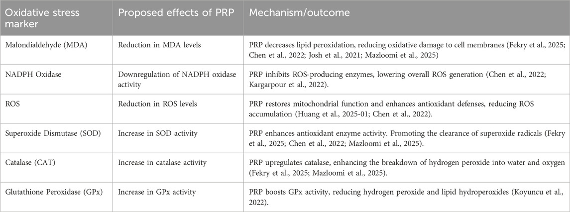
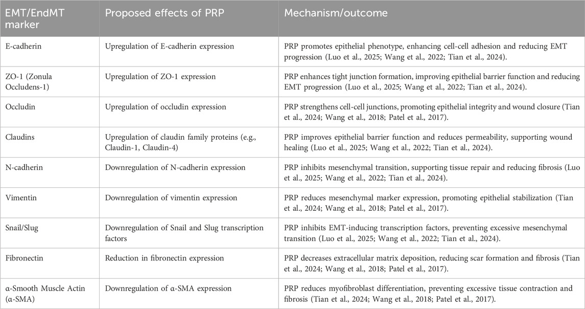
PRP has exhibited considerable promise in regulating oxidative stress markers, which hold paramount importance in the pathophysiological mechanisms underlying diabetic wound healing (Fekry et al., 2025; Huang et al., 2025-01). Table1 concisely outlines the possible impacts of PRP on oxidative stress indicators (Huang et al., 2025-01; Chen et al., 2022; Josh et al., 2021; Mazloomi et al., 2025; Kargarpour et al., 2022). By targeting these oxidative stress markers, PRP addresses the fundamental issues-notably chronic oxidative stress and mitochondriial dysfunction-that contribute to delayed wound healing in diabetes. Furthermore, PRP has demonstrated potential efficacy in modulating EMT/EndMT, processes that are indispensable for effective diabetic wound healing (Table2) (Luo et al., 2025; Wang et al., 2022; Tian et al., 2024; Wang et al., 2018; Patel et al., 2017). The dysregulation of EMT/EndMT is a significant factor impairing wound healing in diabetes, and PRP may aid in restoring balance by exerting influence on crucial markers (Tian et al., 2024).
Evaluation of the hypothesis
While oxidative stress and excessive ROS production play central roles in diabetic wound pathophysiology, ROS also function as essential signaling molecules in normal wound repair. Physiological ROS levels promote cell proliferation, differentiation, and immune defense mechanisms that are critical for proper wound healing (Huo et al., 2009). These molecules activate key pathways including nuclear factor kappa B (NF-κB) and mitogen-activated protein kinases (MAPKs), which coordinate inflammation, angiogenesis, and tissue regeneration (Huo et al., 2009; Lim et al., 2006). Chronic hyperglycemia in diabetes, however, elevates ROS production, causing mitochondrial dysfunction, compromised antioxidant defenses, and persistent oxidative stress (Caturano et al., 2025). Such dysregulation impairs the healing cascade, resulting in delayed wound closure and defective tissue repair. PRP may help rebalance ROS signaling by reducing oxidative damage while maintaining ROS-mediated physiological functions. Clarifying these mechanisms could inform therapeutic strategies to manage oxidative stress in diabetic wounds. Although promising, this hypothesis warrants rigorous evaluation against existing evidence.
The potential impact of PRP on the ROS-EMT/EndMT axis in diabetic wound healing
The absence of proof connecting PRP to EMT/EndMT regulation is acknowledged (Figure 2). The role of PRP in diabetic wound healing has garnered significant attention due to its potential to regulate various biological processes, including the ROS and EMT/EndMT axis. Diabetic wounds are characterized by delayed healing, often attributed to oxidative stress, impaired angiogenesis, and dysfunctional cellular responses. PRP, abundant in growth factors and cytokines, offers therapeutic benefits by addressing these underlying issues. It has been demonstrated that PRP can reduce oxidative stress by enhancing the antioxidant capacity of the wound environment, potentially mitigating the adverse effects of ROS on wound healing (Zhang C. et al., 2019). In the context of diabetic wounds, the dysregulation of EMT and EndMT hinders effective healing. PRP may influence the ROS-EMT/EndMT axis by modulating Wnt/β-catenin, PI3K/Akt, MAPK, NF-κB, and TGF-β signaling pathways (Table 3) (Zhang C. et al., 2019; Ebrahim et al., 2021; Liu et al., 2019; Zhang L. et al., 2023; Ju et al., 2023; Guo et al., 2022; Liu et al., 2024; Fang et al., 2019), promoting a more conducive healing environment. For instance, PRP has been shown to activate the PI3K/Akt pathway, which plays a crucial role in cell survival and proliferation. This activation can counteract the harmful effects of ROS, thereby supporting the cellular functions necessary for effective wound healing (Ebrahim et al., 2021). By addressing oxidative stress and regulating the EMT/EndMT axis, PRP can enhance cellular responses, angiogenesis, and tissue regeneration in diabetic wounds (Zhang C. et al., 2019; Ebrahim et al., 2021; Fang et al., 2024; Karas et al., 2024). However, further research is needed to elucidate the precise mechanisms by which PRP affects these pathways and to optimize its application in clinical settings.
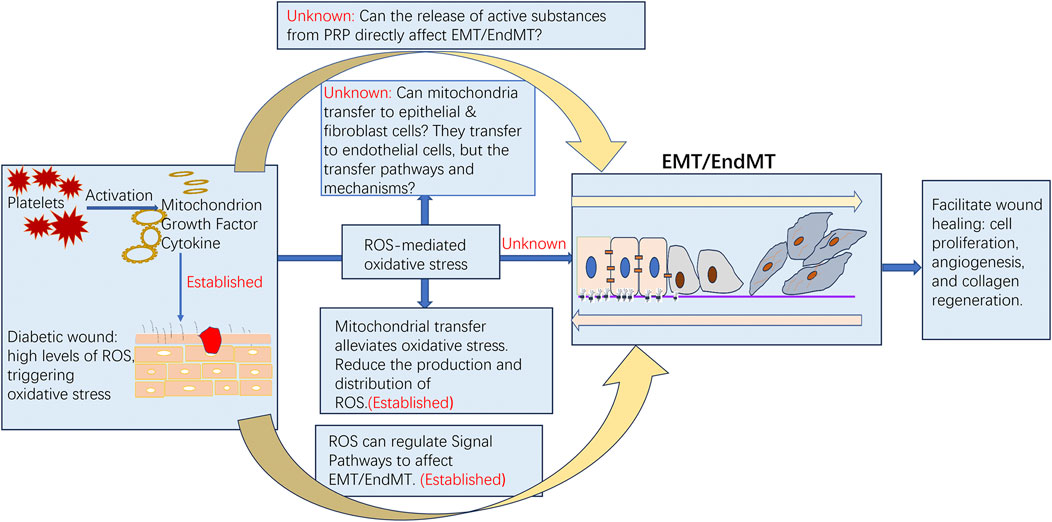
Figure 2. Current research advances and future exploration directions of PRP, ROS, oxidative stress, and EMT/EndMT in the healing process of diabetic wounds.
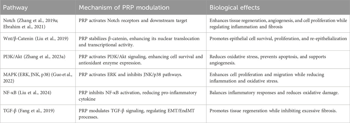
Table 3. Mechanistic analysis of PRP’s modulation of key signaling pathways in diabetic wound healing.
The role of platelet-derived mitochondria in diabetic wound healing within PRP
The role of platelet mitochondria derived from PRP in diabetic wound healing presents a novel treatment paradigm (Figure 2). Mitochondrial transfer generally refers to the movement and exchange of mitochondria between different cells or organelles, potentially playing a significant role in cellular injury repair and energy metabolism. After transfer, the mitochondria integrate into the mitochondrial network of recipient cells through fusion and fission. Jin et al. (2023) demonstrated that platelets serve as important donors of mitochondria, and platelet-derived mitochondria can promote wound healing by reducing cell apoptosis in vascular endothelial cells caused by oxidative stress. They also proposed that ultrasound is a more suitable method for activating platelets to release respiratory mitochondria. Additionally, these mitochondria released by platelets can transfer to mesenchymal stem cells, enhancing their angiogenic capacity through metabolic reprogramming (Levoux et al., 2021). Platelet-derived mitochondria can also bind to neutrophils, promoting enhanced rolling adhesion of neutrophils to the vascular wall and facilitating the activation and migration of inflammatory cells, thereby regulating inflammatory responses and wound healing (Boudreau et al., 2020). These findings further expand our understanding of platelet function and provide new insights into the role of platelet-derived mitochondria in wound healing. However, the specific mechanisms underlying platelet mitochondrial transfer and how transferred mitochondria improve cell function remain to be further explored in this study. Therefore, transferring healthy mitochondria to replace those damaged by oxidative stress in wound cells (including but not limited to mesenchymal stem cells, neutrophils, vascular endothelial cells, epithelial cells, and fibroblasts) represents a potential novel therapeutic approach for chronic diabetic wounds.
The direct impact of PRP on the EMT/EndMT process
While PRP has been shown to have antioxidant properties and to reduce oxidative stress in some studies (Koyuncu et al., 2022; Chen et al., 2022; Josh et al., 2021; Mazloomi et al., 2025; Kargarpour et al., 2022), the direct link between PRP’s ability to modulate EMT/EndMT and its antioxidant effects on ROS-mediated oxidative stress in diabetic wound healing is not well-established (Figure 2). To contextualize this, EMT and EndMT are fundamental processes in tissue repair. During normal wound healing, partial EMT allows epithelial and endothelial cells to adopt a more mesenchymal phenotype, enhancing their migratory capacity to cover the wound bed and initiate angiogenesis (S et al., 2022; Tian et al., 2024; Simon et al., 2014; Gutmann et al., 2021; Lambert and Weinberg, 2021). Conversely, EndMT contributes to the formation of activated fibroblasts and myofibroblasts crucial for extracellular matrix deposition and wound contraction (Miscianinov et al., 2018; Tian et al., 2024). However, in the pathological milieu of diabetic wounds, chronic hyperglycemia and sustained oxidative stress drive excessive and dysregulated EMT/EndMT (S et al., 2022; Miscianinov et al., 2018). This pathological transition promotes fibroblast -to-myofibroblast conversion, excessive collagen deposition, vascular rarefaction, and ultimately fibrosis, which impedes re-epithelialization and functional tissue regeneration (S et al., 2022; Miscianinov et al., 2018; Patel et al., 2017). Thus, therapeutic modulation of EMT/EndMT aims not for complete inhibition, but for restoring its precise spatiotemporal dynamics–promoting beneficial cell migration and angiogenesis while suppressing persistent activation that fuels fibrosis. PRP has been shown to regulate various cellular pathways such as Wnt/β-catenin, MAPK, and NF-κB (Liu et al., 2019; Zhang L. et al., 2023; Ju et al., 2023; Fang et al., 2019), which are key modulators of EMT/EndMT. Additionally, PRP contains a multitude of activators (e.g., TGF-β, PDGF) that can directly influence the EMT/EndMT process (Simon et al., 2014; Gutmann et al., 2021; Lambert and Weinberg, 2021). Based on these observations, it is plausible to infer that PRP may have a potential impact on the EMT/EndMT process, potentially shifting the balance away from the pathological, fibrotic transition observed in diabetes towards a more controlled, healing-promoting transition. For instance, certain growth factors within PRP may inhibit excessive EMT/EndMT activation by modulating related signaling pathways (e.g., counteracting sustained TGF-β/Smad signaling), while others might transiently support necessary mesenchymal characteristics for cell migration.
In summary, while the hypothesis that PRP may accelerate diabetic wound healing by modulating EMT/EndMT through inhibiting ROS-mediated oxidative stress is plausible, it requires further investigation and validation through rigorous scientific research. Studies that specifically investigate the mechanisms by which PRP affects EMT/EndMT and its antioxidant effects on ROS-mediated oxidative stress in diabetic wounds would be valuable in advancing our understanding of this complex process.
Testing the hypothesis
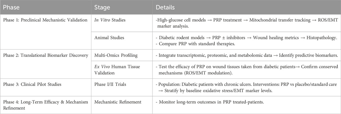
To test the hypothesis, researchers would conduct comprehensive cellular, animal experiments, and clinical trials (Table 4), examining the impact of PRP on ROS levels in diabetic wounds and assess its potential to reduce oxidative stress, evaluating the modulation of EMT/EndMT markers in wound tissues by PRP, investigating the signaling pathways involved in PRP-mediated regulation of EMT/EndMT and wound healing, assessing functional outcomes such as wound size reduction, re-epithelialization, and granulation tissue formation in diabetic animals treated with PRP, and comparing its efficacy with standard wound care therapies to determine if PRP offers additional benefits in accelerating diabetic wound healing.
In vitro experiments designed to investigate the impact of PRP on ROS-mediated oxidative stress and the EMT/EndMT axis
Researchers can create a well-established diabetic rat model (such as those already available in the research field, like the streptozotocin - induced diabetic rat model) and isolate wound - related cells (including endothelial cells, keratinocytes, and fibroblasts) from appropriate biological samples. Typically, the establishment of the diabetic rat model is part of animal experiments. These cells are then cultured in a high-glucose medium to simulate the diabetic environment. Meanwhile, PRP is extracted and prepared from the blood of non-diabetic rats, with its growth factor and mitochondrial content enhanced through ultrasonic treatment. The cells are divided into experimental groups (treated with PRP alone, ROS inhibitors such as N-acetylcysteine, and EMT inhibitors such as EMT inhibitor-1 and EMT inhibitor-3), and a control group (untreated). Subsequently, the migration, proliferation, and invasion capabilities of the cells in each group are evaluated, and the levels of ROS and the expression of EMT/EndMT-related markers are detected. Additionally, the expression levels of target genes and the activity of specific signaling pathways are analyzed, while single-cell sequencing technology is employed to explore changes in the gene expression profile before and after treatment. Existing studies show PRP alleviates oxidative stress in tendon (Tognoloni et al., 2023), intervertebral disc (Lian et al., 2024), and human umbilical vein endothelial cells (HUVECs) (Wei et al., 2024a). In tendon cells, it boosts antioxidant enzyme expression via Nrf2 (Tognoloni et al., 2023). In intervertebral disc cells, it lowers ROS and enhances GPX4 activity (Lian et al., 2024). In high-glucose-damaged HUVECs, it restores angiogenesis, cuts ROS by 47%, and reduces inflammatory cytokines (Wei et al., 2024a).
In animal experiments, the impact of PRP on ROS-mediated oxidative stress and the EMT/EndMT axis can be investigated
Diabetic rat ulcer models are divided into four groups: diabetic control group, PRP treatment group, ROS inhibitor treatment group, and EMT inhibitor treatment group. Regular wound healing assessments (area, time, appearance) and histopathological exams (angiogenesis via CD31/CD34 staining, collagen neoformation via Masson’s/Sirius Red) are conducted. ROS-related proteins and genes are analyzed by Western blot and RT-qPCR. EMT markers (epithelial, endothelial, mesenchymal, and others) are detected by Western blot, immunocytochemistry and immunohistochemistry. Blood glucose, oxidative stress indicators (SOD, GSH-Px, MDA), and inflammatory cytokines (TNF-α, IL-6, IL-1β) are measured. Statistical analysis compares differences across groups and time points, providing insights into wound healing, inflammation, antioxidant status, and EMT processes. Animal experiments have demonstrated that PRP can mitigate lipid peroxidation and ROS accumulation by inhibiting ferroptosis (Zhou et al., 2024). Moreover, PRP promotes angiogenesis through the VEGFA/VEGFR2/ERK signaling pathway (Wei et al., 2024b), while concurrently suppressing high glucose-induced endothelial cell dysfunction and mesenchymal transition (Wei et al., 2024a).
In cellular and animal experiments, the research focuses on the impact of mitochondrial transfer on vascular endothelial cells, keratinocytes, and fibroblasts
Existing research has revealed that in conditions such as diabetes, hypertension, or ischemic diseases, mitochondrial transfer can reverse mitochondrial fission and oxidative stress in endothelial cells (Li G. et al., 2022; Ajoolabady et al., 2024), while also inhibit the abnormal proliferation of keratinocytes and fibroblasts (such as in keloid formation) (Zhang M. et al., 2023). Based on this, an experimental study can be designed to investigate the transfer of mitochondria from platelets to vascular endothelial cells, keratinocytes, and fibroblasts. Vascular endothelial cells, keratinocytes, and fibroblasts can be isolated from animals with diabetic ulcers. Mitochondrial transfer from platelets to cells damaged by diabetic conditions can be examined through coculture experiments. Cells will be cultured in high-glucose medium to simulate diabetic conditions. Fluorescently labeled platelet mitochondria will be cocultured with these cells (experimental group) and with normal glucose-treated cells (control group) to assess mitochondrial transfer. Fluorescence microscopy and confocal microscopy will be used to track intracellular mitochondrial distribution and quantify transfer efficiency at multiple time points.
To evaluate the impact of platelet mitochondrial transfer on oxidative stress and cellular function, four experimental groups will be established: the healthy control group will comprise animals administered normal glucose treatment to establish baseline parameters; the diabetic ulcer model group will include animals subjected to high-glucose treatment to induce ulcerative pathology; in the functional platelet treatment group, diabetic ulcer models will be administered intact platelets to assess mitochondrial transfer efficacy; and the respiration-inhibited platelet treatment group will consist of diabetic ulcer models treated with platelets pre-inhibited by mitochondrial respiration inhibitors to investigate mechanistic dependencies. Skin ulcer tissues will be systematically collected from all groups for integrated transcriptomic and metabolomic sequencing, enabling the identification of differentially expressed redox-related genes and metabolite profiles. These multi-omics analyses will elucidate how platelet mitochondria modulate oxidative stress pathways and facilitate tissue functional recovery, as evidenced by quantitative indicators including wound healing dynamics, mitochondrial membrane potential stability, and antioxidant enzyme activities.
Implications of the hypothesis
The hypothesis presents a novel mechanism for PRP’s therapeutic action in diabetic wound healing, extending beyond its established role as a growth factor delivery system to include oxidative stress regulation and EMT/EndMT modulation. This expanded understanding of PRP’s multifaceted healing properties provides new insights into optimizing diabetic wound treatment. The involvement of EMT/EndMT is particularly significant, as these processes govern cell migration, proliferation, and differentiation during tissue repair—suggesting PRP could strategically manipulate these transitions to accelerate healing. The emphasis on ROS-mediated oxidative stress inhibition further underscores PRP’s potential to restore redox balance, a critical factor impaired in diabetic wounds that often hinders cellular function and tissue regeneration. The proposed mitochondrial transfer mechanism offers an additional dimension, where PRP may restore cellular energy metabolism by replenishing damaged mitochondria in metabolically compromised wound environments. Finally, PRP’s direct regulation of EMT/EndMT balance could prove instrumental in preventing fibrosis while promoting healthy tissue regeneration, addressing a key complication in chronic wound management. Collectively, these mechanisms position PRP as a multifunctional therapeutic approach with potential applications extending beyond diabetic wound healing to other oxidative stress-related pathologies.
Discussion
PRP has demonstrated therapeutic potential for diabetic wounds, primarily due to its high concentration of growth factors essential for tissue repair (Alamdari et al., 2021; Azam et al., 2024; Karmakar et al., 2024; Wang et al., 2022-01; Orban et al. 2022; Saad et al., 2022). A clinical trial reported that diabetic patients treated with PRP demonstrated significantly improved healing rates compared to the control group (Orban et al. 2022), while long-term follow-up data revealed reduced infection rates and lower amputation risks (Saad et al., 2022). A systematic review further substantiates these findings, with meta-analytic data showing PRP increases wound healing likelihood and decreases amputation risk (Saad et al., 2022).
The therapeutic mechanisms of PRP have been implicated to extend beyond established effects on inflammation, angiogenesis, and extracellular matrix remodeling (Ebrahim et al., 2021; Wang et al., 2023; Xu et al., 2020; Lian et al., 2014). Emerging evidence suggests PRP may regulate oxidative stress and influence EMT/EndMT transitions through mitochondrial transfer and ROS scavenging. This polypharmacological approach simultaneously activates multiple regenerative pathways while addressing both oxidative damage and its underlying cause—mitochondrial dysfunction.
While synthetic biomaterials like photothermal hydrogels (Wang et al., 2024) or ATP-responsive prodrug systems (Qi et al., 2024) target specific wound aspects, PRP offers inherent biological complexity without synthetic limitations. However, key questions remain regarding PRP’s precise regulation of EMT/EndMT transitions and ROS modulation. PRP likely influences EMT/EndMT through multiple pathways, including the regulation of oxidative stress via ROS scavenging and mitochondrial transfer, as well as the activation of pro-regenerative signaling pathways such as PI3K/Akt and TGF-β. These actions collectively help balance EMT/EndMT transitions, promoting tissue repair while preventing excessive fibrosis or epithelial dysfunction.
Standardization challenges—including centrifugation protocols, platelet concentration, and leukocyte content—significantly affect therapeutic outcomes (Fadadu et al., 2019; Zhang N. et al., 2019; Popescu et al., 2021). Double-spin centrifugation and calcium chloride activation, for instance, alter growth factor release kinetics and platelet yields. Moreover, it is crucial to acknowledge potential adverse effects associated with PRP therapy. Reported adverse effects include postoperative infections, inflammation, nodule development, allergic reactions, and even blindness, with postoperative infections being the most frequently documented (Arita and Tobita, 2024). Furthermore, long-term safety concerns warrant attention, especially for diabetic patients who face elevated baseline risks for malignancies. The pro-angiogenic properties of growth factors in PRP raise theoretical concerns about potentially exacerbating cancer risk in this population. Therefore, in addition to optimizing PRP formulations, comprehensive evaluation of the therapy’s efficacy and risks is essential. Establishing robust post-treatment follow-up protocols to monitor for potential adverse effects is equally critical.
Conclusion
Collectively, this hypothesis suggests that PRP may act as a multifunctional regulator of diabetic wound pathogenesis by coordinating mitochondrial transfer, ROS-EMT/EndMT modulation, and redox homeostasis within a cohesive therapeutic mechanism. PRP may accelerate healing through a distinct mechanism that targets mitochondrial dysfunction, oxidative stress, and aberrant cellular transitions, surpassing conventional growth factor therapies. Functional platelet-derived mitochondria directly correct metabolic deficits in diabetic wounds, while coordinated suppression of ROS overproduction and precise EMT/EndMT modulation prevents fibrotic progression and restores regenerative capacity. The synergistic effects of mitochondrial bioenergetic recovery, oxidative stress mitigation, and cellular transition regulation collectively counteract the pathological environment perpetuating chronic diabetic wounds. While these findings expand our understanding of PRP’s action mechanisms, further studies must precisely characterize its molecular targets and optimize clinical protocols. Additional research should also explore PRP’s applicability to other oxidative stress-mediated pathologies.
Data availability statement
The original contributions presented in the study are included in the article/supplementary material, further inquiries can be directed to the corresponding authors.
Author contributions
YL: Writing – original draft, Writing – review and editing, Conceptualization. BC: Writing – original draft, Conceptualization, Writing – review and editing. JT: Writing – original draft, Supervision, Conceptualization, Writing – review and editing.
Funding
The author(s) declare that financial support was received for the research and/or publication of this article. This research was supported by the following grants: Guangdong Provincial Administration of Traditional Chinese Medicine (Grant No. 20241357). First Batch of 2024 Social Welfare and Basic Research Projects in Zhongshan City (General Healthcare Projects) (Grant No. 2024B1100). Additional funding was provided by the General Programs of the National Natural Science Foundation of China (Grant Nos. 82172223 and 82372531).
Conflict of interest
The authors declare that the research was conducted in the absence of any commercial or financial relationships that could be construed as a potential conflict of interest.
Generative AI statement
The author(s) declare that Generative AI was used in the creation of this manuscript. In the course of compiling this manuscript, the authors leveraged artificial intelligence to elevate the linguistic quality and readability. Subsequent to its application, the authors meticulously reviewed and edited the content, accepting full accountability for the published material.
Publisher’s note
All claims expressed in this article are solely those of the authors and do not necessarily represent those of their affiliated organizations, or those of the publisher, the editors and the reviewers. Any product that may be evaluated in this article, or claim that may be made by its manufacturer, is not guaranteed or endorsed by the publisher.
References
Ajoolabady, A., Pratico, D., and Ren, J. (2024). Angiotensin II: role in oxidative stress, endothelial dysfunction, and diseases. Mol. Cell Endocrinol. 592, 112309. doi:10.1016/j.mce.2024.112309
Alamdari, N. M., Shafiee, A., Mirmohseni, A., and Besharat, S. (2021). Evaluation of the efficacy of platelet-rich plasma on healing of clean diabetic foot ulcers: a randomized clinical trial in Tehran, Iran. Diabetes Metab. Syndr. 15 (2), 621–626. doi:10.1016/j.dsx.2021.03.005
Arita, A., and Tobita, M. (2024). Adverse events related to platelet-rich plasma therapy and future issues to be resolved. Regen. Ther. 26, 496–501. doi:10.1016/j.reth.2024.07.004
Azam, M. S., Azad, M. H., Arsalan, M., Malik, A., Ashraf, R., and Javed, H. (2024). Efficacy of platelet-rich plasma in the treatment of diabetic foot ulcer. Cureus 16 (5), e60934. doi:10.7759/cureus.60934
Bodnár, E., BakondiE, K. K., Kovács, K., Hegedűs, C., Lakatos, P., Robaszkiewicz, A., et al. (2018). Redox profiling reveals clear differences between molecular patterns of wound fluids from acute and chronic wounds. Oxid. Med. Cell Longev. 2018, 5286785. doi:10.1155/2018/5286785
Borcherding, N., and Brestoff, J. R. (2023). The power and potential of mitochondria transfer. Nature 623 (7986), 283–291. doi:10.1038/s41586-023-06537-z
Boudreau, L., Foulem, R., and Léger, J. (2020). The effects of functional platelet-derived extracellular mitochondria on the inflammatory phenotype of neutrophils. J. Immunol. 204 (1_Suppl. e), 67.23. doi:10.4049/jimmunol.204.supp.67.23
Cai, F., Chen, W., Zhao, R., and Liu, Y. (2023). Mechanisms of Nrf2 and NF-κB pathways in diabetic wound and potential treatment strategies. Mol. Biol. Rep. 50 (6), 5355–5367. doi:10.1007/s11033-023-08392-7
Caturano, A., Rocco, M., Tagliaferri, G., Piacevole, A., Nilo, D., Di Lorenzo, G., et al. (2025). Oxidative stress and cardiovascular complications in type 2 diabetes: from pathophysiology to lifestyle modifications. Antioxidants (Basel) 14 (1), 72. doi:10.3390/antiox14010072
Chen, L., Wu, D., Zhou, L., and Ye, Y. (2022). Platelet-rich plasma promotes diabetic ulcer repair through inhibition of ferroptosis. Ann. Transl. Med. 10 (20), 1121. doi:10.21037/atm-22-4654
Ebrahim, N., Dessouky, A. A., Mostafa, O., Hassouna, A., Yousef, M. M., Seleem, Y., et al. (2021). Adipose mesenchymal stem cells combined with platelet-rich plasma accelerate diabetic wound healing by modulating the notch pathway. Stem Cell Res. Ther. 12 (1), 392. doi:10.1186/s13287-021-02454-y
Fadadu, P. P., Mazzola, A. J., Hunter, C. W., and Davis, T. T. (2019). Review of concentration yields in commercially available platelet-rich plasma (PRP) systems: a call for PRP standardization. Reg. Anesth. Pain 44, 652–659. doi:10.1136/rapm-2018-100356
Fang, D., Jin, P., Huang, Q., Yang, Y., Zhao, J., and Zheng, L. (2019). Platelet-rich plasma promotes there generation of cartilage engineered by mesenchymal stem cells and collagenhydrogel via the TGF-β/SMAD signaling pathway. J. Cell Physiol. 234 (9), 15627–15637. doi:10.1002/jcp.28211
Fang, X., Wang, X., Hou, Y., Zhou, L., Jiang, Y., and Wen, X. (2024). RETRACTED: effect of platelet-rich plasma on healing of lower extremity diabetic skin ulcers: a meta-analysis. Int. Wound J. 21 (4), e14856. doi:10.1111/iwj.14856
Fekry, E., Refaat, G. N., and Hosny, S. A. (2025). Ameliorative potential of carvedilol versus platelet-rich plasma against paclitaxel-induced femoral neuropathy in wistar rats: a light and electron microscopic study. Microsc. Microanal. 31, ozaf002. doi:10.1093/mam/ozaf002
Guo, M. S., Gao, X., Hu, W., Wang, X., Dong, T. T., and Tsim, K. W. K. (2022). Scutellarin potentiates the skin regenerative function of self growth colony, an optimized platelet-rich plasma extract, in cultured keratinocytes through VEGF receptor and MAPK signaling. J. Cosmet. Dermatol-Us 21 (10), 4836–4845. doi:10.1111/jocd.14800
Gutmann, C., Joshi, A., Zampetaki, A., and Mayr, M. (2021). The landscape of coding and noncoding RNAs in platelets. Antioxid. Redox Sign 34 (15), 1200–1216. doi:10.1089/ars.2020.8139
Huang, Z., Gu, Z., Zeng, Y., and Zhang, D. (2025). Platelet-rich plasma alleviates skin photoaging by activating autophagy and inhibiting inflammasome formation. N-S Arch. Pharmacol. 398, 8669–8680. doi:10.1007/s00210-025-03800-0
Huo, Y., Qiu, W. Y., Pan, Q., Yao, Y. F., Xing, K., and Lou, M. F. (2009). Reactive oxygen species (ROS) are essential mediators in epidermal growth factor (EGF)-Stimulated corneal epithelial cell proliferation, adhesion, migration, and wound healing. Exp. EYE Res. 89 (6), 876–886. doi:10.1016/j.exer.2009.07.012
Jafar, H., Hasan, M., Al-Hattab, D., Saleh, M., Ameereh, L. A., Khraisha, S., et al. (2020). Platelet lysate promotes the healing of long-standing diabetic foot ulcers: a report of two cases and invitro study. Heliyon 6 (5), e03929. doi:10.1016/j.heliyon.2020.e03929
Jin, P., Pan, Q., Lin, Y., Dong, Y., Zhu, J., Liu, T., et al. (2023). Platelets facilitate wound healing by mitochondrial transfer and reducing oxidative stress in endothelial cells. Oxid. Med. Cell Longev. 2023, 1–23. doi:10.1155/2023/2345279
Josh, F., Soekamto, T. H., Adriani, J. R., Jonatan, B., Mizuno, H., and Faruk, M. (2021). The combination of stromal vascular fraction cells and platelet-rich plasma reduces malondialdehyde and nitric oxide levels in deep dermal burn injury. J. Inflamm. Res. 14, 3049–3061. doi:10.2147/JIR.S318055
Ju, T., Xiaoying, H., Chenyan, L., and Luo, Z. (2023). Platelet-rich plasma accelerate diabetic wound healing via dynamic modulation of multiple signaling pathways. Med. Hypothes. 176, 111097. doi:10.1016/j.mehy.2023.111097
Karas, R. A., Alexeree, S., Elsayed, H., and Attia, Y. A. (2024). Assessment of wound healing activity in diabetic mice treated with a novel therapeutic combination of selenium nanoparticles and platelets rich plasma. Sci. Rep. 14 (1), 5346. doi:10.1038/s41598-024-54064-2
Kargarpour, Z., Panahipour, L., Miron, R. J., and Gruber, R. (2022). Blood clots versus PRF: activating TGF-β signaling and inhibiting inflammation in vitro. Int. J. Mol. Sci. 23 (11), 5897. doi:10.3390/ijms23115897
Karina, W. K. A., Sobariah, S., Rosliana, I., Rosadi, I., Widyastuti, T., et al. (2019). Evaluation of platelet-rich plasma from diabetic donors shows increased platelet vascular endothelial growth factor release. Stem Cell Investig. 6, 43. doi:10.21037/sci.2019.10.02
Karmakar, N., Sharma, P., and Sandalya, B. (2024). Role of prp in diabetic foot ulcer. Int. J. Adv. Res. (Indore) 12 (03), 1102–1105. doi:10.21474/ijar01/18500
Koyuncu, A., Koç, S., Akdere, Ö. E., Çakmak, A. S., and Gümüşderelioğlu, M. (2022). Investigation of the synergistic effect of platelet-rich plasma and polychromatic light on human dermal fibroblasts seeded chitosan/gelatin scaffolds for wound healing. J. Photochem. B 232, 112476. doi:10.1016/j.jphotobiol.2022.112476
Lambert, A. W., and Weinberg, R. A. (2021). Linking EMT programmes to normal and neoplastic epithelial stem cells. Nat. Rev. Cancer 21 (5), 325–338. doi:10.1038/s41568-021-00332-6
Levoux, J., Prola, A., Lafuste, P., Gervais, M., Chevallier, N., Koumaiha, Z., et al. (2021). Platelets facilitate the wound-healing capability of mesenchymal stem cells by mitochondrial transfer and metabolic reprogramming. Cell Metab. 33 (3), 688–690. doi:10.1016/j.cmet.2021.02.003
Li, Y., Fu, R., Duan, Z., Zhu, C., and Fan, D. (2022a). Artificial nonenzymatic antioxidant MXene nanosheet-anchored injectable hydrogel as a mild photothermal-controlled oxygen release platform for diabetic wound healing. ACS Nano 16 (5), 7486–7502. doi:10.1021/acsnano.1c10575
Li, G., Xu, K., Xing, W., Yang, H., Li, Y., Wang, X., et al. (2022b). Swimming exercise alleviates endothelial mitochondrial fragmentation via inhibiting dynamin-related Protein-1 to improve vascular function in hypertension. Hypertension 79 (10), e116–e128. doi:10.1161/HYPERTENSIONAHA.122.19126
Lian, Z., Yin, X., Li, H., Jia, L., He, X., Yan, Y., et al. (2014). Synergistic effect of bone marrow-derived mesenchymal stem cells and platelet-rich plasma in streptozotocin-induced diabetic rats. Ann. Dermatol 26 (1), 1–10. doi:10.5021/ad.2014.26.1.1
Lian, S. L., Huang, J., Zhang, Y., and Ding, Y. (2024). The effect of platelet-rich plasma on ferroptosis of nucleus pulposus cells induced by erastin. Biochem. Rep. 41, 101900. doi:10.1016/j.bbrep.2024.101900
Lim, Y., Levy, M. A., and Bray, T. M. (2006). Dietary supplementation of N-acetylcysteine enhances early inflammatory responses during cutaneous wound healing in protein malnourished mice. J. Nutr. Biochem. 17 (5), 328–336. doi:10.1016/j.jnutbio.2005.08.004
Liu, Y., Jiang, J., Ye, Y., Li, Z., Tan, M., Zou, J., et al. (2019). Stromal vascular fraction and platelet-rich plasma upregulate vascular endothelial growth factor expression to promote hair growth via the Wnt/β -catenin signaling pathway. Nanosci. Nanotech Let. 11 (12), 1685–1692. doi:10.1166/nnl.2019.3057
Liu, X., Wang, Y., Wen, X., Hao, C., Ma, J., and Yan, L. (2024). Platelet rich plasma alleviates endometritis induced by lipopolysaccharide in mice via inhibiting TLR4/NF-κB signaling pathway. Am. J. Reprod. Immunol. 91 (3), e13833. doi:10.1111/aji.13833
Luo, G., Zhu, S., He, L., Liu, Q., Xu, C., Yao, Q., et al. (2025). Platelets promote metastasis of intrahepatic cholangiocar cinoma through activation of TGF-β/Smad2 pathway. Biochim. Acta Mol. Basis Dis. 1871 (4), 167734. doi:10.1016/j.bbadis.2025.167734
Mazloomi, S., Tayebinia, H., Farimani, M. S., and Ghorbani, M. (2025). A retrospective cohort study investigating the effect of intraovarian platelet-rich plasma therapy on the oxidative state of follicular fluid in women with diminished ovarian reserve. Chonnam Med. J. 61 (1), 46–51. doi:10.4068/cmj.2025.61.1.46
Miscianinov, V., Martello, A., Rose, L., Parish, E., Cathcart, B., Mitić, T., et al. (2018). MicroRNA-148b targets the TGF-β pathway to regulate angiogenesis and endothelial-to-mesenchymal transition during skin wound healing. Mol. Ther. 26 (8), 1996–2007. doi:10.1016/j.ymthe.2018.05.002
Orban, Y. A., Soliman, M. A., Hegab, Y. H., and Alkilany, M. M. (2006). Autologous platelet-rich plasma vs conventional dressing in the management of chronic diabetic foot ulcers. WOUNDS 33, 36–42. doi:10.25270/wnds/2022.3642
Patel, J., Baz, B., Wong, H. Y., Lee, J. S., and Khosrotehrani, K. (2017). Accelerated endothelial to mesenchymal transition increased fibrosis via deleting notch signaling in wound vasculature. J. Invest Dermatol 138 (5), 1166–1175. doi:10.1016/j.jid.2017.12.004
Popescu, M. N., Iliescu, M. G., Beiu, C., Popa, L. G., Mihai, M. M., Berteanu, M., et al. (2021). Autologous platelet-rich plasma efficacy in the field of regenerative medicine: product and quality control. Biomed. Res. Int. 2021, 4672959. doi:10.1155/2021/4672959
Qi, X., Xiang, Y., Li, Y., Wang, J., Chen, Y., Lan, Y., et al. (2024). An ATP-Activated spatiotemporally controlled hydrogel prodrug system for treating multidrug-resistant bacteria-infected pressure ulcers. Bioact. Mater 45, 301–321. doi:10.1016/j.bioactmat.2024.11.029
Qian, Z., WangH, B. Y., Wang, Y., Tao, L., Wei, Y., et al. (2020). Improving chronic diabetic wound healing through an injectable and self-healing hydrogel with platelet-rich plasma release. ACS Appl. Mater Interfaces 12 (50), 55659–55674. doi:10.1021/acsami.0c17142
Shi, Y., Wang, S., Yang, R., Wang, Z., Zhang, W., Liu, H., et al. (2022). ROS promote hypoxia-induced keratinocyte epithelial-mesenchymal transition by inducing SOX2 expression and subsequent activation of Wnt/β-Catenin. Oxid. Med. Cell Longev. 2022, 1084006. doi:10.1155/2022/1084006
Saad, H., Yehia, A., and Osman, G. (2022). The effect of platelet rich plasma (PRP) injection to the wound compared to PRP jel local application compared to classic dressing on diabetic foot healing ulcer. Surg. Res. 4 (2). doi:10.33425/2689-1093.1048
Simon, L. M., Edelstein, L. C., Nagalla, S., Woodley, A. B., Chen, E. S., Kong, X., et al. (2014). Human platelet microRNA-mRNA networks associated with age and gender revealed by integrated plateletomics. Blood 123 (16), e37–e45. doi:10.1182/blood-2013-12-544692
Tian, J., You, H., Ding, J., Shi, D., Long, C., li, Y., et al. (2024). Platelets could be key regulators of epithelial/endothelial-to- mesenchymal transition in atherosclerosis and wound healing. Med. HYPOTHESES 189, 111397. doi:10.1016/j.mehy.2024.111397
Tognoloni, A., Bartolini, D., Pepe, M., Di Meo, A., Porcellato, I., Guidoni, K., et al. (2023). Platelets rich plasma increases antioxidant defenses of tenocytes via Nrf2 signal pathway. Int. J. Mol. Sci. 24 (17), 13299. doi:10.3390/ijms241713299
Wang, Z., Fei, S., Suo, C., Han, Z., Tao, J., Xu, Z., et al. (2018). Antifibrotic effects of hepatocyte growth factor on endothelial-to-mesenchymal transition via transforming growth Factor-Beta1 (TGF-β1)/Smad and Akt/mTOR/P70S6K signaling pathways. Ann. Transpl. 23, 1–10. doi:10.12659/aot.906700
Wang, X., Zhao, S., Wang, Z., and Gao, T. (2022). Platelets involved tumor cell EMT during circulation: communications and interventions. Cell Commun. Signal 5, 82. doi:10.1186/s12964-022-00887-3
Wang, Y., Liu, B., Pi, Y., Hu, L., Yuan, Y., Luo, J., et al. (2022). Risk factors for diabetic foot ulcers mortality and novel negative pressure combined with platelet-rich plasma therapy in the treatment of diabetic foot ulcers. Front. Pharmacol. 13, 1051299. doi:10.3389/fphar.2022.1051299
Wang, Z., Feng, C., Chang, G., Liu, H., and Li, S. (2023). The use of platelet-rich plasma in wound healing and vitiligo: a systematic review and meta-analysis. Skin. Res. Technol. 29 (9), e13444. doi:10.1111/srt.13444
Wang, Y., Liu, K., Wei, W., and Dai, H. (2024). A multifunctional hydrogel with photothermal antibacterial and AntiOxidant activity for smart monitoring and promotion of diabetic wound healing. Adv. Funct. MATER 34, 2402531. doi:10.1002/adfm.202402531
Wei, W., Jiang, T., Hu, F., and Liu, H. (2024a). Tibial transverse transport combined with platelet-rich plasma sustained-release microspheres activates the VEGFA/VEGFR2 pathway to promote microcirculatory reconstruction in diabetic foot ulcer. Growth Factors. 42 (3), 128–144. doi:10.1080/08977194.2024.2407318
Wei, W., Xu, D., Hu, F., Jiang, T., and Liu, H. (2024b). Platelet-rich plasma promotes wound repair in diabetic foot ulcer mice via the VEGFA/VEGFR2/ERK pathway. Growth Factors. 42 (4), 161–170. doi:10.1080/08977194.2024.2422014
Xu, P., Wu, Y., Zhou, L., Yang, Z., Zhang, X., Hu, X., et al. (2020). Platelet-rich plasma accelerates skin wound healing by promoting re-epithelialization. Burns Trauma 8, tkaa028. doi:10.1093/burnst/tkaa028
Zhang, Z., and Zhao, Y. (2024). Enhanced diabetic rat wound healing by platelet-rich plasma adhesion zwitterionic hydrogel. Ann. Plas Surg. 93 (5), 649. doi:10.1097/SAP.0000000000004059
Zhang, C., Zhu, Y., Lu, S., Zhong, W., Wang, Y., and Chai, Y. (2019a). Platelet-rich plasma with endothelial progenitor cells accelerates diabetic wound healing in rats by upregulating the Notch1 signaling pathway. J. Diabetes Res. 2019, 1–12. doi:10.1155/2019/5920676
Zhang, N., Wang, K., Li, Z., and Luo, T. (2019b). Comparative study of different anticoagulants and coagulants in the evaluation of clinical application of platelet-rich plasma (PRP) standardization. Cell Tissue Bank. 20 (1), 61–75. doi:10.1007/s10561-019-09753-y
Zhang, W., Chen, L., Xiong, Y., Panayi, A. C., Abududilibaier, A., Hu, Y., et al. (2021). Antioxidant therapy and antioxidant-related bionanomaterials in diabetic wound healing. Front. BioengBiotechnol 9, 707479. doi:10.3389/fbioe.2021.707479
Zhang, L., Zhang, Q., Lk, C., Wu, L., and Gao, S. (2023a). Kartogenin combined platelet-rich plasma (PRP) promoted tendon-bone healing for anterior cruciate ligament (ACL)Reconstruction by suppressing inflammatory response via targeting AKT/PI3K/NF-κB. Appl. Biochem. 195 (2), 1284–1296. doi:10.1007/s12010-022-04178-y
Zhang, M., Chen, H., Qian, H., and Wang, C. (2023b). Characterization of the skin keloid microenvironment. Cell Commun. Signal 21 (1), 207. doi:10.1186/s12964-023-01214-0
Keywords: platelet-rich plasma, diabetic wound healing, oxidative stress, mitochondrial transfer, EMT/EndMT
Citation: Li Y, Cheng B and Tian J (2025) Platelet-rich plasma may accelerate diabetic wound healing by modulating epithelial/endothelial-mesenchymal transition through inhibiting reactive oxygen species-mediated oxidative stress. Front. Bioeng. Biotechnol. 13:1623780. doi: 10.3389/fbioe.2025.1623780
Received: 06 May 2025; Accepted: 29 July 2025;
Published: 11 August 2025.
Edited by:
Pedro Lei, University at Buffalo, United StatesReviewed by:
Ajay Kumar, Institute of Nano Science and Technology (INST), IndiaHoussam Khaled Al-Koussa, RIKEN Center for Integrative Medical Sciences (IMS), Japan
Sandy R. Botros, Minia University, Egypt
Copyright © 2025 Li, Cheng and Tian. This is an open-access article distributed under the terms of the Creative Commons Attribution License (CC BY). The use, distribution or reproduction in other forums is permitted, provided the original author(s) and the copyright owner(s) are credited and that the original publication in this journal is cited, in accordance with accepted academic practice. No use, distribution or reproduction is permitted which does not comply with these terms.
*Correspondence: Biao Cheng, Y2hlbmdiaWFvY2hlbmdAMTYzLmNvbQ==; Ju Tian, dGlhbi1qdUAxNjMuY29t
 Youan Li1
 Biao Cheng
 Ju Tian