 Yiqi Chen1
 Adili Alimujiang
- 1School of Stomatology, Jinan University, Guangzhou, China
- 2State Key Laboratory of Oral & Maxillofacial Reconstruction and Regeneration, Key Laboratory of Oral Biomedicine Ministry of Education, Hubei Key Laboratory of Stomatology, School & Hospital of Stomatology, Wuhan University, Wuhan, China
Dental mesenchymal stem cells (DMSCs) are important seed cells for oral and maxillofacial tissue regeneration due to their wide availability, multi-directional differentiation potential, strong proliferation ability, maintenance of stemness after multiple proliferations, and immunomodulatory capacity. DMSCs have a wide range of sources, and different subtypes have different properties. They can promote tissue regeneration through direct differentiation into tissue cells, improvement of the local regeneration microenvironment through immunomodulatory functions, and release of various bioactive components. The article reviews the classification of DMSCs, their performance comparisons in recent years, methods of promoting tissue regeneration, and their applications in maxillofacial bone and soft tissue. Furthermore, the review explores DMSCs’ excellent abilities to promote bone tissue repair when supported by various materials and repair maxillofacial soft tissue defects by promoting the regeneration of special structures of teeth such as dentin, cementum, and dental pulp vessels and nerves.
1 Introduction
Oral and maxillofacial defects are often caused by trauma, tumors or congenital diseases. For a long time, traditional repair methods have faced many insurmountable bottlenecks in restoring the function of the oral and maxillofacial region and meeting aesthetic needs. However, in recent years, with the vigorous development of the frontier field of tissue regeneration medicine, a brand-new exploration direction has been opened up for the repair of oral and maxillofacial defects. Among many innovative approaches, dental mesenchymal stem cells (DMSCs) have stood out due to their wide availability, multi-directional differentiation potential, and low immunogenicity, becoming one of the most notable hotspots in current oral medicine research.
Mesenchymal stem cells (MSCs) are a type of non-hematopoietic adult stem cells. They can be isolated from various tissues and organs, including bone marrow, skin, adipose tissue, and umbilical cord. These cells possess the potential for both proliferation and differentiation (Li et al., 2023). DMSCs are a type of adult stem cells among mesenchymal stem cells and can be isolated from dental pulp, periodontal ligament, apical papilla, gingiva, dental follicle and human deciduous teeth. Due to their multi-directional differentiation potential, strong proliferation ability, maintenance of cell stemness after multiple proliferations, and immunomodulatory capacity, they have become an important seed cell in the clinical application of tissue regeneration.
2 The classification of DMSCs
DMSCs are characterized by a diverse array of sources, and their properties vary notably among different subtypes. In the subsequent part, the sources of DMSCs are introduced in Figure 1. Additionally, several common types of DMSCs and their properties are expounded upon. Moreover, Figure 2 illustrates the sources and differentiation potential of DMSCs.
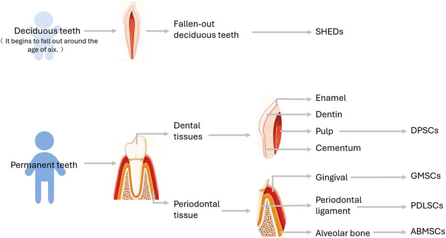
Figure 1. Schematic diagram of the Origin of DMSCs. SHEDs: Stem cells populations from human exfoliated deciduous teeth, DPSCs, Dental pulp stem cells; GMSCs, Gingival mesenchymal stem cells; PDLSCs, Periodontal ligament stem cells; ABMSCs, Alveolar bone mesenchymal stem cells.
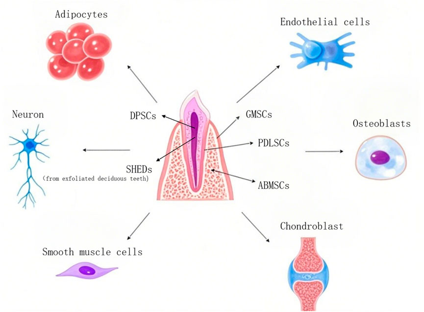
Figure 2. Partial sources and differentiation of dental mesenchymal stem cells. DPSCs, Dental pulp stem cells; SHEDs, Stem cells populations from human exfoliated deciduous teeth; GMSCs, Gingival mesenchymal stem cells; PDLSCs, Periodontal ligament stem cells; ABMSCs, Alveolar bone mesenchymal stem cells.
2.1 Dental pulp stem cells (DPSCs)
Dental pulp refers to the loose connective tissue within the pulp cavity surrounded by dentin, which has functions such as forming dentin, sensing pain, providing nutrition, and resisting bacterial invasion. Dental pulp stem cells are cells isolated from dental pulp with mesenchymal stem cell characteristics. In 2000, Gronthos et al. successfully isolated DPSCs (Gronthos et al., 2000). As the earliest isolated dental stem cells, they revealed that mesenchymal stem cells also exist in dental-related tissues besides bone marrow, adipose tissue, placenta, umbilical cord, etc. Compared with the extraction of bone marrow mesenchymal stem cells which requires anesthesia, DPSCs are easier to obtain and can be isolated from healthy first premolars, third molars, or other teeth extracted for orthodontic purposes, thus causing less ethical disputes (Hwan and Woo, 2022).
With the deepening of research, DPSCs can be traced back to neural crest cells, thus having the ability to differentiate into neurons and glial cells. Under certain environmental conditions, they can also produce capillary-like structures by synthesizing and secreting angiogenic regulators. Based on their potential for neural differentiation and angiogenesis, dental pulp stem cells have become the best seed cells for dental pulp regeneration.
2.2 Periodontal ligament stem cells (PDLSCs)
The periodontal ligament is a dense connective tissue connecting the alveolar bone and cementum. As a connection between the alveolar bone and cementum, it plays a crucial role in resisting and regulating the pressure on teeth during chewing. In 2004, Seo et al. successfully isolated periodontal ligament stem cells (Seo et al., 2004). In vitro experiments have shown that when periodontal tissue is damaged, periodontal ligament stem cells can migrate to the damaged area and repair it, revealing the possibility of periodontal tissue regeneration (Hwan and Woo, 2022).
2.3 Stem cells populations from human exfoliated deciduous teeth (SHEDs)
In 2003, Miura et al. discovered and isolated SHEDs (Miura et al., 2003). Due to the growth and development differences between deciduous teeth and permanent teeth, deciduous dental pulp stem cells exhibit different characteristics from permanent dental pulp stem cells, such as a strong proliferation tendency, higher mineralization ability, and unique osteoinductive ability. Therefore, they have become a new direction for the repair of dental and bone tissues in the future (Forough et al., 2023).
2.4 Gingival mesenchymal stem cells (GMSCs)
GMSCs were first proposed by Zhang et al., in 2009 (Zhang et al., 2009). The gingiva is a tough oral mucosa surrounding the cervical part of the tooth and the alveolar ridge, and it participates in oral mucosal immunity as a mucosal barrier. Studies have found that gingival mesenchymal stem cells have the potential for bone regeneration, neural regeneration, and the regeneration of muscle, cartilage, and synovial tissues, but their osteogenic potential is lower than that of other sources of dental mesenchymal stem cells (Gamilah et al., 2021).
2.5 Other DMSCs
In addition to the above-mentioned stem cells, there are also dental follicle stem cells (DFSCs) (also known as dental follicle progenitor cells, DFPCs), (Karamzadeh et al., 2017), alveolar bone mesenchymal stem cells (ABMSCs), (Mason et al., 2014), and stem cells from apical papilla (SCAPs), etc (Sonoyama et al., 2006).
3 Comparison of various cellular properties of DMSCs
The following table compares the proliferation ability, differentiation potential and immune regulation characteristics of several types of cells (Table 1).
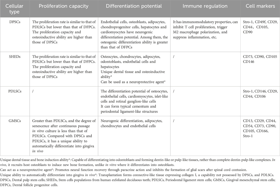
Table 1. Comparison of various properties of DMSCs (Guanlin et al., 2021; Linglu et al., 2021; Lu et al., 2020; Sabbagh et al., 2020; Al-Qadhi et al., 2021; Deng et al., 2024; Kotova et al., 2021; Nowwarote et al., 2020; Abe et al., 2022).
4 The modalities by which dental mesenchymal stem cells facilitate tissue regeneration
Previous studies have shown that dental mesenchymal stem cells mainly promote tissue regeneration through three ways: (1)differentiation into tissue cells; (2) improvement of the local regeneration microenvironment through immunomodulatory functions; (3) promotion of tissue regeneration by releasing various bioactive components.
4.1 Differentiate into tissue cells
During the development of the tooth germ, the enamel organ composed of the ectodermal epithelium undergoes significant morphological changes. In the cap stage, an intermediate layer appears between the inner enamel epithelium and the stellate reticulum. In the late stage, acidic mucopolysaccharides and glycogen deposits can be observed in the cytoplasm of the intermediate layer cells, and they have high alkaline phosphatase activity. Subsequent studies have demonstrated that alkaline phosphatase is involved in the formation of enamel, dentin, and bone. Furthermore, its high enzymatic activity can serve as a marker for cells associated with the development of these tissues (Yu et al., 2015). Stem cell surface markers are often used to routinely identify the differentiation potential of pluripotent mesenchymal stem cells (i.e., to confirm the strength of the stemness of cells). CD51/CD140α and CD271 have been used to isolate and identify neural crest cell progenitors, while the STRO-1/CD146 combination has been used to isolate mesenchymal stem cells (MSCs) located in blood vessels (Mao et al., 2012). Since the success of craniofacial defect repair depends on the ability to separate specific subpopulations of DMSCs with strong differentiation ability into appropriate cell types, it is crucial to identify effective markers for isolating pluripotent DMSCs. The mRNA expression of dental source genes can indicate whether mesenchymal stem cells have differentiated into dental lineage tissue cells.
In the experiment by Alvarez et al. (Ruth et al., 2015), primary dental pulp cells (DPCs) from the dental pulp tissue of adult third molars were obtained in α-modified Eagle’s medium (containing 20% fetal bovine serum (FBS), non-essential amino acids, 100 units/mL of penicillin, and 100 units/mL of streptomycin) in a humidified incubator at 37 °C with 5% CO2 to obtain DMSCs. Then, fluorescence-activated cell sorting (FACS) technology was used to sort out suitable DMSCs. These cells are induced to undergo odontogenesis through a specific tooth-inducing medium or what is known as odontogenic induction medium (OIM). The results (Table 2) showed that various surface markers (including CD51/CD140α, CD271, and STRO-1/CD146 DMSCs) could be isolated from the surface of DMSCs. Their alkaline phosphatase (ALP) activity was measured to determine their differentiation ability, and the mRNA expression of dental source genes at different time points was detected to confirm that DMSCs with surface markers may exhibit the ability to differentiate into dental lineage cells. Furthermore, numerous researchers have uncovered the differentiation potential of DMSCs. Figure 3 illustrates some of the studies on DMSCs - derived differentiated cells.
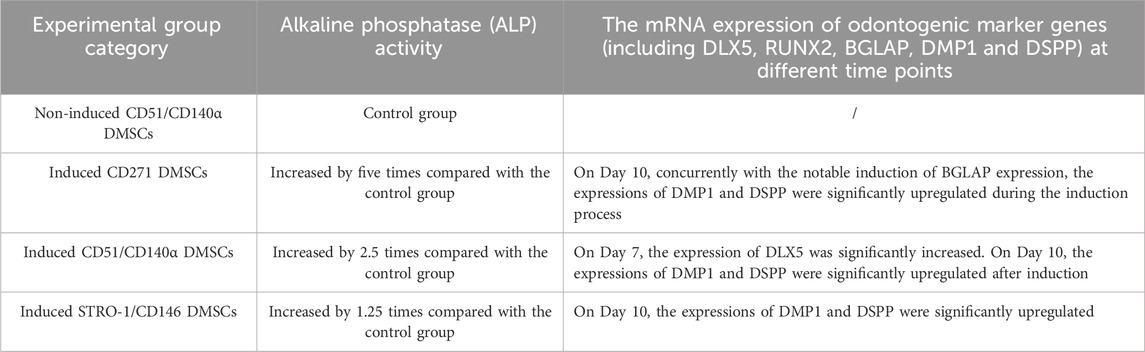
Table 2. ALP activity and expression of dental source marker genes under different induction media cultivation (Ruth et al., 2015).
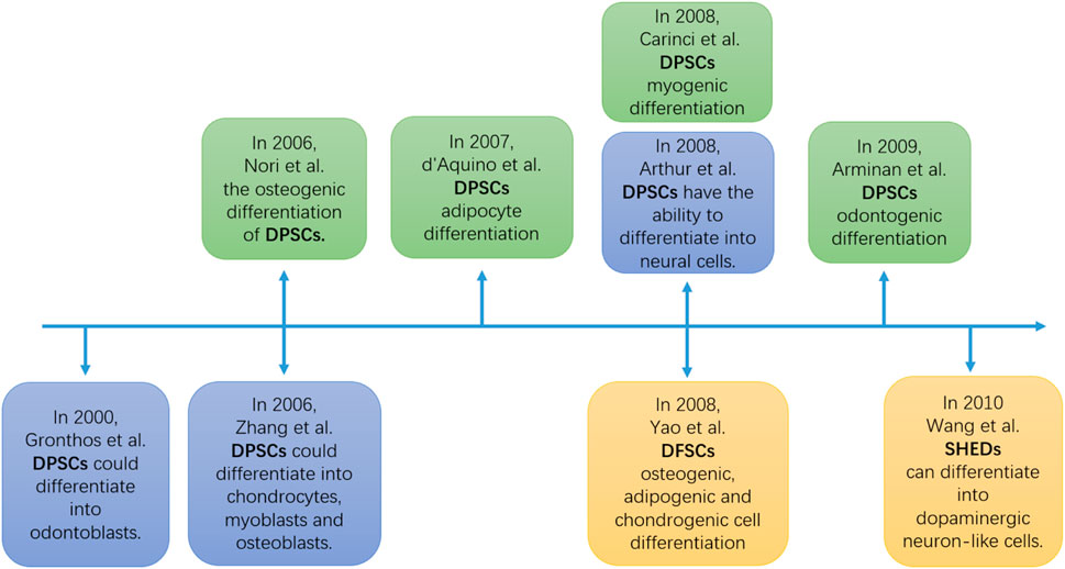
Figure 3. The timeline of the DMSCs differentiation potential experimen (Zhang et al., 2006; Arthur et al., 2008; Nör, 2006; d’Aquino et al., 2007; Carinci et al., 2008; Armiñán et al., 2009; Yao et al., 2008; Wang et al., 2010). DPSCs, Dental pulp stem cells; DFSCs, Dental follicle stem cells; SHEDs, Stem cells populations from human exfoliated deciduous teeth.
4.2 Improve the local regenerative microenvironment through immune regulation function
After various injury factors cause damage to the body’s tissues and cells, sentinel cells around the injured tissue (such as macrophages) mediate the production of inflammatory mediators by recognizing injury factors and necrotic tissue. This is the reason why the body often experiences an inflammatory response in cases of tissue damage and infection (such as fractures, trauma, tumors, etc.). The body can repair damage by confining or eliminating injury factors. However, if tissue cells are chronically exposed to an inflammatory environment (such as without using drugs to block the continuous inflammatory process), the inflammatory mediators in the inflammatory environment will activate the host’s vascular response and leukocyte response. The aggregation of a large number of activated cells and the excessive release of enzymes and chemokines will instead impede the process of tissue repair. Timely blocking the development of the inflammatory response through immune regulation function becomes an important factor for tissue regeneration (Wang et al., 2020).
DMSCs can exert immunomodulatory functions by releasing or inhibiting the expression of factors, influencing the proliferation, differentiation, apoptosis and other physiological processes and morphological functions of T cells and macrophages, thereby improving the local regenerative microenvironment to promote tissue repair and regeneration (Table 3).
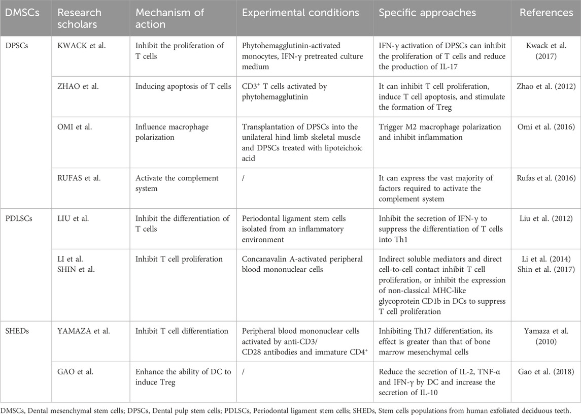
Table 3. Immune regulation pathways of DMSCs.
4.3 By releasing a variety of bioactive components, it promotes tissue regeneration
Dental pulp stem cells share the same characteristics as other mesenchymal stem cells, such as high proliferation capacity and multi-lineage differentiation ability. Besides differentiating into various mesodermal tissue cells, dental pulp stem cells also exhibit differentiation capabilities in non-mesodermal tissues, such as neurons. In experiments, researchers have found that dental pulp stem cells in the culture medium for regeneration secrete various bioactive substances. Ogata et al. discovered that the culture medium derived from dental pulp stem cells contains anti-inflammatory cytokines, IL-10, IL-13, follistatin, TGF-β1, hepatocyte growth factor (HGF), neural cell adhesion molecule-1 (NCAM-1), and other bioactive substances. The presence of these bioactive substances is highly beneficial in reducing inflammation, effectively reducing infection, promoting stem cell proliferation, and improving the tissue repair environment (Kwack et al., 2017).
In addition, Zhang et al. found that DPSCs can secrete neuron-related proteins such as DCX, NeuN and NF200, thereby restoring the neurological function of rats with Alzheimer’s disease (Zhang et al., 2021).
Moreover, Roato et al. demonstrated that both PDLSCs and DPSCs can secrete VEGF and generate substances such as proteoglycans when cultured in osteogenic medium (OM). These secretions play a significant role in promoting angiogenesis (Roato et al., 2025).
Through the secretion of growth factors, chemokines, etc., DMSCs exert a paracrine effect. This effect serves to regulate the microenvironment of the injured area, inhibit cell apoptosis and local inflammatory responses, and facilitate angiogenesis and tissue repair.
5 The application of DMSCs in the repair of maxillofacial bone tissue
In bone tissue repair, DMSCs can promote bone regeneration, enhance bone repair capacity, improve bone quality, and repair bone defects. They can be applied in periodontal disease treatment and alveolar bone repair. The following is a detailed description of the currently well-studied DPSCs in bone tissue repair.
5.1 DPSCs
According to the studies by Amiryaghoubi et al. (2020) and Imanishi et al. (2021), DPSCs were investigated using different materials and scaffolds. The results demonstrated that DPSCs exhibited excellent regenerative performance when supported by hydrogel scaffolds and a mixed scaffold composed of three substances including collagen (COL). This can achieve the goal of micro-repairing mandibular and facial bone defects by efficiently inducing the proliferation of DPSCs and promoting their differentiation into osteoblasts, without inducing inflammation. It is a material that can balance safety and efficiency in mandibular and facial bone regeneration. Meanwhile, Alksne et al. (2022) delved into the biological characteristics of DPSCs themselves and discovered that they can secrete a unique extracellular matrix (ECM) protein network, broadening the understanding of the repair mechanism of DPSCs: this protein network can promote biological behaviors of cells such as adhesion, migration, proliferation or immune regulation by forming a suitable microenvironment, thereby achieving biological processes such as recruiting endogenous stem cells, osteogenesis and angiogenesis (Table 4).
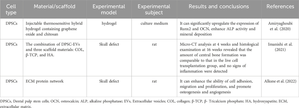
Table 4. Applications of DPSCs in bone tissue repair.
5.2 SHEDs
Hiraki et al. (2020) used a 5 mm critical-sized mouse calvarial defect model to demonstrate that both human exfoliated SHEDs and their conditioned medium (SHED-CM) could significantly promote bone regeneration, with SHED-CM showing a superior effect compared to direct SHEDs application. This study was the first to prove that transplanting only the secretome of SHEDs could achieve bone regeneration comparable to that of live cells without immunosuppression, making it particularly suitable for repairing alveolar clefts in children with cleft lip and palate. Sattary et al. (2022) used polycaprolactone/gelatin/hydroxyapatite nanoparticles (PGH) as a scaffold to culture SHEDs and found that PGH scaffolds seeded with SHEDs could accelerate bone repair, which could be further translated into clinical applications for repairing craniofacial bone defects. Kunwong et al. (2021) further explored the application of SHEDs in a poly (lactic-co-glycolic acid) (PLGA)-bioactive glass composite scaffold. The results showed that a scaffold composed of 10% bioactive glass and PLGA performed well in promoting the adhesion, proliferation, and osteogenic differentiation of SHEDs, and could promote new bone formation at the site of rat alveolar bone defects without inducing inflammatory responses, providing important experimental evidence for the development of new and efficient bone regeneration materials (Table 5).
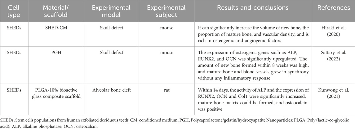
Table 5. Applications of SHEDs in bone tissue repair.
5.3 SCAPs
Cui et al. (2020) demonstrated that injecting berberine (BBR) into the root canals of a rat model of apical periodontitis activated the Wnt/β-catenin signaling pathway in SCAPs, significantly promoting root apex repair in immature teeth with apical periodontitis. This finding suggests that pharmacological activation of SCAPs can drive root reconstruction and may be used for the treatment of root apex defects in children and adolescents. Wu L. et al. (2021) found that a low concentration (0.2 mg/mL) of injectable root canal sealer (iRoot SP) significantly promoted the proliferation, migration, and osteogenic differentiation of SCAPs. By regulating the activity of SCAPs, it provides a favorable microenvironment for the repair of periapical bone tissue and is expected to become an efficient biomaterial for promoting periapical bone regeneration. Ramírez et al. (2024) discovered that both platelet-rich fibrin (PRF) and low-level laser therapy (LLLT) alone could promote the osteogenic differentiation of SCAPs, and their combined use produced a synergistic effect, significantly enhancing the formation of mineralized nodules and the secretion of osteogenic-related proteins (such as BMP-2 and BMP-4) by SCAPs. This study indicates that the combination of PRF and LLLT can serve as an efficient tool for the repair of maxillofacial bone tissue, providing a new combined treatment strategy and holding promise for more effective bone regeneration in clinical practice (Table 6).
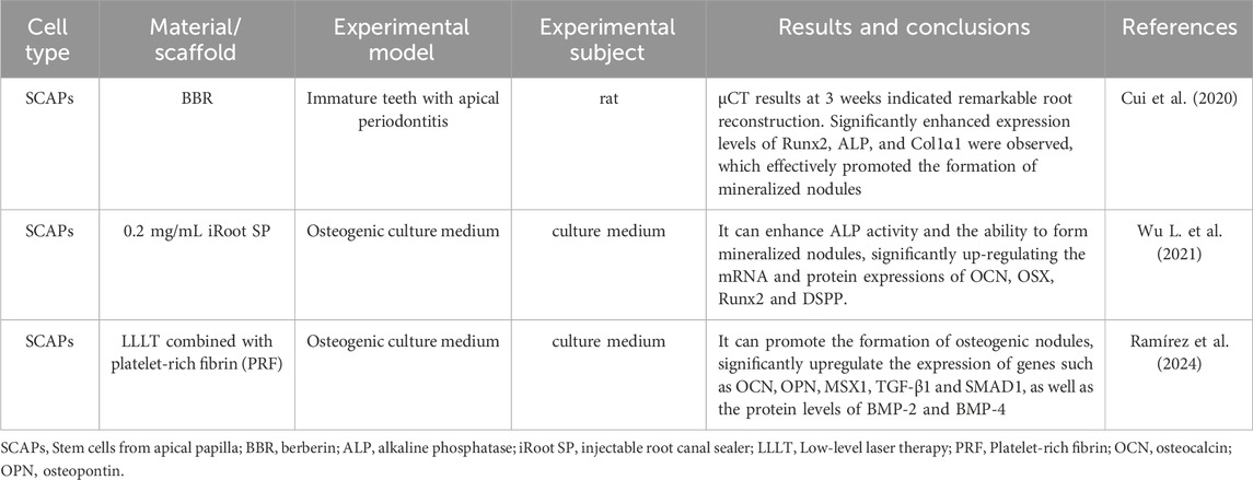
Table 6. Applications of SCAPs in bone tissue repair.
5.4 PDLSCs
Pan et al. (2020) encapsulated PDLSCs in liquid methacrylated Gelatin (GelMA) hydrogel and demonstrated in a rat critical alveolar bone defect model that GelMA hydrogel could effectively fill any irregularly shaped alveolar bone defects and provide a suitable physicochemical microenvironment for the adhesion, viability, and osteogenic differentiation of PDLSCs, protecting PDLSCs from being lost due to bone defects in the early stage of transplantation. PDLSCs encapsulated in GelMA hydrogel exhibited significant alveolar bone regeneration potential. Ha and Choung (2020) proved that methylsulfonylmethane (MSM) could significantly promote the proliferation and osteogenic differentiation of PDLSCs and enhance the regeneration efficiency of PDLSCs in the repair of maxillofacial bone defects. Zhao et al. (2022) demonstrated that exosomes derived from periodontal ligament stem cells (PDLSCs-Exos) could significantly promote the repair of mandibular bone defects. Their combined application with sodium GelMA-Alginate hydrogel (Gel-Alg) could be used to repair initial alveolar bone defects in SD rats, promoting bone regeneration. This is a promising strategy for the repair of mandibular bone defects, breaking through the limitations of traditional stem cell therapy and providing new ideas for future clinical applications (Table 7).
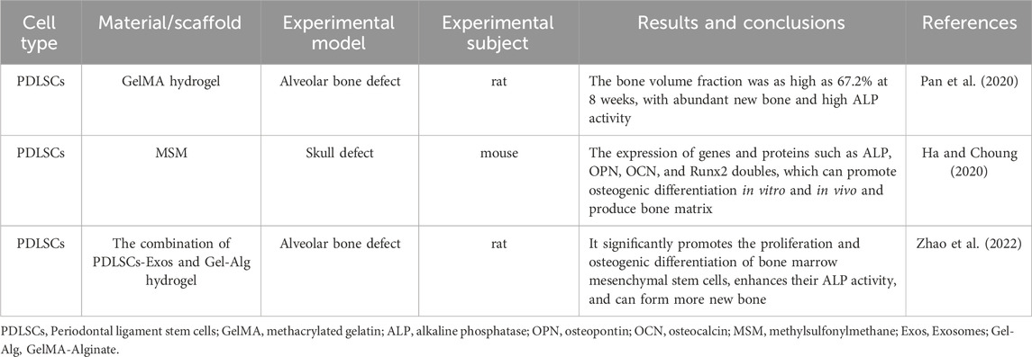
Table 7. Applications of PDLSCs in bone tissue repair.
5.5 DFSCs
Meng et al. (2022) focused on the effects of N-acetylcysteine (NAC) on the osteogenic properties of DFSCs and alveolar bone repair. The study found that treatment with 5 mM NAC significantly enhanced the proliferation ability of DFSCs, reduced the senescence rate, and increased the expression of stem cell-specific markers and immune-related factors. It also promoted the strongest osteogenic differentiation ability. An appropriate concentration of NAC enhanced the osteogenic properties of DFSCs through the PI3K/AKT/ROS signaling pathway, providing clinical potential for alveolar bone regeneration based on stem cells. Chen et al. (2023) revealed the important role of long non-coding RNA HOTAIRM1 in DFSC-mediated bone regeneration. The study found that HOTAIRM1 was induced under hypoxic conditions and activated HIF-1α, which in turn upregulated oxygen-sensing histone demethylases KDM6A/B and inhibited the methyltransferase EZH2. By targeting HIF-1α, HOTAIRM1 regulated the demethylation of H3K27 and promoted osteogenic differentiation of DFSCs. This study provided a new molecular mechanism and potential therapeutic target for the application of DFSCs in the repair of maxillofacial bone tissue, indicating that HOTAIRM1 may promote the osteogenic ability of DFSCs and bone regeneration by regulating the HIF-1α/KDM6/EZH2/H3K27me3 axis (Table 8).
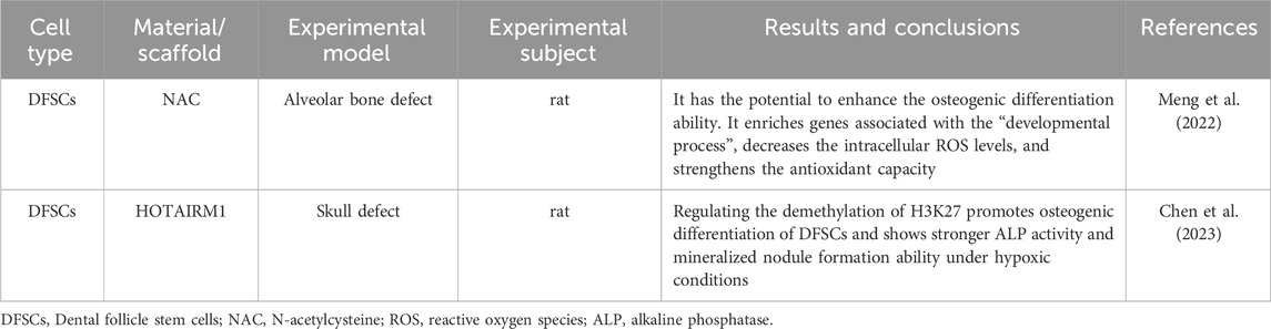
Table 8. Application of DFSCs in bone tissue repair.
6 The application of DMSCs in the repair of soft tissues in the maxillofacial region
The oral and maxillofacial region plays a crucial role in chewing, pronunciation, and facial expressions. However, its high exposure rate makes it vulnerable to trauma and infection. Defects in the soft tissues of the oral and maxillofacial region not only cause functional problems for patients, bringing many inconveniences to their lives, but also seriously affect their appearance and mental health. In recent years, as people’s demands for aesthetic restoration have increased, how to functionally reconstruct the damaged tissues while restoring their appearance as much as possible and achieving good aesthetic restoration results has become the focus of soft tissue repair. Utilizing dental mesenchymal stem cells to achieve soft tissue regeneration in the oral and maxillofacial region will become an effective approach for treating and restoring maxillofacial soft tissues in the future.
6.1 PDLSCs
The periodontal ligament is a connective tissue located between the alveolar bone and cementum, capable of absorbing mechanical pressure exerted on the teeth during chewing. PDLSCs are heterogeneous, clonal, highly proliferative and pluripotent cells that can differentiate into osteoblasts, cementoblasts, chondrocytes and adipocytes, promoting the new formation of periodontal tissues, including periodontal ligament fibers similar to Sharpey fibers, bone and cementum (Queiroz et al., 2021).
You et al. (2024) confirmed through a rat periodontal window defect model that PDLSCs injection could reduce the reverse diversity during periodontal repair and increase the abundance of non-pathogenic bacteria such as Bifidobacterium and Lactobacillus, demonstrating that PDLSCs have the ability to influence the oral microbiota and promote periodontal regeneration. Meanwhile, the studies by Zhao et al. (2020) and Hu et al. (2020) indicated that PDLSCs, when combined with different carriers, could improve periodontal tissue regeneration. They found that the expression of type I collagen, alkaline phosphatase and bone sialoprotein genes was significantly upregulated on the polycaprolactone (PCL) membrane scaffold containing simvastatin (SIM), and the formation of mineralized matrix increased. In the tissue derived from PDLSC sheets combined with HA/TCP, the fibers showed obvious directionality, meeting the functional requirements of the periodontal soft tissue for the ordered structure of fibers. This suggests that PDLSCs materials provide a molecular and structural basis for the repair of periodontal soft tissue, which may contribute to periodontal tissue regeneration (Table 9).
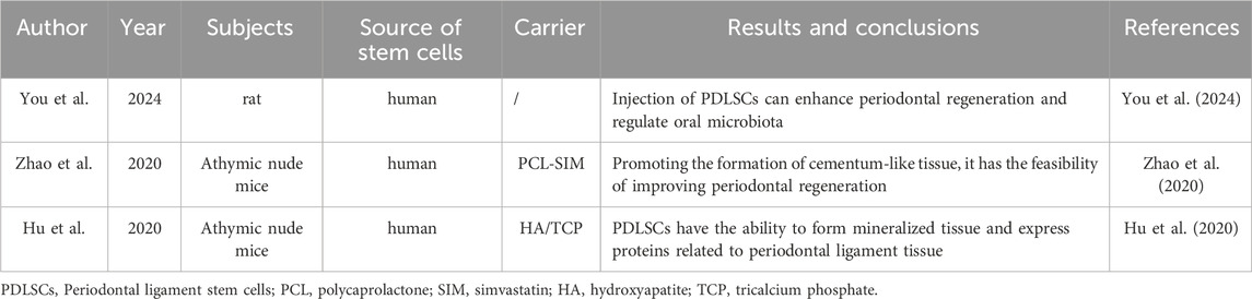
Table 9. Research on PDLSCs in soft tissue repair.
6.2 SCAPs
SCAPs are a type of MSC population residing in the apical region of immature permanent teeth, characterized by high proliferation potential, self-renewal ability, and low immunogenicity (Sonoyama et al., 2006), and thus hold great potential for stem cell therapy.
Combining the in vitro and in vivo experimental results of Jing et al. (2022) on the exosomes of stem cells from apical papillae (SCAP-Exo), it was found that SCAP-Exo demonstrated excellent angiogenic and osteogenic properties, and this ability was still significantly manifested in diabetic (diabetes mellitus, DM) rats. The experiments showed an increase in the number of trabeculae in the alveolar bone of DM rats, a reduction in trabecular space, and a significant improvement in bone healing. This indicates that SCAPs have outstanding potential in promoting periodontal tissue regeneration. Meanwhile, Li et al. (2018) focused on the clinical translation potential of SCAPs, taking the key clinical assessment indicators in periodontitis treatment - probing pocket depth (PPD) and attachment loss (AL) - as the core research objects. Through experiments, it was confirmed that SCAPs could effectively improve the PPD and AL indicators in a porcine periodontitis model. By using clinical assessment data such as PPD and AL, a bridge was built between laboratory research and clinical application, which has a positive effect on the application of SCAPs in soft tissue repair (Table 10).
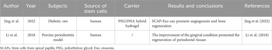
Table 10. Research on SCAPs in soft tissue repair.
6.3 SHEDs
SHEDs are regarded as a promising cell population for cell-based or cell-free therapy and tissue engineering due to their proliferative, pluripotent and immunomodulatory properties. Studies have found that in addition to their osteogenic and odontogenic potential, SHEDs also exhibit remarkable neuroregenerative capabilities (Tsutsui, 2020).
According to the research of Wu M. et al. (2021), it was found that the exosomes of SHEDs (SA-Exo) can upregulate the expression of angiogenesis-related proteins such as VEGF, angiopoietin 2 and PDGF in SHEDs. This reflects the endothelial differentiation potential of SHEDs and also indicates from a histological perspective that SA-Exo can enhance the regeneration of the dentin-pulp complex and angiogenesis. Guo et al. (2020) focused on the regeneration of blood vessels and nerves. Through histological analysis, it was shown that the extracted pulp permanent incisors of miniature pigs implanted with SHEDs contained regenerated pulp tissue with a similar structure to normal pulp. Moreover, the positive expression of platelet endothelial cell adhesion molecule (CD31) and neurofilament (NF) in the regenerated pulp tissue confirmed the regeneration of blood vessels and nerves. In summary, SHEDs have the ability to regenerate host pulp tissue in vivo. The clinical case report by Ghana Shyam Prasad et al. (2019) provided key practical evidence: after treating tooth #46 with SHEDs for apical periodontal lesions, the patient’s periapical radiolucency completely disappeared, and tooth #46 responded positively to the electric pulp test, which may indicate the reinnervation of pulp tissue in the root canal. It can be seen that the application of SHEDs in humans shows the ability to effectively treat apical periodontal lesions of permanent teeth (Table 11).
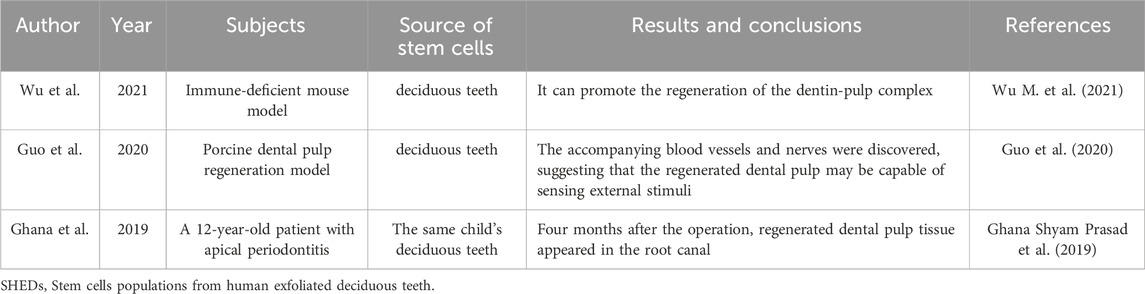
Table 11. Research on SHEDs in soft tissue repair.
6.4 GMSCs
The gingiva is firmly attached to the teeth and jawbone, forming a mucoperiosteum, which has the ability to resist masticatory and minor traumatic forces. It plays a crucial role in maintaining oral health and exhibits a unique fetal-like scarless healing process after injury, suggesting that this tissue may be a source of stem cells (Chen et al., 2012).
Qiu et al. (2020) transplanted GMSCs into periodontal defects in rats and found that periodontal tissue regeneration occurred, with significantly lower expression levels of tumor necrosis factor (TNF-α) and interleukin (IL-1β) compared to the control group, and significantly higher expression of BSP-II and Runx2. This indicates that GMSC-CM may have the effect of inhibiting inflammation, promoting periodontal regeneration, and osteogenic differentiation. Sun et al. (2019) transplanted GMSCs into C57BL/6 J mice with periodontitis and found that the alveolar bone height of the mice increased significantly, and GMSCs were detected in the newly formed periodontal ligament and alveolar bone. This suggests that systemically transplanted GMSCs can return to the periodontal injury site and promote the regeneration and repair of periodontal tissue. Fawzy El-Sayed et al. (2015) transplanted GMSCs into periodontal defects in miniature pigs and found that compared to the control group, they showed better clinical attachment levels, PPD, GR, periodontal attachment levels, cementum regeneration and bone regeneration, as well as shorter junctional epithelium length and better improvement in probing bleeding. This indicates that GMSCs have excellent potential for periodontal regeneration (Table 12).
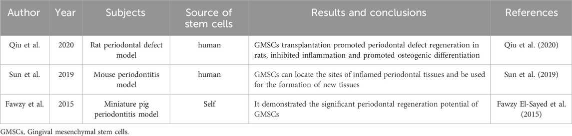
Table 12. Research on GMSCs in soft tissue repair.
7 Summary and prospects
In summary, DMSCs have demonstrated excellent performance in preclinical and clinical studies for the repair of bone and cartilage tissues in the maxillofacial region under the support of various culture media. Despite the promising application prospects of DMSCs, there are still bottlenecks for their large-scale clinical application.
7.1 Challenges in the clinical application of DMSCs
7.1.1 Lack of standardized isolation and culture procedures for DMSCs
One of the challenges in the clinical application of DMSCs is how to obtain a large number of stem cells from tissues. This goal cannot be achieved without the isolation of stem cells, multiple passages, and cryopreservation. Although enzymatic methods are generally applicable in the process of isolating stem cells, there are no unified regulations for the physical operations of tissue separation for different types of DMSCs. Moreover, multiple passages may reduce the differentiation potential of cultured cells, thus leading to the development of various culture media to regulate their performance. Additionally, different cryopreservation strategies, such as controlled-rate freezing and non-controlled-rate freezing, are still controversial (Khaseb et al., 2021).
7.1.2 Difficulties in quality control of DMSCs
Compared with chemically synthesized drugs, the cell culture conditions of DMSCs, including cell passage number and density, may lead to inconsistencies among different batches of DMSCs. Moreover, studies have found that the conditioned medium functions of DMSCs from different sources may be affected by their cell sources (Joo et al., 2018). The age and diseases of DMSC donors may also influence the composition of DMSC culture media (Bhandi et al., 2021).
7.1.3 Rejection and tumorigenic risks
When DMSCs are used for allogeneic transplantation, the risks of immunogenicity and tumorigenicity cannot be ignored. Since DMSCs, such as DPSCs, have unique immunomodulatory functions, if their upregulated genes are enriched in immune and inflammatory-related cell pathways, they often exhibit higher immunogenicity, increasing the risk of immune rejection during allogeneic transplantation. Additionally, the differential expression of tumor-related genes and the presence of undifferentiated induced pluripotent stem cells during the induction and differentiation process of DMSCs may also lead to tumor formation (Taoran et al., 2024).
7.1.4 Cost issues in large-scale production
Exosomes are extracellular vesicles that carry various bioactive molecules such as proteins, genetic material, and lipids, playing a crucial role in intercellular communication and material transport (Lu et al., 2024). Exosomes can be extracted from DMSCs, and studies have found that their functions are highly dependent on the source cells and their physiological states (Huang et al., 2016). In recent years, multiple studies have discovered that exosomes play a role in various aspects such as angiogenesis (Ribeiro et al., 2013) and neural repair (Jarmalavičiūtė et al., 2015) by regulating intracellular signaling pathways. Like other bioactive components, exosomes, as an important component of DMSCs, must be stored under appropriate conditions. Wu J et al. (2021) found that different exosome storage conditions can affect their biological utilization rate. Generally, it is recommended to store them at −80 °C, but this condition is costly (Zhang et al., 2025).
Although a large number of proteins, nucleic acids, lipids and metabolic enzymes have been identified in Exos, little is known about their functions and sorting mechanisms. Moreover, the concentration of effective substances in Exos depends on the surrounding environment and the metabolic state of the host cells, making the condition control in the clinical application of Exos rather complex. Despite extensive research on Exos in chronic wound healing and skin regeneration, the exact molecular mechanisms and roles of Exos in tissue repair and regeneration still need further investigation. In addition, to use Exos as a research tool, extensive research is still needed in the fields of EV biosynthesis, cellular uptake and transport, which still has a long way to go (Hade et al., 2022).
7.2 Prospects for the clinical application of DMSCs
DMSCs, with their multi-directional differentiation and immunomodulatory properties, show great potential in the repair of maxillofacial defects. In the future, through technological optimization and more preclinical and clinical trials, overcoming issues such as quality control and safety, DMSCs may become a core force in regenerative medicine.
Author contributions
YC: Data curation, Formal Analysis, Visualization, Writing – original draft, Writing – review and editing. XM: Data curation, Writing – review and editing. HM: Data curation, Writing – review and editing. PL: Data curation, Writing – review and editing. RH: Conceptualization, Supervision, Writing – review and editing. AA: Conceptualization, Supervision, Writing – review and editing.
Funding
The author(s) declare that no financial support was received for the research and/or publication of this article.
Conflict of interest
The authors declare that the research was conducted in the absence of any commercial or financial relationships that could be construed as a potential conflict of interest.
Generative AI statement
The author(s) declare that no Generative AI was used in the creation of this manuscript.
Any alternative text (alt text) provided alongside figures in this article has been generated by Frontiers with the support of artificial intelligence and reasonable efforts have been made to ensure accuracy, including review by the authors wherever possible. If you identify any issues, please contact us.
Publisher’s note
All claims expressed in this article are solely those of the authors and do not necessarily represent those of their affiliated organizations, or those of the publisher, the editors and the reviewers. Any product that may be evaluated in this article, or claim that may be made by its manufacturer, is not guaranteed or endorsed by the publisher.
References
Abe, S., Kaida, A., Kanemaru, K., Nakazato, K., Yokomizo, N., Kobayashi, Y., et al. (2022). Differences in the stemness characteristics and molecular markers of distinct human oral tissue neural crest-derived multilineage cells. Cell Prolif. 55 (10), e13286. doi:10.1111/cpr.13286
Al-Qadhi, G., Aboushady, I., and Al-Sharabi, N. (2021). The gingiva from the tissue surrounding the bone to the tissue regenerating the bone: a systematic review of the osteogenic capacity of gingival mesenchymal stem cells in preclinical studies. Stem Cells Int. 2021 (1), 1–26. doi:10.1155/2021/6698100
Alksne, M., Kalvaityte, M., Simoliunas, E., Gendviliene, I., Barasa, P., Rinkunaite, I., et al. (2022). Dental pulp stem cell-derived extracellular matrix: autologous tool boosting bone regeneration. Cytotherapy 24 (6), 597–607. doi:10.1016/j.jcyt.2022.02.002
Amiryaghoubi, N., Pesyan, N. N., Fathi, M., and Omidi, Y. (2020). Injectable thermosensitive hybrid hydrogel containing graphene oxide and chitosan as dental pulp stem cells scaffold for bone tissue engineering. Int. J. Biol. Macromol. 162, 1338–1357. doi:10.1016/j.ijbiomac.2020.06.138
Armiñán, A., Gandía, C., Bartual, M., García-Verdugo, J. M., Lledó, E., Mirabet, V., et al. (2009). Cardiac differentiation is driven by NKX2. 5 and GATA4 nuclear translocation in tissue-specific mesenchymal stem cells. Stem Cells Dev. 18, 907–918. doi:10.1089/scd.2008.0292
Arthur, A., Rychkov, G., Shi, S., Koblar, S. A., and Gronthos, S. (2008). Adult human dental pulp stem cells differentiate toward functionally active neurons under appropriate environmental cues. Stem Cells 26, 1787–1795. doi:10.1634/stemcells.2007-0979
Bhandi, S., Al Kahtani, A., Mashyakhy, M., Alsofi, L., Maganur, P. C., Vishwanathaiah, S., et al. (2021). Modulation of the dental pulp stem cell secretory profile by hypoxia induction using cobalt chloride. J. Pers. Med. 11 (4), 247. doi:10.3390/jpm11040247
Carinci, F., Papaccio, G., Laino, G., Palmieri, A., Brunelli, G., D'Aquino, R., et al. (2008). Comparison between genetic portraits of osteoblasts derived from primary cultures and osteoblasts obtained from human pulpar stem cells. J. Craniofac Sur 19, 616–625. doi:10.1097/scs.0b013e31816aabc8
Chen, F., Sun, H., Lu, H., and Yu, Q. (2012). Stem cell-delivery therapeutics for periodontal tissue regeneration. Biomaterials 33 (27), 6320–6344. doi:10.1016/j.biomaterials.2012.05.048
Chen, Z., Gan, L., Chen, X., Zheng, J., Shi, S., Wu, L., et al. (2023). LncRNA HOTAIRM1 promotes dental follicle stem cell-mediated bone regeneration by regulating HIF-1α/KDM6/EZH2/H3K27me3 axis. J. Cell. Physiology 238 (7), 1542–1557. doi:10.1002/jcp.31028
Cui, Y., Xie, J., Fu, Y., Li, C., Zheng, L., Huang, D., et al. (2020). Berberine mediates root remodeling in an immature tooth with apical periodontitis by regulating stem cells from apical papilla differentiation. Int. J. Oral Sci. 12 (1), 18. doi:10.1038/s41368-020-0085-7
Deng, Y., Li, Q., Svoboda, K. K. H., Opperman, L., Ruest, L., and Liu, X. (2024). Gli1+ periodontal mesenchymal stem cells in periodontitis. J. Dent. Res. 103 (3), 279–288. doi:10.1177/00220345231220915
d’Aquino, R., Graziano, A., Sampaolesi, M., Laino, G., Pirozzi, G., De Rosa, A., et al. (2007). Human postnatal dental pulp cells co-differentiate into osteoblasts and endotheliocytes: a pivotal synergy leading to adult bone tissue formation. Cell Death Differ. 14, 1162–1171. doi:10.1038/sj.cdd.4402121
Fawzy El-Sayed, K., Mekhemar, M., Beck-Broichsitter, B., Bähr, T., Hegab, M., Receveur, J., et al. (2015). Periodontal regeneration employing gingival margin-derived stem/progenitor cells in conjunction with IL-1ra-hydrogel synthetic extracellular matrix. J. Of Clin. Periodontology 42 (5), 448–457. doi:10.1111/jcpe.12401
Forough, J. M., Benyamin, P., Ezatolah, K., and Ayyoob, K. (2023). Hopes and opportunities of stem cells from human exfoliated deciduous teeth (SHED) in cartilage tissue regeneration. Front. Bioeng. Biotechnol., 111021024–1021024. doi:10.3389/fbioe.2023.1021024
Gamilah, Q. A., Iman, A., and Niyaz, S. A. (2021). The gingiva from the tissue surrounding the bone to the tissue regenerating the bone: a systematic review of the osteogenic capacity of gingival mesenchymal stem cells in preclinical studies. Stem Cells Int., 20216698100–6698100. doi:10.1155/2021/6698100
Gao, X., Shen, Z., Guan, M., Huang, Q., Chen, L., Qin, W., et al. (2018). Immunomodulatory role of stem cells from human exfoliated deciduous teeth on periodontal regeneration. Tissue Eng. Part A 24 (17-18), 1341–1353. doi:10.1089/ten.tea.2018.0016
Ghana Shyam Prasad, M., Juvva, R., and Babu Duvvi, N. (2019). Towards a new era in the management of large periapical lesion in permanent tooth using stemcells: a 2-year clinical application report. J. Dent. (Shiraz, Iran) 20 (2), 137–140. doi:10.30476/DENTJODS.2019.44925
Gronthos, S., Mankani, M., Brahim, J., Robey, P. G., and Shi, S. (2000). Postnatal human dental pulp stem cells (DPSCs) in vitro and in vivo. Proc. Natl. Acad. Sci. 97 (25), 13625–13630. doi:10.1073/pnas.240309797
Guanlin, Q., Yan, L., Lu, C., Qin, C., Duohong, Z., Chi, Y., et al. (2021). Comparison of osteogenic differentiation potential of human dental-derived stem cells isolated from dental pulp, periodontal ligament, dental follicle, and alveolar bone. Stem Cells Int., 20216631905–6631905. doi:10.1155/2021/6631905
Guo, H., Zhao, W., Liu, A., Wu, M., Shuai, Y., Li, B., et al. (2020). SHED promote angiogenesis in stem cell-mediated dental pulp regeneration. Biochem. and Biophysical Res. Commun. 529 (4), 1158–1164. doi:10.1016/j.bbrc.2020.06.151
Ha, S. H., and Choung, P. H. (2020). MSM promotes human periodontal ligament stem cells differentiation to osteoblast and bone regeneration. Biochem. Biophysical Res. Commun. 528 (1), 160–167. doi:10.1016/j.bbrc.2020.05.097
Hade, M. D., Suire, C. N., Mossell, J., and Suo, Z. (2022). Extracellular vesicles: emerging frontiers in wound healing. Med. Res. Rev. 42 (6), 2102–2125. doi:10.1002/med.21918
Hiraki, T., Kunimatsu, R., Nakajima, K., Abe, T., Yamada, S., Rikitake, K., et al. (2020). Stem cell-derived conditioned media from human exfoliated deciduous teeth promote bone regeneration. Oral Dis. 26 (2), 381–390. doi:10.1111/odi.13244
Hu, L., Zhao, B., Gao, Z., Xu, J., Fan, Z., Zhang, C., et al. (2020). Regeneration characteristics of different dental derived stem cell sheets. J. Of Oral Rehabilitation 47, 66–72. doi:10.1111/joor.12839
Huang, C. C., Narayanan, R., Alapati, S., and Ravindran, S. (2016). Exosomes as biomimetic tools for stem cell differentiation: applications in dental pulp tissue regeneration. Bio Mater. 111, 103–115. doi:10.1016/j.biomaterials.2016.09.029
Hwan, K. K., and Woo, H. L. (2022). Clinical potential of dental pulp stem cells in pulp regeneration: current endodontic progress and future perspectives. Front. Cell Dev. Biol., 10857066–857066. doi:10.3389/fcell.2022.857066
Imanishi, Y., Hata, M., Matsukawa, R., Aoyagi, A., Omi, M., Mizutani, M., et al. (2021). Efficacy of extracellular vesicles from dental pulp stem cells for bone regeneration in rat calvarial bone defects. Inflamm. Regen. 41, 12. doi:10.1186/s41232-021-00163-w
Jarmalavičiūtė, A., Tunaitis, V., Pivoraitė, U., Venalis, A., and Pivoriūnas, A. (2015). Exo- somes from dental pulp stem cells rescue human do- paminergic neurons from 6-hydroxy-dopamine-in- duced apoptosis. Cytotherapy 17 (7), 932–939. doi:10.1016/j.jcyt.2014.07.013
Jing, X., Wang, S., Tang, H., Li, D., Zhou, F., Xin, L., et al. (2022). Dynamically bioresponsive DNA hydrogel incorporated with dual-functional stem cells from apical papilla-derived exosomes promotes diabetic bone regeneration. Acs Appl. Mater. and Interfaces 14 (14), 16082–16099. doi:10.1021/acsami.2c02278
Joo, K., Song, J., Kim, S., Lee, H. S., and Jeon, M. (2018). Cytokine expression of stem cells originating from the apical complex and coronal pulp of immature teeth. J. Endod. 44 (1), 87–92.e1. doi:10.1016/j.joen.2017.08.018
Karamzadeh, R., Baghaban Eslaminejad, M., and Sharifi-Zarchi, A. (2017). Comparative in vitro evaluation of human dental pulp and follicle stem cell commitment. Cell J. 18 (4), 609–618. doi:10.22074/cellj.2016.4727
Khaseb, S., Orooji, M., Pour, M. G., Safavi, S. M., Eghbal, M. J., and Rezai Rad, M. (2021). Dental stem cell banking: techniques and protocols. Cell Biol. Int. 45 (9), 1851–1865. doi:10.1002/cbin.11626
Kotova, A. V., Lobov, A. A., Dombrovskaya, J. A., Sannikova, V. Y., Ryumina, N. A., Klausen, P., et al. (2021). Comparative analysis of dental pulp and periodontal stem cells: differences in morphology, functionality, osteogenic differentiation and proteome. Biomedicines 9 (11), 1606. doi:10.3390/biomedicines9111606
Kunwong, N., Tangjit, N., Rattanapinyopituk, K., Dechkunakorn, S., Anuwongnukroh, N., Arayapisit, T., et al. (2021). Optimization of poly (lactic-co-glycolic acid)-bioactive glass composite scaffold for bone tissue engineering using stem cells from human exfoliated deciduous teeth. Archives oral Biol. 123, 105041. doi:10.1016/j.archoralbio.2021.105041
Kwack, K. H., Lee, J. M., Park, S. H., and Lee, H. W. (2017). Human dental pulp stem cells suppress alloantigen-induced immunity by stimulating T cells to release transforming growth factor beta. J. Endod. 43 (1), 100–108. doi:10.1016/j.joen.2016.09.005
Li, C., Wang, X., Tan, J., Wang, T., and Wang, Q. (2014). The immunomodulatory properties of periodontal ligament stem cells isolated from inflamed periodontal granulation. Cells Tissues Organs 199 (4), 256–265. doi:10.1159/000367986
Li, G., Han, N., Zhang, X., Yang, H., Cao, Y., Wang, S., et al. (2018). Local injection of allogeneic stem cells from apical papilla enhanced periodontal tissue regeneration in minipig model of periodontitis. Biomed Res. Int. 2018, 1–8. doi:10.1155/2018/3960798
Li, J., Wu, Z., Zhao, L., Liu, Y., Su, Y., Gong, X., et al. (2023). The heterogeneity of mesenchymal stem cells: an important issue to be addressed in cell therapy. Stem cell Res. and Ther. 14 (1), 381. doi:10.1186/s13287-023-03587-y
Linglu, J., Yunpeng, Z., Dongfang, L., Zhang, W., Zhang, D., and Xu, X. (2021). Analyses of key mRNAs and lncRNAs for different osteo-differentiation potentials of periodontal ligament stem cell and gingival mesenchymal stem cell. J. Cell. Mol. Med. 25 (13), 6217–6231. doi:10.1111/jcmm.16571
Liu, D., Xu, J., Liu, O., Fan, Z., Liu, Y., Wang, F., et al. (2012). Mesenchymal stem cells derived from inflamed periodontal ligaments exhibit impaired immunomodulation. J. Clin. Periodontol. 39 (12), 1174–1182. doi:10.1111/jcpe.12009
Lu, G., Ying, L., Dixin, C., Yue, P., Liwei, Z., and Mian, W. (2020). Dental tissue-derived human mesenchymal stem cells and their potential in therapeutic application. Stem Cells Int. doi:10.1155/2020/8864572
Lu, H., Zheng, Y., and Wei, Z. (2024). The mechanism of action of dental mesenchymal stem cell exosomes in pulp regeneration. Int. J. Stomatology 51 (04), 467–474. doi:10.7518/gjkq.2024064
Mao, J. J., Robey, P. G., and Prockop, D. J. (2012). Stem cells in the face: tooth regeneration and beyond. Cell Stem Cell 11 (3), 291–301. doi:10.1016/j.stem.2012.08.010
Mason, S., Tarle, S. A., Osibin, W., Kinfu, Y., and Kaigler, D. (2014). Standardization and safety of alveolar bone–derived stem cell isolation. J. Dent. Res. 93 (1), 55–61. doi:10.1177/0022034513510530
Meng, Z., Liu, J., Feng, Z., Guo, S., Wang, M., Wang, Z., et al. (2022). N-acetylcysteine regulates dental follicle stem cell osteogenesis and alveolar bone repair via ROS scavenging. Stem Cell Res. and Ther. 13 (1), 466. doi:10.1186/s13287-022-03161-y
Miura, M., Gronthos, S., Zhao, M., Lu, B., Fisher, L. W., Robey, P. G., et al. (2003). SHED: stem cells from human exfoliated deciduous teeth. Proc. Natl. Acad. Sci. 100 (10), 5807–5812. doi:10.1073/pnas.0937635100
Nör, J. (2006). Buonocore memorial lecture: tooth regeneration in operative dentistry. Oper. Dent. 31, 633–642. doi:10.2341/06-000
Nowwarote, N., Manokawinchoke, J., Kanjana, K., Fournier, B. P., Sukarawan, W., and Osathanon, T. (2020). Transcriptome analysis of basic fibroblast growth factor treated stem cells isolated from human exfoliated deciduous teeth. Heliyon 6 (6), e04246. doi:10.1016/j.heliyon.2020.e04246
Omi, M., Hata, M., Nakamura, N., Miyabe, M., Kobayashi, Y., Kamiya, H., et al. (2016). Transplantation of dental pulp stem cells suppressed inflammation in sciatic nerves by promoting macrophage polarization towards anti-inflammation phenotypes and ameliorated diabetic polyneuropathy. J. Diabetes Investig. 7 (4), 485–496. doi:10.1111/jdi.12452
Pan, J., Deng, J., Yu, L., Wang, Y., Zhang, W., Han, X., et al. (2020). Investigating the repair of alveolar bone defects by gelatin methacrylate hydrogels-encapsulated human periodontal ligament stem cells. J. Mater. Sci. Mater. Med. 31, 3–12. doi:10.1007/s10856-019-6333-8
Qiu, J., Wang, X., Zhou, H., Zhang, C., Wang, Y., Huang, J., et al. (2020). Enhancement of periodontal tissue regeneration by conditioned media from gingiva-derived or periodontal ligament-derived mesenchymal stem cells: a comparative study in rats. Stem Cell Res. and Ther. 11 (1), 42. doi:10.1186/s13287-019-1546-9
Queiroz, A., Albuquerque-Souza, E., Gasparoni, L. M., França, B. N. d., Pelissari, C., Trierveiler, M., et al. (2021). Therapeutic potential of periodontal ligament stem cells. World J. stem cells 13 (6), 605–618. doi:10.4252/wjsc.v13.i6.605
Ramírez, D. G., Inostroza, C., Rouabhia, M., Rodriguez, C. A., Gómez, L. A., Losada, M., et al. (2024). Osteogenic potential of apical papilla stem cells mediated by platelet-rich fibrin and low-level laser. Odontology 112 (2), 399–407. doi:10.1007/s10266-023-00851-8
Ribeiro, M. F., Zhu, H. Y., Millard, R. W., and Fan, G. C. (2013). Exosomes function in pro- and anti-angiogenesis. Curr. An giogenes 2 (1), 54–59. doi:10.2174/22115528113020020001
Roato, I., Orrico, C., Meinardi, S., Pedraza, R., Mosca Balma, A., Baima, G., et al. (2025). The pro-angiogenic potential of periodontal ligament stem cells and dental pulp stem cells: a comparative analysis. Cells 14 (12), 864. doi:10.3390/cells14120864
Rufas, P., Jeanneau, C., Rombouts, C., Laurent, P., and About, I. (2016). Complement C3a mobilizes dental pulp stem cells and specifically guides pulp fibroblast recruitment. J. Endod. 42 (9), 1377–1384. doi:10.1016/j.joen.2016.06.011
Ruth, A., Hye-Lim, L., Christine, H., and Wang, C. Y. (2015). Single CD271 marker isolates mesenchymal stem cells from human dental pulp. Int. J. oral Sci. 7 (4), 205–212. doi:10.1038/ijos.2015.29
Sabbagh, J., Ghassibe-Sabbagh, M., Fayyad-Kazan, M., Al-Nemer, F., Fahed, J. C., Berberi, A., et al. (2020). Differences in osteogenic and odontogenic differentiation potential of DPSCs and SHED. J. Dent. 101, 103413. doi:10.1016/j.jdent.2020.103413
Sattary, M., Kefayat, A., Bigham, A., and Rafienia, M. (2022). Polycaprolactone/Gelatin/Hydroxyapatite nanocomposite scaffold seeded with Stem cells from human exfoliated deciduous teeth to enhance bone repair: in vitro and in vivo studies. Mater. Technol. 37 (5), 302–315. doi:10.1080/10667857.2020.1837488
Seo, B. M., Miura, M., Gronthos, S., Mark Bartold, P., Batouli, S., Brahim, J., et al. (2004). Investigation of multipotent postnatal stem cells from human periodontal ligament. Lancet 364 (9429), 149–155. doi:10.1016/s0140-6736(04)16627-0
Shin, C., Kim, M., Han, J. A., Choi, B., Hwang, D., Do, Y., et al. (2017). Human periodontal ligament stem cells suppress T-cell proliferation via down-regulation of non-classical major histocompatibility complex-like glycoprotein CD1b on dendritic cells. J. Periodontal Res. 52 (1), 135–146. doi:10.1111/jre.12378
Sonoyama, W., Liu, Y., Fang, D., Yamaza, T., Seo, B. M., Zhang, C., et al. (2006). Mesenchymal stem cell-mediated functional tooth regeneration in swine. PloS one 1 (1), e79. doi:10.1371/journal.pone.0000079
Sun, W., Wang, Z., Xu, Q., Sun, H., Liu, X., Yang, J., et al. (2019). The treatment of systematically transplanted gingival mesenchymal stem cells in periodontitis in mice. Exp. And Ther. Med. 17 (3), 2199–2205. doi:10.3892/etm.2019.7165
Taoran, Z., Hou, W., and Wang, M. (2024). Therapeutic potential and application risks of mesenchymal stem cells derived from induced pluripotent stem cells. Chin. J. Biochem. Mol. Biol. 40 (05), 656–663. doi:10.13865/j.cnki.cjbmb.2024.04.1015
Tsutsui, T. W. (2020). Dental pulp stem cells: advances to applications. Stem cells cloning Adv. Appl. 13, 33–42. doi:10.2147/sccaa.s166759
Wang, J., Wang, X., Sun, Z., Wang, X., Yang, H., Shi, S., et al. (2010). Stem cells from human-exfoliated deciduous teeth can differentiate into dopaminergic neuron-like cells. Stem Cells Dev. 19, 1375–1383. doi:10.1089/scd.2009.0258
Wang, Z., Xiaolan, L., and Liu, J. (2020). How to apply the immunomodulatory characteristics of dental mesenchymal stem cells in the treatment of oral diseases. Chin. J. Tissue Eng. Res. 24 (25), 4060–4067. doi:10.3969/j.issn.2095-4344.2100
Wu, L., Xue, K., Hu, G., Du, H., Gan, K., Zhu, J., et al. (2021). Effects of iRoot SP on osteogenic differentiation of human stem cells from apical papilla. BMC Oral Health 21, 407–410. doi:10.1186/s12903-021-01769-9
Wu, M., Liu, X., Li, Z., Huang, X., Guo, H., Guo, X., et al. (2021). SHED aggregate exosomes shuttled miR-26a promote angiogenesis in pulp regeneration via TGF-β/SMAD2/3 signalling. Cell Prolif. 54 (7), e13074. doi:10.1111/cpr.13074
Wu, J, Li, Y., Hu, X., Huang, S., and Xiang, D. X. (2021). Preservation of small extracellular vesicles for functional analysis and therapeutic applications: a comparative evaluation of storage conditions. Drug Deliv. 28 (1), 162–170. doi:10.1080/10717544.2020.1869866
Yamaza, T., Kentaro, A., Chen, C., Liu, Y., Shi, Y., Gronthos, S., et al. (2010). Immunomodulatory properties of stem cells from human exfoliated deciduous teeth. Stem Cell Res. Ther. 1 (1), 5. doi:10.1186/scrt5
Yao, S., Pan, F., Prpic, V., and Wise, G. (2008). Differentiation of stem cells in the dental follicle. J. Dent. Res. 87, 767–771. doi:10.1177/154405910808700801
You, J., Zhang, Q., Qian, L., Shi, Z., Wang, X., Jia, L., et al. (2024). Antibacterial periodontal ligament stem cells enhance periodontal regeneration and regulate the oral microbiome. Stem Cell Res. and Ther. 15 (1), 334. doi:10.1186/s13287-024-03939-2
Yu, Z., Gauthier, P., Tran, Q. T., El-Ayachi, I., Bhatti, F. U., Bahabri, R., et al. (2015). Differential properties of human ALP+ periodontal ligament stem cells vs their ALP- counterparts. J. Stem Cell Res. Ther. 5 (7), 292. doi:10.4172/2157-7633.1000292
Zhang, W., Walboomers, X. F., Shi, S., Fan, M., and Jansen, J. A. (2006). Multilineage differentiation potential of stem cells derived from human dental pulp after cryopreservation. Tissue Eng. 12, 2813–2823. doi:10.1089/ten.2006.12.2813
Zhang, Q., Shi, S., Liu, Y., Uyanne, J., Shi, Y., Shi, S., et al. (2009). Mesenchymal stem cells derived from human gingiva are capable of immunomodulatory functions and ameliorate inflammation-related tissue destruction in experimental colitis. J. Immunol. 183 (12), 7787–7798. doi:10.4049/jimmunol.0902318
Zhang, X. M., Ouyang, Y. J., Yu, B. Q., Li, W., Yu, M. Y., Li, J. Y., et al. (2021). Therapeutic potential of dental pulp stem cell transplantation in a rat model of Alzheimer’s disease. Neural Regen. Res. 16 (5), 893–898. doi:10.4103/1673-5374.297088
Zhang, Z., Chen, O., and Bai, M. (2025). Therapeutic potential of bioactive substances secreted by dental mesenchymal stem cells for bone repair. Chin. J. Tissue Eng. Res. doi:10.12307/2025.571
Zhao, Y., Wang, L., Jin, Y., and Shi, S. (2012). Fas ligand regulates the immunomodulatory properties of dental pulp stem cells. J. Dent. Res. 91 (10), 948–954. doi:10.1177/0022034512458690
Zhao, B., Chen, J., Zhao, L., Deng, J., and Li, Q. (2020). A simvastatin-releasing scaffold with periodontal ligament stem cell sheets for periodontal regeneration. J. of Appl. Biomaterials and Funct. Mater. 18, 2280800019900094. doi:10.1177/2280800019900094
Zhao, Y., Gong, Y., Liu, X., He, J., and Zheng, B. (2022). The experimental study of periodontal ligament stem cells derived exosomes with hydrogel accelerating bone regeneration on alveolar bone defect. Pharmaceutics 14 (10), 2189. doi:10.3390/pharmaceutics14102189
Glossary
DMSCs Dental mesenchymal stem cells
MSCs Mesenchymal stem cells
DPSCs Dental pulp stem cells
PDLSCs Periodontal ligament stem cells
SHEDs Stem cells populations from human exfoliated deciduous teeth
GMSCs Gingival mesenchymal stem cells
DFSCs Dental follicle stem cells
DFPCs Dental follicle progenitor cells
ABMSCs Alveolar bone mesenchymal stem cells
SCAPs Stem cells from apical papilla
DPCs Dental pulp cells
FBS Fetal bovine serum
FACS Fluorescence-activated cell sorting
OIM Odontogenic induction medium
ALP Alkaline phosphatase
HGF Hepatocyte growth factor
NCAM-1 Neural cell adhesion molecule 1
OM Osteogenic medium
EV Extracellular vesicles
COL Collagen
β-TCP β-Tricalcium phosphate
HA Hydroxyapatite
ECM Extracellular matrix
CM Conditioned medium
PGH Polycaprolactone/gelatin/hydroxyapatite Nanoparticles
PLGA Poly(lactic-co-glycolic acid)
BBR Berberin
iRoot SP Injectable root canal sealer
PRF Platelet-rich fibrin
LLLT Low-level laser therapy
OCN Osteocalcin
OPN Osteopontin
GelMA- Methacrylated Gelatin
MSM Methylsulfonylmethane
Exos Exosomes
Gel-Alg GelMA-Alginate
BMSCs Bone marrow mesenchymal stem cells
NAC N-acetylcysteine
ROS Reactive oxygen species
SIM Simvastatin
PCL Polycaprolactone
DM Diabetes mellitus
PEG Polyethylene glycol
PPD Probing pocket depth
GR Gingival recession
AL Adhesion loss
PDGF Platelet-derived growth factor
NF Neurofilament
CCM Concentrated conditioned medium
Keywords: dental mesenchymal stem cells, dental pulp stem cells, periodontalligament stem cells, deciduous tooth pulp stem cells, gingival mesenchymal stemcells, apical papilla stem cells, dental follicle precursor cells, tissue regeneration
Citation: Chen Y, Ma X, Mai H, Lai P, Huang R and Alimujiang A (2025) The application of dental mesenchymal stem cells (DMSCs) in the repair of oral and maxillofacial defects. Front. Bioeng. Biotechnol. 13:1677422. doi: 10.3389/fbioe.2025.1677422
Received: 31 July 2025; Accepted: 19 September 2025;
Published: 03 October 2025.
Edited by:
Qinggang Dai, Shanghai Jiao Tong University, ChinaReviewed by:
Chao Yang, Chengdu Shiliankangjian Biotechnology Co., Ltd., ChinaSara Entezami, Tabriz University of Medical Sciences, Iran
Copyright © 2025 Chen, Ma, Mai, Lai, Huang and Alimujiang. This is an open-access article distributed under the terms of the Creative Commons Attribution License (CC BY). The use, distribution or reproduction in other forums is permitted, provided the original author(s) and the copyright owner(s) are credited and that the original publication in this journal is cited, in accordance with accepted academic practice. No use, distribution or reproduction is permitted which does not comply with these terms.
*Correspondence: Renlong Huang, aHJsMjAyNEBqbnUuZWR1LmNu; Adili Alimujiang, YWxpbXVqaWFuZ2FkaWxpMDcxMkBqbnUuZWR1LmNu