 Karima Ait-Aissa1,2*
 Linette N. Leng1
 Nathanial R. Lindsey1
 Xutong Guo1Denise Juhr1Olha M. Koval1
 Isabella M. Grumbach1,3,4*
- 1Abboud Cardiovascular Research Center, Department of Internal Medicine, Carver College of Medicine, University of Iowa, Iowa City, IA, United States
- 2Department of Biomedical Sciences, Dental College of Medicine, Lincoln Memorial University, Knoxville, TN, United States
- 3Free Radical and Radiation Biology Program, Department of Radiation Oncology, Carver College of Medicine, University of Iowa, Iowa City, IA, United States
- 4Iowa City VA Healthcare System, Iowa City, IA, United States
A Corrigendum on
By Ait-Aissa K, Leng LN, Lindsey NR, Guo X, Juhr D, Koval OM and Grumbach IM (2023). Front Cardiovasc Med. 10:1133315. doi: 10.3389/fcvm.2023.1133315
In the published article, there was an error in Figure 1 as published. The labeling of the subfigures 1B–H is incorrect. The corrected Figure 1 and its caption appear below.
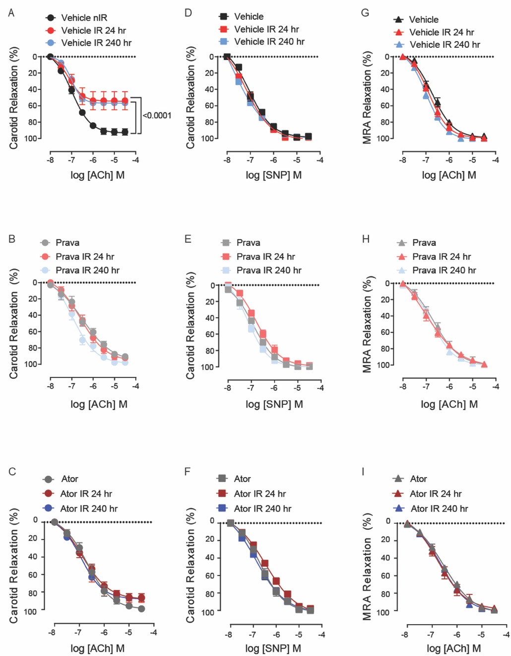
Figure 1. Pravastatin and atorvastatin preserve endothelial function in vivo following head-and-neck IR. (A–C) Effects of statins on endothelium-dependent relaxation of the carotid artery in response to acetylcholine (ACh). C57BL/6J mice were treated with (A) vehicle, (B) pravastatin (Prava), or (C) atorvastatin (Ator) after head-and-neck irradiation (12Gy) or sham treatment, and relaxation was tested at 24 and 240 h after irradiation. (D–F) Effects of statins on endothelium-independent relaxation of the carotid artery in response to sodium nitroprusside (SNP), in mice treated as in A, B, and C, respectively. (G–I) Effects of statins on endothelium-dependent relaxation of mesenteric resistance arteries (MRAs), in mice treated as in A, B, and C, respectively. n = 5 mice per group. p values were determined using repeated measures 2-way ANOVA followed by Tukey's post-hoc test.
The authors apologize for this error and state that this does not change the scientific conclusions of the article in any way. The original article has been updated.
In the published article, there was an error in Figure 3 as published. Images for Vehicle-treated non-irradiated (V-nIR) mitoSOX staining were inadvertently mismatched with those of mitotracker staining and merged images. The corrected Figure 3 and its caption appear below.
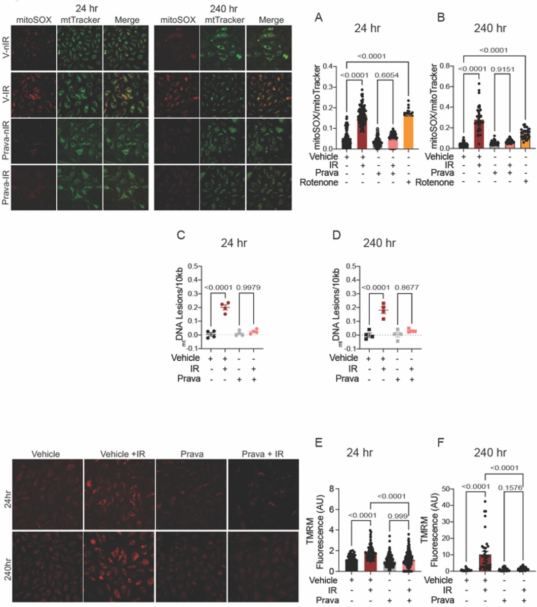
Figure 3. Pravastatin protects against IR-induced mitochondrial damage or hyperpolarization in vitro. All panels compare cells subjected to irradiation (4Gy) after pretreatment with pravastatin (Prava, 10 μM) starting at 18 h before irradiation. (A,B) Representative images and signal integrated density of mitoSOX fluorescence normalized to mitoTracker fluorescence in HCAECs at (A) 24 and (B) 240 h after IR. (C,D) mtDNA lesions in DNA extracted from HUVECs at (C) 24 h and (D) 240 h after IR. (E,F) Representative images and integrated density of mitochondrial membrane potential in HCAECs, as determined by TMRM fluorescence, at (E) 24 h and (F) 240 h after irradiation. Analysis per cell, n = 4 independent experiments. p values were determined by Kruskal–Wallis test.
The authors apologize for this error and state that this does not change the scientific conclusions of the article in any way. The original article has been updated.
In the published article, there was an error in Figure 4 as published. In Figure 4A, images for mitoSox and mtTracker staining of atorvastatin-treated cells in the left upper panels were mistakenly switched. We confirmed that the analysis was performed in the correct panels.
Figures 4C,D: The labeling for the samples treated with atorvastatin and/or IR is incorrect. The correct description for radiation treatment (IR) should be “− + − +” instead of “− + + +”. Figures 4E,F: Images for TMRM in the left lower panel are mislabeled. The correct labeling of the images should read: Vehicle, Vehicle + IR, Atorva, Atorva + IR (instead of Vehicle, Vehicle + IR, Prava, Prava + IR).
The corrected Figure 4 and its caption appear below.
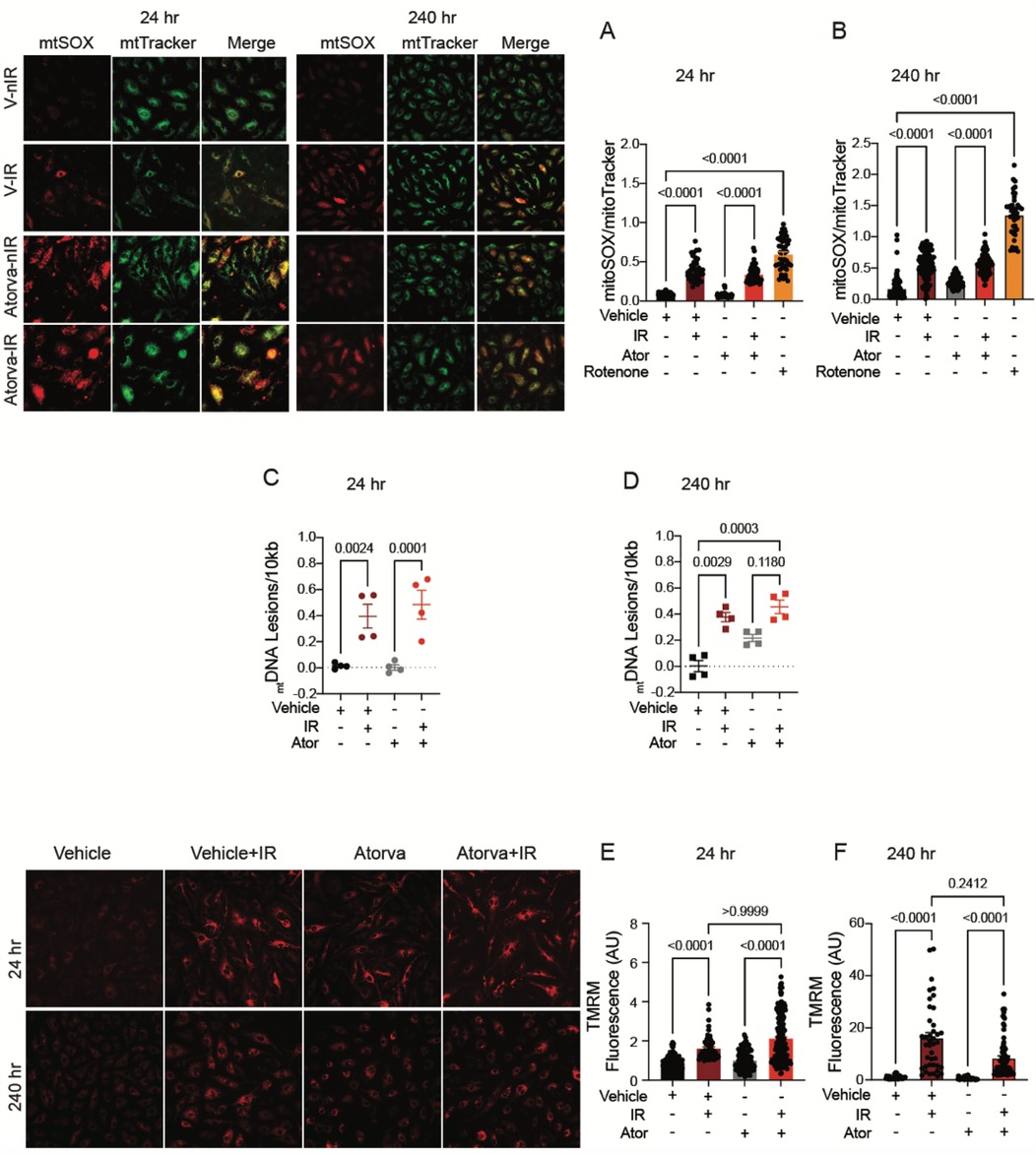
Figure 4. Atorvastatin does not protect against IR-induced mitochondrial damage or hyperpolarization in vitro. All panels compare HCAECs subjected to irradiation (4 Gy) after pretreatment with atorvastatin (Ator, 5 μM) or vehicle. Parameters assessed are: (A,B) Representative images and signal integrated density of MitoSOX fluorescence normalized to MitoTracker fluorescence at (A) 24 and (B) 240 h after irradiation, in cells treated with atorvastatin (5 μM) or vehicle starting 18hr before irradiation. (C,D) Damage to mtDNA as assessed by PCR assay. mtDNA lesions at (C) 24 and (D) 240 h after irradiation, in HUVECs treated with atorvastatin or vehicle starting 18 h before IR. (E,F) Representative images and integrated density of mitochondrial membrane potential, as determined by TMRM fluorescence, at (E) 24 and (F) 240 h after irradiation. Analysis per cell, n = 4 independent experiments, p values by Kruskal–Wallis test.
The authors apologize for this error and state that this does not change the scientific conclusions of the article in any way. The original article has been updated.
In the published article, there was an error in Figure 7 as published. The labeling for the samples treated with atorvastatin and/or IR in Figures 7E–H is incorrect. The correct description for radiation treatment (IR) should be “− + − +” instead of “− + + +”. The corrected Figure 7 and its caption appear below.
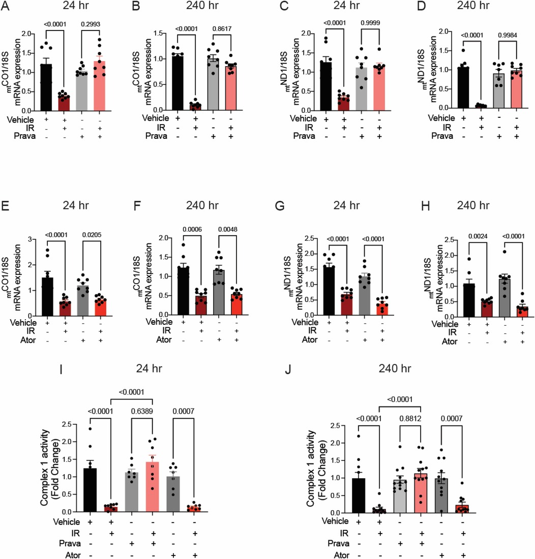
Figure 7. Pravastatin, but not atorvastatin, prevents irradiation-induced reduction of mtDNA transcription and ETC activity. All panels compare HCAECs subjected to irradiation (4Gy) after pretreatment with atorvastatin (Ator, 5 μM, overnight), pravastatin (Prava, 10 μM, overnight) or vehicle. (A–D) Effects of pretreatment with pravastatin (Prava, 10 μM, overnight) on transcriptional activity. (A,B) Quantitative (q)RT-PCR for cytochrome c oxidase I (MT-COI) at (A) 24 and (B) 240 h after irradiation. (C–D) qRT-PCR for NADH-ubiquinone oxidoreductase chain 1 (MT-ND1) at (C) 24 and (D) 240 h after irradiation. (E–H) Effects of pretreatment with atorvastatin (Ator, 5 μM, overnight) on transcriptional activity. (E,F) qRT-PCR for MT-COI at (E) 24 and (F) 240 h after irradiation. (G,H) qRT-PCR for MT-ND1 at (G) 24 and (H) 240 h after irradiation. (I,J) Activity of ETC complex 1, as assessed by fluorometric assay at (I) 24 and (J) 240 h after irradiation. p values by Kruskal–Wallis test.
The authors apologize for this error and state that this does not change the scientific conclusions of the article in any way. The original article has been updated.
In the published article, there was an error in Supplementary Figure 3. The labeling for the samples treated with atorvastatin and/or IR in Supplementary Figures S3E–H is incorrect. The correct description for radiation treatment (IR) should be “− + − +” instead of “− + + +”. The corrected figure and its caption appear below.
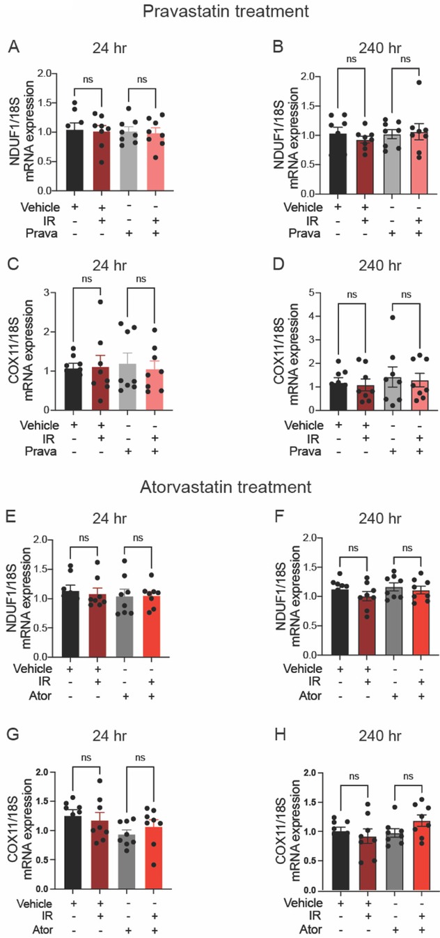
Supplementary Figure 3. Neither pravastatin nor atorvastatin affects IR-induced transcription of nuclear DNA. (A–D) Effects of pretreatment with pravastatin (Prava, 10 μM, 1 h) on nucDNA damage in HCAECs after irradiation (IR, 4 Gy). (A,B) Quantitative (q)RT-PCR for NADH dehydrogenase [ubiquinone] 1 alpha subcomplex subunit 1 [(B), NDUF1], with cDNA normalized to 100 ng at 24 and 240 h after IR. (C,D) qRT-PCR for cytochrome c oxidase 11 [(D), COX11], with cDNA normalized to 100 ng at 24 and 240 h after IR. (E–H) Effects of pretreatment with atorvastatin (5 μM, overnight) on nucDNA damage in HCAECs subjected to IR. (E,F) qRT-PCR for NDUF1 (B), with cDNA normalized to 100 ng at 24 and 240 h after IR. (G,H) qRT-PCR for COX11 (D), with cDNA normalized to 100 ng at 24 and 240 h after IR. Statistical analysis by Kruskal–Wallis test. Ns indicates not significant.
The authors apologize for this error and state that this does not change the scientific conclusions of the article in any way. The original article has been updated.
Publisher's note
All claims expressed in this article are solely those of the authors and do not necessarily represent those of their affiliated organizations, or those of the publisher, the editors and the reviewers. Any product that may be evaluated in this article, or claim that may be made by its manufacturer, is not guaranteed or endorsed by the publisher.
Keywords: radiation therapy, carotid stenosis, endothelium, statin, mitochondria, prevention
Citation: Ait-Aissa K, Leng LN, Lindsey NR, Guo X, Juhr D, Koval OM and Grumbach IM (2025) Corrigendum: Mechanisms by which statins protect endothelial cells from radiation-induced injury in the carotid artery. Front. Cardiovasc. Med. 12:1523371. doi: 10.3389/fcvm.2025.1523371
Received: 5 November 2024; Accepted: 24 March 2025;
Published: 11 April 2025.
Edited and Reviewed by: Masanori Aikawa, Brigham and Women's Hospital and Harvard Medical School, United States
Copyright: © 2025 Ait-Aissa, Leng, Lindsey, Guo, Juhr, Koval and Grumbach. This is an open-access article distributed under the terms of the Creative Commons Attribution License (CC BY). The use, distribution or reproduction in other forums is permitted, provided the original author(s) and the copyright owner(s) are credited and that the original publication in this journal is cited, in accordance with accepted academic practice. No use, distribution or reproduction is permitted which does not comply with these terms.
*Correspondence: Karima Ait-Aissa, a2FyaW1hLmFpdGFpc3NhQGxtdW5ldC5lZHU=; Isabella M. Grumbach, aXNhYmVsbGEtZ3J1bWJhY2hAdWlvd2EuZWR1