 Marta Díaz Zorita Bonilla1,2*
 Monice Timm2
 Mauricio Marciales Daza2
 Auxiliadora Moreno Onorato3Eva Alarcón García3
 Alexandros F. Karakostis4,5
 Javier Escudero Carrillo1
 Luis Arboledas Martínez3Sonia Robles Carrasco3
 Berta Morell6
 Derek Hamilton7
 Helen Dawson1
 Francisco Contreras Cortés3Martin Bartelheim1,2
 Jörg Baten2,8
- 1Institut für Ur- und Frühgeschichte und Archäologie des Mittealters, University of Tübingen, Tübingen, Germany
- 2SFB 1070 RessourceCultures, Tübingen, Germany
- 3Facultad de Filosofía y Letras, Department of Prehistory and Archaeology, University of Granada, Campus Universitario de Cartuja, Granada, Spain
- 4Paleoanthropology, Department of Geosciences, Senckenberg Centre for Human Evolution and Palaeoenvironment, Eberhard Karls University of Tübingen, Tübingen, Germany
- 5Integrative Prehistory and Archaeological Science, University of Basel, Basel, Switzerland
- 6Institución Milá y Fontanals de investigación en Humanidades – CSIS, Barcelona, Spain
- 7Scottish Universities Research Centre, SUERC, Glasgow, Scotland
- 8Department of Economics and Social Sciences, University of Tübingen, Tübingen, Germany
The Biniadrís cave is located in the southern part of the island of Menorca (Spain). It is the fourth funerary cave with preserved materials discovered in Menorca up to now, along with Es Càrritx, Es Mussol, and Es Pas. However, the chronology of Biniadrís is more extensive, covering more than seven centuries, allowing for a study of human practices over the “longue durée”. What makes Biniadrís unique is its large number of bioarchaeological remains, mainly human remains and other organic and inorganic material, in an extraordinary state of preservation. The main goal of this paper is to reconstruct the burial ritual using comprehensive bioarchaeological methods, including 14C radiocarbon dating, to understand the modality and temporality of use of the Biniadrís cave during the pre- and protohistory of the Balearic Islands. The palaeodemographic data derived from our ongoing excavation are described here in detail and point to interesting patterns, such as the equal representation of female and male individuals across all ages, including very young and older individuals. The fact that all individuals are equally represented and integrated into the community in the funerary settings provides insights into the group ideology, which are discussed further. These burial practices are interpreted as a strong expression of community identity at Biniadrís linked to its island setting, as seen from the preservation of the funerary memory over such a long period, which is unique for this cave. The multi-proxy approach employed here to study the bioarchaeological and organic remains contributes to a better understanding of the “longue durée” of the funerary ritual and the reconstruction of human behavior during the Late Bronze and Iron Ages of an island community in the western Mediterranean.
1 Introduction
The Balearic Islands represent an anomaly or paradox being the last archipelago to be permanently settled in the Mediterranean in prehistory, defying the general trend of island colonization in the Neolithic (Dawson, 2014; Cherry and Leppard, 2018). On current knowledge, the earliest direct evidence of human presence in the archipelago cannot be traced before the mid-3rd millennium cal BC (Calvo et al., 2002; Bergadá et al., 2005). However, indirect evidence from pollen analyses has revealed an increase of aridification from the 6th millennium cal BC to the end of the 3rd millennium cal BC (Servera-Vives et al., 2023). Human impact, including deforestation and farming practices, but also a climatic event might have been the potential driving factors for this environmental change. In its initial phase, starting in the mid-3rd millennium, human occupation is represented by open-air sites with diversified associated funerary practices, with single burials found in natural caves, collective burial sites, and dolmens (Gili et al., 2006; Guerrero Ayuso et al., 2007; Calvo et al., 2012; Calvo Trias et al., 2001; Ramis, 2010; De Cet, 2017). The 2nd millennium cal BC is characterized by a more connected Mediterranean and the arrival of new groups, which could have reached the Balearics (Coll et al., 1997). This period represents a period of flourishing (Lull et al., 1999), especially in Menorca and Mallorca (Cherry and Leppard, 2018; De Cet et al., 2017; Ramis, 2014). Several marked changes from the second half of the 2nd millennium BC (1600–1550 cal BC) can be observed in the monumentality based on cyclopean architecture of the subsequent phase know as “Navetiform”. This term refers to the boat-shaped building technique (navetas), a technique that reflects community engagement and group identity (Fornés et al., 2009), and is deeply embedded within the island landscape. Nevertheless, the process of monumentality shifted from the first millennium BC toward other spaces, which were organized and modified by the community (Lull et al., 1999; Portillo et al., 2014).
The cultural dynamics of each island are very distinctive. By the end of the 2nd millennium BC, new social interactions and cultural changes are associated with the Talaiotic culture particularly in Menorca and Mallorca (Galmés-Alba and Calvo-Trias, 2021; Pons, 2017). Therefore, communities on each island were shaping their own identity, as can be seen in the types of sites such as the monumental tower-like monuments (talaiots), sanctuaries, and in domestic spaces (Gornés, 2022). The funerary world remains quite complex and diverse in the Balearics in terms of burial practices. From the Middle Bronze and Early Iron Ages, these varied from individual burial in necropolises to collective burials in natural caves with cyclopean architecture, hypogea, and navetas (Gornés, 2016; Gili et al., 2006). During this time, the increased presence of exogenous material such as ivory and tin is attested and suggests the Balearics were already connected within a larger exchange network in the Mediterranean (Calvo et al., 2012). Regarding the study of funerary practices at natural caves, after the investigation of the three major funerary caves discovered in Menorca—Es Mussol (Lull et al., 1999), Es Càrritx (Lull and Micó Pérez, 1999; Rihuete Herrada, 2003), and Es Pas (Fullola et al., 2007; Armentano and Malgosa, 2003; Armentano et al., 2010)— the Biniadrís cave, in the Calescoves area, is now under investigation and presented in this paper. Concerning the funerary practices within the Calescoves area in later periods, there is a necropolis with more than a hundred caves dated from the 9th to the 2nd century BC (Veny Meliá, 1982; Gornés and Gual, 2000; Gornés, 1997, 2000). From the 4th century BC until the 7th century AD, this area was of great importance for anchorage (Fernández-Miranda, 1991; Belén Deamos and Fernández-Miranda, 1977; Belén Deamos et al., 1979). In the vicinity, there is also the Coberxo Blanc sanctuary linked to navigation (Orfila et al., 2010), a documented shaft with stairs (Veny Meliá, 1982; Plantalamor Massanet, 1991), and other coastal settlements such as Es Castellet (Anglada et al., 2010; Hernández Sanz, 1896–1897; Plantalamor Massanet, 1991; Sánchez López et al., 2013) with contemporary chronologies. These later sites suggest that the island communities were also oriented toward the sea. The extent of isolation and interaction during the period of Biniadrís is therefore another focal aspect of this paper. The Biniadrís cave is located about 2 km inland in the Middle and Upper Miocene platform of the Calescoves area in the southern part of the island of Menorca (Spain) (Figure 1). It is a natural cave located at the Biniadrís gorge, which is part of Menorca's Migjorn platform. It is an endokarst formation, open in the middle toward the upper part of the cliff without water circulation oriented toward the direction of the valley. The formation of the cave is thought to be associated with the evolution of the gorge or a period of sea-level stabilization (Trías, 2004).
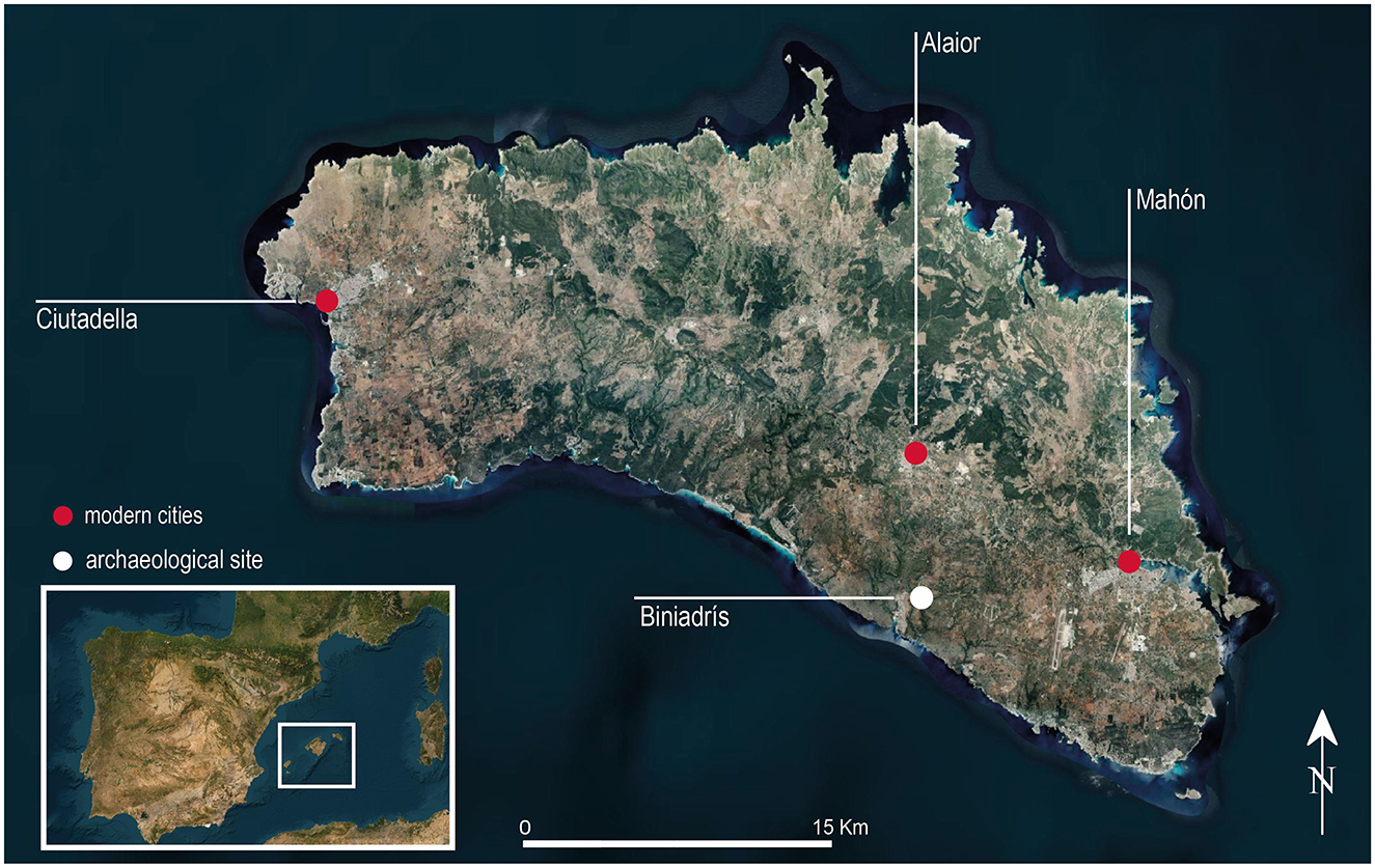
Figure 1. Location of the Biniadrís cave.
Since its discovery (Moreno Onorato et al., 2019), the Biniadrís cave has provided insights into the island's prehistoric occupation due to its exceptional state of preservation of different materials. These findings are similar to those documented at the other funerary caves of the same chronology excavated on the island, such as Es Mussol and Es Càrritx (Lull et al., 1999; Lull and Micó Pérez, 1999; Lull et al., 2014; Rihuete Herrada, 2000), as well as Es Pas (Fullola et al., 2007, 2008). The particularity of Biniadrís resides in the small funerary space, extending around 18 m2 (Figure 2), whose entire surface is full of commingled human bone remains, macro- and microfaunal remains, residues of organic material, and associated items. Due to the exceptional assemblage of human remains and the complexity of the mortuary treatment, the only way to overcome this analysis is throughout the multi-proxy bioarchaeological research designed in this study. The upper layer is made up of around 8 cm of microfaunal remains, which have effectively sealed the evidence for prehistoric activity at the cave since antiquity (Figure 3).
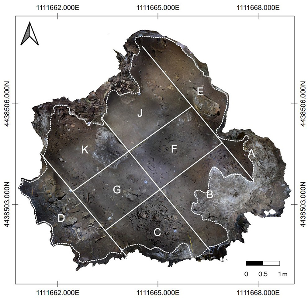
Figure 2. Main plain of the Biniadrís cave with sectors.
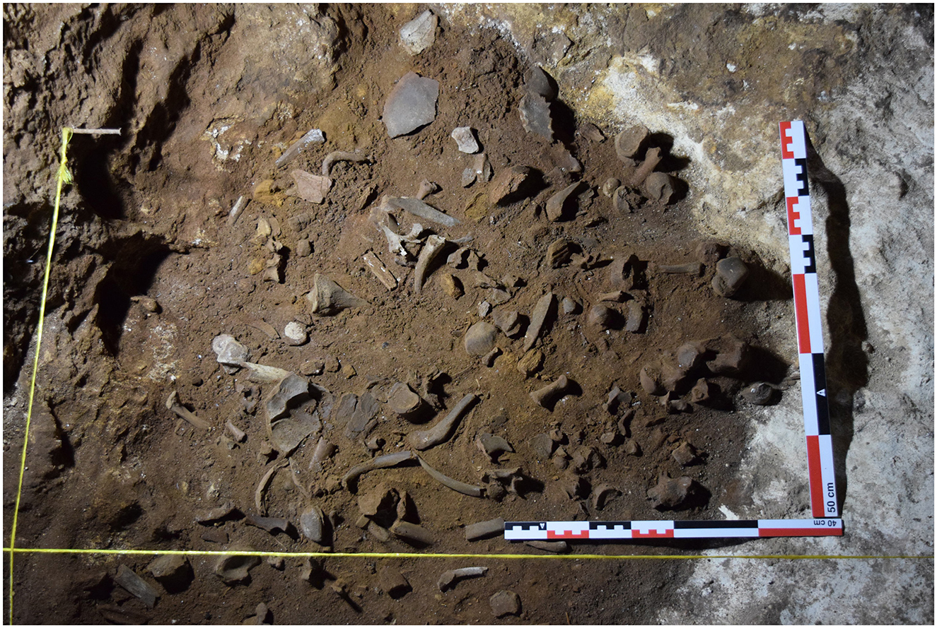
Figure 3. The Biniadrís cave during excavation.
The position of the human remains does not follow any discernible anatomical order, owing to the multiple funerary practices that took place in the cave during its period of use. This is a recurrent funerary practice in Balearic prehistory, which has been observed in megalithic monuments (Aranda Jiménez et al., 2020), but also in ritual caves (Lull et al., 1999). Although the archaeological record has yielded one of the largest commingled bone assemblages for Menorca, the manipulation and subsequent removal of human remains was carefully done. This can be observed in the reduced fragmentation and overall completeness of the osteoarchaeological record. The timing of the use of the cave has been assessed through radiocarbon analysis and the scientific results showed that this cave was in use for at least seven centuries during the Late Bronze and Iron Ages, corresponding to the Naviform, Talaiotic, and post-Talaiotic periods. This phenomenon could be explained as the result of the longue durée of the funerary memory on the island of Menorca, as seen from the transfer and preservation of knowledge through time of the groups who used the Biniadrís cave as a place to bury their ancestors. In this paper, we will also discuss how this exceptional longevity can be seen as an expression of place attachment and community identity related to the island environment (Dawson, 2020). The reconstruction of the paleodemographic profile in addition to the evaluation of health and the activity patterns in conjunction with the radiocarbon and isotope analyses aligns with the principles of Environmental Archaeology.
2 Material and methods
2.1 The funerary space
Biniadrís is a natural cave with minor anthropic modifications but with intentionally designed funerary spaces. Of special interest is the gate of cyclopean construction with a large stone lintel and stone slabs. The architecture is made of a mixed technique of dry stone but also with the use of clay at the bottom of the entrance wall to provide stability. A small corridor with paved stones was flanked by two lateral stones, which give access to an area where the different rituals took place.
The entire surface is covered with remains that are mainly human and associated funerary objects. Within the cave, several spaces functioned differently. For instance, in sector A, many intentionally broken ceramic vessels were found as part of certain funerary rituals. However, at the opposite end, in sector (D), which is an area specifically hidden from the entrance and without direct sunlight, a wooden structure of log slices, planks, and other cut-wood features was surrounded by a selection of one dozen complete skulls (Figure 4). Without a doubt, this is the most spectacular area of the cave. Not only for the singularity of this funerary setting but also for the exceptional state of preservation and its striking ritual significance. There are no other parallels on the island apart from Es Càrritx (Lull et al., 1999; Lull and Micó Pérez, 1999; Lull et al., 2014; Rihuete Herrada, 2000). The combination of the different nature of the organic and inorganic materials plus the practices and symbolism embedded at the Biniadrís cave, has resulted in a space that stands out as sacred and special in Balearic archaeology.
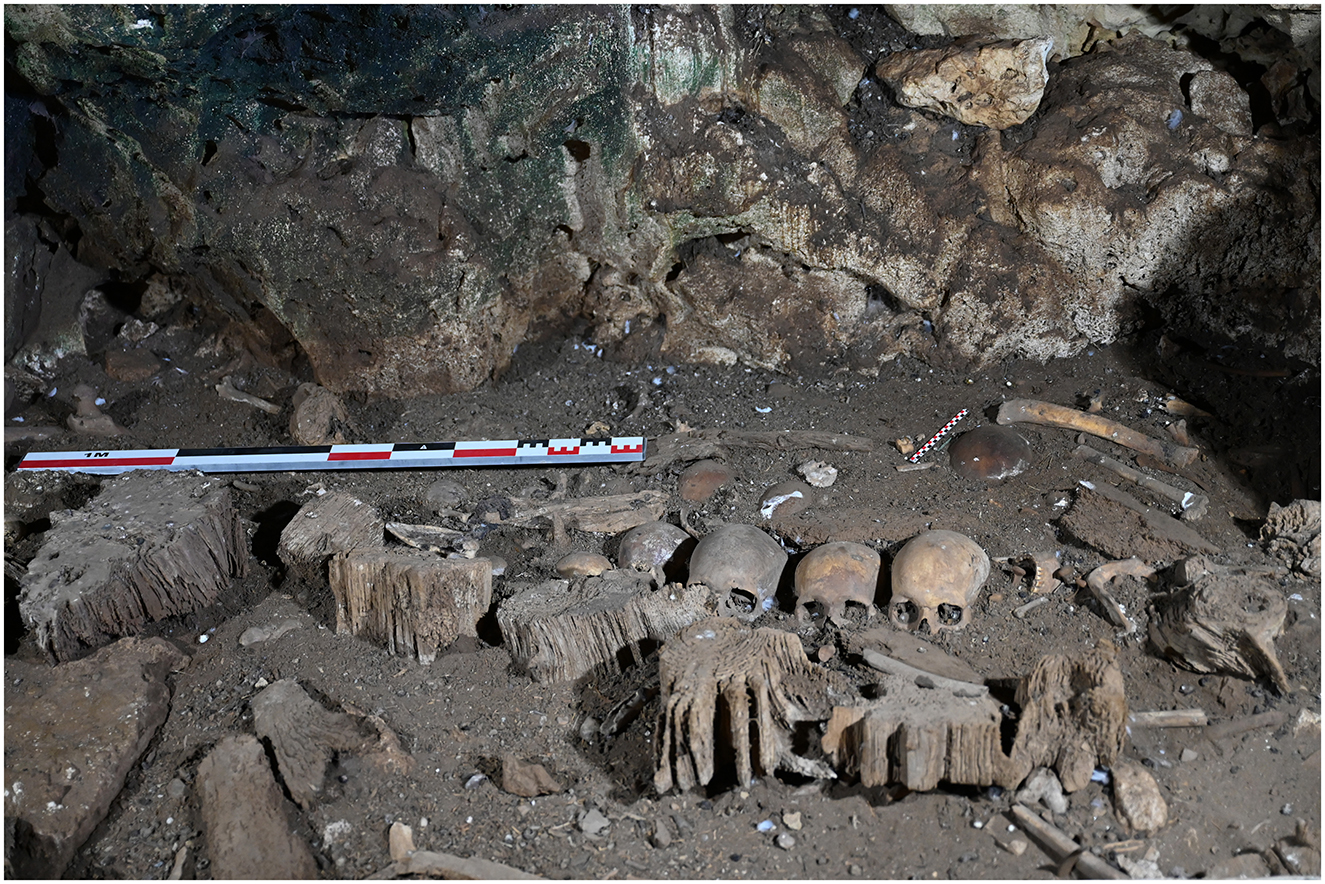
Figure 4. Wooden structure and organization of human skulls (sector D).
2.2 Bioarchaeological analyses
Human remains are the most abundant material found at the Biniadrís cave. Since the remains are commingled, their analysis requires using established calculation methods, such as the minimum number of individuals (MNI), along with others, to investigate the palaeodemographic profile and, therefore, to estimate the population size. These methods are also required to study the health status, disease profiles, and mortuary practices of the community (Osterholtz et al., 2014, p. 35; Konigsberg and Adams, 2014). The MNI-calculation is based on counting elements depending on their relative completeness considering that each fragment must share a unique landmark to avoid double-counting of fragments from the same individual. The analysis of the adult and non-adult individuals followed commonly established standard bioarchaeological methods (Buikstra and Ubelaker, 1994; Ferembach et al., 1979, 1980; Steckel et al., 2005).
Sex and age were determined following established protocols by Buikstra and Ubelaker (1994) and Ferembach et al. (1979, 1980). The specific age estimation focused on the combination of different methods, which included the degenerated morphology of the pelvis, along with the degenerative changes of the acetabulum (Calce, 2012), as well as the morphology of the auricular surface (Lovejoy et al., 1985) and the pubic symphysis (Brooks and Suchey, 1990; Todd, 1920, 1921). In addition, we analyzed the closure of the cranial suturae (Meindl and Lovejoy, 1985; Hermann et al., 1990; Olivier, 1960; Vallois, 1937, modified by Rösing, 1977), as well as the palatine suture closure (Mann et al., 1991) and the first rib morphology (Kunos et al., 1999) and the ossification process of the sternal costae end after Işcan et al. (1984a,b) to estimate age. To assess the mature age classes in this commingled context, the degeneration of the sternal end of the clavicle was used for age analysis after the method of Falys and Prangle (2015) and as a method comparison Szilvássy (1980). Moreover, the age estimation through dental eruption and mineralisation as well as the dental abrasion was included in the study [Lovejoy et al., 1985; Brothwell, 1981; Mays et al., 2022; Murphy, 1959, modified by Smith and Knight (1984); AlQahtani (2008); Ubelaker (1978); Schour and Massler (1941, 1944)].
Sex estimation was done mainly on the two most sexually dimorphic bone elements, the skull and the pelvis (Buikstra and Ubelaker, 1994; Ferembach et al., 1979, 1980; Bruzek, 2002). A good example of the analysis of growth and aging processes is provided by the Spitalfields Project (Molleson and Cox, 1993). In some cases, when almost complete, the method after Murail et al. (1999, 2005) was used to evaluate the difference of the pelvic appearance through metric analysis. Despite its lower accuracy, metric sex assessment through long bone measurements (Bass, 2005) was also carried out. Most sexually dimorphic measurements have been established for the femur and humerus. Therefore, it was decided to compare the vertical diameter of the humeral head as well as the distal breadth (comparable to the bicondylar width) based on Stewart (1979), Spradley (2011), Mall et al. (2001) and Krogman (1962), the maximum diameter of the femoral head and the femur circumference following Pearson (1917/1919), Krogman (1962) and Black (1978) among other metrical methods which are summarized in Bass (2005) and Işcan and Steyn (2013), Cunningham et al. (2016) and Grupe et al. (2015). In addition, the sexual dimorphism of the scapula and clavicle, following the methods published by Bass (2005), was included.
For non-adult human bone remains, the following standard methods on age development were used (Fazekas and Kósa, 1978; Scheuer et al., 2008; Lewis, 2007; Cunningham et al., 2016; Cardoso et al., 2016). The fusing states of the proximal and distal epiphyses follow the methodology of Acsádi and Nemeskéri (1974) as well as Buikstra and Ubelaker (1994). The classification of individuals' sex under the age of 12 years is not recommended at all and is partly problematic, because of a high error rate related to the lack of fully developed sexual dimorphism (Chamberlain, 2016, p. 19). For these reasons, sex determination in non-adult individuals was not pursued in any further analyses. However, it was recorded to complete the data collection and refers to the analyses of Schutkowski (1990).
For a deeper insight into the life of the deceased, metrical data and further analysis (Buikstra and Ubelaker, 1994; Bräuer and Knussmann, 1988; Martin and Saller, 1957; Ruff et al., 2012), pathological changes (Jacobs, 1992; Roberts and Manchester, 2010; Ortner and Aufderheide, 1991; Ortner, 2003; Ortner and Putschar, 1981; Adler, 2006; Aufderheide and Rodríguez-Martin, 1998; Waldron, 2008; Mann and Hunt, 2005) and morphological features on the bones (Alt, 1997; Barnes, 2008; Berry and Berry, 1967; Finnegan and Faust, 1974; Hauser and DeStefano, 1989; Mann et al., 2016) were also recorded, following a practice that corresponds to the standardized analyses of skeletons. Furthermore, the morphological conspicuousness, which occurred particularly due to the robustness of the bone morphology, was investigated in detail (Vilotte, 2006; Henderson, 2009; Henderson et al., 2017; Vilotte et al., 2016; Mariotti et al., 2007; Ruff, 2018; Handling et al., 2010; Cardoso and Henderson, 2013; Bonnet and Ferrari, 2010).
Analysis of the musculo-skeletal stress markers was carried out using a precise and experimentally supported three-dimensional (3D) system of quantification and analysis, known as the Validated Entheses-based Reconstruction of Activity (V.E.R.A.) approach (Karakostis and Lorenzo, 2016; see reviews by Karakostis and Harvati, 2021; Karakostis, 2023). Following the V.E.R.A. 1.0 protocols (Karakostis, 2023), the entheseal 3D area measurements (square mm) were subjected to a principal component analysis (PCA) relying on the correlation matrix (due to varying ranges across variables). Subsequently, the strength of the correlation between the values of each PC axis and body size was evaluated using the vertical head diameter (as body size proxy) and three Pearson's correlation tests. All necessary assumptions for Pearson's test were met, including the normal distribution of the variables (based on Shapiro Wilk's tests) and the absence of extreme outliers (based on boxplots and the interquartile range approach) (Tabachnick and Fidell, 2019). The goal of the V.E.R.A. 1.0 analysis was to identify entheseal variation that might reflect the observed substantial differences in robusticity across bones. In this study, we focused on the humerus, a widely studied bone element with fundamental importance for functional morphology and activity reconstruction.
Given the notable robustness observed in long bone, a total of 63 humeri (29 right, 34 left) were selected for a pilot study. These specimens were analyzed using the established scoring methods of Vilotte et al. (2016) and Mariotti et al., 2007 to establish a comparative framework for the V.E.R.A. 1.0 methodology. Given the limited scope of this pilot project, the decision was made to focus on the right humerus, as isotopic analyses were also conducted on this body side. The primary criterion for the application of the V.E.R.A. 1.0 methodology was age, with specimens restricted to the Middle Adult age category to minimize the potential effects of significant degenerative changes or ongoing ossification/bone growth. This selection process resulted in a sample of 20 isolated and well-preserved right humeri. On each bone, we selected three entheses corresponding to muscle groups that co-activate for various human movements involving the upper limbs (Table 1). Particularly, the entheses used involve the: (a) the lesser tubercle (muscles associated with arm adduction and medial rotation); (b) medial epicondyle (elbow, wrist, and hand flexion); and (c) lateral epicondyle (wrist and hand extension) (Table 2). A total of seven bones bearing notable potentially pathological traits (i.e., enthesopathies) were excluded from the analysis, which finally focused on the remaining 14 bones.
Table 1. List of musculo-skeletal stress markers recorded at Biniadrís cave.
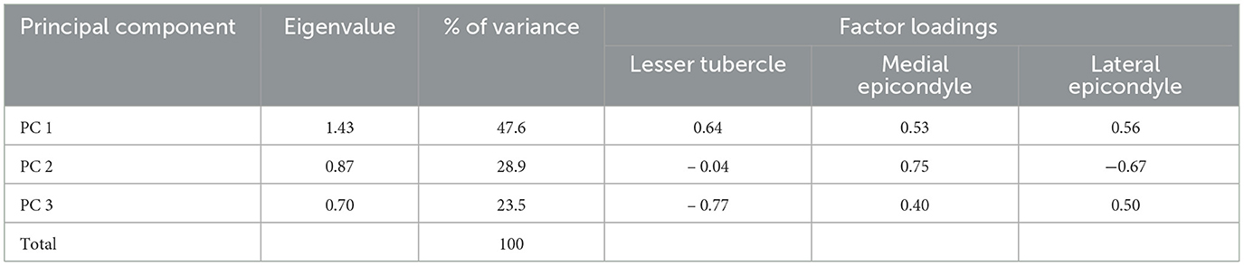
Table 2. Eigenvalues and factor loadings of the principal component analysis (PCA).
The assessment of health and paleopathology requires an independent evaluation of each bone and tooth, following the protocols of Roberts and Connell (2004), Lovell (1997), and Steckel et al. (2006). Different categories of disease were considered. For infectious diseases, the methodology of Roberts and Manchester (2012) was employed. Traumatic injuries and the classification of fractures were analyzed according to Ortner and Putschar (1981), Waldron (2008, p. 139), Roberts and Connell (2004, p. 37) and Redfern and Roberts (2019). Joint disease, with a focus on the prevalent condition of osteoarthritis (OA) affecting synovial joints as described by Jurmain and Kilgore (1995) and was identified through characteristic skeletal alterations such as osteophytes, porosity & pittings, eburnation, new bone formation, and the fusion of joints (Waldron, 2008, p. 27; Rogers and Waldron, 1995; Roberts and Manchester, 2010; Aufderheide and Rodríguez-Martin, 1998; Cockburn et al., 1979; Rogers et al., 1985). The evaluation of each joint is additionally done after Schultz (1988), Steckel et al. (2006) and Sager (1969). Metabolic diseases, as the most complex conditions caused by hormonal or nutritional deficiencies, such as Vitamin D or C deficiency including conditions like cribra orbitalia, porotic hyperostosis and osteoporosis (Brickley and Ives, 2008; Waldron, 2008; Roberts and Manchester, 2010; Brickley and Mays, 2019; Grupe et al., 2015). They were identified based on the criteria outlined by Roberts and Connell (2004) and Stuart-Macadam (1989). The conditions of the cribra orbitalia and porotic hyperostosis were evaluated through different criteria and approaches (Brickley, 2018; Cole and Waldron, 2019; O'Donnell et al., 2020; Rinaldo et al., 2019; Steckel et al., 2006). Finally, oral pathologies and cranial modifications were described for each tooth and cranium, respectively, following the descriptive standards of Buikstra and Ubelaker (1994), for enamel defects (Pinhasi and Mays, 2007; Reid and Dean, 2000, 2006) and general dental diseases (Brothwell, 1981; Roberts and Manchester, 2012; Hillson, 1996, 2001; Aufderheide and Rodríguez-Martin, 1998; Pinhasi and Mays, 2007; Dias and Tayles, 1997).
2.3 Biochemical analysis
2.3.1 δ13C and δ15N
A total of 22 samples were analyzed for diet composition (Table 3) through carbon (δ13C) and nitrogen (δ15N) stable isotopes. Since the burial deposit is commingled, to avoid double sampling of individuals, we based this strategy on the Minimum Number of Individuals (MNI) determined by the right humerus. Fifteen zooarchaeological samples from different trophic-level species from the cave were also analyzed as a protein food intake reference of the local population. Collagen was extracted from all human remains for dietary purposes, as well as to estimate any reservoir effect and for radiocarbon dating.
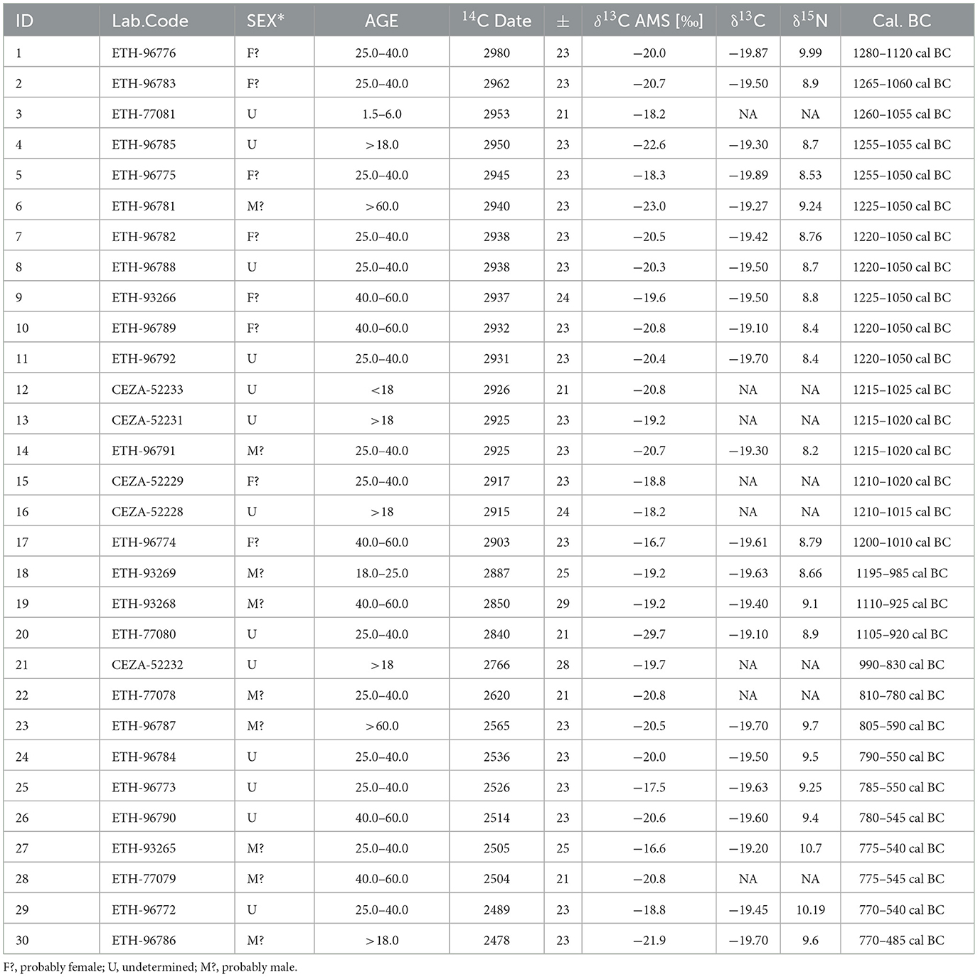
Table 3. Radiocarbon dates (2σ) of the Biniadrís cave, as well as the δ13C and δ15N isotopic values and the estimated percentage of marine consumption.
The analysis of stable isotopes of δ13C and δ15N proved to be a powerful technique to investigate the amount of protein in the diet. The sampling strategy followed in this paper entailed the selection of the long bones to provide information to reconstruct on average the last 10 years of the life of the individual (Richards and Hedges, 1999; Bocherens and Drucker, 2003).
The collagen extraction was performed at the Biogeology Laboratory of the University of Tübingen following the standard procedures (Deniro and Epstein, 1981; Bocherens et al., 1997). Thus, a total of 500 mg of bone was extracted for each sample, followed by a process of cleaning, drying, and crushing.
Afterwards, the samples were put through an elemental analysis in a CHN, Carlo Erba NA 1500, to check the potential bone collagen preservation as inferred from the content of nitrogen (Iacumin et al., 1996). Finally, the bone collagen was extracted using the standard laboratory procedure (Bocherens et al., 1997). The human and animal collagen samples were measured in duplicate in the Institute of Environmental Science and Technology (ICTA) at the Laboratory of the Autonomous University of Barcelona using a Thermo Flash 1112 (Thermo Scientific) elemental analyzer coupled to a Thermo Delta V Advantage MS with a Conflo III interface. The ratios 13C/12C and 15N/14N were measured relative to the V-PDB for carbon and AIR for nitrogen using an international laboratory standard, IAEA 600 (caffeine), which showed an analytical error below 0.2‰ (1σ).
2.3.2 14C dating
A total of 30 radiocarbon dates (Table 3) were obtained from two human bone elements, the humerus and petrous bone, representing samples from different individuals (approximately 25% of the minimum number of individuals). The sampling strategy prioritized long bones with minimal spongy structure to maximize the likelihood of collagen preservation, as well as petrous bones due to their exceptional preservation quality.
Elemental yields for carbon and nitrogen in the extracted collagen ranged from 23.57% to 43.07% and 8.20 to 15.29%, respectively. All samples presented an acceptable C/N ratio ranging from 3.2 to 3.5 with a mean of 3.3. A possible reservoir effect was tested by δ13C & δ15N isotope analyses (Table 3). The dietary values ranged from −19.1% to −19.89% (mean of −19.78%) for δ13C and between 8.2 and 10.19 (mean of 9.79) for δ15N. The same samples that were analyzed for δ13C and δ15N were also sent for radiocarbon analyses (14C AMS). They were measured at two different laboratories: the ETH Laboratory in Zurich (Switzerland) and the CEZA Laboratory in Mannheim (Germany). The 14C dates were calibrated using the terrestrial IntCal20 curve and the OxCal v.4.4. software, as the isotopic values ruled out a potential reservoir effect from consuming marine dietary protein (Reimer et al., 2013; Bronk Ramsey, 2001, 2009, 2013, 2017; Reimer et al., 2020).
The χ2 Test (OxCal's R_Combine function following Ward and Wilson, 1978) and Bayesian Chronological Modeling (Bronk Ramsey, 2009) were used to determine the temporal distribution of the cave's inhumations and their degree of contemporaneity through OxCal v.4.4 software.
3 Results
3.1 Paleodemographic analyses and palaeopathological conditions
The latest excavations yielded 13,182 bone elements. According to the MNI, this corresponded to at least 150 individuals based on the right humerus. From this MNI-calculation, a total of 37 non-adult individuals and 113 adults were identified. The estimation of sex analyses showed a balanced distribution of sex with the different categories of age represented including a high number of non-adult bone elements. Because of the nature of the deposit and the commingled remains, a large amount of bone elements was classified as that of adults without specific age determination. Within the skeletal elements from Biniadrís, numerous anatomical variants showed up, potentially displaying trained muscles.
Based on the highly robust appearance and the long period of use of the cave, the question arose as to whether this represents an entheseal change in the population caused by certain activity/occupation or whether at least two different population groups were present here. As this is a commingled remains context, it is difficult to include several entheses/muscle parts that are connected via several bones. In a preliminary study, the V.E.R.A. 1.0 method was used to investigate the entheseal variation between the (very) robust and gracile individuals regarding the use of the upper arm muscles. Activity reconstruction is important here to provide insight into the reasons for the occurrence of these two robustness groups. The selection of the upper arm was based on its frequent occurrence in the assemblage and its good state of preservation as well as the assumption that this was changed by more obvious factors. Furthermore, it was important to use fully grown individuals that showed no serious pathological changes of the enthesis and in general pathological lesions that could influence the method's results. Individuals were examined that showed a distinctive overall impression (gracile/robust) as well as the aspect of the respective entheses and their expression on the bone. The metric analyses allowed the humeri to be assigned to both male and female individuals.
An additional exception is the relatively high stature, where the individuals showed a body height within a range from 145 cm to 176 cm after the method of Ruff et al. (2012) mentioned in Grupe et al. (2015). The method does not consider sex as a determinant. In addition, there is an overrepresentation of the platymeric appearance in the femur. There are more recognized traits in the post-cranium than in the cranium. Interestingly, accessory facets, such as squatting facets on the tibia occur frequently. Approximately 147 tibiae of all ages are preserved at the distal end, 75 of which (left [n = 42]; right [n = 47]) exhibit a variation of the squatting facet (i.e., single or double-curved). The femur and the humerus display the most interesting morphological variations. Next to the prominent flat proximal shape of the femur, most of the individuals also created a prominent third trochanter as well as several cortical defects at the area of the Tuberositas glutealis. Another morphological change was detected in the distal femoral area with a cortical defect on the popliteal fossa, also known as Charles‘ facet. The identification is generally made by the observation of an irregular porous appearance and, in some cases, upraising and ingoing spots in the restricted area. In the Biniadrís collection, Charles' facet in adults appears infrequently but relatively often enough to warrant further analysis. In some cases, the femur has altered its appearance through the so-called hypotrochanteric fossa. The frequency of this fossa has a relation to age and might be a population-specific variation (Mann et al., 2016, p. 617). Also, the hypotrochanteric fossa might be related to the present “platymeric” appearance, which often appears as a ‘flattening' on the tibiae and femora in this collection. The morphology of the humerus is characterized by the occurrence of bone torsions, cortical defects at the deltoid muscle, and the Crista tuberculi majoris/minoris. This research is still in its beginning phase and further investigation with a larger sample size is needed. However, this preliminary study of the human remains showed that the entheseal differences suggest that the more robust specimens belong to generally large individuals who presented proportionally greater hand and elbow use (flexors and extensors).
Regarding the health condition of the individuals, this is remarkably well identified mainly through the presence of degenerative alterations on the bones, especially for the vertebral column. The pathological findings from Biniadrís show around 1,205 bone elements with pathological changes. Most of these are related to bone changes observed at joints, which is highly correlated to age as well as an overuse of the skeletal elements, mostly located at the articular surfaces and epiphysial ends. This joint change can cause pain and disability for the affected individual, especially in long-living populations. In the Biniadrís collection, a frequent occurrence of degenerative changes is related to long bones but also in a high amount present in the vertebral column. Additionally, frequent in this collection is the occurrence of ossified laminal spurs (ligamenta flava), represented by spurs along the arcus of the thoracic vertebrae. Ossification of this ligament seems to be related to increasing age as well as strenuous activity (Mann and Hunt, 2005, p. 78). Another indication of a high correlation between activity and bone formation in the sample is related to the appearance of these spurs, which is not often combined with other structures, such as osteophytes and inflammations. Next to it, in nearly all vertebrae, Schmorl's depressions occurred. The morphology of this depression resembles circles, letters, or small circular depressions as well as shallow pits mostly centered in the corpus of the vertebra. It occurs primarily in the lower vertebrae, including both thoracic and lumbar vertebrae. Additionally, their occurrence might be related to younger and older individuals as the morphology of the billowing curves on the vertebral corpus indicates. To Roberts and Manchester (2010, p. 140) the etiology of this disease is unknown and traumatic events with the previous influence of underlying infection, osteoporosis and neoplastic diseases can be taken into consideration in this disease development. Concerning oral pathologies, the most frequently occurring diseases were periodontal and alveolar changes. However, a periodontal disease related to antemortem tooth loss is lacking in this assemblage.
As well as the degenerative changes, the most common condition in the palaeopathological analysis is trauma. The human bone remains were carefully dispersed in different sectors of the cave. Interestingly, cranial fragments, as well as nearly complete crania, could be found in all sectors. Four pieces show an unusual morphology. One piece was located near the entrance in sector J and the other three at the opposite side in sector B at the cave wall. These fragments were primarily from the parietal and the occipital bones, all of them showing different affections. Although they are influenced by taphonomy which further complicates examination, the morphology of these spots is reminiscent of trephination (Figure 5), a practice known since very early times. Generally, it is associated with surgery for differential reasons, resulting in the incision of the scalp followed by cutting and removing an area of the skull (Roberts and Manchester, 2010, p. 124). The traces of the probable trephination were found in both sexes but mostly in older individuals and are comparable to the cases from Es Càrritx (Rihuete Herrada, 2003, p. 1999) in their appearance of the shape and the occurrence in different individuals. All circular changes that have occurred require further detailed analysis (paper in preparation).
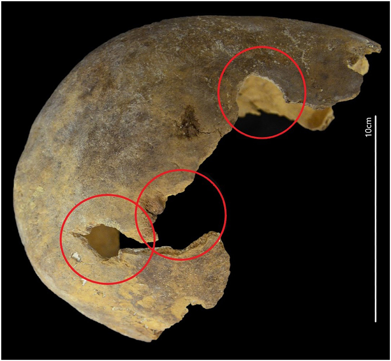
Figure 5. Skull from a mature individual (40–60 years old), probably female, with signs of modification compatible with trephination.
3.2. Entheseal changes
Regarding the 3D analysis of the entheses, as explained in Materials and Methods, a total of 42 entheseal 3D areas (three entheses in 14 humeri) were digitally delineated and quantified on the 3D models using the V.E.R.A. 1.0 protocols (Karakostis and Lorenzo, 2016; Karakostis et al., 2018; Karakostis, 2023). Subsequently, all measurements were subjected to PCA (see extensive V.E.R.A. 1.0-protocol in Karakostis, 2023), to explore multivariate patterns among entheses that may reflect differences among bones in the degree of robusticity. Although this is preliminary data, in the resulting plots, the sex of individuals was labeled using only those specimens with distinct sex scores following the application of widely used metric approaches of sex estimation. Finally, the humeri were separated into different robusticity groups, to determine whether their multivariate 3D entheseal patterns suggest different activities between robust and more gracile individuals. This categorization was attempted using three different strategies: (a) visual assessment of bone overall robusticity; (b) semi-automated K-clustering of robusticity measurements (both diameters of humeral head and epicondylar width); and (c) fully automated clustering of specimens into a blind number of groups based on paired-group linear clustering (see Figure 6). This analysis separated specimens into two groups of robusticity. All specimens in the resulting PCA plots were colored accordingly (blue = more robust; red = more gracile). The latter categorization approach provided the most intriguing results in terms of multivariate entheseal analysis (Figures 7, 8) and was thus the one reported in this study.
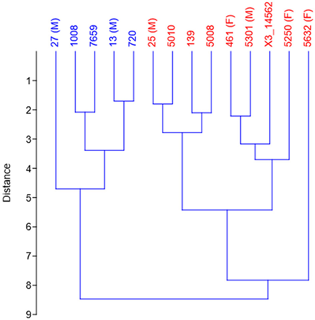
Figure 6. The dendrogram shows the clustering of humeri based on robusticity proxies, using the paired-group linear clustering algorithm (UPGMA). Two main clusters are evident: the first (in blue) includes humeri with greater robusticity, while the second (in red) represents humeri with lower robusticity.
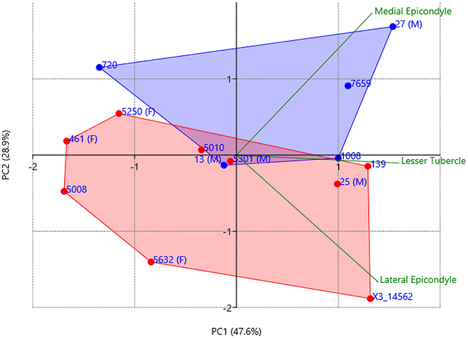
Figure 7. Principal component analysis (PCA) plot displaying the first two principal components (PC1 and PC2), using all three humeral entheseal 3D measurements as variables. The plot highlights the key patterns and differences between more robust (blue) and more gracile (red) individuals, color-labeled following the clustering process presented in Figure 4. The specimen labels indicate the few bones with assessed biological sex as males (M) or females (F).
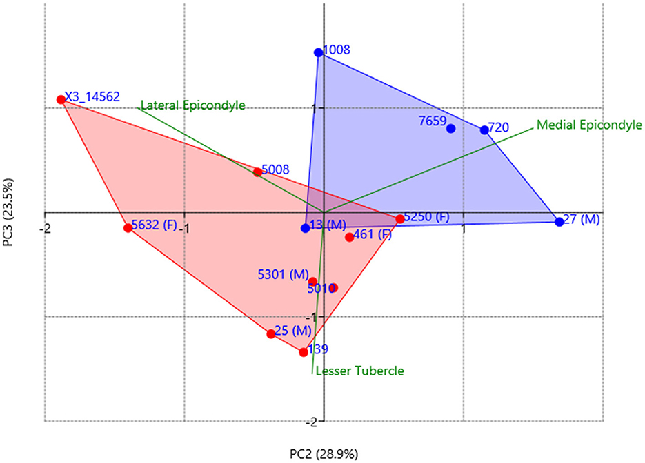
Figure 8. Principal component analysis (PCA) plot displaying the second and third principal components (PC2 and PC3), using all three humeral entheseal measurements as variables. The plot highlights the key patterns and differences between more robust (blue) and more Gracile (red) individuals, color-labeled following the clustering process presented in Figure 4. The specimen labels indicate the few bones with assessed biological sex as males (M) or females (F).
In particular, the multivariate patterns of 3D entheses identified differences between the assigned four biological males and three females in the scores of PC1 (representing 47.6% of total variation). Based on the factor loadings (Table 2 and green vectors in Figure 8), variation on PC1 represents overall entheseal size (i.e., all loadings are positive). In contrast, the relationship between PC2 (28.9% of total variation) and PC3 (23.5% of total variation) provided considerable separation in entheseal patterns between robust bones (blue dots in Figures 7, 8) and more gracile specimens (red dots in Figures 7, 8). Based on the loadings of PC2, robust individuals presented proportionally larger medial epicondyles. Nevertheless, our correlation tests showed a strong association between entheseal patterns represented by PC2 scores and the vertical head diameter of individuals (p-value > 0.01; r-value = 0.67). By contrast, PC3 scores do not seem to be associated with biological sex (see Figures 7, 8) and did not correlate with head diameters. Based on the factor loadings (Table 2 and green vectors in Figure 8), variation on PC3 reflects proportionally greater use of hand, wrist, and elbow muscles (for flexion and extension, represented by the medial and lateral epicondyles) compared to arm adduction via humeral head medial rotation (represented by the lesser tubercle).
3.3. Results of δ13C and δ15N
The stable isotopes results show, in a first approach, the preservation of the collagen in bone through the percentages of carbon and nitrogen. Any sample showing a carbon and nitrogen percentage lower than 20% and 4% respectively was not considered for the analysis. All human and zooarchaeological samples ranged from 33.6% to 44.2% in carbon and from 12.0 to 15.6% in nitrogen. The atomic C/N ratio was 3.3 in every sample, which means that all the samples are acceptable following Van Klinken (1999) standards.
The human remains from the Biniadrís cave (black dots on Figure 9) showed very narrow δ13C delta values ranging from −19.9% to −19.1% and δ15N values between 8.2% and 10.2%. Regarding the zooarchaeological samples, they ranged between −22.1‰ to −19.7‰ for δ13C and from 4.3‰ to 8.9‰ for δ15N. The results show a strong terrestrial diet, excluding any type of marine diet (Figure 9, Table 3). Even though there is a very slight relationship in the increase of carbon and nitrogen stable isotope values, it does not reach values that correspond with marine food. This fits logically with the zooarchaeological data published for the Balearic Islands (Ramis et al., 2017)
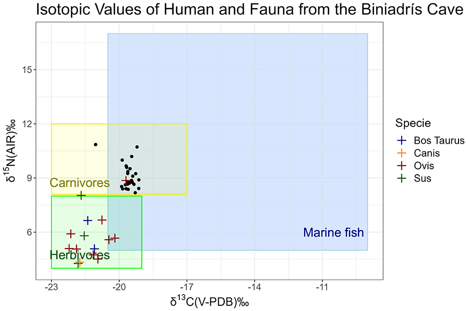
Figure 9. Results of the δ13C and δ15N isotopic values of the human (black dots) and faunal remains from the Biniadrís cave.
The stable carbon and nitrogen isotope analysis revealed a remarkable consistency across the studied samples. Both the human skeletal remains and the faunal specimens suggest a predominant terrestrial dietary pattern, primarily reliant on C3 plants. Despite the slight dietary variability observed within the human samples, the isotopic values remain largely in line with the anticipated terrestrial signatures, suggesting the consumption of cattle, sheep/goat, and pig as seen from the domestic contexts on the island (Ramis et al., 2017). It is worth noting that the samples under scrutiny were sourced from adult individuals, and no differentiation was made based on sex, thus limiting our ability to discern potential dietary biases along gender lines. However, an exhaustive examination of the aggregated terrestrial values suggests that any segregation based on variables such as sex, skeletal robustness, or chronological considerations is unlikely to yield significant deviations within the broader terrestrial dietary spectrum (C3).
Moreover, the consistent isotopic patterns also shed light on the population's behavioral traits concerning the utilization of marine resources in coastal communities. The lack of significant deviations from terrestrial isotopic norms underscores a consistent reliance on terrestrial food sources, despite the proximity to marine environments. This uniformity in dietary behavior, as evidenced by isotopic analysis, suggests a degree of cultural or ecological stability within these populations, with minimal adaptation to marine resource availability (Mannino et al., 2012; Salazar-García et al., 2018). Such findings contribute to our understanding of ancient human dietary habits and the socioecological dynamics influencing resource utilization patterns over time.
3.4. Results of the radiocarbon 14C- AMS analysis
A Single Contiguous Phase Bayesian Model (Figure 10, Amodel 107.3 and Aoverall 107.2) determined a continuous temporal distribution of the cave's inhumations between 1295 and 1175 cal BC and 750–520 cal BC (95% probability) with a span of 445–675 years (95% probability). The radiocarbon results were examined using a χ2 test (Ward and Wilson, 1978), which resulted in seven groups of dates that could have been contemporary (Figure 11). If the groups indicate pulses of use, the oldest one occurred in 1195–1120 cal BC (95% probability; 53.33% of the dated graves, Acomb 191.6), the second one 1190–1055 cal BC (95% probability; 40%, Acomb 174.1), the third one in 1110–1005 cal BC (95% probability; 13.33%, Acomb 85.2), the fourth in 1010–915 cal BC (95% probability; 6.66%, Acomb 67.4), the fifth one in 805–780 cal BC (95% probability; 6.66%, Acomb 75.4), the sixth in 790–590 cal BC (95% probability; 10%, Acomb 121.7) and, the last one in 770–570 cal BC (95% probability; 23.33%. Acomb 106.1).
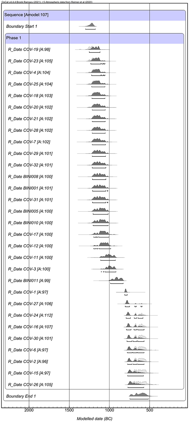
Figure 10. Radiocarbon dates (with 2σ calibration range) for the human population of the Biniadrís cave.
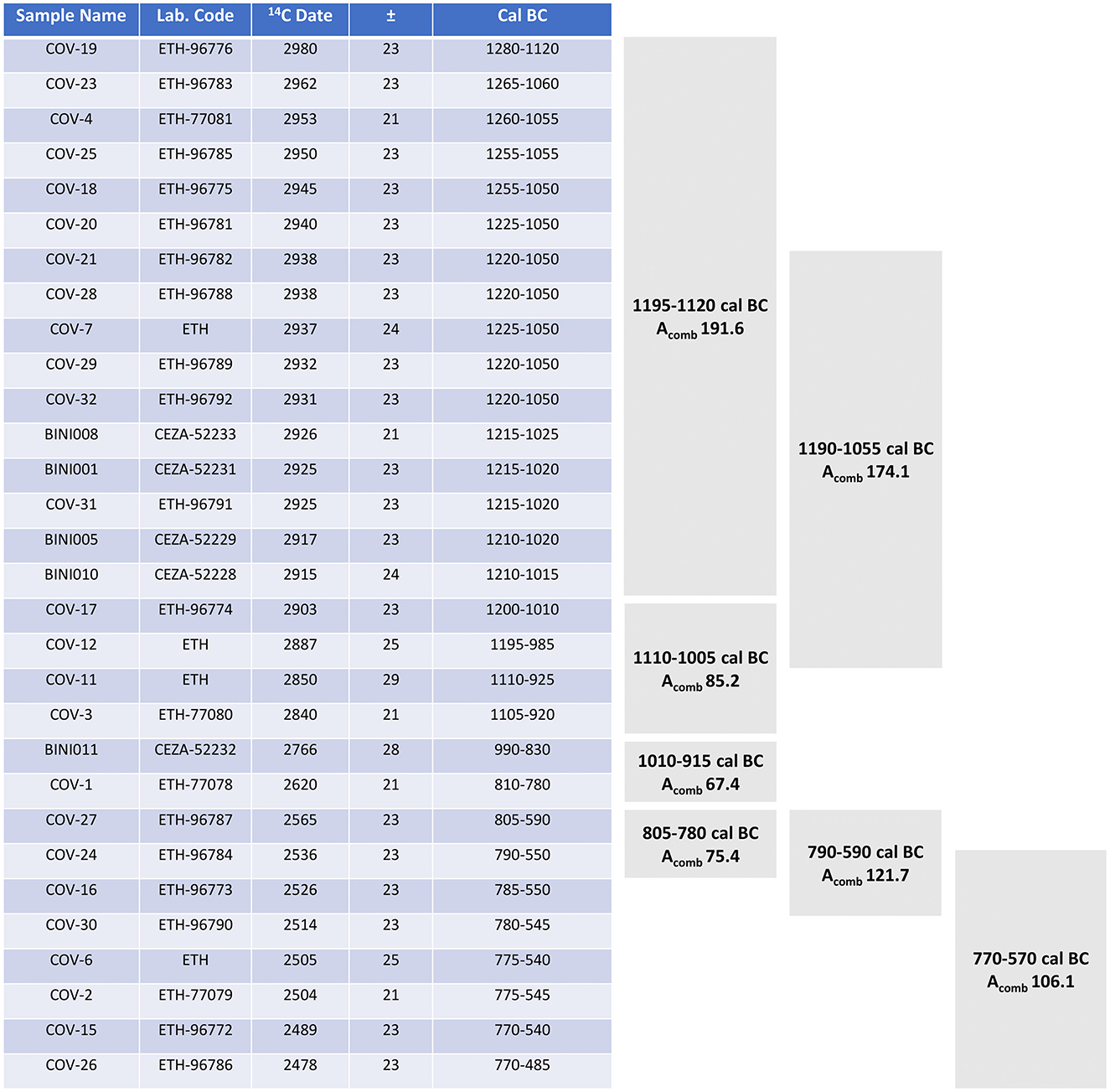
Figure 11. χ2 test of radiocarbon dates (with 2σ calibration range) of the human population the Biniadrís cave.
4 Discussion
During the Late Bronze Age (c. 1,400–800 BC) the funerary ritual at Biniadrís cave is linked to the changes observed in the archaeological record and the habitational and funerary spaces in the Balearic Islands but particularly in Menorca (Lull et al., 1999). During this period, different types of spaces coexisted with different distributions across the Balearics, such as boat-shaped structures known as “navetas,” which initially served as habitational structures but later were used exclusively for collective funerary purposes. Examples include Biniac-L'Argentina (Plantalamor Massanet, 1991), Es Tudons (Fernández-Miranda, 1991), and Rafal Rubí (Roselló-Bordoy, 1979). These navetas were contemporaneous with natural caves or shelters used for similar purposes, such as Cova dels Ossos de Mongofre Nou, Cova de Es Càrritx (Lull et al., 1999; Lull and Micó Pérez, 1999), Cova des Mussol (Lull et al., 1999; Lull and Micó Pérez, 1999), Cova des Pas (Fullola et al., 2008), Biniadrís, and Torre del Ram (Plantalamor Massanet, 1997). The concept of collective burial, evolving from individual burials within an organized and structured deposit, is exemplified at the Biniadrís cave, which contains a human bone collection of over 150 individuals buried in this context. The equal representation of female and male with the inclusion of a large non-adult group is one of the main features of the Biniadrís community. In addition, the results of our preliminary entheseal analysis suggest that the robust humeri of Menorca belonged to relatively larger individuals (represented by high PC2 scores correlating with our body size proxy; see Results) whose habitual upper arm activities involved extensive use of elbows and hands (represented by high PC3 scores; see Table 2). Interestingly, most relatively gracile individuals presented an entheseal pattern reflecting proportionally greater adduction and medial rotation of the arm (reflected in low PC3 scores). Further studies will follow the advanced iteration of the original V.E.R.A. 1.0, which is a semi-automated protocol (using primary MeshLab), and will be helpful to analyse in depth the entheseal changes in the Biniadrís bones (Karakostis, 2024).
Of special importance and one of the main features of the funerary caves, shelters, and hypogea is the site selection. It seems to have played a significant role before the start of their use, with inaccessibility and location, usually far from the settlements, being key components. The interiors are small, circular or oval in shape, with monumentalised entrances, such as the one still preserved at Biniadrís, that are also known from Menorcan settlements. There is a specific burial treatment such as the dyeing of the hair, where the red pigment is characteristic and has been used also in other caves (Lull et al., 1999; Lull and Micó Pérez, 1999; Lull et al., 2014; Rihuete Herrada, 2000). In addition, the cutting of the hair and its deposition in special containers, made this ritual quite elaborate and specific. The funerary ritual is accompanied by the use of plants that seem to be exclusive to some ritual spaces (Guerra-Doce et al., 2023; Riera Mora et al., 2018). There also appears to have been some selection in the production of associated ceramics. Personal belongings accompanying the bodies of the deceased persons are made of metal but also of bone, such as the V-shaped buttons (Altamirano García and Alarcón García, 2018), which is also a recurrent feature that has been found in the western Mediterranean.
These Late Bronze Age communities were attached to their landscape and made good use of their resources (Díaz-Zorita Bonilla et al., 2025), mainly land-based terrestrial sources, practicing agricultural and herding strategies. The scientific analyses applied to the study of subsistence practices showed a human diet clearly based on terrestrial resources of animal and plant proteins, and excluding any appreciable marine component. Similar patterns have been documented at Cova des Pas, where an identical archaeological record and terrestrial-based diets were observed (Sotiriadou et al., 2023).
The 700 years of use of the cave should be understood in a longue durée perspective, especially considering that access to the cave is concealed and difficult, which reinforces the intentionality, strong ties, and collective knowledge and identity of the community that has cared for the funerary space through time. The archaeological record shows a very long use of the cave, where the funerary memory has been preserved for centuries. It is not only the preservation of this memory that is important, but also the archaeological record, which has been organized with great care, with fragmentation kept to a minimum. It is now possible to observe the organization of the community and the reconstruction of group identity, the interaction between its members and the modification of space and objects over time (Hernando, 2002; Sánchez Romero, 2005, 2007; Querol, 2001; Alarcón García et al., 2020). Since the main archaeological material found in the Biniadrís cave is human remains, our research focuses on the human body while also considering everything else that encompasses the ritual understanding of the body as cultural elements (Sánchez Romero, 2008, 2009). The prolonged use of the cave and the longevity of the ritual practices can also be interpreted in terms of place-attachment and place-related identity, a feature of small islands arising from their more bounded character and strong sense of place (Dawson, 2019).
Similarly, the evidence for healing and care among the individuals buried in the cave is a strong indication of a close-knit and autonomous population, as is to be expected on an island. The absence of indicators for a marine diet suggests limited engagement with the sea and greater exploitation of the land, through agriculture and animal husbandry, which “left their mark on the bodies” (Díaz-Zorita Bonilla et al., 2025, p. 128). This is not to say that the community at Biniadrís was completely isolated or inward-looking. While still under study and not included in this paper, the material evidence found at the cave, ranging from organic (e.g., flax, V-perforated bone buttons) to inorganic material (e.g., carnelian beads), points to connections to the Iberian mainland as well as the wider Mediterranean. Alternating phases of increased isolation and connectivity are bound to have occurred over 700 years; thus, the continuity in the use of the same ritual space over seven centuries represents a conscious expression of community identity. The absence of contemporary settlement evidence makes it difficult to evaluate whether Biniadrìs cave served one or more communities and where the individuals buried there lived. Nonetheless, its longevity underscores the considerable effort that went into maintaining it as a focal point in a shifting landscape. The sharing of a limited space, practices, and resources is an important factor in the development of a unique community identity within an island setting. Moreover, the evidence indicates a stable, well-organized, adapted, and cared-for community, managing well at the interface between isolation and connectivity. The development of a collective identity at Biniadrís, which we understand as the expression of community belonging and place-attachment, can be traced from its shared and continued practices and appears amplified by insularity (Calvo-Trias, 2024; Dawson, 2020).
5 Conclusions
The Biniadrís cave represents a sacred space shared by all members of the community, whose actions further reinforced their identity as reflected in the care of their ancestors according to a specific funerary programme. The fact that individuals of all ages, especially young non-adults, had the right to be inhumed in the same space, and the equal representation of female and male adults and the absence of evidence for gendered divisions of labor in the osteological record, attest to the inclusion of all members of the community in the daily activities and also in the funerary world. Additionally, the palaeopathological analysis showed the implementation of care in the past, which along with the setting of the ritual, reinforces the agency and identity of the community through time and space. This longue durée can be further explained by the insular setting of this community, with its limited space and resources, which would reinforce community cohesion. Biniadrís thus provides significant insights into the changing nature of insularity, as seen from our ongoing study of long-term environmental and cultural dynamics at the island and archipelago scale. The exceptional preservation in Biniadrís cave, including the remarkable condition of the human remains, combined with its enduring funerary memory spanning over seven centuries, makes it a remarkable site for understanding the life and death of Menorcan communities during the late prehistoric period in Europe.
Data availability statement
The original contributions presented in the study are included in the article/supplementary material, further inquiries can be directed to the corresponding author.
Author contributions
MDZB: Conceptualization, Data curation, Formal analysis, Funding acquisition, Investigation, Methodology, Project administration, Resources, Supervision, Validation, Visualization, Writing – original draft, Writing – review & editing. MT: Formal analysis, Writing – review & editing. MM: Formal analysis, Writing – review & editing. AM: Writing – review & editing. EA: Writing – review & editing. AK: Formal analysis, Software, Writing – review & editing. JE: Writing – review & editing. LA: Writing – review & editing. SR: Writing – review & editing. BM: Formal analysis, Writing – review & editing. DH: Writing – review & editing. HD: Writing – review & editing. FC: Writing – review & editing. MB: Writing – review & editing. JB: Writing – review & editing.
Funding
The author(s) declare that financial support was received for the research and/or publication of this article. Funding was provided by Fundació Rubió i Turudí, the B06 project at the SFB 1070 RessourceCultures (DFG), Consell Insular de Menorca, the Institut Menorquí d' Estudis and the SFB explorative funds.
Acknowledgments
Thanks to Hervé Bocherens and Dorothée Drucker at the Biogeology Laboratory of the University of Tübingen for helping with the laboratory process and to Eulália Subirá for measuring the δ13C and δ15N samples at the Autonomous University of Barcelona. In addition, we are grateful to the SFB1070 and the Fundació Rubió, for the financial and logistic support during the duration of this project.
Conflict of interest
The authors declare that the research was conducted in the absence of any commercial or financial relationships that could be construed as a potential conflict of interest.
Generative AI statement
The author(s) declare that no Gen AI was used in the creation of this manuscript.
Publisher's note
All claims expressed in this article are solely those of the authors and do not necessarily represent those of their affiliated organizations, or those of the publisher, the editors and the reviewers. Any product that may be evaluated in this article, or claim that may be made by its manufacturer, is not guaranteed or endorsed by the publisher.
References
Acsádi, G. Y., and Nemeskéri, J. (1974). History of human lifespan and mortality. Curr. Anthropol. 15, 495–507. doi: 10.1086/201508
Adler, C-. P. (2006). Knochenkrankheiten: Diagnostik makroskopischer, histologischer und radiologischer Strukturveränderungen am. Skelett. Springer DE.
Alarcón García, E., Onorato, M. A. M., Bonilla, M. D.-Z., and Martínez, L. A. (2020). ¿Quiénes eran? La cueva de Biniedrís (Alaior, Menorca, Islas Baleares). Rev. de Menorca 99, 113–134.
AlQahtani, S. J. (2008). Atlas of Tooth Development and Eruption. Bart's and the London School of Medicine and Dentistry. London: Queen Mary University of London, MClinDent.
Alt, K. W. (1997). Odontologische Verwandschaftsanalyse: Individuelle Charakteristika der Zähne in ihrer Bedeutung für Anthropologie, Archäologie und Rechtsmedizin. Stuttgart: Fischer.
Altamirano García, M., and Alarcón García, E. (2018). Bone tools for the deceased; approaches to the worked osseous assemblage from the Bronze Age funerary cave of Biniadrís (Menorca, Spain). Quat. Int. 472, 108–114. doi: 10.1016/j.quaint.2017.12.052
Anglada, F. M., Ferrer, R., Plantalamor, P., and Ramis, D. (2010). Aixecament planimètric d‘es Castellet (Ciutadella), un assentament prehistòric costaner a la zona occidental de Menorca. Bolletí de la Societat Arqueològica Lulliana. Rev. d'Estudis Hist. 66, 267–277.
Aranda Jiménez, G., Sánchez Romero, M., Díaz-Zorita Bonilla, M., Lozano Medina, A., Escudero Carrillo, J., and Milesi, L. (2020). “Cultural resistance to social fragmentation. The continuity and reuse of megalithic monuments during the Argaric Bronze Age in southeastern Iberia,” in The Matter of Prehistory: Papers in Honor of Antonio Gilman Guillén, eds. P. Díaz del Río, K. Lillios, and I. Sastre Prats (Spanish National Research Council), 213–233.
Armentano, N., Jordana, X., and Malgosa, A. (2010). Aproximación paleodemográfica a una población protohistórica de las Baleares. El yacimiento de la Cova des Pas (Ferreries, Menorca). Rev. de Demografía Hist. 28, 91–108.
Armentano, N., and Malgosa, A. (2003). “Enterramientos primarios versus enterramientos secundarios,” in Antropología y Biodiversidad, Bellaterra Edicions (Bellaterra: Universitat Autònoma de Barcelona), 38–49.
Aufderheide, A. C., and Rodríguez-Martin, C. (1998). The Cambridge Encyclopaedia of Human Paleopathology. Cambridge; New York, NY: Cambridge University Press, 478. doi: 10.1525/aa.2000.102.1.171
Barnes, E. (2008). “Congenital anomalies,” in Advances in Human Palaeopathology, ed. R. Pinhasi (Chichester: Wiley), 329–362. doi: 10.1002/9780470724187.ch15
Bass, W. M. (2005). Human Osteology-A laboratory and field Manual, 5th Edn. Columbia, MO: Missouri Archaeological Society. Special Publication No. 2.
Belén Deamos, M., and Fernández-Miranda, M. (1977). La historia que se ahogó en el mar. Arqueología submarina: investigaciones en la isla de Menorca. Historia 16, 41–54.
Belén Deamos, M., Fernández-Miranda, M., and Cerdà Juan, D. (1979). El fondeadero de Cales Coves: (Menorca, Islas Baleares). Madrid: General Subdirectorate of Archaeology.
Bergadá, M., Guerrero, V. M., and Ensenyat, J. (2005). Primeras evidencias de estabulación en el yacimiento de son Matge (Serra de Tramuntana, Mallorca) a través del registro sedimentario. Mayurqa 30, 153–180.
Berry, A. C., and Berry, R. J. (1967). Epigenetic variation in the human cranium. J. Anat. 101, 361–379.
Black, T. K. (1978). A new method for assessing the sex of fragmentary skeletal remains: femoral shaft circumference. Am. J. Phys. Anthropol. 48, 227–231. doi: 10.1002/ajpa.1330480217
Bocherens, H., Billiou, D., Patou-Mathis, M., Bonjean, D., Otte, M., Mariotti, A., et al. (1997). Paleobiological implications of the isotopic signatures (13C, 15N) of fossil mammal collagen in Scladina cave (Sclayn, Belgium). Quat. Res. 48, 370–380. doi: 10.1006/qres.1997.1927
Bocherens, H., and Drucker, D. (2003). Trophic level isotopic enrichments for carbon and nitrogen in collagen: case studies from recent and ancient terrestrial ecosystems. Int. J. Osteoarchaeol. 13, 46–53. doi: 10.1002/oa.662
Bonnet, N., and Ferrari, S. L. (2010). Exercise and the skeleton: how it works and what it really does. IBMS BoneKey 7, 235–248. doi: 10.1138/20100454
Bräuer, G., and Knussmann, R. (1988). “Grundlagen der Anthropometrie,” in Anthropologie-Handbuch der vergleichenden Biologie des Menschen, Bd. I Wesen und Methoden der Anthropologie, 1. Teil, Wissenschaftstheorie, Geschichte, morphologischer Methoden, ed. R. Knußmann (Stuttgart: Spektrum Akademischer Verlag), 129–159.
Brickley, M. B. (2018). Cribra orbitalia and porotic hyperostosis: a biological approach to diagnosis. Am. J. Phys. Anthropol. 167, 896–902. doi: 10.1002/ajpa.23701
Brickley, M. B., and Ives, R. (2008). The Bioarchaeology of Metabolic Bone Disease. Academic Press. doi: 10.1016/B978-0-12-370486-3.00002-0
Brickley, M. B., and Mays, S. (2019). “Metabolic disease.” in Ortner's Identification of Pathological Conditions in Human Skeletal Remains, 3rd Edn, ed. J. E. Buikstra (Academic Press Elsevier Inc.), 531–566. doi: 10.1016/B978-0-12-809738-0.00015-6
Bronk Ramsey, C. (2001). Development of the radiocarbon calibration program. Radiocarbon 43, 355–363. doi: 10.1017/S0033822200038212
Bronk Ramsey, C. (2009). Bayesian Analysis of Radiocarbon Dates. Radiocarbon 51, 337–60. doi: 10.1017/S0033822200033865
Bronk Ramsey, C. (2013). “Recent and planned developments of the program OxCal. Radiocarbon 55, 720–730. doi: 10.1017/S0033822200057878
Bronk Ramsey, C. (2017). Methods for Summarizing Radiocarbon Datasets. Radiocarbon 59, 1809–1833. doi: 10.1017/RDC.2017.108
Brooks, S., and Suchey, J. M. (1990). Skeletal age determination based on the Os pubis: a comparison of the Acsádi-Nemeskéri and Suchey-Brooks methods. Hum. Evol. 5, 227–238. doi: 10.1007/BF02437238
Brothwell, D. R. (1981). Digging up Bones: The Excavation, Treatment, and Study of Human Skeletal Remains, 3rd Revised Edn. Ithaca, NY; London: Cornell University Press (in cooperation with British Museum [Natural History]).
Bruzek, J. (2002). A Method for visual Determination of Sex, Using the Human Hip Bone. Am. J. Phys. Anthropol. 117, 157–168. doi: 10.1002/ajpa.10012
Buikstra, J. E., and Ubelaker, D. H. (1994). Standards for the data collection from human skeletal remains. Arkansas Arch. Survey Res. Ser. 44:3.
Calce, S. E. (2012). A new method to estimate adult age-at-death using the acetabulum. Am. J. Phys. Anthropol. 148, 11–23. doi: 10.1002/ajpa.22026
Calvo Trias, M., Guerrero, V. M., and Salvá, B. (2001). Arquitectura Ciclópea del Bronce Balear,” in Análisis morfofuncional y desarrollo secuencial, ed. El Tall (Spain: Palma de Mallorca).
Calvo, M., Albero, D., Rosselló, J. G., and Javaloyas, D. (2012). “Re-thinking social hierarchisation and stratification in the Bronze Age of the Balearic Islands,” in The Prehistory of Iberia: Debating Early Social Stratification and the State (Abingdon; New York, NY: Routledge), 170–202.
Calvo, M., Guerrero, V., and Salvá, B. (2002). Los orígenes del poblamiento balear. Una discusión inacabada. Complutum 13, 159–191.
Calvo-Trias, M. (2024). In pursuit of the analytical unit. island archaeology as a case study. Cambridge Archaeol. J. 34, 583–600. doi: 10.1017/S0959774323000501
Cardoso, F. A., and Henderson, C. (2013). The categorisation of occupation in identified skeletal collections: a source of bias? Int. J. Osteoarchaeol. 23, 186–196. doi: 10.1002/oa.2285
Cardoso, H. F. V., Vandergugten, J. M., and Humphrey, L. T. (2016). Age estimation of immature human skeleton remains from the metaphyseal and epiphyseal widths of the long bones in the post-natal period. Am. J. Anthropol. 162, 19–35. doi: 10.1002/ajpa.23081
Cherry, J. F., and Leppard, T. P. (2018). The balearic paradox: why were the Islands colonized so late? Pyrenae 49, 49–70.
Cockburn, A., Duncan, H., and Riddle, J. M. (1979). Arthritis, ancient and modern: guidelines for field workers. Henry Ford Hosp. Med. J. 27, 74–79.
Cole, G., and Waldron, T. (2019). Cribra orbitalia: dissecting an ill-defined phenomenon. Int. J. Osteoarchaeol. 29, 613–621. doi: 10.1002/oa.2757
Coll, Conesa, J., Calvo Trías, M. A., and Guerrero Ayuso, V. M. (1997). El Dolmen de S'Aiga Dolca: sepulcro colectivo del Pretalaiótico. Rev. de Arqueología 18, 18–29.
Cunningham, C., Scheuer, L., and Black, S. (2016). Developmental Juvenile Osteology. San Diego, CA: Academic Press. doi: 10.1016/B978-0-12-382106-5.00003-7
Dawson, H. (2014). Mediterranean Voyages. The Archaeology of Island Colonisation and Abandonment. London: Routledge.
Dawson, H. (2019). “Island archaeology,” in Encyclopedia of Global Archaeology, ed. C. Smith (Switzerland: Springer Nature). doi: 10.1007/978-3-319-51726-1_3280-1
Dawson, H. (2020). Network science and island archeology: advancing the debate. J. Island Coastal Archaeol. 16, 213–230. doi: 10.1080/15564894.2019.1705439
De Cet, M. (2017). Long-term Social development on a mediterranean Island: Menorca between 1600BCE and 1900CE. Verlag Dr. Rudolf Habelt GmbH. (Universitätsforschungen zur prähistorischen Archäologie 303; Human Development in Landscapes 10).
De Cet, M., Lull, V., Micó, R., Rihuete, C., and Risch, R. (2017). “Migration and integration during the bronze and iron ages: the case of Menorca,” in Migration und Integration von der Urgeschichte bis zum Mittelalter, 9. Mitteldeutscher Archäologentag vom 20. bis 22. Oktober 2016 in Halle (Saale), Bd. 17, eds. H. Meller, F. Daim, J. Krause, and R. Risch (Halle (Saale): State Museum of Prehistory).
Deniro, M. J., and Epstein, S. (1981). Influence of diet on the distribution of nitrogen isotopes in animals. Geochim. Cosmochim. Acta. 45, 341–351. doi: 10.1016/0016-7037(81)90244-1
Dias, G., and Tayles, N. (1997). ‘Abscess cavity'-a Misnomer. Int. J. Osteoarchaeol. 7, 548–554. doi: 10.1002/(SICI)1099-1212(199709/10)7:5<548::AID-OA369>3.0.CO;2-I
Díaz-Zorita Bonilla, M., Marciales Daza, M., and Timm, M. (2025). “Use and non-use of resources during the bronze and iron ages of Menorca (Balearic Islands, Spain),” in ResourceCultures. Wiesbaden: Reichert Verlag, 119–129.
Falys, C. G., and Prangle, D. (2015). Estimating age of mature adults from the degeneration of the sternal end of the clavicle. Am. J. Phys. Anthropol. 156, 203–214. doi: 10.1002/ajpa.22639
Ferembach, D., Schwidetzky, I., and Stloukal, M. (1979). Empfehlungen für die alters- und Geschlechtsdiagnose am Skelett. Homo 30, 1–32.
Ferembach, D., Schwidetzky, I., and Stloukal, M. (1980). Recommendations for age and sex diagnosis of skeletons. J. Hum. Evol. 9, 517–549. doi: 10.1016/0047-2484(80)90061-5
Fernández-Miranda, M. (1991). La transición hacia la cultura talayótica en Menorca. Trab Prehist 48, 37–50. doi: 10.3989/tp.1991.v48.i0.512
Finnegan, M., and Faust, M. A. (1974). Variants of the Femur. Bibliography of human and non-human, non-metric variation. Res. Rep. 14, 1–133.
Fornés, J., Javaloyas, D., Salvá, B., Berenguer, C., Matas, F., Servera, G., et al. (2009). “Más que una casa. Los navetiformes de la Edad del Bronce Balear,” in Lspai doméstic i lorganització de la societat a la protohistoria de la Mediterrània occidental (Ier. Mil•leni AC), Actes de la IV Reunió d'Arqueologia de Calafell, vol. 11. Arqueomediterrània, ed. C. Belarte, 323–330.
Fullola, J. M., Guerrero, V. M., Petit, M. A., Calvo, M., Malgosa, A., Armentano, N., et al. (2007). “La Cova del Pas (Ferreries, Menorca):unavanç,” in L'arqueologia a Menorca: eina per al coneixement del passat. Llibres del Patrimoni Històrici Cultural (Menorca: Consell Insular de Menorca), 95–110.
Fullola, J. M., Guerrero, V. M., Petit, M. A., Calvo, M. A., Malgosa Armentano, N., Arnau, P., et al. (2008). La cova des Pas (Ferreries, Menorca): un jaciment capdal en la Prehistòria de les Balears. Unicum 7, 10–16.
Galmés-Alba, A., and Calvo-Trias, M. (2021). Connection architectures across the landscapes: a visibility and network analysis in the island of mallorca during the late bronze age and early bronze age. Cambridge Archaeol. J. 32, 467–487. doi: 10.1017/S0959774321000627
Gili, S., Lull, V., Micó, R., Rihuete, C., and Risch, R. (2006). An island decides: megalithic burial rites on Menorca. Antiquity 80, 829–842. doi: 10.1017/S0003598X0009445X
Gornés, S. (1997). Arqueología de la muerte y cambio social. Análisis e interpretación de la necrópolis de Cales Coves, Menorca. Complutum 7, 91–103.
Gornés, S. (2000). Ipogei del Talaiotico inale: analisi e interpretazionedella necrópolis di Cales Coves, minorca. L'ipogeismo nel Mediterráneo: sviluppo, quadri culturali, Vol. II. (Sassari-Oristano: Università degli Studi di Sassari), 553–571.
Gornés, S. (2016). Sociedad y cambio en Menorca: sistematización de los contextos arqueológicos de las navetas funerarias entre el 1400 y el 850 cal ANE (Tesis doctoral). Universitat Autònoma de Barcelona, Sapin.
Gornés, S. (2022). Talayots y taulas. La evolución de la arquitectura simbólica en la Prehistoria de Menorca entre los siglos XI al II cal ANE. Trabajos de Prehistoria 79, 99–114. doi: 10.3989/tp.2022.12289
Gornés, S., and Gual, J. (2000). El hipogeo XXI de la necrópolis de Cales Coves, Minorca. L'ipogeismo nel Mediterráneo: sviluppo, quadri culturali, Vol. II. (Sassari-Oristano: Università degli Studi di Sassari), 573–590.
Grupe, G., Harbeck, M., and McGlynn, G. C. (2015). Prähistorische Anthropologie. Berlin, Heidelberg: Springer. doi: 10.1007/978-3-642-55275-5
Guerra-Doce, E., Rihuete-Herrada, C., Micó, R., Risch, R., Lull, V., Niemeyer, H. M., et al. (2023). Direct evidence of the use of multiple drugs in Bronze-Age Menorca (Western Mediterranean) from human hair analysis. Sci. Rep. 12:4782. doi: 10.1038/s41598-023-31064-2
Guerrero Ayuso, V. M., Calvo Trias, M., García Rosselló, J., and Gornés Hachero, S. (2007). Prehistoria de las Islas Baleares. Registro arqueológico y evolución social antes de la Edad del Hierro. British Archaeological Reports, International Series 1690.
Handling, M. A., Curtis, A. S., and Miller, S. L. (2010). The origin of the long head of the triceps: a cadaveric study. J. Shoulder Elbow Surg. 19, 69–72. doi: 10.1016/j.jse.2009.06.008
Hauser, G., and DeStefano, G. F. (1989). Epigenetic Variants of the Human Skull. Stuttgart: Schweizerbart'sche Verlagsbuchhandlung.
Henderson, C. (2009). Musculoskeletal Stress Markers in Bioarchaeology: Indicators of Activity Levels or Human Variation? A re-analysis and Interpretation, Durham theses. Durham University, UK.
Henderson, C. Y., Mariotti, V., Santos, F., et al. (2017). The new Coimbra method for recording entheseal changes and the effect of age-at death. BMSAP 29, 140–149. doi: 10.1007/s13219-017-0185-x
Hermann, B., Grupe, G., Hummel, S., Piepenbrink, H., and Schutkowski, H. (1990). Prähistorische Anthropologie- Leitfaden der Feld- und Labormethoden. Berlin, Heidelberg: Springer. doi: 10.1007/978-3-642-61514-6
Hernández Sanz, F. (1896–1897). Antigua población de Cales Coves. Revista de Menorca (Mahón) 1, 160–167.
Hillson, S. (1996). Dental Anthropology. Cambridge: Cambridge University Press. doi: 10.1017/CBO9781139170697
Hillson, S. (2001). Recording dental caries in archaeological human remains. Int. J. Osteoarchaeol. 11 249–289. doi: 10.1002/oa.538
Iacumin, P., Bocherens, H., Mariotti, A., and Longinelli, A. (1996). An isotopic palaeoenvironmental study of human skeletal remains from the Nile Valley. Palaeogeogr. Palaeoclimatol. Palaeoecol. 126, 15–30. doi: 10.1016/S0031-0182(96)00067-3
Işcan, M. Y., Loth, S. R., and Wright, R. (1984a). Age estimation from the rib by phase analysis: white males. J. Forensic Sci. 29, 1094–1104. doi: 10.1520/JFS11776J
Işcan, M. Y., Loth, S. R., and Wright, R. (1984b). Metamorphosis at the sternal rib: a new method to estimate age at death in males. Am. J. Phys. Anthropol. 65, 147–156. doi: 10.1002/ajpa.1330650206
Işcan, M. Y., and Steyn, M. (2013). The Human Skeleton in Forensic Medicine, 2nd & 3rd Edn. Springfield, IL: Charles C. Thomas.
Jacobs, K. (1992). Estimating Femur and Tibia length from Fragmentary Bones. Am. J. Phys. Anthropol. 89, 333–346. doi: 10.1002/ajpa.1330890307
Jurmain, R. D., and Kilgore, L. (1995). Skeletal evidence of osteoarthritis: a paleopathological perspective. Ann. Rheum. Dis. 54, 443–450. doi: 10.1136/ard.54.6.443
Karakostis, F. A. (2023). Statistical protocol for analyzing 3D muscle attachment sites based on the “Validated Entheses-based Reconstruction of Activity” (VERA) approach. Int. J. Osteoarchaeol. 33, 461–474. doi: 10.1002/oa.3196
Karakostis, F. A. (2024). Protocol for the 'Validated Entheses-based reconstruction of activity 20′ (VERA 2.0) Method: semi-automated measurement of 3d entheseal changes. PLOS One 20:e0321479. doi: 10.1371/journal.pone.0321479
Karakostis, F. A., and Harvati, K. (2021). New horizons in reconstructing past human behavior: Introducing the “Tübingen University Validated Entheses-based Reconstruction of Activity” method. Evolution. Anthropol. 30, 185–198. doi: 10.1002/evan.21892
Karakostis, F. A., Hotz, G., Scherf, H., et al. (2018). A repeatable geometric morphometric approach to the analysis of hand entheseal three-dimensional form. Am. J. Anthropol. 166, 246–260. doi: 10.1002/ajpa.23421
Karakostis, F. A., and Lorenzo, C. (2016). Morphometric patterns among the 3D surface areas of human hand entheses. Am. J. Anthropol. 160, 694–707. doi: 10.1002/ajpa.22999
Konigsberg, L. W., and Adams, B. J. (2014). “Estimating the number of individuals represented by commingled human remains: a critical evaluation of methods,” in Commingled Human Remains-Method in Recovery, Analysis, and Identification, eds. B. J. Adams, and J. E. Byrd (Academic Press). doi: 10.1016/B978-0-12-405889-7.00009-5
Krogman, W. M. (1962). The Human Skeleton in Forensic Medicine. 2nd Sub. Springfield, IL: Charles C. Thomas.
Kunos, C. A., Simpson, S. W., Russel, K. F., and Hershkovitz, I. (1999). First rib metamorphosis: its possible utility for human age-at-death estimation. Am. J. Phys. Anthropol. 110, 303–323. doi: 10.1002/(SICI)1096-8644(199911)110:3<303::AID-AJPA4>3.3.CO;2-F
Lewis, M. E. (2007). The Bioarchaeology of Children: Perspectives From Biological and Forensic Anthropology. Cambridge Studies in Biological and Evolutionary Anthropology 50. Cambridge: Cambridge University Press.
Lovejoy, C. O., Meindl, R. S., Pryzbeck, T. R., and Mensforth, R. P. (1985). Chronological metamorphosis of the auricular surface of the ilium: a new method for the determination of adult skeletal age at death. Am. J. Phys. Anthropol. 68, 15–28. doi: 10.1002/ajpa.1330680103
Lovell, N. C. (1997). Trauma analysis in paleopathology. Yearb. Phys. Anthropol. 40, 139–170. doi: 10.1002/(SICI)1096-8644(1997)25+<139::AID-AJPA6>3.0.CO;2-#
Lull, V., and Micó Pérez, R. (1999). Rituales de vida y muerte en la Prehistoria de Menorca: La Cova des Càrritx. Menorca: Consell Insular de Menorca; Ajuntament de Ciutadella.
Lull, V., Micó, R., Rihuete, C., and Risch, R. (1999). La cova des Cárritx y la Cova des Mussol. Ideología y Sociedad, en la Prehistoria de Menorca. Barcelona: Consell Insular de Menorca.
Lull, V., Micó, R., Rihuete, C., and Risch, R. (2014). “Rituales funerarios en Menorca durante la Edad del Bronce,” in La muerte en la Península Ibérica. Casos de estudio, eds. E. Guerra and J. Fernández Manzano. (Valladolid: Universidad de Valladolid), 137–153.
Mall, G., Hubig, M., Büttner, A., Kuznik, J., Penning, R., Graw, M., et al. (2001). Sex determination and estimation of stature from long bones of the arm. Forensic Sci. Int. 117, 23–30. doi: 10.1016/S0379-0738(00)00445-X
Mann, R., Hunt, D. R., and Lozanoff, S. (2016). Photographic Regional Atlas of Non-Metric Traits and Anatomical Variants in the Human Skeleton, 1st Edn, Charles C Thomas Pub Ltd.
Mann, R., Jantz, R. L., Bass, W. M., and Willey, P. S. (1991). Maxillary suture obligation: a visual method for estimating skeletal age. J. Forensic Sci. 36, 781–791. doi: 10.1520/JFS13088J
Mann, R. W., and Hunt, D. R. (2005). Photographic Regional Atlas of Bone Disease. A Guide to Pathologic and Normal Variation in the Human Skeleton, 2. Aufl. Springfield, IL: Charles C. Thomas.
Mannino, M., Catalano, G., Talamo, S., Mannino, G., Di Salvo, R., Schimmenti, V., et al. (2012). Origin and diet of the prehistoric hunter-gatherers on the Mediterranean island of Favignana (Ègadi Islands, Sicily). PLoS One 7:e49802. doi: 10.1371/journal.pone.0049802
Mariotti, V., Facchini, F., and Giovanna Belcastro, M. (2007). The study of entheses: proposal of a standardised scoring method for twenty-three entheses of the postcranial skeleton. Coll. Antropol. 31, 291–313.
Martin, R., and Saller, K. (1957). Lehrbuch der Anthropologie, Bd.1, 3 Auflage. Stuttgart: G. Fischer.
Mays, S., Zakrzewski, S., and Field, S. (2022). The relationship between dental wear and age at death in British archaeological human skeletal remains: a re-evaluation of the ‘Brothwell chart'. J. Archaeol. Sci. Rep. 46:103707. doi: 10.1016/j.jasrep.2022.103707
Meindl, R. S., and Lovejoy, C. O. (1985). Ectocranial suture closure: a revised method for the determination of skeletal age at death based on the lateral anterior sutures. Am. J. Phys. Anthropol. 68, 57–66. doi: 10.1002/ajpa.1330680106
Molleson, T., and Cox, M. (1993). The Spitalfields Project Research Report 86. The Anthropology - The Middling Sort (vol. 2). London: Council for British Archaeology.
Moreno Onorato, A., Alarcón García, E., Díaz-Zorita Bonilla, M., and Arboledas Martínez, L. (2019). El descubrimiento de la cueva de Biniedrís (Alaior, Menorca, España). Rev. de Menorca 98, 271–286.
Murail, P., Bruzek, J., and Braga, J. (1999). A New Approach to Sexual Diagnosis in Past Populations-Practical Adjustments from Van Vark's Procedure. Int. J. Osteoarchaeol. 9, 39–53. doi: 10.1002/(SICI)1099-1212(199901/02)9:1<39::AID-OA458>3.0.CO;2-V
Murail, P., Bruzek, J., Houët, F., and Cunha, E. (2005). DSP: a tool for probabilistic sex diagnosis using worldwide variability in hip-bone measurements. Bull. et mémoires de la Société d'Anthropologie de Paris 17, 167–176. doi: 10.4000/bmsap.1157
Murphy, T. (1959). The changing pattern of dentine exposure in human tooth attrition. Am. J. Phys. Anthropol. 17, 167–178. doi: 10.1002/ajpa.1330170302
O'Donnell, L., Hill, E. C., Anderson, A. S. A., and Edgar, H. J. H. (2020). Cribra orbitalia and porotic hyperostosis are associated with respiratory infections in a contemporary mortality sample from New Mexico. Am. J. Phys. Anthropol. 173, 721–733. doi: 10.1002/ajpa.24131
Orfila, M., Baratta, G., and Mayer, M. (2010). Los Santuarios de Calescoves (Alaior, Menorca): Coberxo blanc y Cova dels Jurats o de l'Esglesia, Informe preliminar. Cuadernos de Prehistoria y Arqueología de la Universidad de Granada 20, 395–428. doi: 10.30827/cpag.v20i0.139
Ortner, D. J. (2003). Identification of Pathological Conditions in Human Skeletal Remains (2nd ed.). San Diego, CA: Academic Press.
Ortner, D. J., and Aufderheide, A. C. (1991). Human Palaeopathology: Current Syntheses and Future Options. Washington, DC: Smithsonian Institution Press. doi: 10.5962/bhl.title.9196
Ortner, D. J., and Putschar, W. G. J. (1981). Identification of Pathological Conditions in Human Skeletal Remains, Smithsonian Contribution to Anthropology. Smithsonian Contributions to Anthropology, No. 28. Washington, DC: Academic Press.
Osterholtz, A. J., Baustian, K. M., Martin, D. L., and Potts, D. T. (2014). “Commingled human skeletal assemblages: integrative techniques in determination of the MNI/MNE,” in Commingled and Disarticulated Human Remains-Working Toward Improved Theory, Method, and Data, eds. A. J. Osterholtz, K. M. Baustian, and D. L. Martin (New York, NY: Springer Science & Business Media), 35–50. doi: 10.1007/978-1-4614-7560-6_3
Pearson, K. (1917/1919). Chapters 1–4. A Study of the Long Bones of the English Skeleton I: The Femur. London: Department of Applied Statistics, Company Research, Memoirs, Biometric Series X, University College, University of London.
Pinhasi, R., and Mays, S. (Eds.). (2007). Advances in Human Palaeopathology. Chichester: Wiley. doi: 10.1002/9780470724187
Plantalamor Massanet, L. L. (1991). L'arquitectura prehistórica y protohistórica de Menorca y el seu marc cultural [The Prehistoric and Protohistoric Architecture of Menorca and Its Cultural Context]. Treballs del Museo de Menorca 12. Maó: Museu de Menorca.
Plantalamor Massanet, L. L. (1997). Prehistoria de las islas Baleares. Espacio, Tiempo y Forma, Serie I, Prehistoria y Arqueologia, 325–389. doi: 10.5944/etfi.10.1997.4660
Pons, G. (2017). The talaiotic culture of the Balearic Islands. Catalan Hist. Rev. 11, 9–23. doi: 10.2436/20.1000.01.143
Portillo, M., Llergo, Y., Ferrer, A., Anglada, M., Plantalamor, L., and Albert, R. M. (2014). Actividades domésticas y molienda en el asentamiento talayótico de Cornia Nou (Menorca, Islas Baleares): resultados del estudio de microfósiles vegetales. Rev. d'Arqueologia de Ponent 24, 311–321.
Ramis (2010). “From colonisation to habitation, early cultural adaptations in the Balearic Bronze age,” in Material Connections in the Ancient Mediterranean-Mobility, Materiality and Identity, eds. P. Van Dommelen, and A. B. Knapp (London: Routledge).
Ramis, D. (2014). “Early island exploitations: productive and subsistence strategies on the prehistoric balearic islands,” in The Cambridge Prehistory of the Bronze and Iron Age Mediterranean, eds. A. B. Knapp, and P. Van Dommelen (Cambridge University Press), 40–56. doi: 10.1017/CHO9781139028387.006
Ramis, D., Anglada, M., Ferrer, A., Plantalamor, L., and Van Strydonck, M. (2017). Faunal introductions to the Balearic Islands in the early 1st millennium cal BC. Radiocarbon 59, 1415–1423. doi: 10.1017/RDC.2017.79
Redfern, R., and Roberts, C. A. (2019). “Trauma,” in Ortner's Identification of Pathological Conditions in Human Skeletal Remains, ed. J. E. Buikstra (London: Academic Press), 211–284. doi: 10.1016/B978-0-12-809738-0.00009-0
Reid, D. J., and Dean, M. C. (2000). Brief communication: the timing of linear hypoplasia on human anterior teeth. Am. J. Phys. Anthropol. 113, 135–139. doi: 10.1002/1096-8644(200009)113:1<135::AID-AJPA13>3.0.CO;2-A
Reid, D. J., and Dean, M. C. (2006). Variation in modern human enamel formation times. J. Hum. Evol. 50, 329–346. doi: 10.1016/j.jhevol.2005.09.003
Reimer, P. J., Austin, W. E. N., Bard, E., Bayliss, A., Blackwell, P. G., Bronk Ramsey, C., et al. (2020). The IntCal20 northern hemisphere radiocarbon age calibration curve (0–55 cal kBP). Radiocarbon 62, 725–757. doi: 10.1017/RDC.2020.41
Reimer, P. J., Bard, E., Bayliss, A., Beck, J. W., Blackwell, P. G., Bronk Ramsey, C., et al. (2013). IntCal13 and marine 13 radiocarbon age calibration curves 0- 50,000 years cal BP. Radiocarbon 55, 1869–1887. doi: 10.2458/azu_js_rc.55.16947
Richards, M. P., and Hedges, R. E. M. (1999). Stable isotope evidence for similarities in the types of marine foods used by Late Mesolithic humans at sites along the Atlantic coast of Europe. J. Archaeol. Sci. 26, 717–722. doi: 10.1006/jasc.1998.0387
Riera Mora, S., Servera-Vives, G., Picornell-Gelabert, L., Cabanis, M., Boi, M., Miras, Y., et al. (2018). “Pollen signatura of a ritual process in the collective burial cave of Cova des Pas (Late Bronze Age, Minorca, Balearic Islands, Spain),” in The Bioarchaeology of Ritual and Religion, eds. A. Livarda, R. Madgwick, and S. Riera Mora (Oxford: Oxbow Books). doi: 10.2307/j.ctvh1dpkx.9
Rihuete Herrada, C. (2000). Dimensiones bio-arqueológicas de los contextos funerarios: estudio de los restos humanos de la necrópolis prehistórica de la cova des Càrritx (Ciutadella, Menorca) (thesis). Spain: Universitat Autònoma de Barcelona.
Rihuete Herrada, C. (2003). “Bio-arqueología de las prácticas funerarias,” in Análisis de la comunidad enterrada en el cementerio prehistórico de la Cova des Càrritx, ca. 1450-800 cal ANE. BAR, International Series, 1161. Ciutadella, Menorca: Oxford.
Rinaldo, N., Zedda, N., Bramanti, B., Rosa, I., and Gualdi-Russo, E. (2019). How reliable is the assessment of Porotic Hyperostosis and Cribra Orbitalia in skeletal human remains? A methodological approach for quantitative verification by means of a new evaluation form. Archaeol. Anthropol. Sci. 11, 3549–3559. doi: 10.1007/s12520-019-00780-0
Roberts, C., and Connell, B. (2004). The Archaeology of Disease (3rd ed.). Ithaca, NY: Cornell University Press. doi: 10.7591/9781501742693
Roberts, C., and Manchester, K. (2010). The Archaeology of Disease, 3rd & 4th Edn. Ithaca, NY: Cornell University Press.
Roberts, C., and Manchester, K. (2012). The Archaeology of Disease (3rd & 4th eds.). Ithaca, NY: Cornell University Press.
Rogers, J., and Waldron, T. (1995). A Field Guide to Joint Disease in Archaeology. New York, NY: Willey.
Rogers, J., Watt, I., and Dieppe, P. (1985). Palaeopathology of spinal osteophytosis, vertebral ankylosis, ankylosing spondylitis, and vertebral hyperostosis. Ann. Rheumatic Dis. 44, 113–120. doi: 10.1136/ard.44.2.113
Roselló-Bordoy, G. (1979). La Cultura Talayótica en Mallorca. 2nd Edn revisada. Spain: Palma de Mallorca.
Rösing, F. W. (1977). Methoden und Aussagemöglichkeiten der anthropologischen Leichenbrandbearbeitung. Archäologie Und Naturwissenschaften 1, 53–80.
Ruff, C. B. (2018). “Chapter 6, Biomechanical analyses of archaeological human skeletons,” in Biological Anthropology of Human Skeleton, 3rd Edn, eds. M. A. Katzenberg and A. L. Grauer (John Wiley & Sons). doi: 10.1002/9781119151647.ch6
Ruff, C. B., Holt, B. M., Niskanen, M., Sladék, V., Berner, M., Garofalo, E., et al. (2012). Stature and body mass estimation from skeletal remains in the European Holocene. American J. Phys. Anthropol. 148, 601–617. doi: 10.1002/ajpa.22087
Sager, P. (1969). Spondylosis cervicalis. Thesis–summary in English and Danish. Copenhagen, 1969. Arsberet Kobenhavns Univ Med Hist Mus. 185–224.
Salazar-García, D., Fontanals-Coll, M., Goude, G., and Subirá, M. (2018). To ‘seafood' or not to ‘seafood'? An isotopic perspective on dietary preferences at the Mesolithic-Neolithic transition in the Western Mediterranean. Quat. Int. 1–14. doi: 10.1016/j.quaint.2017.12.039
Sánchez López, E., Gutiérrez Rodríguez, M., and Orfila Pons, M. (2013). “Los asentamientos costeros de Menorca: el caso de Es Castellet (Calescoves, Alaior),” in (Coords.) V Jornades d'arqueologia de les Illes Balears. Consell de Mallorca, Palma, eds. M. Riera, and J. Cardell, 59–67.
Sánchez Romero, M. (2007). “Actividades de mantenimiento en la Edad del Bronce del sur Peninsular: El cuidado y la socialización de individuos infantiles,” Arqueología de las mujeres y de las relaciones de género, ed. M. Sánchez Romero (Madrid: Complutum), 18, 185–194.
Sánchez Romero, M. (2008). Cuerpos de mujeres: la construcción de la identidad y su manifestación durante la Edad del Bronce. Arenal. Revista De Historia De Las Mujeres 15, 5–29. doi: 10.30827/arenal.v15i1.3023
Sánchez Romero, M. (2009). “La arqueología de las mujeres y las relaciones de género en España: una revisión bibliográfica,” in Los estudios de las mujeres en España y Argentina, eds. M. E. Fernández Fraile, N. Romo Avilés, N. Bonaccorsi, and C. Lagunas (Buenos Aires: Prometeo), 79–102.
Scheuer, L., Black, S., and Schaefer, M. C. (2008). Juvenile Osteology: A Laboratory and Field Manual. Academic Press.
Schour, I., and Massler, M. (1941). Development of the human dentition. J. Am. Dent. Assoc. 28, 1153–1160.
Schour, I., and Massler, M. (1944). Development of the Human Dentition, 2nd Edn. Americal Dental Association.
Schultz, M. (1988). “Palaopathologische Diagnostik,” in Anthropologie. Handbuch Der Vergleichenden Biologie Des Menschen. Bd. 1: Wesen Und Methoden Der Anthropologie. 1. Teil: Wissenschaftstheorie, Geschichte, Morphologische Methoden, ed. R. Knussmann (Heidelberg: Spektrum Akademischer Verlag), 480–496.
Schutkowski, H. (1990). Zur Geschlechtsdiagnose von Kinderskeletten. Morphognostische, metrische und diskriminanzanalytische Untersuchung (Dissertation), Georg-August-Universität zu Göttingen, Germany.
Servera-Vives, G., Mus Amezquita, M., Snitker, G., Florenzano, A., Torri, P., Ruiz, M., et al. (2023). Human-impact gradients through anthropogenic pollen indicators in a mediterranean mosaic landscape (balearic Islands). Sustainability 15:8807. doi: 10.3390/su15118807
Smith, B. G., and Knight, J. K. (1984). An index for measuring the wear of teeth. Brit. Dent. J. 156, 435–438. doi: 10.1038/sj.bdj.4805394
Sotiriadou, T., Tornero, C., Malgosa, A., Sureda, P., and Jordana, X. (2023). Dietary reconstruction of the Bronze Age necropolis of Cova des Pas (Minorca Islan): evidence from d13C and d15N analyses. Archaeol. Anthropol. Sci. 15:11. doi: 10.1007/s12520-022-01707-y
Spradley, M. K. (2011). Sex estimation in forensic anthropology: skull versus postcranial elements. J. Forensic Sci. 56, 289–296. doi: 10.1111/j.1556-4029.2010.01635.x
Steckel, R. H., Larsen, C. S., Sciulli, P. W., and Walker, P. L. (2005). Data Collection Codebook. The Global History of Health Project.
Steckel, R. H., Larsen, C. S., Sciulli, P. W., and Walker, P. L. (2006). Data Collection Codebook. The Global History of Health Project. 1–41.
Stuart-Macadam, P. (1989). Porotic hyperostosis: Relationship between orbital and vault lesions. Am. J. Biol. Anthropol. 80, 187–193. doi: 10.1002/ajpa.1330800206
Szilvássy, J. (1980). Age determination on the sternal articular faces of the clavicula. J. Hum. Evol. 9, 609–610. doi: 10.1016/0047-2484(80)90090-1
Todd, T. W. (1920). Age changes in the pubic bone I: the male white pubis. Am. J. Phys. Anthropol. 3, 285–334. doi: 10.1002/ajpa.1330030301
Todd, T. W. (1921). Age changes in the pubic bone II: the pubis of male negro-white hybrid, III: The pubis of white female, IV: the pubis of female negro-white hybrid. Am. J. Phys. Anthropol. 4, 1–70.
Trías, M. (2004). “El paisatge subterrani meridional de Menorca,” in Història Natural del Migjorn de Menorca: el Medi Físic i l‘Influx Humà, eds. J. J. Fornós, A. Obrador, and V. M. Rosselló (Palma de Mallorca: Societat d'Història Natural de les Balears), 121–128.
Ubelaker, D. H. (1978). Human Skeletal Remains: Excavation, Analysis, Interpretation. Chicago, IL: Aldine Publishing.
Van Klinken, G. J. (1999). Bone collagen quality indicators for palaeodietary and radiocarbon measurements. J. Archaeol. Sci. 26, 687–695. doi: 10.1006/jasc.1998.0385
Vilotte, S. (2006). Connaissances médicales actuelles, cotation des enthesopathies: nouvelle méthode/New scoring systems of enthesopathies based on present medical data. Bull. et Mémoires de la Société d'Anthropologie de Paris 18, 65–85. doi: 10.4000/bmsap.1325
Vilotte, S., Assis, S., Alves Cardoso, F., Henderson, C. Y., Mariotti, V., Milella, M., et al. (2016). In search of consensus: terminology for enthesal changes (EC). Int. J. Paleopathol. 13, 49–55. doi: 10.1016/j.ijpp.2016.01.003
Waldron, T. (2008). Palaeopathology, 1st and 2nd Edn. Cambridge: Cambridge University Press. doi: 10.1017/CBO9780511812569
Keywords: late bronze age, social identity, bioarchaeology, palaeodemography, funerary record, chronology, Western Mediterranean
Citation: Díaz Zorita Bonilla M, Timm M, Marciales Daza M, Moreno Onorato A, Alarcón García E, Karakostis AF, Escudero Carrillo J, Arboledas Martínez L, Robles Carrasco S, Morell B, Hamilton D, Dawson H, Contreras Cortés F, Bartelheim M and Baten J (2025) Island bioarchaeology and ritual knowledge transfer at Biniadrís cave (Menorca, Spain): a longue durée perspective. Front. Environ. Archaeol. 4:1506152. doi: 10.3389/fearc.2025.1506152
Received: 04 October 2024; Accepted: 09 June 2025;
Published: 22 July 2025.
Edited by:
Claudia Speciale, Institut Català de Paleoecologia Humana i Evolució Social (IPHES), SpainReviewed by:
Elena Varotto, Flinders University, AustraliaJonathan Santana-Cabrera, University of Las Palmas de Gran Canaria, Spain
Pau Sureda, Spanish National Research Council (CSIC), Spain
Copyright © 2025 Díaz Zorita Bonilla, Timm, Marciales Daza, Moreno Onorato, Alarcón García, Karakostis, Escudero Carrillo, Arboledas Martínez, Robles Carrasco, Morell, Hamilton, Dawson, Contreras Cortés, Bartelheim and Baten. This is an open-access article distributed under the terms of the Creative Commons Attribution License (CC BY). The use, distribution or reproduction in other forums is permitted, provided the original author(s) and the copyright owner(s) are credited and that the original publication in this journal is cited, in accordance with accepted academic practice. No use, distribution or reproduction is permitted which does not comply with these terms.
*Correspondence: Marta Díaz Zorita Bonilla, bWFydGEuZGlhei16b3JpdGEtYm9uaWxsYUB1bmktdHVlYmluZ2VuLmRl