 Fatemeh Salimi
 Fatemeh Mohammadipanah
- 1Department of Cellular and Molecular Biology, School of Biology, Damghan University, Damghan, Iran
- 2Pharmaceutical Biotechnology Lab, Department of Microbiology, School of Biology and Center of Excellence in Phylogeny of Living Organisms, College of Science, University of Tehran, Tehran, Iran
Age and diabetes related slow-healing or chronic wounds may result in morbidity and mortality through persistent biofilms infections and prolonged inflammatory phase. Nano-materials [metal/metal oxide NPs (39%), lipid vehicles (21%), polymer NPs (19%), ceramic nanoparticles (NPs) (14%), and carbon nanomaterials (NMs) (7%)] can be introduced as a possible next-generation therapy because of either their intrinsic wound healing activity or via carrying bioactive compounds including, antibiotics, antioxidants, growth factor or stem cell. The nanomaterials have been shown to implicate in all four stages of wound healing including hemostasis (polymer NPs, ceramic NPs, nanoceria-6.1%), inflammation (liposome/vesicles/solid lipid NPs/polymer NPs/ceramic NPs/silver NPs/gold NPs/nanoceria/fullerenes/carbon-based NPs-32.7%), proliferation (vesicles/liposome/solid lipid NPs/gold NPs/silver NPs/iron oxide NPs/ceramic NPs/copper NPs/self-assembling elastin-like NPs/nanoceria/micelle/dendrimers/polymer NPs-57.1%), remodeling (iron oxide NPs/nanoceria-4.1%). Natural compounds from alkaloids, flavonoids, retinoids, volatile oil, terpenes, carotenoids, or polyphenolic compounds with proven antioxidant, anti-inflammatory, immunomodulatory, or antimicrobial characteristics are also well known for their potential to accelerate the wound healing process. In the current paper, we survey the potential and properties of nanomaterials and microbial compounds in improving the process of wound and scar healing. Finally, we review the potential biocompounds for incorporation to nano-material in perspective to designate more effective or multivalent wound healing natural or nano-based drugs.
Introduction
Wounding disrupts the typical structure and function of the tissues and causes hemorrhage, vessel contraction via blood coagulation, activation of complement, and inflammation (Robson et al., 2001). Chronic, hard-to-heal wounds lead to a high rate of morbidity and mortality (Natarajan et al., 2000), and due to their considerable prevalence in the aged population, wound morbidity will have an immense social and economic impact in the future (Natarajan et al., 2000; Robson et al., 2001). This paper is a survey on the potential and properties of nanomaterials and microbial compounds in improving the process of wound and scar healing. The potential of biocompounds for incorporation to nano-material in perspective to the designation of more effective or multivalent wound healing natural or nano-based drugs is overviewed. In addition, the concerns on toxicity, aggregation and disintegration of the nanomaterial are also discussed in this review. According to the records on wound healing activity of nano and microbial-based substances, it prospects that some of the discussed substances in this review can be considered as future drug candidates.
Types of Wounds
The severity of the wound can be varied from a slight fracture in the skin, which is confined to the epithelial layer (closed wounds) or can be extended into the subcutaneous tissue (open wounds). Wounds may also result from physico-chemical damages or pathological processes of a disease like diabetes (Alonso et al., 1996). Immediately after the injury, the complicated process of healing begins, which is involved of several steps including hemorrhage, coagulation, acute inflammation, the proliferation of connective tissue and parenchyma cells, synthesis of extracellular matrix (ECM), and profound changes of ECM composition (Skover, 1991; Lawrence, 1998; Hart, 2002; Toy, 2005).
Acute wounds are spontaneously repaired during coordinated and highly regulated processes in approximate 5–10 days till the structure and function of injured tissues are restored (Lazarus et al., 1994; Robson et al., 2001). If healing processes is compromised or cannot be completed in the organized normal healing process, the postponed wound repairing or hard to heal chronic wound may occur owing to extension or discontinuation of each phase, which leads to chronic wounds (Szycher and Lee, 1992; Robson et al., 2001). Disturbance factors in non-healing wounds that interferes with one or more wound healing phases include infection, exudate, hypoxia in tissue, necrosis, and high amount of inflammatory cytokines. The structure and function of injured tissue cannot be revived, and such wounds frequently relapse. Because of a postponed, incomplete, or uncoordinated healing process, pathologic inflammation occurs in these non-healing wounds (Degreef, 1998; Vanwijck, 2001). Based on the contamination level, wounds can be divided into three types of aseptic, contaminated, and septic wounds (Komarčević, 2000; Vanwijck, 2001; Strecker-McGraw et al., 2007).
SKIN Wound Healing Process
Healing of wounds in the skin is a complex, evolutionarily conserved, significantly coordinated, and the precisely programmed event of healing the impaired tissue to restores its lost integrity. It necessitates a sequence of physiological and biochemical phenomena in four sequential, integrated, and sometimes overlapping phases, including hemostasis, inflammation, proliferation, and remodeling (Gosain and DiPietro, 2004). To have an optimal healing process, several critical events should have occurred during these four phases, including quick hemostasis; appropriate inflammation; differentiation, proliferation, and migration of mesenchymal cells to the wound site; proper angiogenesis; immediate re-epithelialization; and synthesis, cross-linking, and alignment of collagen to provide strength to the healing tissue. Keratinocytes, fibroblasts, immune, endothelial, and progenitor cells are some of the cells involved in the above phases. The interaction of these cells with each other and ECM are tightly controlled by some bioactive molecules and mediators such as the interleukin family, multiple growth factors, chemokines, and cytokines specified for every phase.
Hemostatic Events
The tissue injury leads to the release of thromboxane A2 and prostaglandin 2-alpha in the site of injury and vasoconstriction, which limits the bleeding, activates coagulation, and maintenance of vessel integrity (Sinno and Prakash, 2013). In the following several molecular events including, platelet activation, adhesion, aggregation, and clot formation, proceed by the activated complement cascades. Activated platelets by secreting several chemokines, including histamine, epidermal growth factor (EGF), fibrinogen, fibronectin, serotonin, von Willebrand factor, and platelet-derived growth factor (PDGF) along with thrombocytes are involved in the formation of the clot and stabilization of the wound (Heldin and Westermark, 1988). The generated fibrin network reestablishes homeostasis and forms a barrier against the microbial cells and organizes the critical temporary matrix for cell migration in consequent healing phases. The clot and surrounding wound tissues liberate pro-inflammatory cytokines and growth factors including, transforming growth factor (TGF)-β, PDGF, fibroblast growth factor (FGF), and EGF. Macrophages and fibroblasts are attracted and activated by the action of platelets to the wound site (Gosain and DiPietro, 2004; Janis and Attinger, 2006; Campos et al., 2008).
Inflammation
The activated complement cascade, platelet degranulation, and bacterial products lead to capillary vasodilatation and local release of histamine at the end of the hemostasis phase. This attracts the migration of inflammatory cells including neutrophils, macrophages, lymphocytes, and skin gamma-delta T-cells to the wound. Activated neutrophils by pro-inflammatory cytokines, such as IL-1β, tumor necrosis factor-alpha (TNF-α), and IFN-γ (interferon-gamma) enhance the expression of adhesion molecules, which facilitate their diapedesis and interaction with endothelial cells for transmission (Gonzalez et al., 2016). Neutrophils, as primary activated and recruited cells, scavenge different cell debris, degrade the invaders by producing proteases, lysosomal enzymes, and antimicrobial compounds such as reactive oxygen species (ROS), cationic peptides, and proteases (Gurtner et al., 2008). Activated macrophages release interleukins, TNF-α, TGF-β, PDGF, and vascular endothelial growth factors that recruit and activate additional leukocytes. They also stimulate fibroblasts and keratinocytes to initiate angiogenesis and formation of granulation tissue. These events lead to transmission into the proliferative phase and tissue regeneration (Brown, 1995; Clark and Henson, 2013). Macrophages also facilitate the decontamination of the wound spot by degrading the apoptotic cells, phagocytosis, and secretion of multiple enzymes like collagenases. Endothelial cells are activated by a secreted factor of inflammatory cells, which lead to the production of PDGF, TGF beta, FGF, and vascular endothelial growth factor (VEGF) and provoke the generation of granulation tissue (Meszaros et al., 2000; Mosser and Edwards, 2008).
Proliferative Stage
The aim of the next stage of wound healing, the proliferative phase, is to diminish the trauma area of the tissue, which can be restored by the re-epithelialization, angiogenesis, granulation tissue formation, collagen deposition, and provisional matrix deposition processes. This phase begins within the first 48 h and can continue to 14 days (Li et al., 2007).
The angiogenesis as a collaborative process involves endothelial, and fibroblast cells, FGF, TGF-β, vascular endothelial growth factor, angiopoietin 1, angiotrofin, angiogenin, TNF-α, and thrombospondin and required oxygen, nutrients, and essential growth factors are provided through newly formed blood vessels (Folkman and D’Amore, 1996; Iruela-Arispe and Dvorak, 1997; Risau, 1997; Gurtner et al., 2008). Further angiogenesis and fibroplasia can be stimulated via released growth factors by macrophages (Sinno and Prakash, 2013). In the following, collagen, glycosaminoglycans, and proteoglycans as significant components of the ECM, including are produced (Gosain and DiPietro, 2004; Campos et al., 2008).
Remodeling
At the end of proliferation and synthesis of ECM, wound repairing will enter the final phase, remodeling, that commence three weeks after injury and can last for years (Brown et al., 1988). For this purpose and to maintain a normal level of vascular density, many newly formed capillaries in the previous phase are regressed (Gosain and DiPietro, 2004; Campos et al., 2008). One of the crucial properties of the remodeling phase is significant changes in ECM, including degradation of type III collagen, hyaluronic, fibronectin acid, and synthesis of type I collagen to consequently provide the maximum tensile strength (Mir et al., 2018). Physical contraction of the wound has been facilitated by contractile fibroblasts after forming a monolayer of keratinocytes on the wound surface (Gosain and DiPietro, 2004; Campos et al., 2008).
Involved Targets in the Wound Healing Process
The activity and function of immune cells, fibroblasts, keratinocytes, ECM, cytokines, growth factors, reactive oxygen species, and various inflammatory mediators involved in the wound healing process can be considered as targets for drug discovery designs (Tsala et al., 2013; desJardins-Park et al., 2018; Kiritsi and Nyström, 2018). The cellular activity begins in hemostasis as the first phase of wound healing. Thus, compounds enhancing the blood vessel integrity or activating platelets can reduce the duration of bleeding, thereby represent wound healing activities (Rodriguez-Merchan, 2012).
As mentioned previously, migration, differentiation, and proliferation of immune cells, epithelial cells, fibroblasts, vascular endothelial cells, and their functions are critical for proper wound repair. Their delayed or disrupted process led to chronic or non-healing wounds. The compounds which can induce or accelerate these vital steps can be further investigated as future potential wound-healing drugs. For instance, it has been shown that nifedipine and amlodipine, as a calcium channel blockers, increase the strength of skin tensile, enhance the wound contraction rate and also partially reverse the steroid-induced suppressed wound healing in rats by affecting the metabolism of cellular calcium, which regulates the keratinocytes differentiation, ECM and collagen production (Bhasker et al., 2004). Further, due to the critical role of ECM in adhesion, migration, proliferation, and differentiation, any compounds which suppress the ECM degradation in disturbing conditions may be valuable as a lead compound with wound healing property. MMPs from the CCN family are also significant targets as they elicit other cell-specific responses using several mechanisms, including expression of growth factors, cytokines, MMPs, and ECM proteins. Therefore, compounds affecting MMP expression in some diseases with deregulated MMP expression may also show wound healing activity (Jun and Lau, 2011).
The elevated levels of generated ROS during inflammation by immune and fibroblast cells can inhibit the microbial pathogens and, in parallel, impose series of adverse impacts on preceding wound healing phases and lead to severe tissue damage and even neoplastic transformation. Therefore, compounds with radical scavenging effect can accelerate the healing process in delayed wound healing (Auf Dem Keller et al., 2000). Finally, compounds with a stimulation effect on angiogenesis may also improve the function of skin regeneration (Figure 1) (Majewska and Gendsazewska-Darmach, 2011).
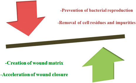
FIGURE 1. The principle aim of therapeutic strategies used in wound healing.
Current Wound Repair Regimes
Current therapeutic strategies leading to the acceleration of the wound healing process are depicted in Figure 2. Some of the current therapies include split-thickness autograft, autograft using donor keratinocytes, autograft using cultured epithelial cells or stem cells; wound dressings using chitosan, hyaluronic acid, collagen, and silicon, delivery of growth factors or platelet-rich plasma, and debridement. The privilege and constraints of these therapies are surveyed in Figure 2 (Han and Ceilley, 2017).
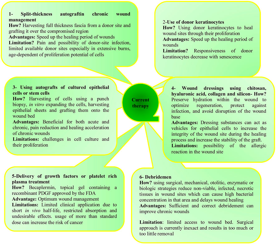
FIGURE 2. Some of the current therapies used in the healing of wounds along with their advantages and limitations.
Wound Healing Using Microorganisms
Although various activities of microbial products have been investigated (Salimi et al., 2018a; Salimi et al., 2018b; Salimi et al., 2019), their wound healing activities are less explored compared to their plant equivalent. Until now, healing activity of whole probiotic cells on burning, gastrointestinal, non-healing wounds, and scars has been proven. Wound healing activities of these microorganisms are related to their cell wall fragments, exopolysaccharide, antimicrobial, and anti-inflammation compounds, which can induce exceptional responses of the immune system in the skin and vitalize barrier functions of the skin (Figure 3) (Lew and Liong, 2013; Lukic et al., 2017; Shirzad et al., 2018).
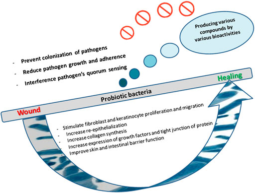
FIGURE 3. Various strategies elicited by probiotic bacteria that can facilitate the wound healing process.
Probiotic bacteria such as lactobacilli and bifidobacteria improve the wound healing process in the GI tract by activating the epithelial cells, stimulating proliferation and/or migration of fibroblast, increasing the synthesis of collagen, and affecting innate immune components of the intestinal barrier (Lew and Liong, 2013; Lukic et al., 2017). It has been shown that Lactobacillus reuteri can accelerate the wound-healing process via the up-regulation of the neuropeptide hormone oxytocin (Poutahidis et al., 2013).
Some researchers have been reported the wound healing activities of probiotics using a variety of experimental models like acetic acid-induced ulcers, full-thickness wounds, a hairless mouse model of UVB stimulated skin photo-aging and intestinal anastomoses. This promotion of the wound healing process by probiotics is attributed to the induction of β-defensin (Schlee et al., 2008) and expression of TGF-β (Dharmani et al., 2013), vascular endothelial growth factor (Dharmani et al., 2013), EGF, EGF receptor activity (EGFR), insulin-like growth factor (IGF) (Fordjour et al., 2010; Wang et al., 2014) and hypoxia-inducible factor 2α (HIF-2α) (Zhao et al., 2015). Also, probiotic bacteria improve tight barrier function in primary human keratinocytes through increasing the expression of tight junction protein in these cells, e.g., Lb. rhamnosus GG and Bifidobacterium longum increased tight junction function through the expression of claudin 1, zonula occludens 1, and occludin in keratinocytes infected with Staphylococcus aureus (Karczewski et al., 2010; Yang et al., 2015).
Probiotic bacteria such as Lb. rhamnosus GG and Lb. reuteri also enhances the re-epithelialization through the induction of chemokines or augmented keratinocyte migration and cellular proliferation. For instance, Lb. rhamnosus GG enhances the expression of the chemokine CXCL2 and its receptor CXCR2 that stimulates keratinocyte proliferation and migration during the normal process of wound healing (Mohammedsaeed et al., 2015).
Probiotic bacteria, via competitive exclusion and production of antibacterial compounds, can reduce the adherence and growth of pathogen ones, respectively (Prince et al., 2012). Hence some probiotic bacteria can prevent infections in cutaneous wounds, e.g., a combination of LAB and yeasts in kefir improved wound healing by producing antimicrobial compounds (Huseini et al., 2012). Reuterin (3- hydroxypropionaldehyde) is a well-known antimicrobial compound generated by Lb. reuteri that is supposed to impose its effect by oxidization of thiol groups in the target pathogens. Notably, reuterin can remarkably inhibit the growth of pathogenic gut bacteria, without affecting beneficial microbiota. Reuterin also exhibits antimicrobial activity on Staphylococcus as a common pathogen of chronic wounds (Schaefer et al., 2010). Lb. reuteri and Lb. rhamnosus GG inhibit the growth of S. aureus in infected keratinocytes through suppressing the primary adhesion of this pathogen to keratinocytes. By enhancing phagocytosis, Lb. plantarum prevented wound colonization by P. aeruginosa S. aureus, and S. epidermidis in a burn mouse model (Prince et al., 2012; Mohammedsaeed et al., 2015). The mechanism of IL-8 level regulating and modulating the entry and activity of PMNs migrating from peripheral blood to the ulcer enables the Lb. plantarum to inhibit the colonization of the pathogen (Peral et al., 2010).
Probiotic bacteria also disrupt the pathogenic agents through interfering with quorum sensing of pathogens usually found in chronic wounds). Especially, Lb. plantarum was supposed to inhibit the synthesis of QS signaling molecules (acyl-homoserine- lactone) by P. aeruginosa, together with the decline of biofilm (Valdez et al., 2005). Also, some probiotic bacteria produce several other metabolites including, hyaluronic acid, sphingomyelinase, lipoteichoic acid, alginate, diacetyl, and acetic acid, to stimulate the wound healing (Chong et al., 2005; Kogan et al., 2007). Hyaluronic acid acts as a matrix in mammalian to preserve the original structure of the epidermal layer against infections. Furthermore, hyaluronic acid affects the proliferation and differentiation of cells and immobilizes water in tissues (Weindl et al., 2004). Hyaluronic acid also accelerates the healing process via its antioxidant activity (Trabucchi et al., 2002). The considerable therapeutic activity of exogenous hyaluronic acid on wounds including, preserving moisture in wound sites, promoting migration of epithelial cells, regeneration, and remodeling processes, encourage its large scale production (Anilkumar et al., 2011). In this regard, microbial sources can be a suitable option for the production of hyaluronic acid. Additionally, microbial sources have lower undesired and interfering compounds such as proteins and nucleotides compared to animal sources. Streptococcus thermophiles, Streptococcus zooepidemicus, and Lactobacillus rhamnosus FTDC 8313, Lactobacillus gasseri FTDC 8131, and Pasteurella multocida are among known instance of hyaluronic acid-producing bacteria (Izawa et al., 2009; Liu et al., 2009; Lew et al., 2013). The recombinant hyaluronic acid has also been produced in genetic engineered Bacillus subtilis 168 and Lactococcus lactis LL-NAB and transformed Streptococcus thermophilus YIT2084 strains (Widner et al., 2005; Chien and Lee, 2007).
A family of ceramides and phosphorylcholine can be generated by the activity of sphingomyelinase from glucosylceramide and sphingomyelin precursors (Jensen et al., 2005). The action of this enzyme is critical for the skin barrier function (Choi and Maibach, 2003). Bacteria, yeast, and mammalian cells produce sphingomyelinase. Streptococcus thermophiles, lactobacillus, and bifidobacteria strains are microbial sources of sphingomyelinase. Since divalent metal ions can improve binding affinity between the sphingomyelinase and sphingomyelin, therefore, the production of sphingomyelinase from Streptococcus thermophiles can be enhanced via adding divalent metal ions into the growth culture (Di Marzio et al., 1999; Lew and Liong, 2012).
Lipoteichoic acid induces tolerances via protecting against the overproduction of proinflammatory cytokines. Lipoteichoic acid, through the induction of toll-like-receptor, stimulates skin defense against microbial threats, leading to a release of the antimicrobial peptides like β-defensins and cathelicidins. Lactobacillus plantarum, lactobacillus, and bifidobacterial strains are lipoteichoic acid-producing bacteria (Schauber and Gallo, 2008; Lew and Liong, 2012; Kim et al., 2013).
In addition to the mentioned compounds, bacteria such as Azotobacter vinelandii ATCC 9046 can accelerate wound healing by producing alginate with improved binding affinity compared to marine alginate (Fischer et al., 2017). Further, it was shown that EPS derived lactic acid bacteria prevented ultraviolet-induced skin damage in hairless mice (Morifuji et al., 2017).
Nanomaterial and Wound Healing
Many nano-based products have been introduced for their specific wound healing activity, and some of them are currently under clinical investigation. Nanomaterials with tissue regeneration ability have been developed with different structures including, nanoparticles, nanospheres, nanocapsules, nanoemulsions, nanocarriers, and nanocolloids (Naskar and Kim, 2020). The nanomaterials can be applied in two distinct principles. First, they can possess intrinsic wound healing properties including carbon-based nanoparticles (Table 1), metallic/metal oxide nanomaterials like silver, gold, copper, titanium, terbium, and zinc, nonmetallic nanomaterials such as graphene, and metalloid-based nanoparticles like silica (Table 2). Second, they can be used as carriers including, nanospheres, nanocapsules, nanoemulsions, nanocarriers, and nanocolloids, liposomes, micelles, vesicles, solid lipid nanoparticles, and nanofibers for therapeutic agents like various growth factors, cytokines, thrombin, nitric oxide (NO), antibiotics, angiogenic factors, opioids or even stem cells that can accelerate healing of chronic wounds (Figure 4) (Table 3) (Tran et al., 2009; Tocco et al., 2012; Nam et al., 2015; Urie et al., 2018).
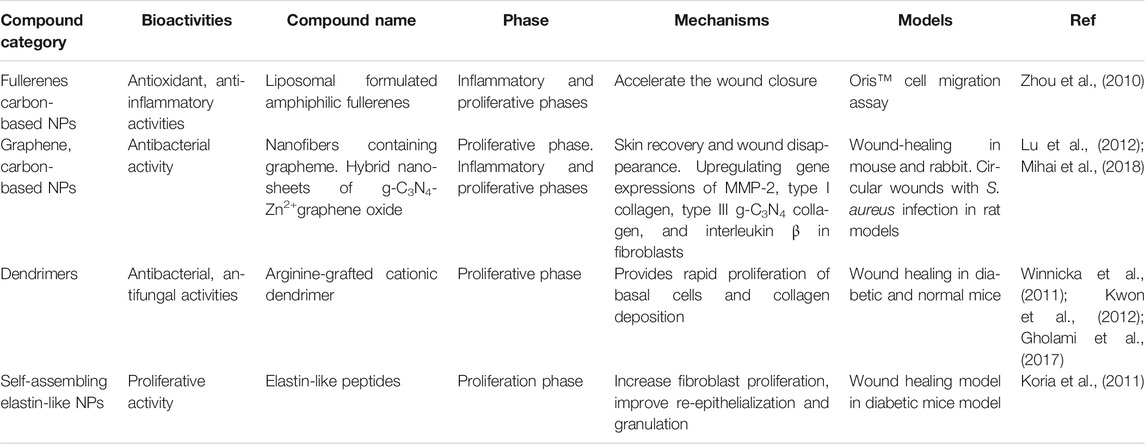
TABLE 1. Intrinsic wound healing activities of carbon-based nano-material.
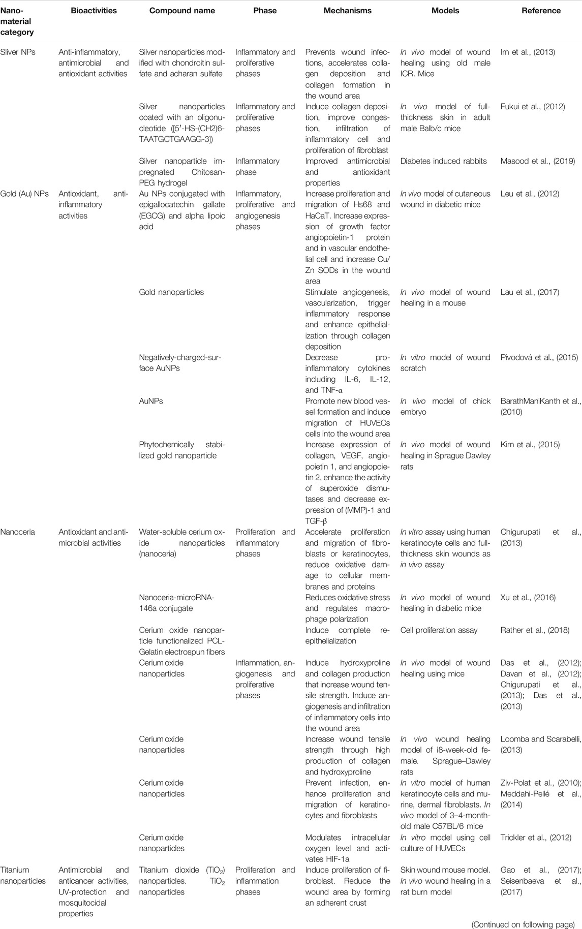
TABLE 2. Intrinsic wound healing activities of metal-based nano-products.
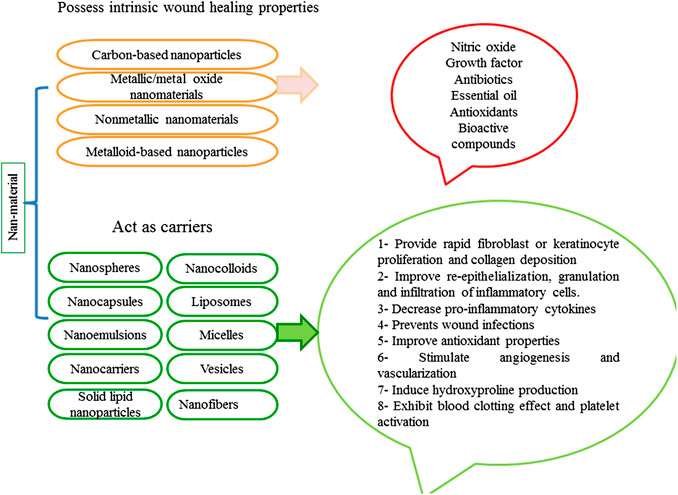
FIGURE 4. Various strategies elicited by nano-based material that can accelerate the wound healing process.
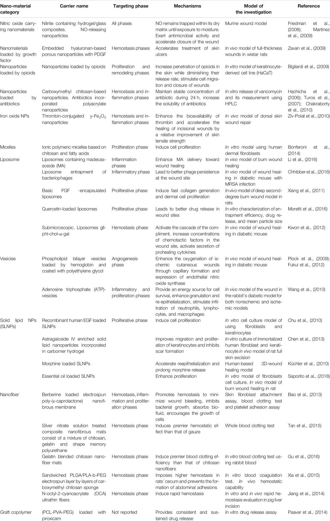
TABLE 3. The nano-products with wound healing activity via carrying nitric oxide, growth factors, thrombin, nitric oxide, antibiotics, angiogenic factors, and opioids.
These carriers control the release rate of therapeutic agents and increase their solubility, prolong their effect in the specified location and reduce the number of required doses, ultimately decrease the risk of development of antibiotic-resistant microorganisms through stabilizing protein structure and their biological activity, protecting proteins from inactivation by proteolytic enzymes in the wound site and regulating the drug release and increase their half-life (Tran et al., 2009; Tocco et al., 2012; Nam et al., 2015; Urie et al., 2018).
Nevertheless, in some cases, the property of chronic wound environments like being the high proteolytic, low frequency of growth factor receptors, and signaling molecules has been limited their application. Also, gene encapsulation using electro-spun nano-fibrous meshes in preparing wound-dressing materials has shown promising results. Using this approach, the expression of a target gene involved in regeneration can be enhanced or reduced. Nanofibres also can support cell adhesion, proliferation, and differentiation and provide sufficient oxygen and water. This potential is attributed to their permeability and prevention of bacterial infections in the wound site by excluding bacterial penetration. Polymeric nanomaterials like chitosan, cellulose, gelatin, dendrimers in different forms including, hydrogels, membranes, films, sponges, and scaffolds due to their antimicrobial, re-epithelialization, immune modulation, superior permeability, and being non-toxic characteristics have been applied as wound dressings or as delivery vectors to treat wounds. They can guarantee a moist wound environment via taking up a considerable amount of liquid (Mihai et al., 2019). The following are some of the studies that revealed wound healing properties of nano-based materials.
Antibacterial characteristics and low toxicity of metal nanoparticles like silver, gold, and zinc make them ideal options for integration in wound dressings like nanocoatings (Mihai et al., 2018). Reduced toxicity of AgNPs can be related to their increased surface-to-volume ratio and controlled release, which leads to their efficacy in a lower concentration. Wound healing activity of AgNPs can be related to their role in modulating the release of the anti-inflammatory cytokine, promoting wound closure and contractility, inducing the differentiation of myofibroblasts from normal fibroblasts, stimulating epidermal re-epithelialization and finally inhibiting bacterial pathogen growth. Generated sulfuric bonds with either microbial cell membrane proteins or thiol groups of various enzymes result in apoptosis of microbial cells. Applying AgNPs along with different antimicrobial drugs like tetracycline can considerably reduce bacterial contamination in tissue layers in in vivo model, so it promotes the healing process (Mihai et al., 2019). The following are some of the studies that revealed wound healing of AgNPs.
Lu et al., synthesized sponge-like nanoAg/ZnO-loaded chitosan composite dressing through the lyophilization process and subsequent incorporation of Ag/ZnO nanocomposites in synthesized sponge structure. The synthesized composite dressing exhibited enhanced blood clotting and antibacterial activity, promoted wound healing, re-epithelialization, and collagen deposition and showed very low toxicity (Lu et al., 2017). Khatami et al., reported that green synthesized Ag, ZnO, and Ag/ZnO via Prosophis fracta and coffee showed significant antibacterial activity against Acinetobacter baumannii and Pseudomonas aeruginosa, which may have the potential to apply in treating diabetic or burn wounds (Khatami et al., 2018). Yu et al., developed a new silkworm cocoon-based wound film wound dressing and, via reducing the ability of silk sericin incorporated Ag nanoparticles in synthesized film. This film promoted the healing process of infected wounds in New Zealand White rabbits and the reconstruction of the intact and thickened epidermis in impaired wound tissue during 14 days (Yu et al., 2017). Alipour et al. showed wound healing properties of silver nanoparticles embedded in electrospun nanofibers containing polyvinylalcohol (PVA), polyvinylpyrrolidone (PVP), pectin (PEC), and Mafenide acetate (MF). They showed the incorporation of silver nanoparticles into PVA/PVP/PEC/MF matrix had a remarkable effect on wound healing in New Zealand white rabbits (Alipour et al., 2019). Zhou et al. synthesized ultrafine silver/silver chloride anchored on reduced graphene oxide with stability and bactericidal activity. In vivo analysis showed that this nanocatalyst could accelerate the regeneration of the epidermis. Therefore has the potential to repair burn wounds (Zhou et al., 2016).
AuNPs can impose their healing activity though antioxidant or bactericidal activity, including targeting the bacterial cell wall and their DNA (Nethi et al., 2019). Akturk et al. showed wound healing ability of nanocomposite collagen scaffolds incorporating gold nanoparticles (AuNPs). Incorporation of AuNPs into cross-linked scaffolds enhanced their stability against enzymatic degradation and increased the tensile strength. The collagen sponge AuX group suppressed the inflammation. Neovascularization was also significant in collagen sponge AuX (Akturk et al., 2016). Li et al. synthesized chitosan incorporated Au-Ag NPs as wound dressing (CS-Au-Ag). The release of silver ions was occurred faster, in the higher amount, and a more durable manner in CS-Au-Ag in comparison to CS-Ag. Also, CS-Au-Ag exhibited increased antibacterial activity and low cytotoxicity. According to their results, CS-Au-Ag broadly promoted wound healing compared to CS-Ag (Li et al., 2017). Wang and coworkers prepared chitosan (CS) film modified with arginine (Arg) and gold NPs (AuNPs). The modification of Arg and AuNPs improved the hydrophilicity, mechanical strength, and antibacterial properties of the film, which in turn provided an enhanced ideal environment for cell adhesion and proliferation. The CS-Arg/AuNP dressing accelerated wound closure, re-epithelialization, and collagen deposition (Wang et al., 2020).
It has been revealed that zinc oxide nanoparticles exhibited a considerable antimicrobial activity. They can induce perforations in the bacterial cell membraneand accelerate the migration of keratinocyte, and consequently, re-epithelialization. In Khorasani et al. study it has been shown that polyvinyl (alcohol)/chitosan/nano zinc oxide nanocomposite with no toxicity and antibacterial activity can treat the wounds sufficiently (Khorasani et al., 2019). Ahmed et al. synthesized nanofiber mats composed of a combination of chitosan, polyvinyl alcohol (PVA), and Zinc oxide (ZnO). The results showed that chitosan/PVA/ZnO nanofibrous membranes had a more inhibitory effect on E. coli, P. aeruginosa, B. subtilis, and S. aureus than chitosan/PVA nanofibrous membranes. Also, chitosan/PVA/ZnO nanofibrous membranes imposed more antioxidant potential and wound healing activity than chitosan/PVA nanofibrous mats (Ahmed et al., 2018).
Copper, via promoting VEGF, upregulating expression of integrin, stabilizing extracellular matrix proteins, fibrinogen, and collagen formation imposes its role in the wound healing process. Tao et al., reported a composite hydrogel consists of methacrylate-modified gelatin (Gel-MA), and N,N-bis(acryloyl)-cystamine (BACA)-chelated Cu, which showed antimicrobial activity against Staphylococcus aureus and Escherichia coli. It also stimulated NIH-3T3 fibroblast proliferation and chronic wound healing process of the S. aureus-infected model (Tao et al., 2019).
Terbium hydroxide nanoparticles (TbNPs) also affected angiogenesis, viability, proliferation, and migration of endothelial cells. So they can accelerate wound healing (Nethi et al., 2019). In addition, silica has been used to generate more effective wound dressing materials to treat wounds. Since these nanoparticles with a positive charge are easily absorbed by the fibroblast cells, release silicic acid, and ultimately stimulate wound healing (Nethi et al., 2019). Nonmetallic inorganic nanoparticles like iodine nanoparticles showed significant inhibitory effects on bacterial growth and biofilm formation at very low concentrations and wound healing ability in in vivo model (Viswanathan et al., 2017).
Biocompounds Incorporated Into Nanomaterials for Wound Healing
Antibiotics are currently used to inhibit colonization and the growth of microbial pathogens in the wound site. However quick removement antimicrobial agents from the bloodstream and their degradation rate limit their efficiency. In this regard, local administration of antimicrobial agents including, Ciprofloxacin, silver sulfadiazine, tetracycline, gentamycin via antibiotics incorporated polymers is being applied. This approach provides a controlled release of antibiotics and can make an aseptic space at the wound site (Miguel et al., 2019; Fang et al., 2020). Liu and collaborators synthesized ciprofloxacin loaded electrospun hydrophobic poly (lactic-co-glycolic acid) (PLGA) fibrous mats modified with hydrophilic sodium alginate (ALG) microparticles. The results showed that ALG enhanced the ciprofloxacin release rate from the PLGA fibrous mats. Aso, ALG decreased the stiffness of PLGA fibrous mats to efficiently protect the wound site (Liu et al., 2018). Mahmoud and coworkers prepared norfloxacin-loaded scaffolds to treat wounds through mixing collagen and two different types of chitosan. It was observed that the tissue regeneration duration in the norfoloxacin-loaded collagen/chitosan scaffolds was faster than that of non-treated wounds in Albino rats. (Mahmoud and Salama, 2016). In another study, mesoporous silica MCM-41 was incorporated into carboxymethylcellulose hydrogel. Then Tetracycline and methylene blue were loaded to the prepared nanocomposite. This nanocomposite showed an improved in vitro water vapor, erosion, swelling, oxygen permeability, and antimicrobial activity (Namazi et al., 2016).
Various growth factors and cytokines including, platelet-derived growth factor (PDGF), fibroblast growth factor (FGF), epidermal growth factor (EGF), transforming growth factor-β (TGF- β), and vascular endothelial growth factor (VEGF), control wound healing phases like modulation of the inflammatory response, angiogenesis remodeling, and the reepithelialization processes. Several drawbacks including, little stability, removement through exudation, limited taking up via the skin, side effects of their high concentration in the administrated sites has been limited their topical application. So synthesizing growth factor loaded polymers can be considered as a promising approach in the wound healing process (Miguel et al., 2019). Lord and coworkers prepared plasmid DNA encoding perlecan domain I and VEGF189 loaded chitosan scaffolds. These scaffolds improved dermal wound healing in normal and diabetic rats. (Lord et al., 2017). In another study, thiolated heparin, and diacrylated poly (ethylene glycol) was prepared and loaded with human epidermal growth factor. A sustained release profile of hEGF was observed. Applying this hydrogel sheet improved wound closure in comparison with the non-treated control group (Goh et al., 2016).
Vitamins, especially Vit-A, C, and E, can improve the wound healing process. Enhancing the population of macrophages and monocytes at the wound site, stimulating the collagen synthesis, and re-epithelialization can be achieved via Vit-A. Vit-E also via antioxidant and anti-inflammatory properties, and the ability to accelerate the angiogenesis can improve the wound healing. In this regard, Voss et al. synthesized vitamin C (VitC) and/or propolis (Prop) loaded cellulose-based films. These films can control the release of vitamin C and possess antimicrobial ability against Escherichia coli and Staphylococcus aureus. Treated diabetic mice with the Cel-PVA/VitC/Prop treatment showed accelerated wound healing (Voss et al., 2018).
The inflammatory and hemostasis phases usually occur simultaneously to coincide losses of blood and fluid and eliminate dead tissues and prevent microbial contamination. In inflammation, monocytes, macrophages, and neutrophils act as wound cleaners via eliminating all dead cells, degraded extracellular matrix, and bacteria from the wound site. They also produce growth factors that attract other cells like smooth muscle cells and fibroblasts and into the injured area. Although extended inflammatory processes, chronic inflammation, generates excessive inflammatory mediators, cytotoxic enzymes, and free radical species, which postpone the physiological healing mechanisms and harm the surrounding tissue. So, the anti-inflammatory compounds incorporated wound dressings are considered as a promising approach to treat wounds (Miguel et al., 2019). Morgado et al. synthesized ibuprofen-β-cyclodextrins loaded poly (vinyl alcohol)/chitosan. According to their results, β-cyclodextrins provided a controlled drug release from the hydrogels that is the main property for applying them in wound management. Moreover, these hydrogels accelerate skin healing (Morgado et al., 2017).
Finally, many bioactive compounds like plant or microbial extracts and essential oils (Miguel et al., 2019), curcumin (Karri et al., 2016; Zahiri et al., 2020), propolis (Voss et al., 2018), and superoxide dismutase (Zhang et al., 2018) have been incorporated into a wound dressing to improve the rate of the healing process. Active agents in extracts including, terpenoids, terpenes, and aromatic and aliphatic compounds via antimicrobial, anti-inflammation, and antioxidative activities, can accelerate various phases of wound healing. Wound dressing can enhance the efficiency, bioavailability, stability, and solubility of these compounds (Miguel et al., 2019).
Toxicity of NANO-BASED Material for Wound Healing
It has been reported that AuNPs increase the growth rate and differentiation of keratinocytes, which in higher doses can cause cell toxicity. Also, applying ZnONPs in high concentrations can lead to mitochondrial in dysfunction of keratinocytes, which causing the release of lactate dehydrogenase. They also may produce radical species and prevent expression and consequently, production of superoxide dismutase and glutathione peroxidase genes in keratinocytes. These events can result in oxidative stress of cell membranes and cell apoptosis. Also, ZnONPs may associate with carcinogenic transformations (Yang et al., 2009).
Future Perspective
According to the considerable number of reported wound healing activity of nano and microbial-based substances, it is evident that some of these substances can be considered as future drug candidates. Despite the wound healing activities of introduced compounds via nanotechnology and microbiology, their application has some limitations and risks. Mainly, the small size of nanomaterial can lead to increased interparticle friction and sticking or raised chemical reactivity due to their increased surface area, which can result in undesired reactions like unwanted entering into the blood-brain barrier, initiation of blood coagulation, production of reactive oxygen species (Zhou et al., 2017). Generated oxidative stress can lead to biomolecules and subsequent severe cell damage (Yang et al., 2009).
It has been shown that PAMAM’s initiate uncontrolled autophagic cell death (De Jong and Borm, 2008; Li et al., 2015). Because of their small size, they may have a highly increased clearance rate that limits drug delivery. Also, in some conditions, nanomaterial can disintegrate or aggregate and lose their healing activities and become toxic substances (Singh and Nehru, 2008). Moreover, the chemical synthesis of NPs has some limitations like high costs, energy consumption, and producing poisonous by-products. So, green synthesis of nano-based materials using plants or microorganisms derived compounds as a non-polluting and cost-effective approach has gained popularity.
On the other hand chemical complexity of natural compounds, low yield of their production in crude extract, time of cost consuming process of their extraction, purification and identification, their incompatibility with high-throughput screening, high probability of rediscovery of known compounds; makes the discovery of novel microbial compounds and scales up of their industrial production rather challenging (Lam, 2007). Despite the mentioned constraints, the therapeutic potential of nano and microbial driven compounds is undeniable, and mainly their toxicity concern should be resolved to develop promising safe therapeutic strategies in the future.
Author Contributions
FS collected the data and drafted the manuscript. FM gave the outline and edited the paper.
Conflict of Interest
The authors declare that the research was conducted in the absence of any commercial or financial relationships that could be construed as a potential conflict of interest.
References
Ahmed, R., Tariq, M., Ali, I., Asghar, R., Khanam, P. N., Augustine, R., et al. (2018). Novel electrospun chitosan/polyvinyl alcohol/zinc oxide nanofibrous mats with antibacterial and antioxidant properties for diabetic wound healing. Int. J. Biol. Macromol. 120, 385–393. doi:10.1016/j.ijbiomac.2018.08.057
Akturk, O., Kismet, K., Yasti, A. C., Kuru, S., Duymus, M. E., Kaya, F., et al. (2016). Collagen/gold nanoparticle nanocomposites: a potential skin wound healing biomaterial. J. Biomater. Appl. 31 (2), 283–301. doi:10.1177/0885328216644536
Alipour, R., Khorshidi, A., Shojaei, A. F., Mashayekhi, F., and Moghaddam, M. J. M. (2019). Skin wound healing acceleration by Ag nanoparticles embedded in PVA/PVP/Pectin/Mafenide acetate composite nanofibers. Polym. Test. 79, 106022. doi:10.1016/j.polymertesting.2019.106022
Alonso, J. E., Lee, J., Burgess, A. R., and Browner, B. D. (1996). The management of complex orthopedic injuries. Surg. Clin. 76 (4), 879–903. doi:10.1016/s0039-6109(05)70486-2
Anilkumar, T., Muhamed, J., Jose, A., Jyothi, A., Mohanan, P., and Krishnan, L. K. (2011). Advantages of hyaluronic acid as a component of fibrin sheet for care of acute wound. Biologicals. 39 (2), 81–88. doi:10.1016/j.biologicals.2011.01.003
Auf Dem Keller, U., Kümin, A., Braun, S., and Werner, S. (2000). “Reactive oxygen species and their detoxification in healing skin wounds. J. Investig. Dermatol. Symp. Proc. 11, 106–111. doi:10.1038/sj.jidsymp.5650001
Augustine, R., Dominic, E. A., Reju, I., Kaimal, B., Kalarikkal, N., and Thomas, S. (2014). Electrospun polycaprolactone membranes incorporated with ZnO nanoparticles as skin substitutes with enhanced fibroblast proliferation and wound healing. RSC Adv. 4 (47), 24777–24785. doi:10.1039/C4RA02450H
Bao, J., Yang, B., Sun, Y., Zu, Y., and Deng, Y. (2013). A berberine-loaded electrospun poly-(ε-caprolactone) nanofibrous membrane with hemostatic potential and antimicrobial property for wound dressing. J. Biomed. Nanotechnol. 9 (7), 1173–1180. doi:10.1166/jbn.2013.1629
BarathManiKanth, S., Kalishwaralal, K., Sriram, M., Pandian, S. R. K., Youn, H.-s., Eom, S., et al. (2010). Anti-oxidant effect of gold nanoparticles restrains hyperglycemic conditions in diabetic mice. J. Nanobiotechnol. 8 (1), 16. doi:10.1186/1477-3155-8-16
Barui, A. K., Veeriah, V., Mukherjee, S., Manna, J., Patel, A. K., Patra, S., et al. (2012). Zinc oxide nanoflowers make new blood vessels. Nanoscale. 4 (24), 7861–7869. doi:10.1039/c2nr32369a
Bhasker, H., Udupa, S. L., and Udupa, A. L. (2004). Effect of nifedipine and amlodipine on wound healing in rats. Indian J. Physicol. Pharmacol. 48 (1), 111–114.
Bigliardi, P. L., Tobin, D. J., Gaveriaux‐Ruff, C., and Bigliardi‐Qi, M. (2009). Opioids and the skin–where do we stand?. Exp. Dermatol. 18 (5), 424–430. doi:10.1111/j.1600-0625.2009.00844.x
Bonferoni, M., Sandri, G., Dellera, E., Rossi, S., Ferrari, F., Mori, M., et al. (2014). Ionic polymeric micelles based on chitosan and fatty acids and intended for wound healing. Comparison of linoleic and oleic acid. Eur. J. Pharm. Biopharm. 87 (1), 101–106. doi:10.1016/j.ejpb.2013.12.018
Brown, G. L., Curtsinger, L. J., White, M., Mitchell, R. O., Pietsch, J., Nordquist, R., et al. (1988). Acceleration of tensile strength of incisions treated with EGF and TGF-beta. Ann. Surg. 208 (6), 788. doi:10.1097/00000658-198812000-00019
Campos, A. C., Groth, A. K., and Branco, A. B. (2008). Assessment and nutritional aspects of wound healing. Curr. Opin. Clin. Nutr. Metab. Care. 11 (3), 281–288. doi:10.1097/MCO.0b013e3282fbd35a
Chakraborty, S. P., Sahu, S. K., Mahapatra, S. K., Santra, S., Bal, M., Roy, S., et al. (2010). Nanoconjugated vancomycin: new opportunities for the development of anti-VRSA agents. Nanotechnology. 21 (10), 105103. doi:10.1088/0957-4484/21/10/105103
Chen, X., Peng, L.-H., Shan, Y.-H., Li, N., Wei, W., Yu, L., et al. (2013). Astragaloside IV-loaded nanoparticle-enriched hydrogel induces wound healing and anti-scar activity through topical delivery. Int. J. Pharm. 447 (1-2), 171–181. doi:10.1016/j.ijpharm.2013.02.054
Chhibber, S., Kaur, J., and Kaur, S. (2018). Liposome entrapment of bacteriophages improves wound healing in a diabetic mouse MRSA infection. Front. Microbiol. 9, 561. doi:10.3389/fmicb.2018.00561
Chien, L.-J., and Lee, C.-K. (2007). Hyaluronic acid production by recombinant Lactococcus lactis. Appl. Microbiol. Biotechnol. 77 (2), 339–346. doi:10.1007/s00253-007-1153-z
Chigurupati, S., Mughal, M. R., Okun, E., Das, S., Kumar, A., McCaffery, M., et al. (2013). Effects of cerium oxide nanoparticles on the growth of keratinocytes, fibroblasts and vascular endothelial cells in cutaneous wound healing. Biomaterials. 34 (9), 2194–2201. doi:10.1016/j.biomaterials.2012.11.061
Choi, M. J., and Maibach, H. I. (2003). Role of ceramides in skin stress: ultraviolet light, tape stripping and crowding. Exog. Dermatol. 2 (6), 286–294. doi:10.1159/000081565
Chong, B. F., Blank, L. M., Mclaughlin, R., and Nielsen, L. K. (2005). Microbial hyaluronic acid production. Appl. Microbiol. Biotechnol. 66 (4), 341–351. doi:10.1007/s00253-004-1774-4
Chu, Y., Yu, D., Wang, P., Xu, J., Li, D., and Ding, M. (2010). Nanotechnology promotes the full‐thickness diabetic wound healing effect of recombinant human epidermal growth factor in diabetic rats. Wound Repair and Regeneration. 18 (5), 499–505. doi:10.1111/j.1524-475X.2010.00612.x
Clark, R. A. F., and Henson, P. M. (2013). The molecular and cellular biology of wound repair. Berlin, Germany: Springer Science and Business Media. doi:10.1007/978-1-4615-1795-5
Das, S., Dowding, J. M., Klump, K. E., McGinnis, J. F., Self, W., and Seal, S. (2013). Cerium oxide nanoparticles: applications and prospects in nanomedicine. Nanomedicine. 8 (9), 1483–1508. doi:10.2217/nnm.13.133
Das, S., Singh, S., Dowding, J. M., Oommen, S., Kumar, A., Sayle, T. X., et al. (2012). The induction of angiogenesis by cerium oxide nanoparticles through the modulation of oxygen in intracellular environments. Biomaterials. 33 (31), 7746–7755. doi:10.1016/j.biomaterials.2012.07.019
Davan, R., Prasad, R., Jakka, V. S., Aparna, R., Phani, A., Jacob, B., et al. (2012). Cerium oxide nanoparticles promotes wound healing activity in in-vivo animal model. J. Bionanoscience. 6 (2), 78–83. doi:10.1016/j.biomaterials.2012.07.019
De Jong, W. H., and Borm, P. J. (2008). Drug delivery and nanoparticles: applications and hazards. Int. J. Nanomed. 3 (2), 133. doi:10.2147/ijn.s596
Degreef, H. J. (1998). How to heal a wound fast. Dermatol. Clin. 16 (2), 365–375. doi:10.1016/s0733-8635(05)70019-x
desJardins-Park, H. E., Foster, D. S., and Longaker, M. T. (2018). Fibroblasts and wound healing: an update. Regen Med. 13 (5), 491–495. doi:10.2217/rme-2018-0073
Dharmani, P., De Simone, C., and Chadee, K. (2013). The probiotic mixture VSL# 3 accelerates gastric ulcer healing by stimulating vascular endothelial growth factor. PLoS One. 8 (3), e58671. doi:10.1371/journal.pone.0058671
Di Marzio, L., Cinque, B., De Simone, C., and Cifone, M. G. (1999). Effect of the lactic acid BacteriumStreptococcus thermophilus on ceramide levels in human KeratinocytesIn vitro and stratum corneum in vivo. J. Invest. Dermatol. 113 (1), 98–106. doi:10.1046/j.1523-1747.1999.00633.x
Fang, Q., Yao, Z., Feng, L., Liu, T., Wei, S., Xu, P., et al. (2020). Antibiotic-loaded chitosan-gelatin scaffolds for infected seawater immersion wound healing. Int. J. Biol. Macromol. 159, 1140-1155. doi:10.1016/j.ijbiomac.2020.05.126
Fischer, M., Gebhard, F., Hammer, T., Zurek, C., Meurer, G., Marquardt, C., et al. (2017). Microbial alginate dressings show improved binding capacity for pathophysiological factors in chronic wounds compared to commercial alginate dressings of marine origin. J. Biomater. Appl. 31 (9), 1267–1276. doi:10.1177/0885328217702173
Folkman, J., and D’Amore, P. A. (1996). Blood vessel formation: what is its molecular basis?. Cell. 87 (7), 1153–1155. doi:10.1016/s0092-8674(00)81810-3
Fordjour, L., D’souza, A., Cai, C., Ahmad, A., Valencia, G., Kumar, D., et al. (2010). Comparative effects of probiotics, prebiotics, and synbiotics on growth factors in the large bowel in a rat model of formula-induced bowel inflammation. J. Pediatr. Gastroenterol. Nutr. 51 (4), 507–513. doi:10.1097/MPG.0b013e3181df5ff2
Friedman, A. J., Han, G., Navati, M. S., Chacko, M., Gunther, L., Alfieri, A., et al. (2008). Sustained release nitric oxide releasing nanoparticles: characterization of a novel delivery platform based on nitrite containing hydrogel/glass composites. Nitric Oxide. 19 (1), 12–20. doi:10.1016/j.niox.2008.04.003
Fukui, T., Kawaguchi, A. T., Takekoshi, S., Miyasaka, M., and Tanaka, R. (2012). Liposome‐encapsulated hemoglobin accelerates skin wound healing in mice. Artif. Organs. 36 (2), 161–169. doi:10.1111/j.1525-1594.2011.01371.x
Gao, Y., Han, Y., Cui, M., Tey, H. L., Wang, L., and Xu, C. (2017). ZnO nanoparticles as an antimicrobial tissue adhesive for skin wound closure. J. Mater. Chem. B. 5 (23), 4535–4541. doi:10.1039/c7tb00664k
Gholami, M., Mohammadi, R., Arzanlou, M., Dourbash, F. A., Kouhsari, E., Majidi, G., et al. (2017). In vitro antibacterial activity of poly (amidoamine)-G7 dendrimer. BMC Infectious Diseases. 17 (1), 395. doi:10.1186/s12879-017-2513-7
Goh, M., Hwang, Y., and Tae, G. (2016). Epidermal growth factor loaded heparin-based hydrogel sheet for skin wound healing. Carbohydr. Polym. 147, 251–260. doi:10.1016/j.carbpol.2016.03.072
Gonzalez, A. C. d. O., Costa, T. F., Andrade, Z. d. A., and Medrado, A. R. A. P. (2016). Wound healing-A literature review. An. Bras. Dermatol. 91 (5), 614–620. doi:10.1590/abd1806-4841.20164741
Gosain, A., and DiPietro, L. A. (2004). Aging and wound healing. World Journal of Surgery. 28 (3), 321–326. doi:10.1007/s00268-003-7397-6
Gu, B. K., Park, S. J., Kim, M. S., Lee, Y. J., Kim, J.-I., and Kim, C.-H. (2016). Gelatin blending and sonication of chitosan nanofiber mats produce synergistic effects on hemostatic functions. Int. J. Biol. Macromol. 82, 89–96. doi:10.1016/j.ijbiomac.2015.10.009
Gurtner, G. C., Werner, S., Barrandon, Y., and Longaker, M. T. (2008). Wound repair and regeneration. Nature. 453 (7193), 314–321. doi:10.1038/nature07039
Hachicha, W., Kodjikian, L., and Fessi, H. (2006). Preparation of vancomycin microparticles: importance of preparation parameters. Int. J. Pharm. 324 (2), 176–184. doi:10.1016/j.ijpharm.2006.06.005
Han, G., and Ceilley, R. (2017). Chronic wound healing: a review of current management and treatments. Adv. Ther. 34 (3), 599–610. doi:10.1007/s12325-017-0478-y
Hart, J. (2002). Inflammation 1: its role in the healing of acute wounds. J. Wound Care. 11 (6), 205–209. doi:10.12968/jowc.2002.11.6.26411
Heldin, C.-H., and Westermark, B. (1988). “Role of platelet-derived growth factor in vivo,” in The molecular and cellular biology of wound repair. Berlin, Germany: Springer), 249–273. doi:10.1007/978-1-4899-0185-9_7
Huseini, H. F., Rahimzadeh, G., Fazeli, M. R., Mehrazma, M., and Salehi, M. (2012). Evaluation of wound healing activities of kefir products. Burns. 38 (5), 719–723. doi:10.1016/j.burns.2011.12.005
Im, A.-R., Kim, J. Y., Kim, H.-S., Cho, S., Park, Y., and Kim, Y. S. (2013). Wound healing and antibacterial activities of chondroitin sulfate-and acharan sulfate-reduced silver nanoparticles. Nanotechnology. 24 (39), 395102. doi:10.1088/0957-4484/24/39/395102
Iruela-Arispe, M. L., and Dvorak, H. (1997). Angiogenesis: a dynamic balance of stimulators and inhibitors. Thromb. Haemostasis. 78 (1), 672–677. doi:10.1055/s-0038-1657610
Izawa, N., Hanamizu, T., Iizuka, R., Sone, T., Mizukoshi, H., Kimura, K., et al. (2009). Streptococcus thermophilus produces exopolysaccharides including hyaluronic acid. J. Biosci. Bioeng. 107 (2), 119–123. doi:10.1016/j.jbiosc.2008.11.007
Janis, J., and Attinger, C. (2006). The basic science of wound healing. Plast. Reconstr. Surg. 117 (7 Suppl. l), 12S–34S. doi:10.1097/01.prs.0000225430.42531.c2
Jensen, J. M., Förl, M., Winoto‐Morbach, S., Seite, S., Schunck, M., Proksch, E., et al. (2005). Acid and neutral sphingomyelinase, ceramide synthase, and acid ceramidase activities in cutaneous aging. Exp. Dermatol. 14 (8), 609–618. doi:10.1111/j.0906-6705.2005.00342.x
Jiang, K., Long, Y.-Z., Chen, Z.-J., Liu, S.-L., Huang, Y.-Y., Jiang, X., et al. (2014). Airflow-directed in situ electrospinning of a medical glue of cyanoacrylate for rapid hemostasis in liver resection. Nanoscale. 6 (14), 7792–7798. doi:10.1039/c4nr01412j
Jun, J.-I., and Lau, L. F. (2011). Taking aim at the extracellular matrix: CCN proteins as emerging therapeutic targets. Nat. Rev. Drug Discov. 10 (12), 945. doi:10.1038/nrd3599
Karczewski, J., Troost, F. J., Konings, I., Dekker, J., Kleerebezem, M., Brummer, R.-J. M., et al. (2010). Regulation of human epithelial tight junction proteins by Lactobacillus plantarum in vivo and protective effects on the epithelial barrier. Am. J. Physiol. Gastrointest. Liver Physiol. 298 (6), G851–G859. doi:10.1152/ajpgi.00327.2009
Karri, V. V. S. R., Kuppusamy, G., Talluri, S. V., Mannemala, S. S., Kollipara, R., Wadhwani, A. D., et al. (2016). Curcumin loaded chitosan nanoparticles impregnated into collagen-alginate scaffolds for diabetic wound healing. Int. J. Biol. Macromol. 93, 1519–1529. doi:10.1016/j.ijbiomac.2016.05.038
Kaushik, M., Niranjan, R., Thangam, R., Madhan, B., Pandiyarasan, V., Ramachandran, C., et al. (2019). Investigations on the antimicrobial activity and wound healing potential of ZnO nanoparticles. Appl. Surf. Sci. 479, 1169–1177. doi:10.1016/j.apsusc.2019.02.189
Khatami, M., Varma, R. S., Zafarnia, N., Yaghoobi, H., Sarani, M., and Kumar, V. G. (2018). Applications of green synthesized Ag, ZnO and Ag/ZnO nanoparticles for making clinical antimicrobial wound-healing bandages. Sustainable Chemistry and Pharmacy. 10, 9–15. doi:10.1016/j.scp.2018.08.001
Khorasani, M. T., Joorabloo, A., Adeli, H., Mansoori-Moghadam, Z., and Moghaddam, A. (2019). Design and optimization of process parameters of polyvinyl (alcohol)/chitosan/nano zinc oxide hydrogels as wound healing materials. Carbohydr. Polym. 207, 542–554. doi:10.1016/j.carbpol.2018.12.021
Kim, J. E., Lee, J., Jang, M., Kwak, M. H., Go, J., Kho, E. K., et al. (2015). Accelerated healing of cutaneous wounds using phytochemically stabilized gold nanoparticle deposited hydrocolloid membranes. Biomaterials science. 3 (3), 509–519. doi:10.1039/c4bm00390j
Kim, J. Y., Kim, H., Jung, B. J., Kim, N.-R., Park, J. E., and Chung, D. K. (2013). Lipoteichoic acid isolated from Lactobacillus plantarum suppresses LPS-mediated atherosclerotic plaque inflammation. Mol. Cell. 35 (2), 115–124. doi:10.1007/s10059-013-2190-3
Kiritsi, D., and Nyström, A. (2018). The role of TGFβ in wound healing pathologies. Mechanisms of ageing and development. 172, 51–58. doi:10.1016/j.mad.2017.11.004
Kogan, G., Šoltés, L., Stern, R., and Gemeiner, P. (2007). Hyaluronic acid: a natural biopolymer with a broad range of biomedical and industrial applications. Biotechnol. Lett. 29 (1), 17–25. doi:10.1007/s10529-006-9219-z
Koria, P., Yagi, H., Kitagawa, Y., Megeed, Z., Nahmias, Y., Sheridan, R., et al. (2011). Self-assembling elastin-like peptides growth factor chimeric nanoparticles for the treatment of chronic wounds. Proc. Natl. Acad. Sci. Unit. States Am. 108 (3), 1034–1039. doi:10.1073/pnas.1009881108
Küchler, S., Wolf, N. B., Heilmann, S., Weindl, G., Helfmann, J., Yahya, M. M., et al. (2010). 3D-wound healing model: influence of morphine and solid lipid nanoparticles. J. Biotechnol. 148 (1), 24–30. doi:10.1016/j.jbiotec.2010.01.001
Kumar, S., Lakshmanan, V.-K., Raj, M., Biswas, R., Hiroshi, T., Nair, S. V., et al. (2013). Evaluation of wound healing potential of β-chitin hydrogel/nano zinc oxide composite bandage. Pharmaceut. Res. 30 (2), 523–537. doi:10.1007/s11095-012-0898-y
Kwon, M. J., An, S., Choi, S., Nam, K., Jung, H. S., Yoon, C. S., et al. (2012). Effective healing of diabetic skin wounds by using nonviral gene therapy based on minicircle vascular endothelial growth factor DNA and a cationic dendrimer. J. Gene Med. 14 (4), 272–278. doi:10.1002/jgm.2618
Lam, K. S. (2007). New aspects of natural products in drug discovery. Trends Microbiol. 15 (6), 279–289. doi:10.1016/j.tim.2007.04.001
Lau, P., Bidin, N., Islam, S., Shukri, W. N. B. W. M., Zakaria, N., Musa, N., et al. (2017). Influence of gold nanoparticles on wound healing treatment in rat model: photobiomodulation therapy. Laser Surg. Med. 49 (4), 380–386. doi:10.1002/lsm.22614
Lazarus, G. S., Cooper, D. M., Knighton, D. R., Margolis, D. J., Percoraro, R. E., Rodeheaver, G., et al. (1994). Definitions and guidelines for assessment of wounds and evaluation of healing. Wound Repair Regen. 2 (3), 165–170. doi:10.1046/j.1524-475X.1994.20305.x
Leu, J.-G., Chen, S.-A., Chen, H.-M., Wu, W.-M., Hung, C.-F., Yao, Y.-D., et al. (2012). The effects of gold nanoparticles in wound healing with antioxidant epigallocatechin gallate and α-lipoic acid. Nanomed. Nanotechnol. Biol. Med. 8 (5), 767–775. doi:10.1016/j.nano.2011.08.013
Lew, L.-C., Gan, C.-Y., and Liong, M.-T. (2013). Dermal bioactives from lactobacilli and bifidobacteria. Ann. Microbiol. 63 (3), 1047–1055. doi:10.1007/s13213-012-0561-1
Lew, L. C., and Liong, M. T. (2013). Bioactives from probiotics for dermal health: functions and benefits. J. Appl. Microbiol. 114 (5), 1241–1253. doi:10.1111/jam.12137
Lew, L., and Liong, M. (2012). Divalent ions of Mn and Mg improved sphingomyelinase activity of Lactobacillus rhamnosus FTDC 8313. Biosci. Biotechnol. Biochem.
Li, J., Chen, J., and Kirsner, R. (2007). Pathophysiology of acute wound healing. Clin. Dermatol. 25 (1), 9–18. doi:10.1016/j.clindermatol.2006.09.007
Li, Q., Lu, F., Zhou, G., Yu, K., Lu, B., Xiao, Y., et al. (2017). Silver inlaid with gold nanoparticle/chitosan wound dressing enhances antibacterial activity and porosity, and promotes wound healing. Biomacromolecules. 18 (11), 3766–3775. doi:10.1021/acs.biomac.7b01180
Li, Y., Zhu, H., Wang, S., Qian, X., Fan, J., Wang, Z., et al. (2015). Interplay of oxidative stress and autophagy in PAMAM dendrimers-induced neuronal cell death. Theranostics. 5 (12), 1363. doi:10.7150/thno.13181
Li, Z., Liu, M., Wang, H., and Du, S. (2016). Increased cutaneous wound healing effect of biodegradable liposomes containing madecassoside: preparation optimization, in vitro dermal permeation, and in vivo bioevaluation. Int. J. Nanomed. 11, 2995. doi:10.2147/IJN.S105035
Liu, L., Du, G., Chen, J., Zhu, Y., Wang, M., and Sun, J. (2009). Microbial production of low molecular weight hyaluronic acid by adding hydrogen peroxide and ascorbate in batch culture of Streptococcus zooepidemicus. Bioresour. Technol. 100 (1), 362–367. doi:10.1016/j.biortech.2008.05.040
Liu, X., Nielsen, L. H., Kłodzińska, S. N., Nielsen, H. M., Qu, H., Christensen, L. P., et al. (2018). Ciprofloxacin-loaded sodium alginate/poly (lactic-co-glycolic acid) electrospun fibrous mats for wound healing. Eur. J. Pharm. Biopharm. 123, 42–49. doi:10.1016/j.ejpb.2017.11.004
Loomba, L., and Scarabelli, T. (2013). Metallic nanoparticles and their medicinal potential. Part II: aluminosilicates, nanobiomagnets, quantum dots and cochleates. Ther. Deliv. 4 (9), 1179–1196. doi:10.4155/tde.13.74
Lord, M. S., Ellis, A. L., Farrugia, B. L., Whitelock, J. M., Grenett, H., Li, C., et al. (2017). Perlecan and vascular endothelial growth factor-encoding DNA-loaded chitosan scaffolds promote angiogenesis and wound healing. J. Contr. Release. 250, 48–61. doi:10.1016/j.jconrel.2017.02.009
Lu, B., Li, T., Zhao, H., Li, X., Gao, C., Zhang, S., et al. (2012). Graphene-based composite materials beneficial to wound healing. Nanoscale. 4 (9), 2978–2982. doi:10.1039/c2nr11958g
Lu, Z., Gao, J., He, Q., Wu, J., Liang, D., Yang, H., et al. (2017). Enhanced antibacterial and wound healing activities of microporous chitosan-Ag/ZnO composite dressing. Carbohydr. Polym. 156, 460–469. doi:10.1016/j.carbpol.2016.09.051
Lukic, J., Chen, V., Strahinic, I., Begovic, J., Lev‐Tov, H., Davis, S. C., et al. (2017). Probiotics or pro‐healers: the role of beneficial bacteria in tissue repair. Wound Repair Regen. 25 (6), 912–922. doi:10.1111/wrr.12607
Mahmoud, A. A., and Salama, A. H. (2016). Norfloxacin-loaded collagen/chitosan scaffolds for skin reconstruction: preparation, evaluation and in-vivo wound healing assessment. Eur. J. Pharmaceut. Sci. 83, 155–165. doi:10.1016/j.ejps.2015.12.026
Majewska, I., and Gendaszewska-Darmach, E. (2011). Proangiogenic activity of plant extracts in accelerating wound healing-a new face of old phytomedicines. Acta Biochimica Polonica. 58 (4), 449. doi:10.18388/abp.2011_2210
Martinez, L. R., Han, G., Chacko, M., Mihu, M. R., Jacobson, M., Gialanella, P., et al. (2009). Antimicrobial and healing efficacy of sustained release nitric oxide nanoparticles against Staphylococcus aureus skin infection. J. Invest. Dermatol. 129 (10), 2463–2469. doi:10.1038/jid.2009.95
Masood, N., Ahmed, R., Tariq, M., Ahmed, Z., Masoud, M. S., Ali, I., et al. (2019). Silver nanoparticle impregnated chitosan-PEG hydrogel enhances wound healing in diabetes induced rabbits. Int. J. Pharm. 559, 23–36. doi:10.1016/j.ijpharm.2019.01.019
Meddahi-Pellé, A., Legrand, A., Marcellan, A., Louedec, L., Letourneur, D., and Leibler, L. (2014). Organ repair, hemostasis, and in vivo bonding of medical devices by aqueous solutions of nanoparticles. Angew. Chem. Int. Ed. 53 (25), 6369–6373. doi:10.1002/anie.201401043
Meszaros, A. J., Reichner, J. S., and Albina, J. E. (2000). Macrophage-induced neutrophil apoptosis. J. Immunol. 165 (1), 435–441. doi:10.4049/jimmunol.165.1.435
Miguel, S. P., Sequeira, R. S., Moreira, A. F., Cabral, C. S., Mendonça, A. G., Ferreira, P., et al. (2019). An overview of electrospun membranes loaded with bioactive molecules for improving the wound healing process. Eur. J. Pharm. Biopharm. 139, 1–22. doi:10.1016/j.ejpb.2019.03.010
Mihai, M. M., Dima, M. B., Dima, B., and Holban, A. M. (2019). Nanomaterials for wound healing and infection control. Materials. 12 (13), 2176. doi:10.3390/ma12132176
Mihai, M. M., Preda, M., Lungu, I., Gestal, M. C., Popa, M. I., and Holban, A. M. (2018). Nanocoatings for chronic wound repair—modulation of microbial colonization and biofilm formation. Int. J. Mol. Sci. 19 (4), 1179. doi:10.3390/ijms19041179
Mir, M., Ali, M. N., Barakullah, A., Gulzar, A., Arshad, M., Fatima, S., et al. (2018). Synthetic polymeric biomaterials for wound healing: a review. Progress in biomaterials. 7 (1), 1–21. doi:10.1007/s40204-018-0083-4
Mohammedsaeed, W., Cruickshank, S., McBain, A. J., and O’Neill, C. A. (2015). Lactobacillus rhamnosus GG lysate increases re-epithelialization of keratinocyte scratch assays by promoting migration. Sci. Rep. 5, 16147. doi:10.1038/srep16147
Moretti, E., Mazzi, L., Bonechi, C., Salvatici, M. C., Iacoponi, F., Rossi, C., et al. (2016). Effect of quercetin-loaded liposomes on induced oxidative stress in human spermatozoa. Reprod. Toxicol. 60, 140–147. doi:10.1016/j.reprotox.2016.02.012
Morgado, P. I., Miguel, S. P., Correia, I. J., and Aguiar-Ricardo, A. (2017). Ibuprofen loaded PVA/chitosan membranes: a highly efficient strategy towards an improved skin wound healing. Carbohydr. Polym. 159, 136–145. doi:10.1016/j.carbpol.2016.12.029
Morifuji, M., Kitade, M., Fukasawa, T., Yamaji, T., and Ichihashi, M. (2017). Exopolysaccharides isolated from milk fermented with lactic acid bacteria prevent ultraviolet-induced skin damage in hairless mice. Int. J. Mol. Sci. 18 (1), 146. doi:10.3390/ijms18010146
Mosser, D. M., and Edwards, J. P. (2008). Exploring the full spectrum of macrophage activation. Nat. Rev. Immunol. 8 (12), 958. doi:10.1038/nri2448
Nam, G., Rangasamy, S., Purushothaman, B., and Song, J. M. (2015). The application of bactericidal silver nanoparticles in wound treatment. Nanomater. Nanotechnol. 5, 5–23. doi:10.5772/60918
Namazi, H., Rakhshaei, R., Hamishehkar, H., and Kafil, H. S. (2016). Antibiotic loaded carboxymethylcellulose/MCM-41 nanocomposite hydrogel films as potential wound dressing. Int. J. Biol. Macromol. 85, 327–334. doi:10.1016/j.ijbiomac.2015.12.076
Naskar, A., and Kim, K.-s. (2020). Recent advances in nanomaterial-based wound-healing therapeutics. Pharmaceutics. 12 (6), 499. doi:10.3390/pharmaceutics12060499
Natarajan, S., Williamson, D., Stiltz, A. J., and Harding, K. (2000). Advances in wound care and healing technology. Am. J. Clin. Dermatol. 1 (5), 269–275. doi:10.2165/00128071-200001050-00002
Nethi, S. K., Das, S., Patra, C. R., and Mukherjee, S. (2019). Recent advances in inorganic nanomaterials for wound-healing applications. Biomaterials science. 7 (7), 2652–2674. doi:10.1039/c9bm00423h
Paaver, U., Tamm, I., Laidmäe, I., Lust, A., Kirsimäe, K., Veski, P., et al. (2014). Soluplus graft copolymer: potential novel carrier polymer in electrospinning of nanofibrous drug delivery systems for wound therapy. BioMed Res. Int. 2014, 789765. doi:10.1155/2014/789765
Peral, M., Rachid, M., Gobbato, N., Martinez, M. H., and Valdez, J. (2010). Interleukin-8 production by polymorphonuclear leukocytes from patients with chronic infected leg ulcers treated with Lactobacillus plantarum. Clin. Microbiol. Infect. 16 (3), 281–286. doi:10.1111/j.1469-0691.2009.02793.x
Pivodová, V., Franková, J., Galandáková, A., and Ulrichová, J. (2015). In vitro AuNPs’ cytotoxicity and their effect on wound healing. Nanobiomedicine. 2, 7. doi:10.5772/61132
Plock, J. A., Rafatmehr, N., Sinovcic, D., Schnider, J., Sakai, H., Tsuchida, E., et al. (2009). Hemoglobin vesicles improve wound healing and tissue survival in critically ischemic skin in mice. Am. J. Physiol. Heart Circ. Physiol. 297 (3), H905–H910. doi:10.1152/ajpheart.00430.2009
Poutahidis, T., Kearney, S. M., Levkovich, T., Qi, P., Varian, B. J., Lakritz, J. R., et al. (2013). Microbial symbionts accelerate wound healing via the neuropeptide hormone oxytocin. PLoS One. 8 (10), e78898. doi:10.1371/journal.pone.0078898
Prince, T., McBain, A. J., and O’Neill, C. A. (2012). Lactobacillus reuteri protects epidermal keratinocytes from Staphylococcus aureus induced cell death by competitive exclusion, Appl. Environ. Microbiol. 78 (15), 5119-5126. doi:10.1128/AEM.00595-12
Raguvaran, R., Manuja, B. K., Chopra, M., Thakur, R., Anand, T., Kalia, A., et al. (2017). Sodium alginate and gum acacia hydrogels of ZnO nanoparticles show wound healing effect on fibroblast cells. Int. J. Biol. Macromol. 96, 185–191. doi:10.1016/j.ijbiomac.2016.12.009
Rather, H. A., Thakore, R., Singh, R., Jhala, D., Singh, S., and Vasita, R. (2018). Antioxidative study of cerium oxide nanoparticle functionalised PCL-Gelatin electrospun fibers for wound healing application. Bioactive materials. 3 (2), 201–211. doi:10.1016/j.bioactmat.2017.09.006
Robson, M. C., Steed, D. L., and Franz, M. G. (2001). Wound healing: biologic features and approaches to maximize healing trajectories. Curr. Probl. Surg. 38 (2), 72–140. doi:10.1067/msg.2001.111167
Rodriguez‐Merchan, E. (2012). Surgical wound healing in bleeding disorders. Haemophilia. 18 (4), 487–490. doi:10.1111/j.1365-2516.2012.02760.x
Salimi, F., Hamedi, J., Motevaseli, E., and Mohammadipanah, F. (2019). Coexistence of anticoagulant and anti-vascular calcification activities in Kribbella sp. UTMC 267 metabolites. Iran. J. Pharm. Res. 18 (1), 459–468.
Salimi, F., Hamedi, J., Motevaseli, E., and Mohammadipanah, F. (2018a). Isolation and screening of rare Actinobacteria, a new insight for finding natural products with antivascular calcification activity. J. Appl. Microbiol. 124 (1), 254–266. doi:10.1111/jam.13605
Salimi, F., Jafari‐Nodooshan, S., Zohourian, N., Kolivand, S., and Hamedi, J. (2018b). Simultaneous anti‐diabetic and anti‐vascular calcification activity of Nocardia sp. UTMC 751. Lett. Appl. Microbiol. 66 (2), 110–117. doi:10.1111/lam.12833
Saporito, F., Sandri, G., Bonferoni, M. C., Rossi, S., Boselli, C., Cornaglia, A. I., et al. (2018). Essential oil-loaded lipid nanoparticles for wound healing. Int. J. Nanomed. 13, 175. doi:10.2147/IJN.S152529
Schaefer, L., Auchtung, T. A., Hermans, K. E., Whitehead, D., Borhan, B., and Britton, R. A. (2010). The antimicrobial compound reuterin (3-hydroxypropionaldehyde) induces oxidative stress via interaction with thiol groups. Microbiology. 156 (6), 1589–1599. doi:10.1099/mic.0.035642-0
Schauber, J., and Gallo, R. L. (2008). Antimicrobial peptides and the skin immune defense system. J. Allergy Clin. Immunol. 122 (2), 261–266. doi:10.1016/j.jaci.2008.03.027
Schlee, M., Harder, J., Köten, B., Stange, E., Wehkamp, J., and Fellermann, K. (2008). Probiotic lactobacilli and VSL# 3 induce enterocyte β‐defensin 2. Clin. Exp. Immunol. 151 (3), 528–535. doi:10.1111/j.1365-2249.2007.03587.x
Seisenbaeva, G. A., Fromell, K., Vinogradov, V. V., Terekhov, A. N., Pakhomov, A. V., Nilsson, B., et al. (2017). Dispersion of TiO 2 nanoparticles improves burn wound healing and tissue regeneration through specific interaction with blood serum proteins. Sci. Rep. 7 (1), 15448. doi:10.1038/s41598-017-15792-w
Shirzad, M., Hamedi, J., Motevaseli, E., and Modarressi, M. H. (2018). Anti-elastase and anti-collagenase potential of Lactobacilli exopolysaccharides on human fibroblast. Artificial cells, nanomedicine, and biotechnology. 46 (Suppl. 1), 1051–1061. doi:10.1080/21691401.2018.1443274
Singh, O. P., and Nehru, R. (2008). Nanotechnology and cancer treatment. Asian J. Exp. Sci. 22 (2), 6. doi:10.2147/IJN.S33838
Sinno, H., and Prakash, S. (2013). Complements and the wound healing cascade: an updated review. Plastic surgery international, 2013, 146764. doi:10.1155/2013/146764
Skover, G. (1991). Cellular and biochemical dynamics of wound repair, wound environment in collagen regeneration. Clin. Podiatr. Med. Surg. 8, 723–756.
Strecker-McGraw, M. K., Jones, T. R., and Baer, D. G. (2007). Soft tissue wounds and principles of healing. Emerg. Med. Clin. 25 (1), 1–22. doi:10.1016/j.emc.2006.12.002
Szycher, M., and Lee, S. J. (1992). Modern wound dressings: a systematic approach to wound healing. J. Biomater. Appl. 7 (2), 142–213. doi:10.1177/088532829200700204
Tan, L., Hu, J., Huang, H., Han, J., and Hu, H. (2015). Study of multi-functional electrospun composite nanofibrous mats for smart wound healing. Int. J. Biol. Macromol. 79, 469–476. doi:10.1016/j.ijbiomac.2015.05.014
Tao, B., Lin, C., Deng, Y., Yuan, Z., Shen, X., Chen, M., et al. (2019). Copper-nanoparticle-embedded hydrogel for killing bacteria and promoting wound healing with photothermal therapy. J. Mater. Chem. B. 7 (15), 2534–2548. doi:10.1039/c8tb03272f
Tocco, I., Zavan, B., Bassetto, F., and Vindigni, V. (2012). Nanotechnology-based therapies for skin wound regeneration. J. Nanomater. 2012, 1687-4110. doi:10.1155/2012/714134
Toy, L. (2005). Matrix metalloproteinases: their function in tissue repair. J. Wound Care. 14 (1), 20–22. doi:10.12968/jowc.2005.14.1.26720
Trabucchi, E., Pallotta, S., Morini, M., Corsi, F., Franceschini, R., Casiraghi, A., et al. (2002). Low molecular weight hyaluronic acid prevents oxygen free radical damage to granulation tissue during wound healing. Int. J. Tissue React. 24 (2), 65–71.
Tran, P. A., Zhang, L., and Webster, T. J. (2009). Carbon nanofibers and carbon nanotubes in regenerative medicine. Adv. Drug Deliv. Rev. 61 (12), 1097–1114. doi:10.1016/j.addr.2009.07.010
Trickler, W. J., Lantz, S. M., Schrand, A. M., Robinson, B. L., Newport, G. D., Schlager, J. J., et al. (2012). Effects of copper nanoparticles on rat cerebral microvessel endothelial cells. Nanomedicine. 7 (6), 835–846. doi:10.2217/nnm.11.154
Tsala, D. E., Amadou, D., and Habtemariam, S. (2013). Natural wound healing and bioactive natural products. Phytopharmacology. 4 (3), 532–560.
Turos, E., Shim, J.-Y., Wang, Y., Greenhalgh, K., Reddy, G. S. K., Dickey, S., et al. (2007). Antibiotic-conjugated polyacrylate nanoparticles: new opportunities for development of anti-MRSA agents. Bioorg. Med. Chem. Lett. 17 (1), 53–56. doi:10.1016/j.bmcl.2006.09.098
Urie, R., Ghosh, D., Ridha, I., and Rege, K. (2018). Inorganic nanomaterials for soft tissue repair and regeneration. Annu. Rev. Biomed. Eng. 20, 353–374. doi:10.1146/annurev-bioeng-071516-044457
Valdez, J., Peral, M., Rachid, M., Santana, M., and Perdigon, G. (2005). Interference of Lactobacillus plantarum with Pseudomonas aeruginosa in vitro and in infected burns: the potential use of probiotics in wound treatment. Clinical microbiology and infection. 11 (6), 472–479. doi:10.1111/j.1469-0691.2005.01142.x
Vanwijck, R. (2001). Surgical biology of wound healing. Bull. Mem. Acad. R. Med. Belg. 156 (3–4), 175–184; discussion 185.
Viswanathan, K., Babu, D. B., Jayakumar, G., and Raj, G. D. (2017). Anti-microbial and skin wound dressing application of molecular iodine nanoparticles. Mater. Res. Express. 4 (10), 104003. doi:10.1088/2053-1591/aa91e5
Voss, G. T., Gularte, M. S., Vogt, A. G., Giongo, J. L., Vaucher, R. A., Echenique, J. V., et al. (2018). Polysaccharide-based film loaded with vitamin C and propolis: a promising device to accelerate diabetic wound healing. Int. J. Pharm. 552 (1–2), 340–351. doi:10.1016/j.ijpharm.2018.10.009
Wang, J., Wan, R., Mo, Y., Li, M., Zhang, Q., and Chien, S. (2010). Intracellular delivery of adenosine triphosphate enhanced healing process in full-thickness skin wounds in diabetic rabbits. Am. J. Surg. 199 (6), 823–832. doi:10.1016/j.amjsurg.2009.05.040
Wang, K., Qi, Z., Pan, S., Zheng, S., Wang, H., Chang, Y., et al. (2020). Preparation, characterization and evaluation of a new film based on chitosan, arginine and gold nanoparticle derivatives for wound-healing efficacy. RSC Adv. 10 (35), 20886–20899. doi:10.1039/D0RA03704D
Wang, L., Cao, H., Liu, L., Wang, B., Walker, W. A., Acra, S. A., et al. (2014). Activation of EGF receptor mediates mucin production stimulated by p40, a Lactobacillus rhamnosus GG-derived protein. J. Biol. Chem, 289, 20234. doi:10.1074/jbc.M114.553800
Weindl, G., Schaller, M., Schäfer-Korting, M., and Korting, H. (2004). Hyaluronic acid in the treatment and prevention of skin diseases: molecular biological, pharmaceutical and clinical aspects. Skin Pharmacol. Physiol. 17 (5), 207–213. doi:10.1159/000080213
Widner, B., Behr, R., Von Dollen, S., Tang, M., Heu, T., Sloma, A., et al. (2005). Hyaluronic acid production in Bacillus subtilis. Appl. Environ. Microbiol. 71 (7), 3747–3752. doi:10.1128/AEM.71.7.3747-3752.2005
Winnicka, K., Sosnowska, K., Wieczorek, P., Sacha, P. T., and Tryniszewska, E. (2011). Poly (amidoamine) dendrimers increase antifungal activity of clotrimazole. Biol. Pharm. Bull. 34 (7), 1129–1133. doi:10.1248/bpb.34.1129
Xia, Q., Liu, Z., Wang, C., Zhang, Z., Xu, S., and Han, C. C. (2015). A biodegradable trilayered barrier membrane composed of sponge and electrospun layers: hemostasis and antiadhesion. Biomacromolecules. 16 (9), 3083–3092. doi:10.1021/acs.biomac.5b01099
Xiang, Q., Xiao, J., Zhang, H., Zhang, X., Lu, M., Zhang, H., et al. (2011). Preparation and characterisation of bFGF-encapsulated liposomes and evaluation of wound-healing activities in the rat. Burns. 37 (5), 886–895. doi:10.1016/j.burns.2011.01.018
Xu, J., Zgheib, C., Hodges, M. M., Hu, J., El Kasmi, K. C., Das, S. S., et al. (2016). Nanoceria-MicroRNA-146a conjugate improves wound healing by reducing reactive oxygen species and regulating macrophage polarization. J. Am. Coll. Surg. 223 (4), e157. doi:10.1016/j.jamcollsurg.2016.08.400
Yang, F., Wang, A., Zeng, X., Hou, C., Liu, H., and Qiao, S. (2015). Lactobacillus reuteri I5007 modulates tight junction protein expression in IPEC-J2 cells with LPS stimulation and in newborn piglets under normal conditions. BMC Microbiology. 15 (1), 32. doi:10.1186/s12866-015-0372-1
Yang, H., Liu, C., Yang, D., Zhang, H., and Xi, Z. (2009). Comparative study of cytotoxicity, oxidative stress and genotoxicity induced by four typical nanomaterials: the role of particle size, shape and composition. J. Appl. Toxicol. 29 (1), 69–78. doi:10.1002/jat.1385
Yu, K., Lu, F., Li, Q., Chen, H., Lu, B., Liu, J., et al. (2017). In situ assembly of Ag nanoparticles (AgNPs) on porous silkworm cocoon-based wound film: enhanced antimicrobial and wound healing activity. Sci. Rep. 7 (1), 1–13. doi:10.1038/s41598-017-02270-6
Zahiri, M., Khanmohammadi, M., Goodarzi, A., Ababzadeh, S., Farahani, M. S., Mohandesnezhad, S., et al. (2020). Encapsulation of curcumin loaded chitosan nanoparticle within poly (ε-caprolactone) and gelatin fiber mat for wound healing and layered dermal reconstitution. Int. J. Biol. Macromol. 153, 1241–1250. doi:10.1016/j.ijbiomac.2019.10.255
Zavan, B., Vindigni, V., Vezzù, K., Zorzato, G., Luni, C., Abatangelo, G., et al. (2009). Hyaluronan based porous nano-particles enriched with growth factors for the treatment of ulcers: a placebo-controlled study. J. Mater. Sci. Mater. Med. 20 (1), 235–247. doi:10.1007/s10856-008-3566-3
Zhang, L., Ma, Y., Pan, X., Chen, S., Zhuang, H., and Wang, S. (2018). A composite hydrogel of chitosan/heparin/poly (γ-glutamic acid) loaded with superoxide dismutase for wound healing. Carbohydr. Polym. 180, 168–174. doi:10.1016/j.carbpol.2017.10.036
Zhao, C., Fu, Q., Song, W., Zhang, D., Ahati, J., Pan, X., et al. (2015). Calcifying cyanobacterium (Nostoc calcicola) reactor as a promising way to remove cadmium from water. Ecol. Eng. 81, 107–114. doi:10.1016/j.ecoleng.2015.04.012
Zhou, Y., Chen, R., He, T., Xu, K., Du, D., Zhao, N., et al. (2016). Biomedical potential of ultrafine Ag/AgCl nanoparticles coated on graphene with special reference to antimicrobial performances and burn wound healing. ACS Appl. Mater. Interfaces. 8 (24), 15067–15075. doi:10.1021/acsami.6b03021
Zhou, Y., Peng, Z., Seven, E. S., and Leblanc, R. M. (2017). Crossing the blood-brain barrier with nanoparticles. J. Contr. Release. 270, 290–303. doi:10.1016/j.jconrel.2017.12.015
Zhou, Z., Joslin, S., Dellinger, A., Ehrich, M., Brooks, B., Ren, Q., et al. (2010). A novel class of compounds with cutaneous wound healing properties. J. Biomed. Nanotechnol. 6 (5), 605–611. doi:10.1166/jbn.2010.1157
Keywords: chronic wounds, skin regeneration, wound healing mechanisms, nanomaterials, microbial metabolites
Citation: Salimi F and Mohammadipanah F (2021) Nanomaterials Versus The Microbial Compounds With Wound Healing Property. Front. Nanotechnol. 2:584489. doi: 10.3389/fnano.2020.584489
Received: 21 July 2020; Accepted: 14 December 2020;
Published: 28 January 2021.
Edited by:
Yogendra Kumar Mishra, University of Southern Denmark, DenmarkReviewed by:
Mubarak A Mujawar, Florida International University, United StatesYiqun Zhou, University of Miami, United States
Alina Maria Holban, University of Bucharest, Romania
Copyright © 2021 Salimi and Mohammadipanah\. This is an open-access article distributed under the terms of the Creative Commons Attribution License (CC BY). The use, distribution or reproduction in other forums is permitted, provided the original author(s) and the copyright owner(s) are credited and that the original publication in this journal is cited, in accordance with accepted academic practice. No use, distribution or reproduction is permitted which does not comply with these terms.
*Correspondence: Fatemeh Salimi, Zi5zYWxpbWlAZHUuYWMuaXI=; Fatemeh Mohammadipanah, Zm1vaGFtbWFkaXBhbmFoQHV0LmFjLmly