 Sedat Gündoğdu1*†
 Muhittin Onur Akça2†
 Mehmet Gursoy3†
 Murat Yılmaz4†
 Xiaoyu Zhang5,6†
 Andrés Rodríguez-Seijo7,8†
 Aisha Bibi9†
 Maria Luisa Di Gioia10†
 Milica Velimirovic5†
- 1Department of Basic Science, Faculty of Fisheries, Cukurova University, Adana, Türkiye
- 2Department of Soil Science and Plant Nutrition, Faculty of Agriculture, Ankara University, Ankara, Türkiye
- 3Chemical Engineering Department, Konya Technical University, Konya, Türkiye
- 4Department of Chemistry and Chemical Processing Technologies, Bahçe Vocational School, Osmaniye Korkut Ata University, Osmaniye, Türkiye
- 5Flemish Institute for Technological Research (VITO), Mol, Belgium
- 6Toxicological Center, University of Antwerp (UA), Antwerpen, Belgium
- 7Department of Plant Biology and Soil Science, Faculty of Sciences, University of Vigo, Ourense, Spain
- 8Instituto de Agroecoloxía e Alimentación (IAA), University of Vigo, Ourense, Spain
- 9Aston Institute of Photonics Technologies (AIPT), Aston University, Birmingham, United Kingdom
- 10Department of Pharmacy, Health and Nutritional Sciences, University of Calabria, Rende, Italy
Microplastics (MPs) pollution has increasingly been recognized as a critical environmental issue impacting terrestrial ecosystems, particularly soil matrices. This review comprehensively evaluates existing identification techniques for MPs in soil, highlighting the complexities associated with soil matrices, such as heterogeneity, organic matter content, and diverse particle sizes. Current methods, including sieving, filtration, density separation, chemical digestion, and spectroscopic analysis (e.g., FTIR, Raman spectroscopy), are critically assessed for efficiency, reliability, and applicability. Our analysis identifies significant methodological inconsistencies across studies, emphasizing the urgent need for standardized analytical protocols to enable reliable comparative assessments. Recommendations include the implementation of stringent quality assurance/quality control measures to mitigate cross-contamination and enhance data quality. Given the projected increase in global plastic production and consequent MPs pollution, it is imperative to develop standardized, scalable, and cost-effective methodologies for monitoring MPs in soil environments.
1 Introduction
Plastics have become indispensable in numerous applications and industries in daily life. Although many modern plastic polymers were discovered over a century ago, large-scale use in most everyday applications did not begin until the 1950s–1970s. Currently, global plastic production exceeds 450 million tons per year and is utilized in thousands of applications, including automobile tires, clothing, agriculture, tobacco filters, packaging materials, and various consumer goods. Plastics offer exceptional properties such as malleability, lightweight structure, and cost-effective production. However, the widespread use of plastics, particularly in single-use applications, has resulted in materials with a lifespan of less than two hours before disposal (1–3).
While global plastic production is projected to double by 2045, there has been no corresponding increase in recycling or recovery rates. To date, less than 10% of all plastics ever produced have been recycled or reused, whereas over 90% have been either incinerated for energy or released into the environment without adequate waste management (3). Since the beginning of large-scale commercial plastic production in the 1950s, the accumulation of plastic waste in the environment has steadily increased (4). Early research and public awareness primarily focused on plastic pollution in marine ecosystems, which contributed to the widespread misconception that plastic pollution is exclusively a marine issue. This perspective also influenced international policy; for example, the United Nations’ initiation of the Global Plastic Treaty was largely driven by concerns over marine plastic pollution (1). However, plastic waste poses a broader environmental threat—it is now well-documented not only in the hydrosphere but also as a significant contaminant in terrestrial (pedosphere) and biological (biosphere) systems.
The pedosphere, which interfaces with the hydrosphere, biosphere, and atmosphere, plays a critical role in mediating interactions between these environmental compartments. Increasing evidence suggests that challenges in plastic waste management have led to the accumulation of plastics in terrestrial ecosystems, particularly agricultural soils, through multiple pathways such as the use of plastic mulching films, application of sewage sludge, atmospheric deposition, littering, and road runoff including tire wear particles. This accumulation poses potential risks to human health through the food system (4, 5). Recent studies indicate that terrestrial environments may contain up to 20 times more plastic than oceanic systems (6, 7).
The term “microplastics” was first introduced in 2004 to describe plastic debris (<20 μm in diameter) found in marine environments. Today, plastic waste is classified by size into macroplastics (>25 mm), mesoplastics (25 mm–5 mm), microplastics (1 μm-5mm), and nanoplastics (<1 μm) across all environmental matrices (3, 8). Among these, microplastics (MPs) have garnered significant scientific attention in recent years and are further classified as either primary or secondary MPs based on their origin. Primary MPs are manufactured as MPs such as microbeads and nurdles, whilst secondary MPs are resulted from the fragmentation of macroplastics. MPs in the environment originate from a wide range of sources and exhibit diverse sizes, shapes, and polymer compositions. Since the early 2010s, research on MPs has intensified (Figure 1), and it is estimated that between 10 and 40 million tons of MPs are released into the environment annually (3).
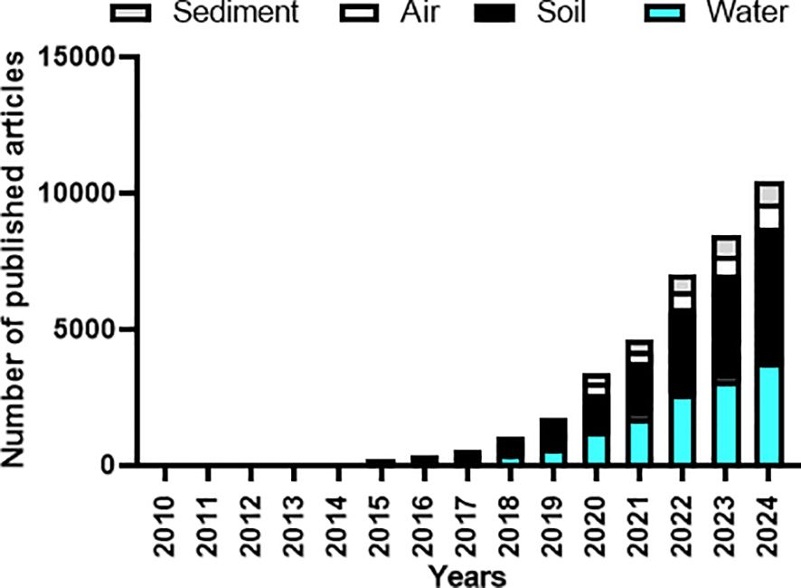
Figure 1. Global scientific publication on MP contamination under different ecosystems (water and soil ecosystems, sediments and atmosphere). The bars represent the number of publications. Only peer-reviewed articles published between 2010 and 2024 have been selected. Reviews, book chapters or notes have been excluded. A literature survey was performed on the Scopus® database using the following keywords: “MPs” combined with “water”, “soil”, “sediment” or “air”. Source: Updated from Daghighi et al. (9). Copyright the authors, licensed under CC BY-NC-ND 4.0.
Studies comparing MPs pollution in areas with varying levels of human activity have found significantly lower MPs concentrations in less anthropogenically influenced regions, such as forests, compared to highly industrialized and urbanized zones like landfills (6, 10). While naturally preserved ecosystems, including agricultural lands and forests, typically exhibit lower MPs levels, long-range atmospheric transport can introduce MPs even into remote areas (11).
Extensive research on MPs in aquatic environments has led to the development of analytical methods primarily suited for water-based matrices. As a result, common MPs extraction techniques rely on filtration and sieving, making them more effective for aquatic systems (12, 13).
Unlike aquatic matrices, soil matrices require more extensive sample preparation protocols. Analysis of MPs in soil matrices have several challenges due to soil complexity and heterogeneity, with several issues related to organic matter content and digestion process, different ranges of soil particle sizes, and the interference of non-plastic or natural materials such as natural fibers from vegetation (14, 15). To date, no standard method is available for sampling (collected soil quantities are very variable from some grams to kilos), pre-treatment, extraction and organic matter digestion and/or identification MPs techniques; once each soil is different with different complexities and challenges, that made that each soil can need a different protocol. Additionally, laboratory contamination is a big problem once MPs are present in the indoor air or come from laboratory items, which increases some additional challenges.
Microplastics contamination in the soil presents considerable obstacles, although it also creates opportunities for interdisciplinary study that integrates environmental science, analytical chemistry, and soil science. Comprehending the mechanisms via which MPs affect soil characteristics, including water retention, nutrient availability, and microbial activity, is essential for mitigating their enduring effects on soil health and agricultural sustainability. Moreover, international policy initiatives, such as the elimination of microbeads and single-use plastics, highlight the pressing necessity for standardized and rigorous procedures to monitor soil contamination. In light of the anticipated doubling of global plastic output by 2045, the establishment of economical, precise, and scalable methods for the identification and quantification of MPs in soils is essential. This study seeks to tackle these urgent concerns by aggregating and assessing existing approaches and offering recommendations to enhance research in this vital domain of environmental science (16, 17). Therefore, this study aims to systematically recompile, evaluate and discuss appropriate methods for the identification and quantification of MPs in the soil matrix.
2 Microplastics in soil
Microplastics are characterized as synthetic organic polymer particles with dimensions < 5 mm that can either arise from primary sources (e.g., industrial pellets) or secondary sources (e.g., plastic waste byproduct) (18). These particles enter soil ecosystems through farm practices (e.g. plastic mulching, sewage sludge application) as well as diffuse sources (e.g. atmospheric deposition, runoff) (19–21).
Microplastics have been widely detected in agricultural lands, forests, urban and industrial soils, grasslands, sediments, wetlands, and especially in soils near landfills, making soil the largest sink for MPs (6, 22–24). Recent reviews found that the highest MPs occurrence in soils ranged from 0 to 106 items kg-1 and 0 to 104 mg kg-1, respectively, with polyethylene, polystyrene and polypropylene the most frequent plastic polymers (24, 25). As an example, the highest reported amount has been indicated by Guo et al. (26) for a landfill surface soil (Hangzhou, Zhejiang province, China) with up to 3.57 x 10⁶ items kg-1.
In general, the occurrence of MPs in soil environments is generally higher in surface layers, depending on related factors; however, high-density MPs may penetrate deeper soil layers (27–29). This underscores the importance of sampling subsurface layers (> ± 25 cm soil depth) in studies on the distribution of MPs in soil once the majority of studies have been focused on surface layers (i.e., 0–10 or 0–20 cm). Nevertheless, deep profiling is essential to reveal a comprehensive MPs profile across soil depths and to understand the historical distribution of MPs, a question highlighted for the vertical distribution of plastics in agricultural and urban areas (i.e. 30, 31). In soil systems, plastics are being subjected to degradation and can migrate vertically and horizontally through the soil profile, usually due to agricultural activities (i.e. ploughing), bioturbation by soil organisms and/or leaching (9, 24).
While the distribution of MPs in soil may vary among regions with similar land use types, the abundance of MPs across different land use types can also be affected by regional variations (32). For instance, in the Loess Plateau of northwestern China, MPs abundance was reported to be highest in orchards, followed by agricultural fields and greenhouses. Akca (6) concluded that open dumps and scrapyards significantly contribute to MPs accumulation in soil. Additionally, while the amount of MPs decreases with soil depth in orchards and greenhouses, it increases with depth in agricultural fields (33). However, in the Qinghai-Tibet Plateau, greenhouse soils exhibited the highest MPs abundance, followed by mulched and unmulched agricultural lands, with abundance decreasing as soil depth increased (32). The number of MPs in soils with different land uses is comparable, and the number of MPs within the medium-size range is generally higher than that of other size classes at the same site.
In general, the amount and mass of MPs with different types, sizes, shapes, and compositions in deeper soil layers across land use types and environmental conditions still warrant further investigation once comparison studies are sometimes not possible due to the differences in sampling, extraction and identification methods. Future research should focus on understanding the distribution, driving processes, and fate of MPs through long-term field observations based on standardized methodologies (29).
3 Soil sampling
Microplastic contamination of soil is an environmental problem, yet currently, there are no standardized sampling methods for soils. Sampling is the first phase of monitoring MPs in soil. Before MPs analysis in the laboratories, samples are collected, stored, and preprocessed (34). The importance of soil sampling strategies is reflected in research data on the effects of soil properties, land management practices, and environmental conditions on the collected data, which show that representative data collection is less dependent on equipment and is more dependent on soil sampling strategies. Main types and guidance for soil sampling for MPs analysis following a broad review of existing knowledge (14, 15, 35).
Soil is a long-term sink of MPs, their transportation and representative sampling must be accounted for, as representative sampling is important when trying to draw significant conclusions from the data collected (5, 21).
Lack of standardization on approaches used to determine the extent of soil MPs contamination hinders study comparisons and produces inconsistent estimates (36–38). Sampling site selection depends on the research purpose, accessibility, and known sources of MPs pollution (37, 39, 40). Methods that rely on design, for instance, random, stratified, and systematic sampling, are often employed to describe spatial variability (10, 15, 35). Random sampling is simple, whereas a stratified design allows sites to be as sub-regions based on, for example, criteria such as vegetation type or land-use type (41). Sampling sites close to roads, plastic mulch fields, or urban sites usually have higher MPs concentrations (23, 40, 42).
Important accumulation zones, such as hollows or ridges, where MPs are deposited through surface water runoff or wind deceleration, need to be considered as well. Indeed, studies highlight the necessity to enhance sampling strategies for these environmental challenges (43, 44). Soil is a three-dimensional matrix; therefore, MPs deposition and build-up in situ can vary significantly. Therefore, the specific site chosen for soil sampling to study MP pollution should reflect the spatial distribution of MPs since these can change dramatically over short distances in the soil. Second, the management history of the sample location must be checked when selecting a specific location of interest, assuring the use of a whole field to a spatially explicit system. MPs distribution changes with depth in soils. Surface soil (0–10 or 0–20 cm) sampling is more common because MPs accumulate in the top few centimeters due to a potentially low vertical transport. However, as previously mentioned, plastics can migrate horizontally and vertically due to agricultural management, biota activity or leaching. Therefore, deeper sampling (>20 cm) is necessary to study long-term MPs migration (14, 15). Depending on the depth and volume requirements of the samples, the common tools used were stainless steel shovels, augers, and core samplers (45).
Sampling and storing tools that are not plastic (i.e., aluminum bags or foil, glass bottles or paper) are recommended to avoid contamination. Field quality controls such as clean equipment and field blanks for potential contamination during sampling are critical (15). The amount of soil collected is determined by the need for an analysis and variability in the site. Composite samples, which are formed by pooling subsamples, can efficiently capture spatial heterogeneity while alleviating analytic burden (46, 47). For instance, Zhang et al. (29) used composite sampling by taking multiple subsamples in each plot and bulking them into one sample. An estimate for the number of total sampling points at each site (single point samples or composite samples) could be made from a statistical power analysis. Yet, the number of replicates and the volume of the sample are highly restricted by sample processing and subsequent analytical methods, which remain the bottleneck of MPs quantification in any environmental compartments (48). Thus, at this stage, no recommendation can be made on the minimum of sampling points per spatial unit to be used.
Soil sample sizes have varied enormously, from 50 g to more than 4 kg, with greater sample sizes giving a better representation of heterogeneity (49). After homogenization, a smaller portion (e.g., 15–50 g) is used for analysis (21, 45), while making sure there is enough material left for parallel tests and potential backups. So far, not enough is known for us to be able to recommend the least amount sufficient for representative soil samples in any of these models, but it must at least surpass the mass or volume of the reference unit; otherwise, it leads to an unjustifiable extrapolation. Item per kg(dry weight) would be the most meaningful reference unit (mean dry weight was determined from aliquots of the sample according to a standard procedure, e.g. (50). In this regard, one must also consider water content and additional aliquots so that the field samples should be much greater than 1 kg.
Soil sampling for MPs comes with its obstacles, being as the complex soil matrix and MPs heterogeneity. MPs bind to soil organic matter or are embedded in aggregates, complicating extraction procedures (51). This represents the outline of challenges in (i) representative sampling; sample design should adjust the potential of spatial variability of MPs; (ii) quality assurance, preventing MPs contamination from tools, clothing, and ambient air; (iii) documentation, specifically documenting sampling depth, location, and conditions (35, 51). Standardized protocols for soil MPs sampling are still a major problem for reproducibility. Establishing common methods (including standardized operating procedures [SOPs]) is essential to ensure the comparability of the studies (15, 39, 45). This includes developing SOPs for sampling design, the depth of sampling, tools, storage, and contaminant prevention. Advancements in in situ detection methods and automated samplers are very promising in improving soil MPs sampling. Highly effective soil sampling is central to the progress in understanding the terrestrial pollution of MPs. While progress has been made, we need standardized protocols to ensure the reliability of data urgently.
In summary, some of the important challenges and solutions to advance our understanding of MPs impacts on soil health are proposed to encourage innovative scientific approaches to quantify better and reduce the effects of MPs on soil health (15, 18).
4 Extraction of microplastics from soil
The extraction of MPs from soil is essential for evaluating their distribution, composition, and potential environmental hazards. In contrast to water-based systems, where MPs may be more readily isolated, soil poses greater hurdles owing to its intricate matrix, variable particle size, and robust interactions between MPs and organic or inorganic substances (17, 52). Diverse extraction techniques have been devised to tackle these issues, roughly categorized into nature-based solutions and conventional technologies (53). Nature-based solutions employ eco-friendly methods, like bio-coagulants, microbial remediation, and plant-based filtering, to reduce chemical utilization while enhancing MPs recovery (17, 54). Conversely, conventional procedures depend on known physical and chemical processes, such as density separation, sieving, and oxidative digestion, to effectively isolate MPs from soil samples (55–57). Every method possesses distinct advantages and limitations for efficiency, selectivity, and sustainability (17, 58). This section provides a summary of recent research on MPs extraction from soil, classified into nature-based and conventional methods.
4.1 Conventional methodologies
Conventional extraction methods utilize physical, chemical, and physicochemical procedures to isolate MPs from soil (Figure 2). These approaches are extensively employed in laboratory research and environmental monitoring owing to their efficacy and reliability. The most commonly applied techniques include density-dependent separation, organic matter removal followed by density separation, density-based flotation, and pressure leaching combined with chemical treatments (Table 1). Density-dependent separation typically uses saturated salt solutions such as NaCl, ZnCl2, and NaBr, achieving high recovery rates (approximately 95%) for both small (10–100 μm) and large (100–5000 μm) MPs (61). However, this method has limitations in detecting micro/nanoplastics smaller than 10 μm due to the resolution constraints of spectroscopy methods like FTIR and Raman micro-spectroscopy (Figure 2).
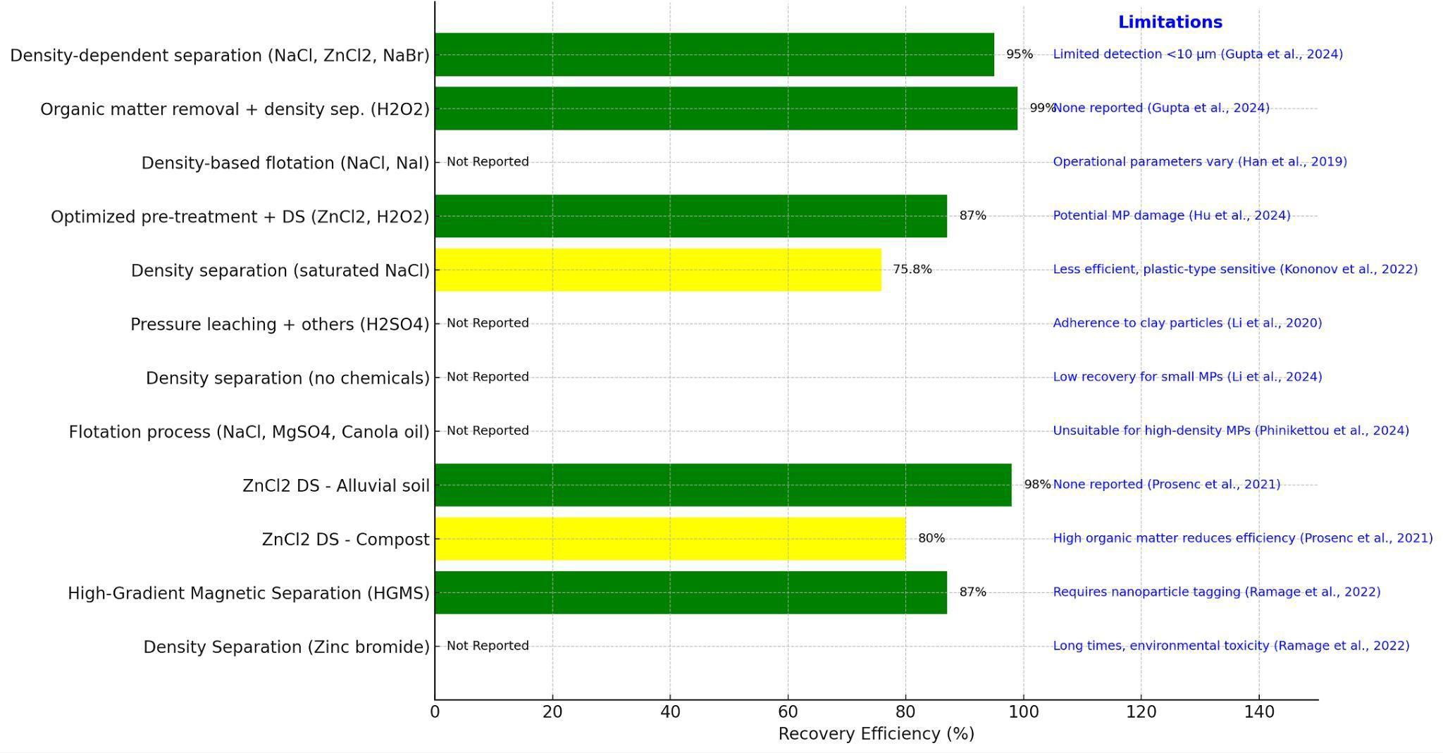
Figure 2. Visual representation of recovery efficiencies for various MP extraction methods from soil and sediment. Methods achieving high recovery efficiencies (>85%) are shown in green, moderate and low efficiencies (70–85%) in yellow. Method-specific limitations, accompanied by relevant citations, are annotated on the right side of the chart to highlight critical considerations in their practical application.
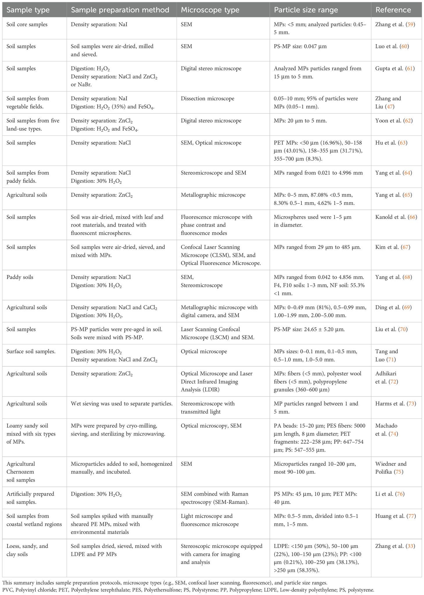
Table 1. Overview of studies employing electron microscopy and related imaging techniques for the identification and characterization of MPs in soil environments.
Furthermore, NaCl solutions, though economically viable and safe, have been reported to be unsuitable for extracting high-density plastics such as PVC and PET, reflecting varying extraction efficiencies based on plastic density and soil properties (Figure 2). Organic matter removal coupled with density separation commonly employs hydrogen peroxide (H2O2) to effectively digest organic matter, resulting in recovery efficiencies of up to 99% (61). This procedure provides clearer supernatants, significantly facilitating MPs recovery. Nevertheless, prolonged oxidative digestion may damage some MPs, thereby affecting the accuracy of identification and quantification. More complex methods, such as the combination of pressure leaching, flotation, electrostatic adsorption, and concentrated sulfuric acid carbonization, offer simplicity and effectiveness for microsized particles. However, these methods struggle with the extraction of MPs adhering strongly to clay particles.
4.2 Nature-based solvents
Conventional analytical techniques for MPs detection often lack environmental sustainability due to their reliance on hazardous reagents (e.g., Fenton’s reagent, zinc chloride), substantial waste production, and high energy consumption (58, 78). Incorporating Green Analytical Chemistry (GAC) principles can address these issues by reducing toxic reagent use, minimizing waste, and lowering energy consumption (79–81).
In pursuit of a sustainable and efficient method for MPs extraction from sediments, Bellasi et al. (82) improved density separation using a NaCl-sucrose mixture, achieving effective separation of MPs while maintaining environmental safety and economic feasibility, though issues with smaller particles and viscosity remain. The method offers an effective and safe strategy for MPs separation while maintaining economic feasibility. Although effective for MPs in the 500 µm to 3 mm range, further research is necessary to optimize its application for smaller particles and mitigate issues related to the solution’s viscosity. Additionally, challenges such as MPs retrieval from the viscous medium must be addressed.
Recently, Rede et al. (83) introduced a QuEChERS-inspired (Quick, Easy, Cheap, Effective, Rugged, and Safe) extraction method combining oxidative digestion, density separation with NaCl (density: 1.2 g cm-³), and centrifugation, achieving high recovery but encountering limitations like prolonged processing and inadequate extraction for dense plastics such as PVC. This modified density separation method combined with centrifugation was implemented to enhance sustainability and efficiency while reducing reagent consumption and waste generation (Table 2; 95). Recovery experiments using certified soils spiked with standard MPs demonstrated a recovery rate exceeding 91% for larger MPs (3–5 mm) and over 69% for smaller MPs (15–300 µm).
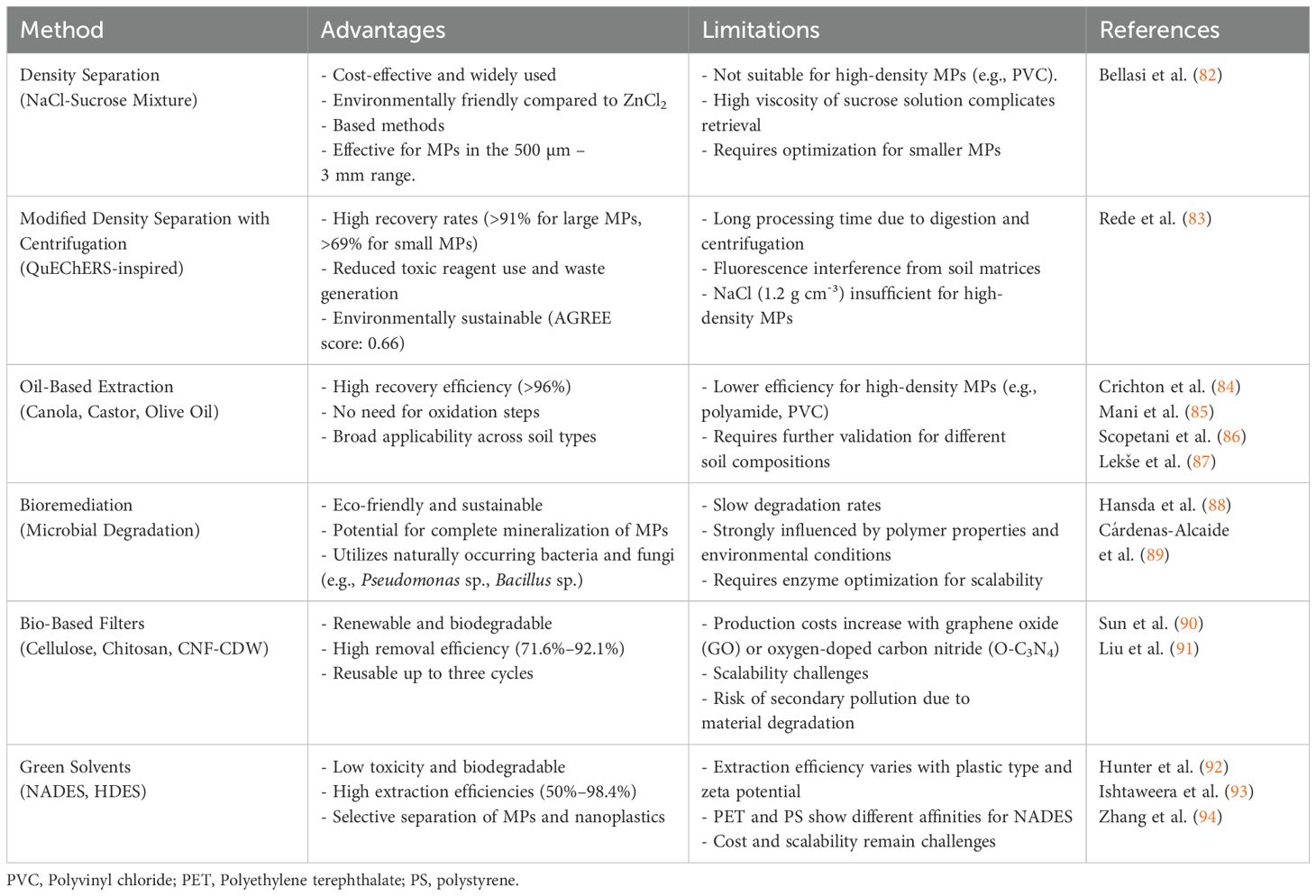
Table 2. Comparison of different natural-based methods for MPs extraction.
Alternative oil-based extraction methods demonstrated promising results, such as canola oil (84), castor oil (85), and olive oil (86, 87), though challenges remain in extracting high-density MPs (Table 2). Pre-treatment with Fenton’s reagent facilitated organic matter removal, and extraction was performed using polytetrafluoroethylene (PTFE) cylinders. To validate the method, soil and compost samples were spiked with six MPs: polyethylene (PE), polystyrene (PS), PVC, polycarbonate (PC), polyethylene terephthalate (PET), and polyurethane (PU).
Bioremediation offers eco-friendly alternatives, utilizing microbial degradation processes involving bacteria, fungi (Pseudomonas sp., Bacillus sp., and Aspergillus sp.), and enzymes such as PETases and laccases (89, 96). However, efficiency depends on polymer characteristics and environmental conditions, indicating the need for further optimization.
Despite the increasing volume of research on MPs contamination in soils, many analytical techniques are adapted from methods originally designed for aquatic environments. These adaptations often fail to fully account for the complexities of soil matrices, leading to challenges in sample preparation and analysis. Developing green, renewable, and cost-effective materials for MPs removal remains a significant challenge. The use of bio-based filters and green solvents is emerging as a promising approach for MPs extraction across various environments. Bio-based filters using biodegradable materials like cellulose and chitosan demonstrate potential for MPs removal (97, 98). Sun et al. (90) evaluated the potential of biodegradable chitin-based sponges enhanced with graphene oxide (GO) and oxygen-doped carbon nitride (O-C3N4) for efficient MPs removal from water. The sponges exhibited high removal efficiencies (71.6%–92.1%) for MPs (~1 µm) with varying surface charges, primarily through adsorption mechanisms such as electrostatic interactions, hydrogen bonding, and π-π interactions. These materials demonstrated biocompatibility, mechanical durability, and reusability for up to three cycles, while also being biodegradable in soil. However, incorporating GO and O-C3N4 increases production costs, posing scalability challenges. Furthermore, rapid degradation of these materials may lead to secondary pollution through the release of potentially harmful byproducts. These factors emphasize the need for further optimization, particularly for large-scale applications and real-world implementation.
Liu et al. (91) introduced cellulose nanofiber-coated delignified wood (CNF-CDW) as an efficient MPs filter. Wood, an abundant and biodegradable natural resource, is widely used in construction, energy storage, water remediation, and flexible electronics. Its porous structure, composed of cellulose, hemicellulose, and lignin, provides an ideal network for filtration and adsorption processes. Balsa wood was selected as a renewable raw material, and various delignification methods were applied to enhance its surface area and pore structure, optimizing MPs removal efficiency. The CNF-CDW filter effectively removed PS MPs, with performance influenced by CNF concentration, CaCl2 concentration, and wood chip thickness. Optimizing CNF-wood interactions under diverse environmental conditions could further enhance filtration efficiency.
Green solvents offer environmentally benign alternatives to traditional chemical solvents, which often contribute to pollution. These solvents facilitate the extraction of MPs from contaminated soil, water, and sediments while exhibiting low toxicity and biodegradability. Among them, Deep Eutectic Solvents (DES) have garnered attention due to their tunability, biodegradability, and lower toxicity compared to conventional solvents. DES are typically formed by combining naturally occurring compounds, such as choline chloride and urea, to create eutectic mixtures with reduced melting points.
Recent research has explored the use of hydrophobic Natural Deep Eutectic Solvents (NADES) as novel extraction media for micro- and nanoplastics. Hydrophobic NADES are particularly promising due to their low toxicity and cost-effectiveness. In 2023, three hydrophobic NADES were investigated for MPs and nanoplastic extraction (92). Experimental studies demonstrated extraction efficiencies of 50%–93% for PET, PS, and polylactic acid (PLA) from water samples. Extraction rates depended on particle size, zeta potential, and specific interactions between NADES molecules and plastic surfaces. Molecular dynamics simulations revealed strong interactions between PLA and PET with NADES, particularly in decanoic acid:menthol (1:1 and 1:2) systems, resulting in high extraction efficiencies. PET exhibited slightly lower performance at the two-hour extraction mark, except in thymol:menthol (1:1), where it matched PLA. Conversely, PS showed minimal interactions with NADES, leading to reduced extraction rates. Ishtaweera et al. (93) introduced a highly efficient nanoplastic extraction method using hydrophobic deep eutectic solvents (HDES) via liquid-liquid extraction. Among ten tested HDES systems, three eutectic mixtures—1:2 tetrabutylammonium bromide:decanoic acid, 1:2 tetraoctylammonium bromide:decanoic acid, and 1:1 thymol:menthol—demonstrated exceptional performance, achieving 98.4% extraction efficiency for polystyrene nanoparticles (100–1000 nm). Further advancements in HDES technology involve the incorporation of lignin-derived HDES, leveraging lignin abundance and renewability for enhanced sustainability. Zhang et al. (94) synthesized and evaluated five lignin-derived HDESs for nanoplastic extraction. The study identified Thymol-2,6-dimethoxyphenol (1:2) as the most promising candidate due to its stability and superior extraction efficiency for PET and PS nanoplastics. The primary extraction mechanisms involved hydrophobic interactions, hydrogen bonding, and π-π interactions. While adsorption onto HDES was the rate-limiting step for most plastics, diffusion of PET NPs toward the HDES phase became the limiting factor in dilute solutions. This study underscores the potential of lignin-derived HDES for scalable nanoplastic remediation.
Natural Deep Eutectic Solvents (NADES) and HDES present promising, sustainable alternatives to conventional MPs extraction methods. Their integration with natural filtration or bioremediation techniques could significantly reduce environmental footprints and enhance plastic pollution remediation. By refining these green solvent systems and optimizing their application in real-world scenarios, researchers can advance more effective and scalable solutions for tackling MPs contamination.
5 Microplastic identifications
The identification of MPs is typically accomplished through two distinct methodologies. The first method is based on morphological characterization, while the second method is based on chemical characterization (Figure 3). Morphological characterization techniques are indicated for various analyses, including size determination, optical properties, depth profiling, component distribution, and detailed surface morphology. Chemical characterization pathways are differentiated based on the intent to quantify chemical species and the presence of interfering substances (water, pigments, or organic compounds), leading to specific analytical techniques such as μ-FTIR, Raman spectroscopy, chromatography, and thermal analysis.
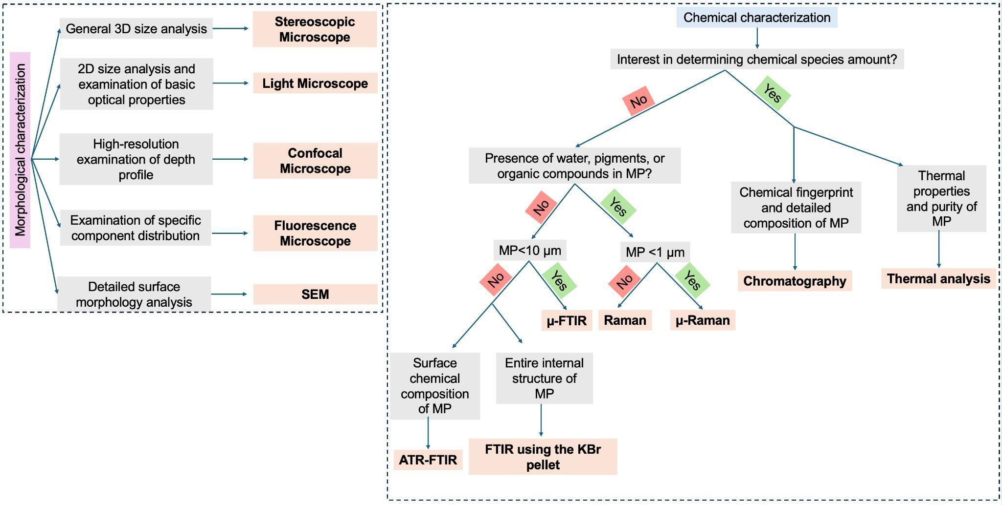
Figure 3. Flowchart illustrating the methodological approaches for morphological and chemical characterization of MPs.
5.1 Microscopic identification
5.1.1 Light microscopy
MPs ranging in size from 1 to 5 mm are generally identified through visual inspection via the naked eye or an optical microscope (99; Figure 3). Their distinction of MPs from non-plastic materials is based on physical characteristics such as color, shape, and light transmission properties (99). Optical microscopy facilitates the identification of smaller MPs within this size range that may not be accurately detected by the naked eye and has been recognized as an economical method to identify MPs by forming a magnified image of the sample using image contrast of the light reflected from the sample. However, visual inspection has been reported to have an error rate ranging from 20% (100) to 70% (101), with the error rate increasing with decreasing MPs size. This is particularly due to the difficulty in differentiating potential MPs with certainty from sand grains, natural biopolymers such as chitin etc., leading to a high number of false positives and negatives (102). Furthermore, optical microscopes are diffraction-limited, contributing to the inaccurate identification of particles with sizes closer to the diffraction limit. Thus, visual inspection is not used as a stand-alone technique. Instead, it is commonly used as a pre-sorting/pre-selection tool prior to a more detailed and accurate chemical analysis. Nevertheless, researchers have also devised mechanisms to avoid false positive identification of MPs through visual inspection by prodding uncertain particles with the needle (103), using hot needle tests (103) and subjecting the MPs to high temperatures for short duration (33).
5.1.2 Fluorescence microscopy
Staining samples with fluorescent dyes is a widely employed approach to facilitate the identification of MPs in environmental samples using fluorescence microscopy (Figure 3). The inherently hydrophobic nature of most plastic polymers enables their affinity for lipophilic fluorescent dyes; however, this property is not exclusive to plastics and may also be shared by other hydrophobic non-plastic particles present in complex environmental matrices, making selective staining of MPs challenging. The problem is further compounded by the absence of standardized staining protocols, resulting in methodological variability across studies (104). The limited availability of suitable fluorescent dyes represents a significant challenge for reliable MPs detection, with Nile Red being the most commonly employed stain due to its strong affinity for hydrophobic polymer surfaces (105). Although the application of Nile Red (NR) staining in aqueous samples is well-established, it presents challenges such as dye precipitation and aggregate formation in water, which can lead to false-positive fluorescence signals (106). These issues, along with concerns regarding dye stability over time, have significant implications for protocols involving marine sample analysis (105). However, NR-based methods have also been adapted and validated for solid environmental matrices, including soils and sludge. For instance, Kang et al. (107) demonstrated the effectiveness of a modified NR plate method (NR-P), which enhances MP detection in complex matrices like sewage sludge, where conventional staining often fails due to the presence of organic matter. Similarly, the NR technique has been successfully applied in terrestrial samples such as street dust, sludge, and soils with high organic content, following appropriate pretreatments (108, 109). Despite its limitations, NR staining combined with robust sample preparation and fluorescence microscopy continues to be a widely used approach for detecting MPs across various environmental matrices, including both aquatic and terrestrial systems.
5.1.3 Electron and other microscopy techniques
Unlike optical microscopy, electron microscopy is not limited by the diffraction limit; thus, very small particles which cannot be identified accurately using an optical microscope can be visualized through an electron microscope because, in contrast to the former, in electron microscope sample illumination is done using a high-energy electron beam as compared to visible light – the shorter wavelength of the electron beam results in higher resolution (110, 111; Figure 3). Visualization of samples using electron microscopy requires the surface to be electrically conductive. Therefore, an additional sample preparation step is often necessary, wherein the particles of interest are coated with a thin metallic layer to enhance their conductivity, thereby improving image quality. Electron microscopes are broadly classified based on their imaging principles into scanning electron microscopes (SEM), transmission electron microscopes (TEM), and reflection electron microscopes (REM). Among these, SEM is the most widely employed, either as a standalone technique or in conjunction with chemical characterization methods such as Raman spectroscopy or Fourier-transform infrared (FTIR) spectroscopy, to detect and identify MPs within the size range of 1 µm to 1 mm (112).
Various types of microscopes are used for identifying MPs, each with distinct advantages and disadvantages (Figure 3; Table 1). Scanning Electron Microscopes (SEM) offer high-resolution imaging and detailed surface morphology analysis but are expensive and require extensive sample preparation. Digital stereo microscopes are user-friendly and cost-effective for three-dimensional observations but lack the resolution needed for smaller MPs. Optical microscopes are widely accessible and simple to use, yet their resolution is limited for particles smaller than a few micrometers. Metallographic microscopes are excellent for analyzing the structure of metallic contaminants within MPs but are specialized and less versatile. Fluorescence microscopes enable the differentiation of MPs from other materials by using dyes or autofluorescence, though their application is limited to fluorescent particles. Confocal Laser Scanning Microscopes (CLSM) provide 3D imaging and better depth resolution but are expensive and time-intensive. SEM combined with Raman spectroscopy (SEM-Raman) integrates the strengths of high-resolution imaging and molecular characterization, though it is cost-prohibitive and requires skilled operation. Finally, while light microscopes and fluorescence microscopes are widely used for the initial screening of MPs, they cannot provide the molecular-level insights or high resolution that advanced techniques like SEM or SEM-Raman can deliver. Each method must be chosen based on the size, composition, and detail required for MPs analysis. Studies on the identification of MPs using electron microscopes are summarized in Table 1.
5.2 Polymer analysis
5.2.1 FTIR
Fourier-transform infrared spectroscopy (FTIR) is the most used method for identifying MPs (113, 114; Table 3). This method is used to accurately find out what chemicals are in MPs, which makes it easier to tell the difference between polymers and other chemicals in the sample (113). FTIR analysis produces an infrared spectrum for each MPs. The basis is predicated on the principle that the spectrometer signal, referred to as infrared absorption bands of the polymer under examination, is dependent upon changes in the permanent dipole moment of a chemical bond, thereby making it sensitive to the polar functional groups included in various plastic polymers. FTIR is a surface-based method that needs no plastic particles in any film, coating, or substance in order to identify it correctly (127). External variables, such as the presence of organic materials or water, can affect its spectral quality (128). Prior to FTIR analysis, samples must undergo complete drying to prevent moisture from affecting identification accuracy (129). Under precisely controlled experimental circumstances, the FTIR method has efficiently evaluated the degradation of MPs (130). Weathering processes, such as photooxidation from sunlight exposure, often cause degradation, leading to structural alterations and fragmentation (131). The first stages of this process include the formation of new C–O, C=O, and O–H bonds during oxidation on the exposed MPs surfaces, a phenomenon that may be seen and quantified by IR spectral analysis (132).
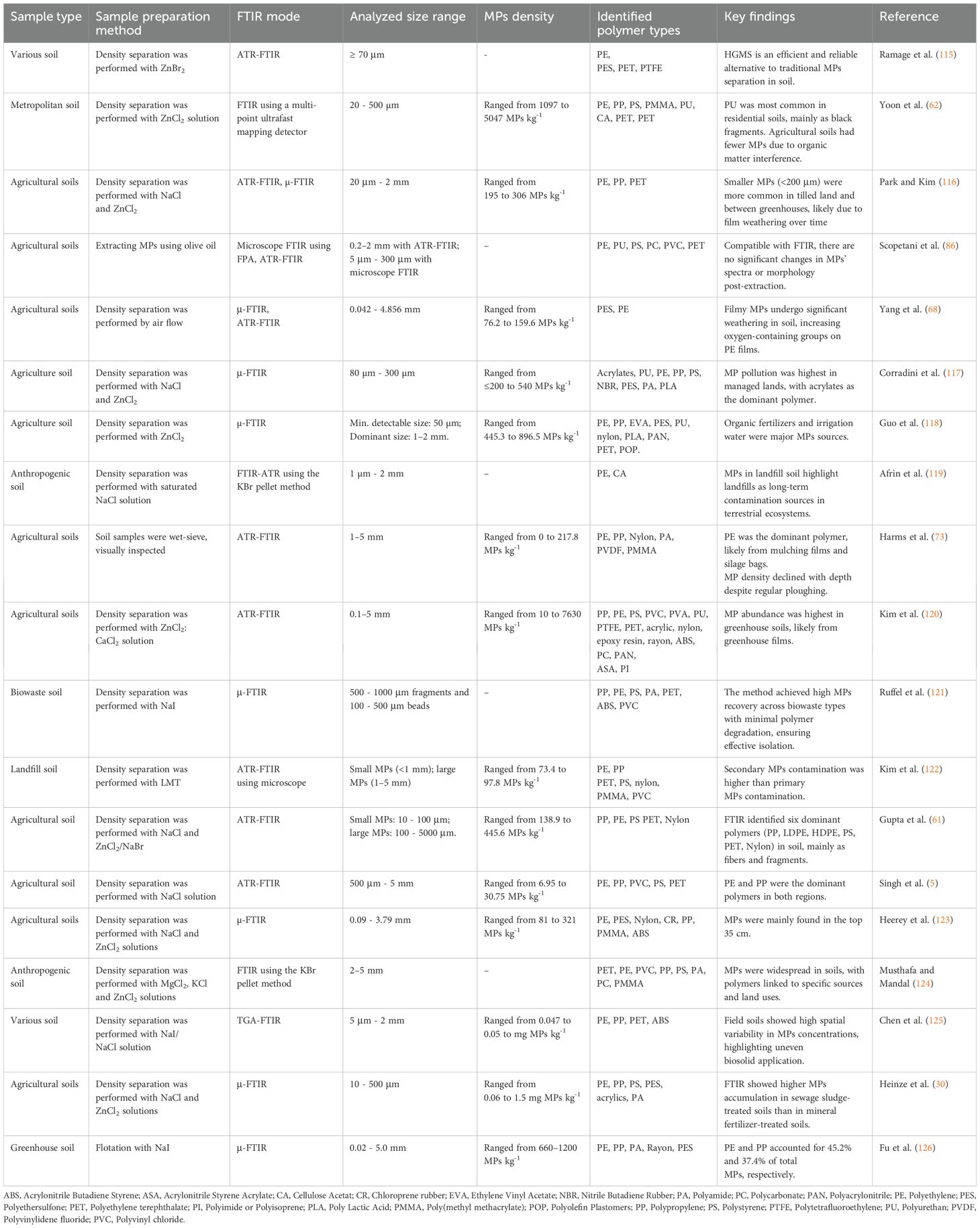
Table 3. Summary of studies employing Fourier Transform Infrared (FTIR) spectroscopy for MPs identification in various soil matrices, detailing sample preparation methods, FTIR modes, analyzed size ranges, MPs densities, identified polymer types, and key findings.
Users of FTIR can change basic settings like spectral range, spectral resolution, number of scans per spectrum, measurement method, and background sample collection (133). The spectral resolution, commonly ranging from 32 to 4 cm-1, indicates the quantity of information produced at each collecting point, with a standard resolution of 4 cm-1 (133). Typically, once the sample has been included, the analyst utilizes a spectral range of 3800–900 cm-1 at a resolution of 8 cm-1, performing 6–30 sample scans (134). Data obtained using FTIR can be compared with an online spectrum library including polymer reference libraries, user-generated libraries, or scientific journals (135). FTIR analysis indicates the most likely polymer identification of the particle (136).
The micro Fourier Transform Interferometer (μ-FTIR) devices provide spatial resolutions of up to 5 μm (137). This technique requires a minimum sample thickness of 150 nm (137). In general, FTIR is very consistent and does not change based on sample color, fluorescence, or other factors. This means it can be used to find MPs with a diameter of more than 20 µm or materials with strong polar functional groups (138). The FTIR spectroscopy has been extended over the years to investigate particles measuring 10–20 μm by methods like μ-FTIR, attenuated total reflectance FTIR (ATR-FTIR), and focal plane array FTIR spectroscopy (FPA-FTIR) (139, 140). Each method has its own benefits: μ-FTIR can look at MPs bigger than 10 μm, ATR-FTIR is used for particles bigger than 500 μm, and FPA-FTIR makes it easier for machines to find MPs bigger than 20 μm (141). Each method has specific benefits and limitations, hence, the optimal methodology is dependent on the sample matrix (129).
Specular reflection, transmission, and attenuated total reflection are the three primary modes of Fourier transform infrared spectroscopy (142). The operational mode may be flexibly selected depending on the attributes of the samples. The specular reflection technique measures the energy that the particle’s surface reflects, as opposed to the energy that the particle transmits. This technique offers the advantage of requiring minimal sample preparation; however, it is applicable entirely to particles having a flat and reflective surface. This technique may not be suitable for weathered MPs with irregular shapes. The transmission technique requires setting a MPs particle between the infrared beam and the detector. The infrared beam of light transmits through the sample, with the detector determining the transmitted light to generate a spectrum. Transmission generates high-quality spectra from various polymer types, including soluble, thin, and dark polymers and powders (143). Attenuated Total Reflection exists in two forms: single-reflection ATR and multi-reflection ATR. Both techniques utilize a crystal as the internal reflection element. Typically, diamond, zinc selenide, or germanium make up the crystal due to their hardness, durability, and elevated reflective index. For both types of ATR to work, the particle must be in close contact with the crystal, which is placed between the source and the detector (143). This mode creates a steady wave that makes it easier to prepare small samples and get better data on opaque and irregularly shaped MPs, such as MPs that are as small as the IR beam aperture and MPs that are bigger than 500 μm (144, 145).
The main goal of doing FTIR analysis is to determine the polymer composition of the measured MPs. This method makes it easier to learn important things about where these particles come from and tells the difference between synthetic polymers and non-synthetic materials in the MPs count (Table 3).
5.2.2 Raman spectroscopy
Similar to Fourier-transform infrared (FTIR) spectroscopy, Raman spectroscopy is widely employed for polymer identification in MPs research (146; Table 4). This technique utilizes laser beams to analyze the distinct molecular and atomic structures of polymers by measuring the frequencies of backscattered light. Compared to FTIR, Raman spectroscopy offers superior spatial resolution and enhanced sensitivity to nonpolar functional groups (155). With a detection limit as low as 1 μm—significantly smaller than that of FTIR—Raman spectroscopy enables high-resolution imaging, allowing for MPs quantification in the range of 2.2 × 10⁴ to 6.9 × 10⁵ particles per kilogram (33, 156). Raman imaging is achieved by collecting spectral data from individual pixels, facilitating detailed visualization of MPs particles (157).
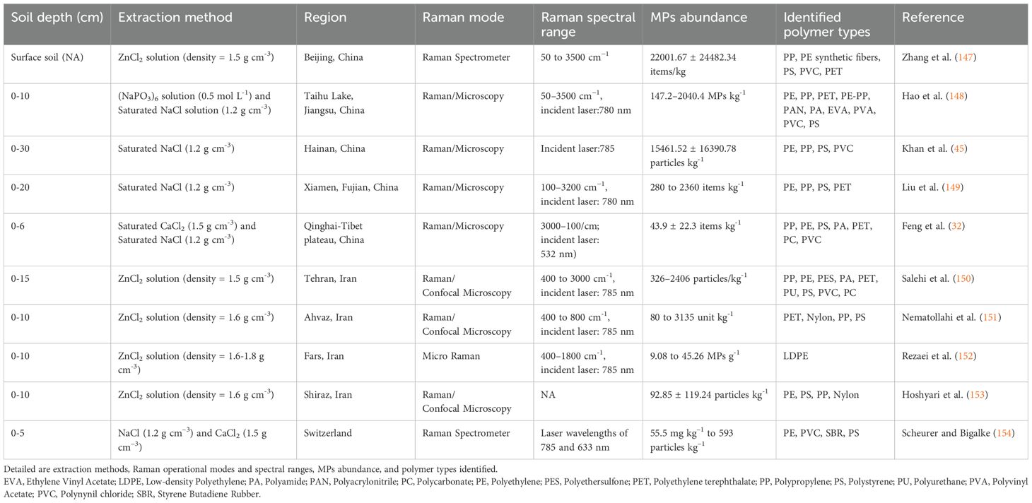
Table 4. Summary of studies employing Raman spectroscopy for MPs identification across various soil depths and regions.
Raman spectroscopy presents several advantages in MPs analysis, including (i) low sensitivity to water, allowing for the examination of wet samples, (ii) the capability to analyze non-transparent and dark-colored particles, and (iii) reduced dependency on particle shape and thickness (158, 159). Raman spectroscopy offers several advantages; however, it also faces notable limitations in MPs identification, particularly due to fluorescence interference from biological, organic, and inorganic contaminants, which can hinder its application to real environmental samples (12, 110). Consequently, pre-purification of samples is often required, increasing the overall analysis time compared to FTIR (159). Furthermore, additives within MPs and surface-bound contaminants can lead to overlapping Raman spectra, making polymer identification more challenging (160). The use of monochromatic laser light sources in Raman spectrometers may also induce photochemical or thermal degradation of MPs polymers, further hindering accurate analysis (161). Furthermore, Raman spectroscopy requires manual selection of regions for imaging, making the detection process time-consuming and technically demanding (162).
Optimized Raman techniques, such as micro-Raman (μ-Raman), have been developed to enhance MPs analysis. Similar to μ-FTIR, μ-Raman integrates an optical microscope, enabling the spatial and chemical characterization of plastic particles at a spatial resolution of 1 μm, even for particles as small as 10 μm (163). While Raman spectroscopy is a powerful tool for MPs detection, the development of standardized protocols is essential for its widespread application. A multidisciplinary approach is required to optimize the entire analytical workflow, from sample collection to spectral acquisition, to ensure accuracy and reliability in MPs identification.
5.2.3 Photoluminescence spectroscopy
Photoluminescence spectroscopy (PLS) is an emerging analytical technique in the field of MPs detection that works on the PL principle, i.e. optical excitation of a material will be followed by the emission of light at a longer wavelength than the excitation source due to energy loss seen by the excited carriers in internal conversion processes (164). In 2018, the first report of PLS measurements were made on the quantification of MPs in the sand samples collected from a beach in Italy for commercial PS samples and partially oxidized low-density PE by using an excitation range of 200–775 nm with emission spectra recorded in the range of 200–800 nm (165). In another piece of research, (164), successfully identified seven different types of MPs, PS, PE, PP, PET, polymethyl methacrylate (PMMA), polycarbonate (PC) and polyvinylidene fluoride (PVDF) using a simple 405 nm laser diode by monitoring the emission spectra recorded in the 400–800 nm range. It is worth mentioning that the emission peaks for all the different types of MPs were observed in the 10 nm wavelength window of 455–465 nm. Another study (166) that used PLS for the detection of MPs as small as 200 μm reported that the emission spectra are dependent on the excitation wavelength, experimentally identifying optimal excitation wavelengths of 360 nm for PS and PET, and 370 nm for PP. This finding underscores a critical consideration in PLS: the necessity of determining the optimal excitation wavelength for each polymer type to ensure optimal detection and characterization.
5.2.4 Chromatographic technology
Except for vibration spectroscopy technologies, such as FTIR and Raman, chromatography-based technologies are also powerful tools for analyzing MPs (Table 5). The chromatographic analytical methods are flexible to be coupled with other techniques, which can endow different features to analyze MPs. Over the last few years, the amount of published articles applying chromatographic methods to analyze MPs has increased (174). The chromatographic methods mainly include Gas Chromatography (GC), Liquid Chromatography (LC) and Gel-permeation Chromatography (GPC), and GC is the most used one to couple with other technologies to characterize MPs. Gas chromatography coupled with mass spectrometry (GC–MS) plays an important role in environmental analysis. In GC-MS, samples are first separated in GC, and then MS, as the detector can identify the compounds separated in GC. Nowadays, GC-MS is very well used in studying the pollutants in various environmental sources, such as air, water, soil, sludge and biological samples (174).
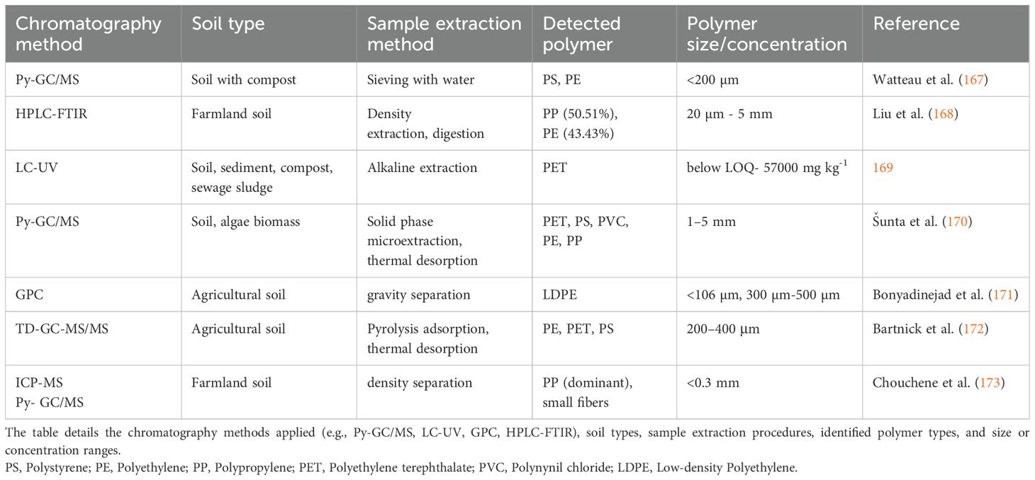
Table 5. Overview of studies utilizing chromatography-based techniques for MPs identification in various soil and sediment matrices.
Among different GC-MS technologies, Pyrolysis-gas chromatography/mass spectrometry (Py-GC/MS) is preferred to be utilized by many researchers thanks to its high sensitivity, which enables it to characterize MPs with small sizes (Table 5). This technique employs heat to degrade polymeric material in a controlled manner in an inert environment (i.e., one without oxygen). The polymer degradation products can then be analyzed using gas chromatography according to their size and polarity before being examined by a mass spectrometric detector (112). The resulting chromatographic fingerprints can be evaluated using well-known reference collections of known polymers. When optimized and with state-of-the-art equipment, this technology is superior to FTIR and Raman spectroscopy because it can characterize particles less than 10 µg, and the use of thermal analysis in conjunction with GC/MS allows the separation and analysis of chemical additives as well as the polymeric material (175).
Compared with GC technology, LC technique is less commonly used on MPs analysis in environmental samples as can be seen in the literature. MS and ultraviolet detector (UV) are the main detectors which are used to couple with LC for MPs analysis. Recently, ultra-high sensitivity in the quantitative and qualitative detection of MPs has been attained by developments in LC-MS/MS, which is especially useful for examining nanoscale fragments in complex atmospheric environments where traditional techniques are insufficient (176). Instead of using MS/MS, high resolution mass spectrometry (HRMS) coupled with LC is able to reach the instrumental limit of detection (ILOD) of 20 pg and methods limits of detection and quantification around 30 pg L−1 and 100 pg L−1, respectively and this LC-HRMS method is equipped with an atmospheric pressure photoionization source (APPI) (177). In 2020, Müller et al. (169) first presented the LC-UV method to determine the mass content of PET in soil samples, and this method is more robust and cost-effective than LC-MS/MS, considering the ultra-high vacuum required for MS measurement is not required, and sticky char contaminations and other MS maintenance procedures are not possible.
Gel Permeation Chromatography (GPC) is a widely used technique in MPs research for determining the molecular weight and polymer distribution of MPs in various environmental samples, including soils, water, and sediments. GPC works by separating polymers based on their size, with larger molecules eluting first and smaller molecules following behind, based on their ability to penetrate the pores of a stationary phase (178). The determination of position and concentration of functional groups in polystyrene was reached by Warner et al. (179) showed that the carbonyl groups of copolymers were evenly distributed on the polymeric chain. The GPC enables researchers to assess the environmental ageing of MPs by measuring polymer chain length changes, which can indicate fragmentation, oxidation, or chemical alteration. A recent study used GPC coupled with ultraviolet detection (GPC-UV) to detect and quantify PS-MPs in soil samples efficiently, improving extraction efficiency with an HCl-assisted method (76). Another study showed that GPC coupled with fluorescence detection enables semi-quantitative and selective identification of common MPs in marine sediments. By utilizing fluorescence detection at 260/280 nm and 370/420 nm excitation/emission wavelengths, the method successfully distinguished PS from partially degraded polyolefins (LDPEox) (165).
Chromatographic techniques, including GC-MS, LC-MS, and GPC, are also widely used for analyzing MPs in soil. These methods help identify polymer types, quantify contamination levels, and assess degradation. Py-GC/MS and LC-MS can provide precise identification and quantification of polymers, even at low concentrations. The GPC shows great ability to assess the degradation of MPs based on analyzing the molecular weight differences. With chromatographic technologies, the quantification of MPs is also reachable and even to a low limit. Plus, chromatographic techniques are able to be coupled with various detectors, which gives them flexibility to fit specific samples. However, extensive sample preparation may be needed before MPs in soil samples analysis with chromatographic technologies, such as density separation, filtration and chemical digestion, to remove the organic interferences. Techniques like Py-GC/MS, are destructive analysis, which makes analysis of individual particle morphology unreachable. Chromatographic technologies provide us the opportunity to analyze MPs in soil with high accuracy of both identification and quantification and they can be applied in other environmental sample analyses as well. However, it may require extensive sample preparation, high-cost instrumentation, and complementary techniques for a comprehensive assessment. Combining chromatography with spectroscopic (FTIR, Raman) and microscopic methods enhances MPs detection and characterization.
5.2.5 Thermal analysis
Thermal analysis techniques are powerful tools applied to identify and quantify MPs in complex matrices like soil. Recently, there has been an increasing number of publications applying thermal technologies to characterize MPs. These methods rely on the thermal degradation of polymers, which produce characteristic gases or degradation profiles that can be detected and analyzed afterwards. There are various techniques included in thermal technology, such as py-GC-MS), thermogravimetry (TGA), hyphenated TGA such as TGA-mass spectrometry (TGA-MS), TGA-thermal desorption-gas chromatography-mass spectrometry (TGA-TD-GC-MS), TGA-differential scanning calorimetry (DSC) and DSC (180; Table 6).
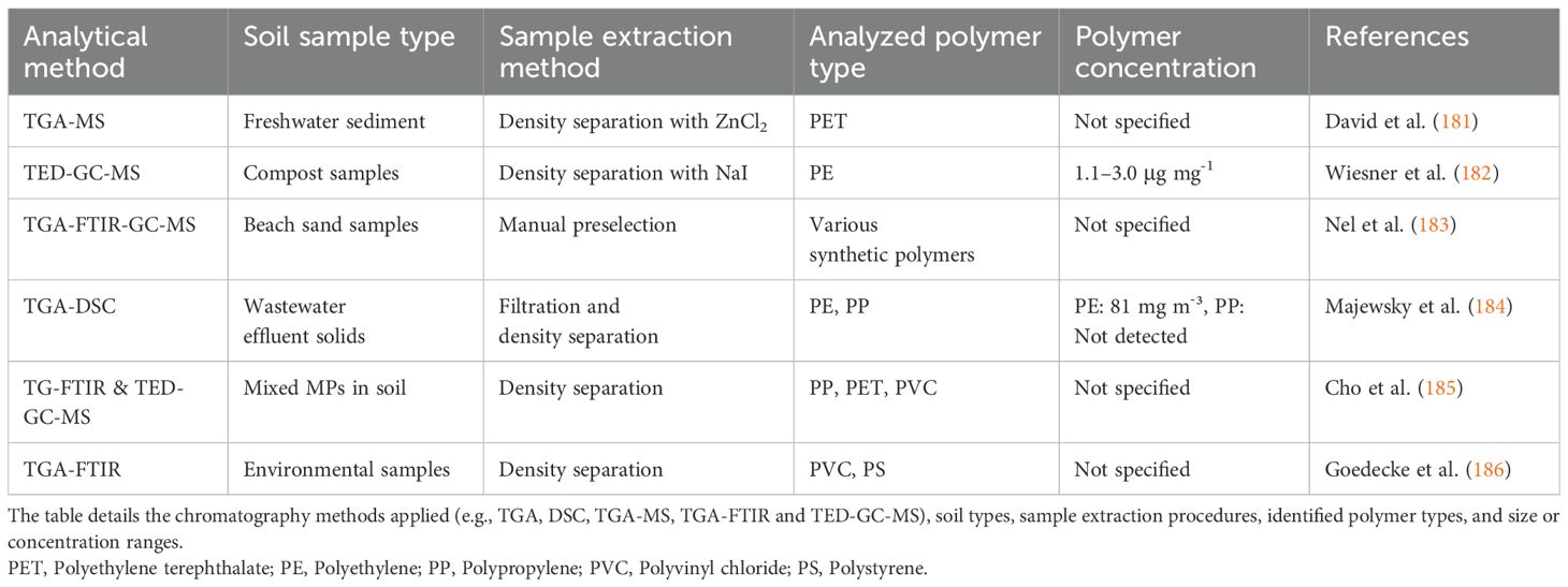
Table 6. Overview of studies utilizing thermal-based techniques for MPs identification in various soil and sediment matrices.
TGA can reach quick screening of MPs, and it is considered a straightforward method for identifying polymers with easy or even no sample preparation treatments (180). TGA works by gradually heating samples in the controlled atmosphere (nitrogen or air) while consistently recording the weight of samples. Considering that different components decompose or volatilize in corresponding characteristic temperatures, the mass loss would appear in different temperature ranges. Therefore, the information of polymers can be directly provided based on the mass losses in specific temperature ranges. Another advantage of using TGA is its flexibility of coupling with various technologies, such as MS, or FTIR. By coupling these technologies and detecting thermal degradation products, analysis of MPs in complex matrices can be achieved (186). Yu et al. (187) employed TGA-FTIR to identify and quantify MPs in mussels, seawater and soil. The polymers of PVC, PA and PS were successfully quantified; however, PE, PP and PET were not distinguishable and cannot be reliably determined by this method. Still, this method is straightforward and cost-effective (187). Dang et al. (188) proved that TGA-FTIR coupled with chemometrics can be applied to perform fast identification and quantification of PS in three matrices: water, liquid skimmed milk and ground coffee 2024. With their method, the organic removal steps are not necessary, though the less the complex matrices, the higher the sensitivity it has (188). David et al. (181) succeeded in using TGA-MS to direct PET in soil samples which were spiked MPs recycled from PET bottles. De la Fuente et al. (189) reached swift and accurate identification and quantification of PS and PE in organic amendments with minimal sample pre-treatment and no limitations on particle sizes. Liu et al. (190) developed the method of TGA-FTIR-GC/MS to study the mussels from coastal China. Four types of polymers, PE, PP, PVC and PS were quantified, and PE was found to be the most abundant type of MPs in mussels (190).
Differential Scanning Calorimetry (DSC) is another well-used thermal technology for MPs studies. DSC is a rapid, simple, and low-cost technology (191), and it investigates thermal properties of polymers, for instance, melting temperature and glass transition temperature (192). With the specific polymer temperatures, DSC presents the possibility to identify and quantify the Semi-crystalline polymer (PE, PP and PS) of MPs (193). In 2023, DSC was suggested for the detection of semi-crystalline polymer-based MPs by ISO 24187:2023 (194). The real plastic wastes, including ground lids (PP and PS), bottle tops (HDPE) and PET bottles, were analyzed with DSC by Lee et al. (34) and the masses of different polymers were obtained by applying Gaussian fitting model for amorphous PS and Asym 2 sig fitting model for semi-crystalline polymers (PET, PP, and HDPE) with minor range of errors (34). The TGA and DSC can be coupled as TGA-DSC technology. Abbasi et al. (195) applied TGA-DSC method to study a real aquatic environment, Maharloo Lake and its rivers, and the results showed that hydrogenation of TGA and DSC can help increase data quality of MPs, including concentrations and polymer type. Majewsky et al. (184) applied TGA-DSC to analyze two wastewater effluent samples from a municipal wastewater treatment plant. The samples contained 240 mg m-³ and 1540 mg m-³ of solid particles ranging from 12 µm to 1 mm. Of these, 34% (81 mg m-³) and 17% (257 mg m-³), respectively, were identified as PE. Nevertheless, they found that PE and PP can be clearly observed as separated peaks, and other selected polymers (PVC, PA, PES, PET, and PU) suffered from overlapping transition temperatures (184). Except for TGA and DSC-based techniques, TED-GC/MS allow for the analysis of entire samples, such as filters containing collected solids, without extensive handling. This reduces the risk of sample loss and contamination, ensuring more accurate results (196).
Thermal analysis techniques (e.g., TGA, DSC, TED-GC-MS) offer powerful capabilities for MPs research, enabling both quantitative analysis and chemical characterization across diverse environmental matrices. Unlike microscopy or spectroscopy, these methods provide unique insights into polymer composition, degradation behavior, and additive content through mass loss profiles, thermal transitions, and evolved gas analysis. However, critical challenges remain: (1) the absence of standardized protocols hinders reproducibility and inter-study comparisons, and (2) current thermal methods lack the sensitivity to detect nanoplastics (<1 μm), leaving a key gap in understanding the full environmental impact of plastic pollution.
6 Quality assurance and quality control
Quality assurance and quality control (QA/QC) are crucial in MPs research to ensure the accuracy, reliability, and comparability of results. Given the pervasive nature of MPs, contamination during sampling, processing, and analysis is a significant issue (197, 198). Without rigorous QA/QC measures, studies risk overestimating or underestimating MPs concentrations, leading to misleading conclusions about environmental and health impacts (Figure 4).
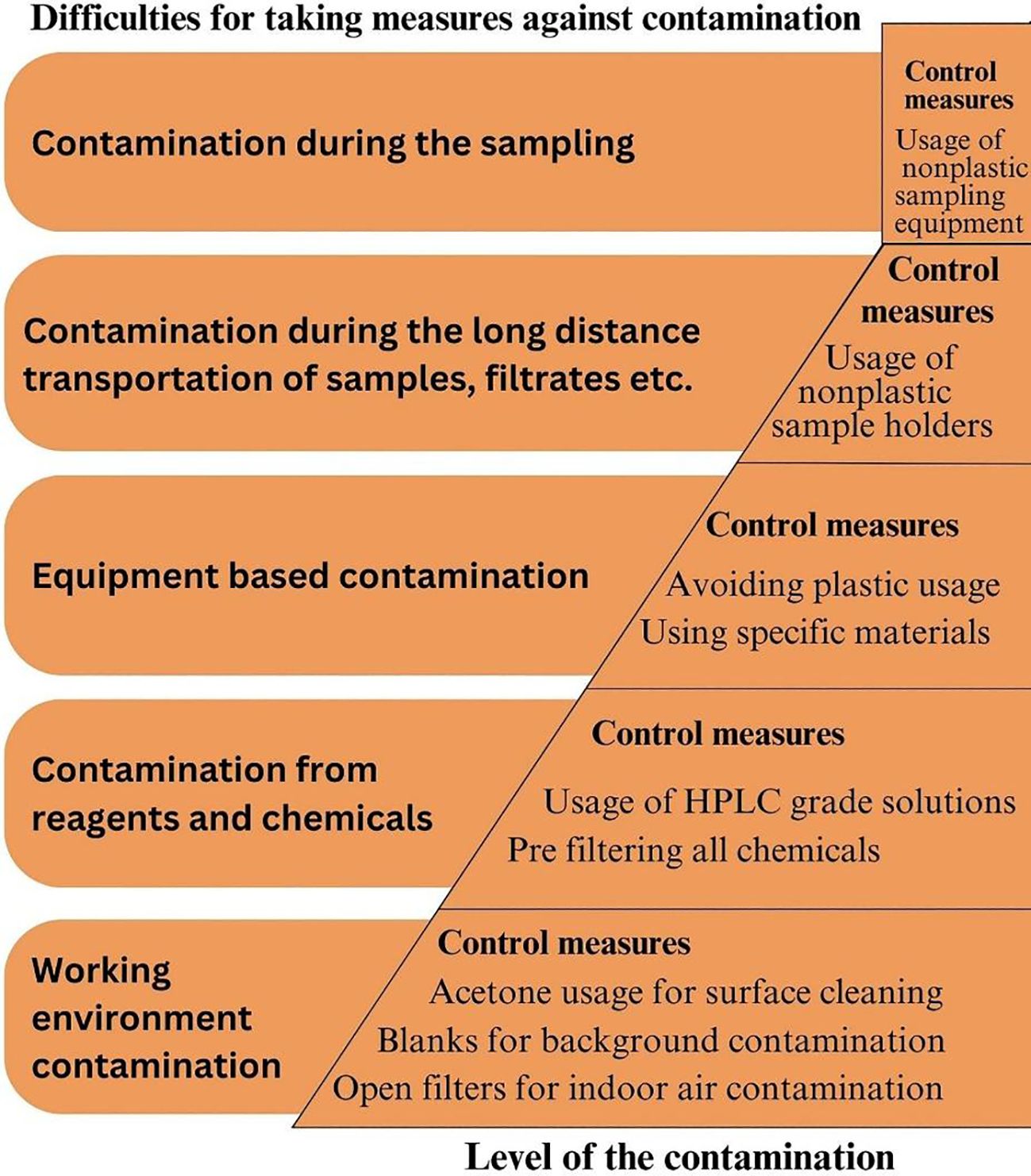
Figure 4. Hierarchy of contamination risks in MP research and corresponding control measures. The figure outlines major contamination sources—ranging from sampling to laboratory environment—and presents mitigation strategies such as the use of non-plastic materials, pre-filtering chemicals, and implementing blank controls to ensure data integrity.
One of the primary concerns in MPs studies is the potential for contamination, which can lead to overestimated concentrations and misleading conclusions. Common errors in MPs studies include airborne contamination, the use of plastic equipment, inconsistent methodologies, subjective identification techniques and lack of cleanliness of working environments. Airborne MPs, especially synthetic fibers, can contaminate samples if proper controls are not implemented. Many studies lack procedural blanks or fail to account for contamination from laboratory environments. The use of plastic-based lab materials can introduce synthetic particles, affecting data accuracy.
Furthermore, the absence of standardized methods in sampling, digestion, and polymer identification makes it difficult to compare results across studies. To minimize contamination, several measures should be implemented. Conducting work in controlled environments such as clean rooms, using procedural and field blanks, and avoiding plastic equipment are key strategies. Protective measures such as wearing natural fiber lab coats, using HEPA filters, and filtering all reagents help to reduce contamination risks. Standardized sample processing and employing spectroscopic techniques for polymer identification enhance the reliability of results. The adoption of rigorous QA/QC measures ensures that data on MPs are accurate and can be used to assess their environmental and health impacts effectively.
7 Conclusions and recommendations
The current state of MPs research in soil demonstrates considerable variability in methodology, resulting in challenges to data comparability and reliability. To address these issues, we strongly recommend developing internationally standardized analytical protocols tailored specifically for soil matrices. Future research must incorporate robust QA/QC frameworks, including the systematic use of blanks and controls to minimize cross-contamination. Enhanced methodological transparency and consistency will significantly improve data quality, facilitate comparative studies, and bolster policy-making efforts aimed at mitigating MPs pollution. Future research should concentrate on maximizing environmentally sustainable alternatives to improve the extraction efficiency of MPs from soil, while also enhancing the selectivity of conventional methods. Employing hybrid procedures that merge physical and chemical methods may enhance recovery rates and reduce contamination hazards. Furthermore, progress in automation and the standardization of extraction procedures is crucial for guaranteeing reproducibility across various soil types.
Utilizing multi-technique methodologies, such as SEM-Raman or fluorescence-assisted imaging, enhances the microscopic detection of MPs form and composition. Subsequent research ought to investigate AI-enhanced image analysis to augment detection precision and minimize manual processing duration. Standardized protocols for microscopy-based identification will improve comparability between investigations and augment data dependability. However, existing pyrolysis-based techniques for MPs detection possess limitations, including incomplete polymer identification due to interference from complex soil matrices, potential polymer degradation, and challenges in accurately quantifying low-abundance polymers. Similarly, conventional extraction methods often face limitations in effectively isolating smaller-sized and lower-density MPs due to their adherence to organic matter and soil particles, resulting in lower recovery rates and potential contamination risks.
Additionally, emphasis should be placed on enhancing interdisciplinary collaboration among environmental scientists, chemists, soil scientists, and policy-makers to foster comprehensive and integrated research approaches. Efforts must also be dedicated to understanding the long-term impacts of MPs on soil health, microbial communities, and agricultural productivity, thereby informing sustainable land management practices. Moreover, international collaborative initiatives should be encouraged to share knowledge, harmonize research methods, and standardize reporting practices, ensuring cohesive global efforts in tackling MPs contamination in terrestrial ecosystems.
Recent advances in machine learning (ML) and automated high-throughput systems offer promising avenues for overcoming long-standing analytical challenges in MP and NP detection. The integration of ML with spectroscopic techniques—particularly FTIR and Raman spectroscopy—enables efficient and accurate spectral pattern recognition, substantially reducing human error and processing time (199). These methods can autonomously extract multidimensional spectral features, improving detection sensitivity even in complex environmental matrices. Furthermore, AI-enhanced chemical imaging workflows now facilitate reliable NP mapping and quantification through algorithmic image processing and Gaussian surface fitting (200). Although these technologies remain underutilized in soil environments compared to aquatic systems, ongoing developments suggest they will play a crucial role in future environmental monitoring. A detailed comparative review of ML models and automated workflows is warranted to establish standard protocols and evaluate their robustness across various environmental compartments.
Although this study provides essential insights into MP contamination in selected soil environments, it does not encompass comparative evaluations across diverse soil types or propose a fully tiered analytical protocol; nonetheless, we underscore the need for standardized, matrix-sensitive workflows and recommend that future studies develop and validate tiered frameworks—ranging from rapid screening methods to high-resolution spectroscopic analyses—to improve methodological consistency, reproducibility, and applicability across environmental conditions and regulatory contexts.
Author contributions
SG: Visualization, Writing – original draft, Formal Analysis, Funding acquisition, Methodology, Supervision, Investigation, Conceptualization, Writing – review & editing. MA: Formal Analysis, Writing – original draft, Investigation, Writing – review & editing, Conceptualization. MG: Writing – original draft, Visualization, Investigation. MY: Writing – original draft, Investigation, Writing – review & editing. XZ: Writing – review & editing, Investigation, Writing – original draft. AR: Methodology, Investigation, Writing – review & editing, Writing – original draft. AB: Writing – review & editing, Investigation, Writing – original draft. MD: Investigation, Writing – review & editing, Writing – original draft. MV: Writing – review & editing, Writing – original draft, Investigation.
Funding
The author(s) declare that financial support was received for the research and/or publication of this article. SG is supported by project number FBA-2023-15165 from the Scientific Research Projects unit of Çukurova University (Türkiye). AR-S thanks the Ministerio de Ciencia e Innovación and the European Union Next Generation EU/PRTR for the postdoc grant Juan de la Cierva Incorporation 2020 (IJC2020-044197-I/MCIN/AEI/10.13039/501100011033). MV acknowledges the European Union project InPlasTwin, grant agreement No. 101160289.
Acknowledgments
This article/publication is based upon work from COST Action CA20101 Plastics monitoRIng detectiOn RemedIaTion recoverY—PRIORITY, supported by COST (European Cooperation in Science and Technology), www.cost.eu.
Conflict of interest
The authors declare that the research was conducted in the absence of any commercial or financial relationships that could be construed as a potential conflict of interest.
Generative AI statement
The author(s) declare that no Generative AI was used in the creation of this manuscript.
Publisher’s note
All claims expressed in this article are solely those of the authors and do not necessarily represent those of their affiliated organizations, or those of the publisher, the editors and the reviewers. Any product that may be evaluated in this article, or claim that may be made by its manufacturer, is not guaranteed or endorsed by the publisher.
References
1. Bergmann M, Almroth BC, Brander SM, Dey T, Green DS, Gundogdu S, et al. A global plastic treaty must cap production. Science. (2022) 376:469–70. doi: 10.1126/science.abq0082
2. Baztan J, Jorgensen B, Carney Almroth BM, Bergmann M, Farrelly T, Muncke J, et al. Primary Plastic Polymers: urgently needed upstream reduction. Cambridge Prisms: Plastics. (2024) 2:e7. doi: 10.1017/plc.2024.8
3. Thompson RC, Courtene-Jones W, Boucher J, Pahl S, Raubenheimer K, and Koelmans AA. Twenty years of microplastic pollution research-what have we learned? Science. (2024) 386:eadl2746. doi: 10.1126/science.adl2746
4. Gündoğdu S, Bour A, Köşker AR, Walther BA, Napierska D, Mihai F, et al. Review of microplastics and chemical risk posed by plastic packaging on the marine environment to inform the Global Plastic Treaty. Sci Total Environ. (2024) 946:174000. doi: 10.1016/j.scitotenv.2024.174000
5. Singh S, Chakma S, Alawa B, Kalyanasundaram M, and Diwan V. Identification, characterization, and implications of microplastics in soil–a case study of Bhopal, central India. J Hazard Mater Adv. (2023) 9:100225. doi: 10.1016/j.hazadv.2022.100225
6. Akça MO. Microplastic accumulation in soils around open dumping and scrapyard sites in Türkiye. Soil Use Manage. (2025) 41:e70021. doi: 10.1111/sum.70021
7. Wang YF, Liu YJ, Fu YM, Xu JY, Zhang TL, Cui HL, et al. Microplastic diversity increases the abundance of antibiotic resistance genes in soil. Nat Commun. (2024) 15:9788. doi: 10.1038/s41467-024-54237-7
8. Napper IE and Thompson RC. Plastics and the environment. Ann Rev Environ Res. (2023) 48:55–79. doi: 10.1146/annurev-environ-112522-072642
9. Daghighi E, Shah T, Wainkwa Chia R, Lee J, Shang J, and Rodríguez-Seijo A. The forgotten impacts of plastic contamination on terrestrial micro- and mesofauna: A call for research. Environ Res. (2023) 231:116227. doi: 10.1016/j.envres.2023.116227
10. Weber CJ. Plastics in soil description and surveys–practical considerations and field guide. Front Soil Sci. (2022) 2:917490. doi: 10.3389/fsoil.2022.917490
11. Allen S, Allen D, Phoenix VR, Le Roux G, Durántez Jiménez P, Binet S, et al. Atmospheric transport and deposition of microplastics in a remote mountain catchment. Nat Geosci. (2019) 12:339–44. doi: 10.1038/s41561-019-0335-5
12. Prata JC, da Costa JP, Duarte AC, and Rocha-Santos TA. Methods for sampling and detection of microplastics in water and sediment: A critical review. TrAC Trends Anal Chem. (2019) 110:150–9. doi: 10.1016/j.trac.2018.10.029
13. ISO. ISO/DIS 16094-2. 2024. Water quality — Analysis of microplastic in water Part 2: Vibrational spectroscopy methods for waters with low content of suspended solids including drinking water. Switzerland: ISO (International Organization for Standardization (2024). Available at: https://www.iso.org/standard/84460.html (Accessed April, 2025).
14. Möller JN, Löder MGJ, and Laforsch C. Finding microplastics in soils: A review of analytical methods. Environ Sci Technol. (2020) 54:2078–90. doi: 10.1021/acs.est.9b04618
15. Chia RW, Lee J, Cha J, and Rodríguez-Seijo A. Methods of soil sampling for microplastic analysis: a review. Environ Chem Lett. (2024) 22:227–38. doi: 10.1007/s10311-023-01652-9
16. Rillig MC, Ziersch L, and Hempel S. Microplastic transport in soil by earthworms. Sci Rep. (2017) 7:1362. doi: 10.1038/s41598-017-01594-7
17. He D, Luo Y, Lu S, Liu M, Song Y, and Lei L. Microplastics in soils: Analytical methods, pollution characteristics and ecological risks. TrAC Trends Anal Chem. (2018) 109:163–72. doi: 10.1016/j.trac.2018.10.006
18. Zhang Y, Kang S, Allen S, Allen D, Gao T, and Sillanpää M. Atmospheric microplastics: a review on current status and perspectives. Earth Sci Rev. (2020) 203:103118. doi: 10.1016/j.earscirev.2020.103118
19. Sun D, Li H, Wang E, He W, Hao W, Yan C, et al. An overview of the use of plastic-film mulching in China to increase crop yield and water-use efficiency. Natl Sci Rev. (2020) 7:1523–6. doi: 10.1093/nsr/nwaa146
20. Stubbins A, Law KL, Muñoz SE, Bianchi TS, and Zhu L. Plastics in the earth system. Science. (2021) 373:51–5. doi: 10.1126/science.abb0354
21. Weber CJ and Opp C. Spatial patterns of mesoplastics and coarse microplastics in floodplain soils as resulting from land use and fluvial processes. Environ pollut. (2020) 267:115390. doi: 10.1016/j.envpol.2020.115390
22. Büks F and Kaupenjohann M. Global concentrations of microplastics in soils - a review. Soil. (2020) 6:649–62. doi: 10.5194/soil-6-649-2020
23. Akça MO, Gündoğdu S, Akca H, Delialioğlu RA, Aksit C, Turgay OC, et al. An evaluation on microplastic accumulations in Turkish soils under different land uses. Sci Total Environ. (2024) 911:168609. doi: 10.1016/j.scitotenv.2023.168609
24. En-Nejmy K, El Hayany B, Al-Alawi M, Jemo M, Hafidi M, and El Fels L. Microplastics in soil: A comprehensive review of occurrence, sources, fate, analytical techniques and potential impacts. Ecotox Environ Saf. (2024) 288:117332. doi: 10.1016/j.ecoenv.2024.117332
25. Kang Q, Zhang K, Dekker SC, and Mao J. Microplastics in soils: A comprehensive review. Sci Total Environ. (2025) 960:178298. doi: 10.1016/j.scitotenv.2024.178298
26. Guo S, Wu Z, Li X, Shen D, Shentu J, Lu L, et al. Microplastic, a possible trigger of landfill sulfate reduction process. Sci Total Environment. (2024) 906:167662. doi: 10.1016/j.scitotenv.2023.167662
27. Gundogdu S, Mihai FC, Fischer EK, Blettler MC, Turgay OC, Akça MO, et al. Micro and nano plastics in groundwater systems: A review of current knowledge and future perspectives. TrAC Trends Anal Chem. (2023) 165:117119. doi: 10.1016/j.trac.2023.117119
28. Gündoğdu S, Köşker AR, Akça H, Akça MO, Harada N, and Turgay OC. Microplastics in food production and agricultural environments. In: Avino P, Di Fiore C, and Farris S, editors. Microplastics in Agriculture and Food Science. 125 London Wall, London EC2Y 5AS, United Kingdom: Academic Press (2025). p. 285–307. doi: 10.1016/B978-0-443-22210-8.00022-5
29. Zhang M, Zheng Y, Li J, Liu K, Wang H, Gu H, et al. Distribution characteristics of microplastics in soil of Loess Plateau in northwest China and their relationship with land use type. Sci Total Environ. (2023) 868:161674. doi: 10.1016/j.scitotenv.2023.161674
30. Heinze WM, Steinmetz Z, Klemmensen NDR, Vollertsen J, and Cornelis G. Vertical distribution of microplastics in an agricultural soil after long-term treatment with sewage sludge and mineral fertiliser. Environ pollut. (2024) 356:124343. doi: 10.1016/j.envpol.2024.124343
31. Beaurepaire M, de Oliveira T, Gasperi J, Tramoy R, Saad M, Tassin B, et al. Stock and vertical distribution of microplastics and tire and road wear particles into the soils of a high-traffic roadside biofiltration swale. Environ pollut. (2025) 373:126092. doi: 10.1016/j.envpol.2025.126092
32. Feng S, Lu H, and Liu Y. The occurrence of microplastics in farmland and grassland soils in the Qinghai-Tibet plateau: different land use and mulching time in facility agriculture. Environ pollut. (2021) 279:116939. doi: 10.1016/j.envpol.2021.116939
33. Zhang S, Yang X, Gertsen H, Peters P, Salánki T, and Geissen V. A simple method for the extraction and identification of light density microplastics from soil. Sci Total Environ. (2018) 616-617:1056–65. doi: 10.1016/j.scitotenv.2017.10.213
34. Lee J, Yoon S, Jang T, Choi JH, Kim N, Kim HO, et al. A facile approach to microplastic identification and quantification using differential scanning calorimetry. Sci Total Environ. (2024) 957:177456. doi: 10.1016/j.scitotenv.2024.177456
35. Dorau K, Hoppe M, Rückamp D, Köser J, Scheeder G, Scholz K, et al. Status quo of operation procedures for soil sampling to analyze microplastics. Micropl Nanopl. (2023) 3:15. doi: 10.1186/s43591-023-00063-5
36. Brewer R, Peard J, and Heskett M. A critical review of discrete soil sample data reliability: part 2—implications. Soil Sediment Contam. (2017) 26:23–44. doi: 10.1080/15320383.2017.1244172
37. Hyde K, Ma W, Obal T, Bradshaw K, Carlson T, Mamet S, et al. Incremental sampling methodology for petroleum hydrocarbon contaminated soils: volume estimates and remediation strategies. Soil Sediment Contam. (2019) 28:51–64. doi: 10.1080/15320383.2018.1529736
38. Li S, Li Z, Xue J, Chen S, Li H, Ji J, et al. Pollution and distribution of microplastics in grassland soils of Qinghai-Tibet Plateau. China Toxics. (2023) 11:86. doi: 10.3390/toxics11010086
39. Petrovskaia A, Ryzhakov G, and Oseledets I. Optimal soil sampling design based on the maxvol algorithm. Geoderma. (2021) 402:115362. doi: 10.1016/j.geoderma.2021.11536
40. Sarkar A, Deb S, Ghosh S, Mandal S, Quazi SA, Kushwaha A, et al. Impact of anthropogenic pollution on soil properties in and around a town in Eastern India. Geoderma Reg. (2022) 28:e00462. doi: 10.1016/j.geodrs.2021.e00462
41. Zhang Y, Gao T, Kang S, Shi H, Mai L, Allen D, et al. Current status and future perspectives of microplastic pollution in typical cryospheric regions. Earth Sci Rev. (2022) 226:103924. doi: 10.1016/j.earscirev.2022.103924
42. Yang L, Kang S, Wang Z, Luo X, Guo J, Gao T, et al. Microplastic characteristic in the soil across the Tibetan Plateau. Sci Total Environ. (2022) 828:154518. doi: 10.1016/j.scitotenv.2022.154518
43. Wang G, Singh M, Wang J, Xiao L, and Guan D. Effects of marine pollution, climate, and tidal range on biomass and sediment organic carbon in Chinese mangrove forests. Catena. (2021) 202:105270. doi: 10.1016/j.catena.2021.105270
44. Tian L, Jinjin C, Ji R, Ma Y, and Yu X. Microplastics in agricultural soils: sources, effects, and their fate. Curr Opin Environ Sci. (2022) 25:100311. doi: 10.1016/j.coesh.2021.100311
45. Khan MA, Huang Q, Khan S, Wang Q, Huang J, Fahad S, et al. Abundance, spatial distribution, and characteristics of microplastics in agricultural soils and their relationship with contributing factors. J Environ Manage. (2023) 328:117006. doi: 10.1016/j.jenvman.2022.117006
46. Xu G, Yang L, Xu L, and Yang J. Soil microplastic pollution under different land uses in tropics, southwestern China. Chemosphere. (2022) 289:133176. doi: 10.1016/j.chemosphere.2021.133176
47. Zhang GS and Liu YF. The distribution of microplastics in soil aggregate fractions in southwestern China. Sci Total Environ. (2018) 642:12–20. doi: 10.1016/j.scitotenv.2018.06.004
48. Miller ME, Kroon FJ, and Motti CA. Recovering microplastics from marine samples: A review of current practices. Mar pollut Bull. (2017) 123:6–18. doi: 10.1016/j.marpolbul.2017.08.058
49. Huerta Lwanga E, Mendoza Vega J, Ku Quej V, Chi J de lA, Sanchez del Cid L, Chi C, et al. Field evidence for transfer of plastic debris along a terrestrial food chain. Sci Rep. (2017) 7:1–7. doi: 10.1038/s41598-017-14588-2
50. ISO. ISO 11465:1993. Soil quality — Determination of dry matter and water content on a mass basis — Gravimetric method. Switzerland: ISO (International Organization for Standardization (1993). Available at: https://www.iso.org/standard/20886.html (Accessed April, 2025 ).
51. Priya AK, Jalil AA, Dutta K, Rajendran S, Vasseghian Y, Qin J, et al. Microplastics in the environment: recent developments in characteristic, occurrence, identification and ecological risk. Chemosphere. (2022) 298:134161. doi: 10.1016/j.chemosphere.2022.134161
52. Fojt J, David J, Přikryl R, Řezáčová V, and Kučerík J. A critical review of the overlooked challenge of determining micro-bioplastics in soil. Sci Total Environ. (2020) 745:140975. doi: 10.1016/j.scitotenv.2020.140975
53. Junhao C, Xining Z, Xiaodong G, Li Z, Qi H, and Siddique KHM. Extraction and identification methods of microplastics and nanoplastics in agricultural soil: A review. J Environ Manage. (2021) 294:112997. doi: 10.1016/j.jenvman.2021.112997
54. Rani M, Ducoli S, Depero LE, Prica M, Tubi A, Ademovic Z, et al. A complete guide to extraction methods of microplastics from complex environmental matrices. Molecules. (2023) 28:5710. doi: 10.3390/molecules28155710
55. Han X, Lu X, and Vogt. RD. An optimized density-based approach for extracting microplastics from soil and sediment samples. Environ pollut. (2019) 254:113009. doi: 10.1016/j.envpol.2019.113009
56. Kononov A, Hishida M, Suzuki K, and Harada N. Microplastic extraction from agricultural soils using canola oil and unsaturated sodium chloride solution and evaluation by incineration method. Soil Systems. (2022) 6:54. doi: 10.3390/soilsystems6020054
57. Hu H, Qiang L, Xu J, Li G, Cheng J, Zhong X, et al. Enhanced microplastic retrieval efficiency from cultivated soil samples through optimized pre-treatment in density-based extraction. Soil Till Res. (2024) 242:106134. doi: 10.1016/j.still.2024.106134
58. Prosenc F, Leban P, Šunta U, and Kralj MB. Extraction and identification of a wide range of microplastic polymers in soil and compost. Polymers. (2021) 13:4069. doi: 10.3390/polym13234069
59. Zhang M, Zhao Y, Qin X, Jia W, Chai L, Huang M, et al. Microplastics from mulching film is a distinct habitat for bacteria in farmland soil. Sci Total Environ. (2019) 688:470–8. doi: 10.1016/j.scitotenv.2019.06.108
60. Luo Y, Zhang Y, Xu Y, Guo X, and Zhu L. Distribution characteristics and mechanism of microplastics mediated by soil physicochemical properties. Sci Total Environ. (2020) 726:138389. doi: 10.1016/j.scitotenv.2020.138389
61. Gupta E, Mishra VK, Patel A, and Srivastava PK. A modified methodology for extraction and quantification of microplastics in soil. NanoImpact. (2024) 35:100525. doi: 10.1016/j.impact.2024.100525
62. Yoon JH, Kim BH, and Kim KH. Distribution of microplastics in soil by types of land use in metropolitan area of Seoul. Appl Biol Chem. (2024) 67:15. doi: 10.1186/s13765-024-00869-8
63. Hu B, Guo P, Han S, Jin Y, Nan Y, Deng J, et al. Distribution characteristics of microplastics in the soil of mangrove restoration wetland and the effects of microplastics on soil characteristics. Ecotoxicology. (2022) 31:1120–36. doi: 10.1007/s10646-022-02561-3
64. Yang J, Li L, Li R, Xu L, Shen Y, Li S, et al. Microplastics in an agricultural soil following repeated application of three types of sewage sludge: A field study. Environ pollut. (2021) 289:117943. doi: 10.1016/j.envpol.2021.117943
65. Yang X, Zhang Z, and Guo X. Impact of soil structure and texture on occurrence of microplastics in agricultural soils of karst areas. Sci Total Environ. (2023) 902:166189. doi: 10.1016/j.scitotenv.2023.166189
66. Kanold E, Rillig M, and Antunes PM. Microplastics and phagotrophic soil protists: evidence of ingestion. Soil Org. (2021) 93:2. doi: 10.25674/so93iss2id160
67. Kim SW, Jeong SW, and An YJ. Microplastics disrupt accurate soil organic carbon measurement based on chemical oxidation method. Chemosphere. (2021) 276:130178. doi: 10.1016/j.chemosphere.2021.130178
68. Yang J, Song K, Tu C, Li L, Feng Y, Li R, et al. Distribution and weathering characteristics of microplastics in paddy soils following long-term mulching: a field study in Southwest China. Sci Total Environ. (2023) 858:159774. doi: 10.1016/j.scitotenv.2022.159774
69. Ding L, Zhang S, Wang X, Yang X, Zhang C, Qi Y, et al. The occurrence and distribution characteristics of microplastics in the agricultural soils of Shaanxi Province, in north-western China. Sci Total Environ. (2020) 720:137525. doi: 10.1016/j.scitotenv.2020.137525
70. Liu J, Qin J, Zhu L, Zhu K, Liu Z, Jia H, et al. The protective layer formed by soil particles on plastics decreases the toxicity of polystyrene microplastics to earthworms (Eisenia fetida). Environ Int. (2022) 162:107158. doi: 10.1016/j.envint.2022.107158
71. Tang KHD and Luo Y. Abundance and characteristics of microplastics in the soil of a higher education institution in China. Trop Aquat Soil pollut. (2023) 3:1–14. doi: 10.53623/tasp.v3i1.152
72. Adhikari K, Pearce CI, Sanguinet KA, Bary AI, Chowdhury I, Eggleston I, et al. Accumulation of microplastics in soil after long-term application of biosolids and atmospheric deposition. Sci Total Environment. (2024) 912:168883. doi: 10.1016/j.scitotenv.2023.168883
73. Harms IK, Diekötter T, Troegel S, and Lenz M. Amount, distribution and composition of large microplastics in typical agricultural soils in Northern Germany. Sci Total Environ. (2021) 758:143615. doi: 10.1016/j.scitotenv.2020.143615
74. MaChado AAS, Lau CW, Kloas W, Bergmann J, Faltin E, Becker R, et al. Microplastics can change soil properties and affect plant performance. Environ Sci Technol 53. (2019) 10:6044–52. doi: 10.1021/acs.est.9b01339
75. Wiedner K and Polifka S. Effects of microplastic and microglass particles on soil microbial community structure in an arable soil (Chernozem). Soil. (2020) 6:315–24. doi: 10.5194/soil-6-315-2020
76. Li G, Pei Z, Li Y, Yang R, Wang P, Liang Y, et al. A high-precision, effective method for extraction and identification of small-sized microplastics from soil. Talanta. (2024) 272:125802. doi: 10.1016/j.talanta.2024.125802
77. Huang AC, Nelson C, Elliott JE, Guertin DA, Ritland C, Drouillard K, et al. River otters (Lontra canadensis) “trapped” in a coastal environment contaminated with persistent organic pollutants: Demographic and physiological consequences. Environ pollut. (2018) 238:306–16. doi: 10.1016/j.envpol.2018.03.035
78. Debraj D and Lavanya M. Microplastics everywhere: A review on existing methods of extraction. Sci Total Environ. (2023) 893:164878. doi: 10.1016/j.scitotenv.2023.164878
79. Sajid M and Justyna P. Green analytical chemistry metrics: a review. Talanta. (2022) 238:123046. doi: 10.1016/j.talanta.2021.123046
80. Pham DT, Park HJ, M.S. Mohamed DF, Kim PG, Tarafdar A, and Kwon JH. Greenness of analytical methods for isolation and identification of microplastics in environmental and food samples - A critical review. TrAC Trends Anal Chem. (2023) 167:117263. doi: 10.1016/j.trac.2023.117263
81. Prasad Kasa V, Thomas AP, Bordoloi S, De Bhowmick G, Dubey BK, and Sarmah AK. Microplastics in soil: Current status and evaluation of the greenness of various analytical methods of identification. Green Anal Chem. (2022) 3:100038. doi: 10.1016/j.greeac.2022.100038
82. Bellasi A, Binda G, Pozzi A, Boldrocchi G, and Bettinetti R. The extraction of microplastics from sediments: an overview of existing methods and the proposal of a new and green alternative. Chemosphere. (2021) 278:130357. doi: 10.1016/j.chemosphere.2021.130357
83. Rede D, Vilarinho R, Moreira JA, Nizzetto L, Delerue-Matos C, and Cruz Fernandes V. Screening for microplastics in agricultural soils: Applying green chemistry principles in extraction and analysis. Environ pollut. (2025) 367:125550. doi: 10.1016/j.envpol.2024.125550
84. Crichton EM, Noeül M, Gies EA, and Ross PS. A novel, density-independent and FTIR- compatible approach for the rapid extraction of microplastics from aquatic sediments. Anal Methods. (2017) 9:1419–28. doi: 10.1039/C6AY02733D
85. Mani T, Frehland S, Kalberer A, and Burkhardt-Holm P. Using castor oil to separate microplastics from four different environmental matrices. Anal Methods. (2019) 11:1788–94. doi: 10.1039/C8AY02559B
86. Scopetani C, Chelazzi D, Mikola J, Leiniö V, Heikkinen R, Cincinelli A, et al. Olive oil-based method for the extraction, quantification and identification of microplastics in soil and compost samples. Sci Total Environ. (2020) 733:139338. doi: 10.1016/j.scitotenv.2020.139338
87. Lekše N, Žgajnar Gotvajn A, Zupančič M, and Griessler Bulc T. Oil-based extraction as an efficient method for the quantification of microplastics in environmental samples. Environ Sci Eur. (2024) 36:68. doi: 10.1186/s12302-024-00898-6
88. Hansda A, Keshari Chand S, Pradhan B, Chand S, Shukla AK, and Rout PR. Toxicological impacts and microbial-mediated degradation processes of microplastics. J Hazard Toxic Radioact Waste. (2024) 29:2. doi: 10.1061/JHTRBP.HZENG-1400
89. Cárdenas-Alcaide MF, Godínez-Alemán JF, González-González RB, Iqbal HMN, and Parra-Saldívar R. Environmental impact and mitigation of micro(nano)plastics pollution using green catalytic tools and green analytical methods. Green Anal Chem. (2022) 3:100031. doi: 10.1016/j.greeac.2022.100031
90. Sun C, Wang Z, Zheng H, Chen L, and Li F. Biodegradable and re-usable sponge materials made from chitin for efficient removal of microplastics. J Hazard Mater. (2021) 420:126599. doi: 10.1016/j.jhazmat.2021.126599
91. Liu X, Li MC, Lu Y, Li Z, Liu C, Liu Z, et al. Cellulose nanofiber-coated delignified wood as an efficient filter for microplastic removal. Prog Nat Sci: Mater Int. (2024) 34:162–71. doi: 10.1016/j.pnsc.2024.02.010
92. Hunter JR, Qiao Q, Zhang Y, Crofcheck C, and Shi J. Green solvent mediated extraction of micro- and nano-plastic particles from water. Sci Rep. (2023) 13:10585. doi: 10.1038/s41598-023-37490-6
93. Ishtaweera P, Ray CL, Filley W, Cobb G, and Baker GA. Nanoplastics extraction from water by hydrophobic deep eutectic solvents. ACS Appl Eng Mater. (2024) 2:1460–6. doi: 10.1021/acsaenm.4c00159
94. Zhang Y, Hunter JR, Ullah A, Shao Q, and Shi J. Lignin derived hydrophobic deep eutectic solvents for the extraction of nanoplastics from water. J Hazard Mater. (2024) 467:133695. doi: 10.1016/j.jhazmat.2024.133695
95. Pena-Pereira F, Wojnowski W, and Tobiszewski M. AGREE—analytical GREEnness metric approach and software. Anal Chem. (2020) 92:10076–82. doi: 10.1021/acs.analchem.0c01887
96. Magalhães S, Alves L, Medronho B, Romano A, and Rasteiro M. Microplastics in ecosystems: from current trends to bio-based removal strategies. Molecules. (2020) 25:3954. doi: 10.3390/molecules25173954
97. Picó Y and Barceló D. Analysis of microplastics and nanoplastics: how green are the methodologies used? Curr Opin Green Sustain Chem. (2021) 31:100503. doi: 10.1016/j.cogsc.2021.100503
98. Picó Y and Barceló D. Micro(Nano)plastic analysis: a green and sustainable perspective. J Hazard Mater Adv. (2022) 6:100058. doi: 10.1016/j.hazadv.2022.100058
99. Bibi A, Can A, Pant U, Hardiman G, Hill D, Elliott C, et al. A review on state-of-the-art detection techniques for micro-and nano-plastics with prospective use in point-of-site detection. Compr Anal Chem. (2023) 101:143–96. doi: 10.1016/bs.coac.2022.11.003
100. Eriksen M, Mason S, Wilson S, Box C, Zellers A, Edwards W, et al. Microplastic pollution in the surface waters of the Laurentian Great Lakes. Mar pollut Bull. (2013) 77:177–82. doi: 10.1016/j.marpolbul.2013.10.007
101. Hidalgo-Ruz V, Gutow L, Thompson RC, and Thiel M. Microplastics in the marine environment: a review of the methods used for identification and quantification. Environ Sci Technol. (2012) 46:3060–75. doi: 10.1021/es2031505
102. Löder MGJ and Gerdts G. Methodology used for the detection and identification of microplastics—A critical appraisal. In: Bergmann M, Gutow L, and Klages M, editors. Marine Anthropogenic Litter. Springer International Publishing, Cham (2015). p. 201–27. doi: 10.1007/978-3-319-16510-3_8
103. Beckingham B, Apintiloaiei A, Moore C, and Brandes J. Hot or not: systematic review and laboratory evaluation of the hot needle test for microplastic identification. Micropl Nanopl. (2023) 3:8. doi: 10.1186/s43591-023-00056-4
104. Gao Z, Wontor K, and Cizdziel JV. Labeling microplastics with fluorescent dyes for detection, recovery, and degradation experiments. Molecules. (2022) 27:7415. doi: 10.3390/molecules27217415
105. Meyers N, Catarino AI, Declercq AM, Brenan A, Devriese L, Vandegehuchte M, et al. Microplastic detection and identification by Nile red staining: Towards a semi-automated, cost- and time-effective technique. Sci Total Environ. (2022) 823:153441. doi: 10.1016/j.scitotenv.2022.153441
106. Ainé L, Jacquin J, Breysse C, Colin C, Andanson J-M, and Delor-Jestin F. Microplastics and nanoplastics detection using flow cytometry: Challenges and methodological advances with fluorescent dye application. MethodsX. (2025) 14:103200. doi: 10.1016/j.mex.2025.103200
107. Kang H, Park S, Lee B, Ahn J, and Kim S. Modification of a nile red staining method for microplastics analysis: A nile red plate method. Water. (2020) 12:3251. doi: 10.3390/w12113251
108. Shruti VC, Pérez-Guevara F, Roy PD, and Kutralam-Muniasamy G. Analyzing microplastics with Nile Red: Emerging trends, challenges, and prospects. J hazardous materials. (2022) 423:127171. doi: 10.1016/j.jhazmat.2021.127171
109. Erni-Cassola G, Gibson MI, Thompson RC, and Christie-Oleza JA. Lost, but found with Nile red: a novel method for detecting and quantifying small microplastics (1 mm to 20 μm) in environmental samples. Environ Sci Technol. (2017) 51:13641–8. doi: 10.1021/acs.est.7b04512
110. Rocha-Santos T and Duarte AC. A critical overview of the analytical approaches to the occurrence, the fate and the behavior of microplastics in the environment. TrAC Trends Anal Chem. (2015) 65:47–53. doi: 10.1016/j.trac.2014.10.011
111. Crawford CB and Quinn B. Chapter 10 - Microplastic identification techniques. In: Crawford CB and Quinn B, editors. Microplastic Pollutants. 125 London Wall, London EC2Y 5AS, United Kingdom: Elsevier (2017). p. 219–67. doi: 10.1016/B978-0-12-809406-8.00010-4
112. Fries E, Dekiff JH, Willmeyer J, Nuelle MT, Ebert M, and Remy D. Identification of polymer types and additives in marine microplastic particles using pyrolysis-GC/MS and scanning electron microscopy. Environ Sci: Process Impacts. (2013) 15:1949–56. doi: 10.1039/C3EM00214D
113. Arenas-Lago D, Santás-Miguel V, and Rodríguez-Seijo A. Current methodology for extraction, separation, identification, and quantification of microplastics in terrestrial systems. In: Núñez-Delgado A and Arias-Estévez M, editors. Emerging Pollutants in Sewage Sludge and Soils. The Handbook of Environmental Chemistry, vol. 114 . Springer, Cham (2022). p. 267–87. doi: 10.1007/698_2022_859
114. De-la-Torre GE, Dioses-Salinas DC, Severini MDF, López ADF, Dobaradaran S, Toapanta TYA, et al. Chapter 4 - Aims of the analysis and the choices of analytical methods. In: Shi H and Sun C, editors. Analysis of Microplastics and Nanoplastics. 125 London Wall, London EC2Y 5AS, United Kingdom: Elsevier (2025). p. 63–77. doi: 10.1016/B978-0-443-15779-0.00001-8
115. Ramage SJFF, Pagaling E, Haghi RK, Dawson LA, Yates K, Prabhu R, et al. Rapid extraction of high- and low-density microplastics from soil using high gradient magnetic separation. Sci Total Environ. (2022) 831:154912. doi: 10.1016/j.scitotenv.2022.154912
116. Park SY and Kim CG. A comparative study on the distribution behavior of microplastics through FT-IR analysis on different land uses in agricultural soils. Environ Res. (2022) 215:114404. doi: 10.1016/j.envres.2022.114404
117. Corradini F, Casado F, Leiva V, Huerta-Lwanga E, and Geissen V. Microplastics occurrence and frequency in soils under different land uses on a regional scale. Sci Total Environ. (2021) 752:141917. doi: 10.1016/j.scitotenv.2020.141917
118. Guo S, Zhang J, Liu J, Guo N, Zhang L, Wang S, et al. Organic fertilizer and irrigation water are the primary sources of microplastics in the facility soil, Beijing. Sci Total Environ. (2023) 895:165005. doi: 10.1016/j.scitotenv.2023.165005
119. Afrin S, Uddin MK, and Rahman MM. Microplastics contamination in the soil from Urban Landfill site, Dhaka, Bangladesh. Heliyon. (2020) 6:e05572. doi: 10.1016/j.heliyon.2020.e05572
120. Kim SK, Kim JS, Lee H, and Lee HJ. Abundance and characteristics of microplastics in soils with different agricultural practices: Importance of sources with internal origin and environmental fate. J Hazard Mater. (2021) 403:123997. doi: 10.1016/j.jhazmat.2020.123997
121. Ruffell H, Pantos O, Robinson B, and Gaw S. A method for the extraction of microplastics from solid biowastes including biosolids, compost, and soil for analysis by µ-FTIR. MethodsX. (2024) 12:102761. doi: 10.1016/j.mex.2024.102761
122. Kim WK, Park H, Ishii K, and Ham GY. Investigation on microplastics in soil near landfills in the Republic of Korea. Sustainability. (2023) 15:12057. doi: 10.3390/su151512057
123. Heerey L, O’Sullivan JJ, Bruen M, Turner J, Mahon AM, Murphy S, et al. Export pathways of biosolid derived microplastics in soil systems - Findings from a temperate maritime climate. Sci Total Environ. (2023) 888:164028. doi: 10.1016/j.scitotenv.2023.164028
124. Musthafa JM and Mandal BK. Laboratory designed portable device for density separation and characterization of microplastics in environmental soil samples. Lett Appl NanoBioScience. (2024) 13:127. doi: 10.33263/LIANBS133.127
125. Chen M, Coleman B, Gaburici L, Prezgot D, Jakubek ZJ, Sivarajah B, et al. Identification of microplastics extracted from field soils amended with municipal biosolids. Sci Total Environ. (2024) 907:168007. doi: 10.1016/j.scitotenv.2023.168007
126. Fu F, Sun Y, Yang D, Zhao L, Li X, Weng L, et al. Combined pollution and soil microbial effect of pesticides and microplastics in greenhouse soil of suburban Tianjin, Northern China. Environ pollut. (2024) 340:122898. doi: 10.1016/j.envpol.2023.122898
127. Okoffo ED, O’Brien S, Ribeiro F, Burrows SD, Toapanta T, Rauert C, et al. Plastic particles in soil: state of the knowledge on sources, occurrence and distribution, analytical methods and ecological impacts. Environ Sci Process Impacts. (2021) 23:240–74. doi: 10.1039/d0em00312c
128. Cabernard L, Roscher L, Lorenz C, Gerdts G, and Primpke S. Comparison of Raman and Fourier transform infrared spectroscopy for the quantification of microplastics in the aquatic environment. Environ Sci Technol. (2018) 52:13279–88. doi: 10.1021/acs.est.8b03438
129. Huang Z, Hu B, and Wang H. Analytical methods for microplastics in the environment: a review. Environ Chem Lett. (2023) 21:383–401. doi: 10.1007/s10311-022-01525-7
130. Du Z, Li G, Ding S, Song W, Zhang M, Jia R, et al. Effects of UV-based oxidation processes on the degradation of microplastic: Fragmentation, organic matter release, toxicity and disinfection byproduct formation. Water Res. (2023) 237:119983. doi: 10.1016/j.watres.2023.119983
131. Andrady AL. Weathering and fragmentation of plastic debris in the ocean environment. Mar pollut Bull. (2022) 180:113761. doi: 10.1016/j.marpolbul.2022.113761
132. Sandt C, Waeytens J, Deniset-Besseau A, Nielsen-Leroux C, and Réjasse A. Use and misuse of FTIR spectroscopy for studying the bio-oxidation of plastics. Spectrochim Acta A Mol Biomol Spectrosc. (2021) 258:119841. doi: 10.1016/j.saa.2021.119841
133. Andrade JM, Ferreiro B, López-Mahía P, and Muniategui-Lorenzo S. Standardization of the minimum information for publication of infrared-related data when microplastics are characterized. Mar pollut Bull. (2020) 154:111035. doi: 10.1016/j.marpolbul.2020.111035
134. Schymanski D, Oßmann BE, Benismail N, Boukerma K, Dallmann G, von der Esch E, et al. Analysis of microplastics in drinking water and other clean water samples with micro-Raman and micro-infrared spectroscopy: minimum requirements and best practice guidelines. Anal Bioanal Chem. (2021) 413:5969–94. doi: 10.1007/s00216-021-03498-y
135. Xu JL, Thoma KV, Luo Z, and Gowen AA. FTIR and Raman imaging for microplastics analysis: State of the art, challenges and prospects. TrAC Trends Anal Chem. (2019) 119:115629. doi: 10.1016/j.trac.2019.115629
136. Truchet DM, López AD, Ardusso MG, Rimondino GN, Buzzi NS, Malanca FE, et al. Microplastics in bivalves, water and sediments from a touristic sandy beach of Argentina. Mar pollut Bull. (2021) 173:113023. doi: 10.1016/j.marpolbul.2021.113023
137. Mallikarjunachari G and Ghosh P. Analysis of strength and response of polymer nano thin film interfaces applying nanoindentation and nanoscratch techniques. Polymer. (2016) 90:53–66. doi: 10.1016/j.polymer.2016.02.042
138. Gu W, Yang G, Liu Y, Mao Y, Li H, Ai H, et al. Treatment and detection methods of microplastics from environmental media: a review. J Civ Environ Eng. (2020) 42:135–43. doi: 10.11835/j.issn.2096-6717.2019.134
139. Boughattas I, Hattab S, Zitouni N, Mkhinini M, Missawi O, Bousserrhine N, et al. Assessing the presence of microplastic particles in Tunisian agriculture soils and their potential toxicity effects using Eisenia andrei as bioindicator. Sci Total Environ. (2021) 796:148959. doi: 10.1016/j.scitotenv.2021.148959
140. Huang D, Xu Y, Lei F, Yu X, Ouyang Z, Chen Y, et al. Degradation of polyethylene plastic in soil and effects on microbial community composition. J Hazard Mater. (2021) 416:126173. doi: 10.1016/j.jhazmat.2021.126173
141. Weisser J, Beer I, Hufnagl B, Hofmann T, Lohninger H, Ivleva NP, et al. From the well to the bottle: identifying sources of microplastics in mineral water. Water. (2021) 13:841. doi: 10.3390/w13060841
142. Tagg AS, Sapp M, Harrison JP, and Ojeda JJ. Identification and quantification of microplastics in wastewater using focal plane array-based reflectance micro-FT-IR imaging. Anal Chem. (2015) 87:6032–40. doi: 10.1021/acs.analchem.5b00495
143. Samandra S, Alwan WS, Hind A, Ellis AV, and Clarke BO. Identification of microplastics using spectroscopic methods. In: Shi H and Sun C, editors. Analysis of Microplastics and Nanoplastics. 125 London Wall, London EC2Y 5AS, United Kingdom: Elsevier (2025). p. 183–205. doi: 10.1016/B978-0-443-15779-0.00020-1
144. Okoffo ED, O’Brien S, O’Brien JW, Tscharke BJ, and Thomas KV. Wastewater treatment plants as a source of plastics in the environment: a review of occurrence, methods for identification, quantification and fate. Environ Sci: Water Res Technol. (2019) 5:1908–31. doi: 10.1039/C9EW00428A
145. Shim WJ, Hong SH, and Eo SE. Identification methods in microplastic analysis: a review. Anal Meth. (2017) 9:1384–91. doi: 10.1039/C6AY02558G
146. Chakraborty I, Banik S, Biswas R, Yamamoto T, Noothalapati H, and Mazumder N. Raman spectroscopy for microplastic detection in water sources: a systematic review. Int J Environ Sci Technol. (2023) 20:10435–48. doi: 10.1007/s13762-022-04505-0
147. Zhang M, Liu L, Xu D, Zhang B, Li J, and Gao B. Small-sized microplastics (< 500 μm) in roadside soils of Beijing, China: Accumulation, stability, and human exposure risk. Environ pollut. (2022) 304:119121. doi: 10.1016/j.envpol.2022.119121
148. Hao Y, Sun H, Zeng X, Dong G, Kronzucker HJ, Min J, et al. Smallholder vegetable farming produces more soil microplastics pollution than large-scale farming. Environ pollut. (2023) 317:120805. doi: 10.1016/j.envpol.2022.120805
149. Liu X, Lin H, Xu S, Yan Y, Yu R, and Hu G. Occurrence, distribution, and characteristics of microplastics in agricultural soil around a solid waste treatment center in southeast China. J Soil Sedim. (2023) 23:936–46. doi: 10.1007/s11368-022-03341-6
150. Salehi Z, Hashemi SH, and Flury M. Micro-and mesoplastics in farmlands with different irrigation water sources. Water Air Soil pollut. (2023) 234:267. doi: 10.1007/s11270-023-06289-6
151. Nematollahi MJ, Keshavarzi B, Mohit F, Moore F, and Busquets R. Microplastic occurrence in urban and industrial soils of Ahvaz metropolis: a city with a sustained record of air pollution. Sci Total Environ. (2022) 819:152051. doi: 10.1016/j.scitotenv.2021.152051
152. Rezaei M, Abbasi S, Pourmahmood H, Oleszczuk P, Ritsema C, and Turner A. Microplastics in agricultural soils from a semi-arid region and their transport by wind erosion. Environ Res. (2022) 212:113213. doi: 10.1016/j.envres.2022.113213
153. Hoshyari E, Hassanzadeh N, Keshavarzi B, Jaafarzadeh N, and Rezaei M. Characterization of microplastic, metals associated and ecological risk assessment in the topsoil of shiraz metropolis, south west of Iran. Chemosphere. (2023) 335:139060. doi: 10.1016/j.chemosphere.2023.139060
154. Scheurer M and Bigalke M. Microplastics in Swiss floodplain soils. Environ Sci Technol. (2018) 52:3591–8. doi: 10.1021/acs.est.7b06003
155. Lenz R, Enders K, Stedmon CA, Mackenzie DM, and Nielsen TG. A critical assessment of visual identification of marine microplastic using Raman spectroscopy for analysis improvement. Mar pollut Bull. (2015) 100:82–91. doi: 10.1016/j.marpolbul.2015.09.026
156. Sun J, Dai X, Wang Q, van Loosdrecht MCM, and Ni BJ. Microplastics in wastewater treatment plants: detection, occurrence and removal. Water Res. (2019) 152:21–37. doi: 10.1016/j.watres.2018.12.050
157. Cai H, Xu EG, Du F, Li R, Liu J, and Shi H. Analysis of environmental nanoplastics: Progress and challenges. Chem Eng J. (2021) 410:128208. doi: 10.1016/j.cej.2020.128208
158. Lu HC, Ziajahromi S, Neale PA, and Leusch FD. A systematic review of freshwater microplastics in water and sediments: recommendations for harmonisation to enhance future study comparisons. Sci Total Environ. (2021) 781:146693. doi: 10.1016/j.scitotenv.2021.146693
159. Yang L, Zhang Y, Kang S, Wang Z, and Wu C. Microplastics in freshwater sediment: A review on methods, occurrence, and sources. Sci Total Environ. (2021) 754:141948. doi: 10.1016/j.scitotenv.2020.141948
160. Oßmann BE, Sarau G, Holtmannspötter H, Pischetsrieder M, Christiansen SH, and Dicke W. Small-sized microplastics and pigmented particles in bottled mineral water. Water Res. (2018) 141:307–16. doi: 10.1016/j.watres.2018.05.027
161. Sullivan GL, Gallardo JD, Jones EW, Hollliman PJ, Watson TM, and Sarp S. Detection of trace sub-micron (nano) plastics in water samples using pyrolysis-gas chromatography time of flight mass spectrometry (PY-GCToF). Chemosphere. (2020) 249:126179. doi: 10.1016/j.chemosphere.2020.126179
162. Araujo CF, Nolasco MM, Ribeiro AMP, and Ribeiro-Claro PJA. Identification of microplastics using Raman spectroscopy: latest developments and future prospects. Water Res. (2018) 142:426–40. doi: 10.1016/j.watres.2018.05.060
163. Man Thaiba B, Sedai T, Bastakoti S, Karki A, Kc A, Khadka G, et al. A review on analytical performance of micro- and nanoplastics analysis methods. Arab J Chem. (2023) 16:104686. doi: 10.1016/j.arabjc.2023.104686
164. Ornik J, Sommer S, Gies S, Weber M, Lott C, Balzer JC, et al. Could photoluminescence spectroscopy be an alternative technique for the detection of microplastics? First experiments using a 405 nm laser for excitation. Appl Phys B. (2020) 126:1–7. doi: 10.1007/s00340-019-7360-3
165. Biver T, Bianchi S, Carosi MR, Ceccarini A, Corti A, Manco E, et al. Selective determination of poly(styrene) and polyolefin microplastics in sandy beach sediments by gel permeation chromatography coupled with fluorescence detection. Mar pollut Bull. (2018) 136:269–75. doi: 10.1016/j.marpolbul.2018.09.024
166. Iqrar SA, Bibi A, Chinnambedu Murugesan R, Hill D, and Rozhin A. Excitation–emission fluorescence mapping analysis of microplastics that are typically pollutants. Photochem. (2024) 4:488–500. doi: 10.3390/photochem4040030
167. Watteau F, Dignac MF, Bouchard A, Revallier A, and Houot S. Microplastic detection in soil amended with municipal solid waste composts as revealed by transmission electronic microscopy and pyrolysis/GC/MS. Front Sustain Food Syst. (2018) 2:81. doi: 10.3389/fsufs.2018.00081
168. Liu M, Lu S, Song Y, Lei L, Hu J, Lv W, et al. Microplastic and mesoplastic pollution in farmland soils in suburbs of Shanghai, China. Environ pollut. (2018) 242:855–62. doi: 10.1016/j.envpol.2018.07.051
169. Müller A, Goedecke C, Eisentraut P, Piechotta C, and Braun U. Microplastic analysis using chemical extraction followed by LC-UV analysis: a straightforward approach to determine PET content in environmental samples. Environ Sci Eur. (2020) 32:1–10. doi: 10.1186/s12302-020-00358-x
170. Šunta U, Trebše P, and Kralj MB. Simply applicable method for microplastics determination in environmental samples. Molecules. (2021) 26:1840. doi: 10.3390/molecules26071840
171. Bonyadinejad G, Salehi M, and Herath A. Investigating the sustainability of agricultural plastic products, combined influence of polymer characteristics and environmental conditions on microplastics aging. Sci Total Environ. (2022) 839:156385. doi: 10.1016/j.scitotenv.2022.156385
172. Bartnick R, Rodionov A, Oster SDJ, Löder MG, and Lehndorff E. Plastic quantification and polyethylene overestimation in agricultural soil using large-volume pyrolysis and TD-GC-MS/MS. Environ Sci Technol. (2024) 58:13047–55. doi: 10.1021/acs.est.3c10101
173. Chouchene K, Nacci T, Modugno F, Castelvetro V, and Ksibi M. Soil contamination by microplastics in relation to local agricultural development as revealed by FTIR, ICP-MS and pyrolysis-GC/MS. Environ pollut. (2022) 303:119016. doi: 10.1016/j.envpol.2022.119016
174. Ainali NM, Kalaronis D, Kontogiannis A, Evgenidou E, Kyzas GZ, Yang X, et al. Microplastics in the environment: Sampling, pretreatment, analysis and occurrence based on current and newly-exploited chromatographic approaches. Sci Total Environ. (2021) 794:148725. doi: 10.1016/j.scitotenv.2021.148725
175. Peters CA, Hendrickson E, Minor EC, Schreiner K, Halbur J, and Bratton SP. Pyr-GC/MS analysis of microplastics extracted from the stomach content of benthivore fish from the Texas Gulf Coast. Mar pollut Bull. (2018) 137:91–5. doi: 10.1016/j.marpolbul.2018.09.049
176. Jia B, Zhu K, Bai Z, Abudula A, Liu B, Yan J, et al. Non targeted and targeted LC-MS/MS insights into the composition and concentration of atmospheric microplastics and additives: Impacts and regional changes of sandstorms in Shanghai and Hohhot, China. Sci Total Environ. (2024) 953:176254. doi: 10.1016/j.scitotenv.2024.176254
177. Schirinzi GF, Llorca M, Seró R, Moyano E, Barceló D, Abad E, et al. Trace analysis of polystyrene microplastics in natural waters. Chemosphere. (2019) 236:124321. doi: 10.1016/j.chemosphere.2019.07.052
178. Johnson JF and Porter RS. Gel permeation chromatography. Prog Polym. Sci. (1970) 2:203–56. doi: 10.1016/S0079-6700(70)80005-3
179. Warner SL, Howell BA, Smith PB, Dais VA, and Priddy DB. Use of GPC-UV to determine the location of functional groups in styrenic polymers. J Appl Polym. Sci. (1992) 45:461–9. doi: 10.1002/app.1992.070450311
180. Peñalver R, Arroyo-Manzanares N, López-García I, and Hernández-Córdoba M. An overview of microplastics characterization by thermal analysis. Chemosphere. (2020) 242:125170. doi: 10.1016/j.chemosphere.2019.125170
181. David J, Steinmetz Z, Kucerik J, and Schaumann GE. Quantitative analysis of poly (ethylene terephthalate) microplastics in soil via thermogravimetry–mass spectrometry. Anal Chem. (2018) 90:8793–9. doi: 10.1021/acs.analchem.8b00355
182. Wiesner Y, Bednarz M, Braun U, Bannick CG, Ricking M, and Altmann K. A promising approach to monitor microplastic masses in composts. Front Environ Chem. (2023) 4:1281558. doi: 10.3389/fenvc.2023.1281558
183. Nel HA, Chetwynd AJ, Kelly CA, Stark C, Valsami-Jones E, Krause S, et al. An untargeted thermogravimetric analysis-fourier transform infrared-gas chromatography-mass spectrometry approach for plastic polymer identification. Environ Sci Technol. (2021) 55:8721–9. doi: 10.1021/acs.est.1c01085
184. Majewsky M, Bitter H, Eiche E, and Horn H. Determination of microplastic polyethylene (PE) and polypropylene (PP) in environmental samples using thermal analysis (TGA-DSC). Sci Total Environ. (2016) 568:507–11. doi: 10.1016/j.scitotenv.2016.06.017
185. Cho MH, Song YJ, Rhu CJ, and Go BR. Pyrolysis process of mixed microplastics using TG-FTIR and TED-GC-MS. Polymers. (2023) 15:241. doi: 10.3390/polym15010241
186. Goedecke C, Dittmann D, Eisentraut P, Wiesner Y, Schartel B, Klack P, et al. Evaluation of thermoanalytical methods equipped with evolved gas analysis for the detection of microplastic in environmental samples. J Anal Appl Pyrolysis. (2020) 152:104961. doi: 10.1016/j.jaap.2020.104961
187. Yu J, Wang P, Ni F, Cizdziel J, Wu D, Zhao Q, et al. Characterization of microplastics in environment by thermal gravimetric analysis coupled with Fourier transform infrared spectroscopy. Mar Poll Bull. (2019) 145:153–60. doi: 10.1016/j.marpolbul.2019.05.037
188. Dang TT, Sogut E, Uysal-Unalan I, and Corredig M. Quantification of polystyrene microplastics in water, milk, and coffee using thermogravimetry coupled with Fourier transform infrared spectroscopy (TGA-FTIR). Chemosphere. (2024) 368:143777. doi: 10.1016/j.chemosphere.2024.143777
189. de la Fuente MA, Marhuenda-Egea FC, Ros M, Pascual JA, Saez-Tovar JA, Martinez-Sabater E, et al. Thermogravimetry coupled with mass spectrometry successfully used to quantify polyethylene and polystyrene microplastics in organic amendments. Environ Res. (2022) 213:113583. doi: 10.1016/j.envres.2022.113583
190. Liu Y, Li R, Yu J, Ni F, Sheng Y, Scircle A, et al. Separation and identification of microplastics in marine organisms by TGA-FTIR-GC/MS: A case study of mussels from coastal China. Environ pollut. (2021) 272:115946. doi: 10.1016/j.envpol.2020.115946
191. Barkmann-Metaj L, Weber F, Bitter H, Wolff S, Lackner S, Kerpen J, et al. Quantification of microplastics in wastewater systems of German industrial parks and their wastewater treatment plants. Sci Total Environ. (2023) 881:163349. doi: 10.1016/j.scitotenv.2023.163349
192. Schick C. Differential scanning calorimetry (DSC) of semicrystalline polymers. Anal Bioanal Chem. (2009) 395:1589–611. doi: 10.1007/s00216-009-3169-y
193. Abdel Ghani SA, El-Sayed AAM, Ibrahim MIA, Ghobashy MM, Shreadah MA, and Shabaka S. Characterization and distribution of plastic particles along Alexandria beaches, Mediterranean Coast of Egypt, using microscopy and thermal analysis techniques. Sci Total Environ. (2022) 834:155363. doi: 10.1016/j.scitotenv.2022.155363
194. ISO. ISO 24187:2023. Principles for the analysis of microplastics present in the environment. Switzerland: ISO (International Organization for Standardization (2023). Available at: https://www.iso.org/standard/78033.html (Accessed April, 2025 ).
195. Abbasi S, Razeghi N, Yousefi MR, Podkościelna B, and Oleszczuk P. Microplastics identification in water by TGA–DSC Method: Maharloo Lake, Iran. Environ Sci pollut Res. (2023) 30:67008–18. doi: 10.1007/s11356-023-27214-8
196. Sorolla-Rosario D, Llorca-Porcel J, Pérez-Martínez M, Lozano-Castelló D, and Bueno-López A. Microplastics’ analysis in water: Easy handling of samples by a new Thermal Extraction Desorption-Gas Chromatography-Mass Spectrometry (TED-GC/MS) methodology. Talanta. (2023) 253:123829. doi: 10.1016/j.talanta.2022.123829
197. Prata JC, Reis V, da Costa JP, Mouneyrac C, Duarte AC, and Rocha-Santos T. Contamination issues as a challenge in quality control and quality assurance in microplastics analytics. J Hazard Mater. (2021) 403:123660. doi: 10.1016/j.jhazmat.2020.123660
198. Shruti VC and Kutralam-Muniasamy G. Blanks and bias in microplastic research: Implications for future quality assurance. Trends Env Anal Chem. (2023) 38:e00203. doi: 10.1016/j.teac.2023.e00203
199. Xie L, Ma M, Ge Q, Liu Y, and Zhang L. Machine learning advancements and strategies in microplastic and nanoplastic detection. Environ Sci Technol. (2025) 59:8885–99. doi: 10.1021/acs.est.4c11888
Keywords: microscopy, nature-based solvents, pyrolysis, microplastic detection, soil extraction
Citation: Gündoğdu S, Akça MO, Gursoy M, Yılmaz M, Zhang X, Rodríguez-Seijo A, Bibi A, Di Gioia ML and Velimirovic M (2025) Microplastics in soil: a comprehensive review of analytical techniques. Front. Soil Sci. 5:1614075. doi: 10.3389/fsoil.2025.1614075
Received: 28 April 2025; Accepted: 18 June 2025;
Published: 09 July 2025.
Edited by:
João Frias, Atlantic Technological University, IrelandReviewed by:
Martina Vitkova, Czech University of Life Sciences Prague, CzechiaLeilei Xiang, Chinese Academy of Sciences (CAS), China
Copyright © 2025 Gündoğdu, Akça, Gursoy, Yılmaz, Zhang, Rodríguez-Seijo, Bibi, Di Gioia and Velimirovic. This is an open-access article distributed under the terms of the Creative Commons Attribution License (CC BY). The use, distribution or reproduction in other forums is permitted, provided the original author(s) and the copyright owner(s) are credited and that the original publication in this journal is cited, in accordance with accepted academic practice. No use, distribution or reproduction is permitted which does not comply with these terms.
*Correspondence: Sedat Gündoğdu, c2d1bmRvZ2R1QGN1LmVkdS50cg==
†ORCID: Sedat Gündoğdu, orcid.org/0000-0002-4415-2837
Muhittin Onur Akça, orcid.org/0000-0003-4540-9371
Mehmet Gursoy, orcid.org/0000-0003-2275-9096
Murat Yılmaz, orcid.org/0000-0002-6465-6960
Xiaoyu Zhang, orcid.org/0009-0006-3238-1380
Andrés Rodríguez-Seijo, orcid.org/0000-0003-4868-3069
Aisha Bibi, orcid.org/0000-0002-2911-7427
Maria Luisa Di Gioia, orcid.org/0000-0001-5469-5459
Milica Velimirovic, orcid.org/0000-0002-3233-8837