 Alice M. Jacob†
 Alexander M. C. Böhner†Lucia D. BeisselLukas OelmeierMark BornAnna-Maria OdenthalChristoph EndlerAndreas HenkelMatthäus ReinertSebastian NowakNarine Mesropyan
 Alexander IsaakClaus C. Pieper
 Julian A. Luetkens
 Daniel Kuetting*
- Clinic for Diagnostic and Interventional Radiology, University Hospital Bonn, Bonn, Germany
Introduction: The introduction of augmented reality (AR) in medical education has been demonstrated to improve learning of medical students and young clinicians. Use of AR is often linked to Head Mounted Displays (HMD), whose high costs and expertise demands make them less widely applicable.
Methods: The open-source application Medical Imaging XR (MIXR), developed by Medicalholodeck™, was used to visualize computed tomography examinations in augmented reality on mobile devices, including smartphones and tablets. Cases were presented during Radiology lectures. Clinical cases relevant to each lecture topic—thorax, abdomen, gynecology, musculoskeletal system, and emergency medicine—were selected from our local picture archiving system. The cases were demonstrated on mobile devices during a radiological lecture, either in person or via video conference. To evaluate the educational experience, students provided feedback through a structured questionnaire.
Results: Medical students received a demonstration with AR in the lecture and were asked to answer a questionnaire. 47 students replied to the questionnaire. Students rated their experience with MIXR in a positive manner. 36.2% of students considered AR images to be very comprehensible, whereas only 21.3% considered conventional images as very comprehensible. 32 out of 47 considered that AR demonstration contribute to better understanding of the case as whole. 91.8% of students would like to continue using AR in the studies and 80.9% would like to have similar tools available for future interactions with patients.
Conclusion/Discussion: Introduction of AR in radiology lectures is well received by medical students, who would be interested in continuing using AR tools in their studies and future career. Using mobile device-based AR proved viable, contributing to democratization of AR, in a low-cost manner and with no need for extended expertise.
Introduction
Medical education has seen significant advances over the past decade, driven by the adoption of innovative teaching tools that leverage digital and virtual platforms. Traditional medical education often relies on lectures, textbooks, and static 2D images, which, while informative, can lack the immersive and interactive elements necessary for fostering deep understanding (Lang et al., 2024). As medical education faces increasing demand for innovative approaches that cater to the evolving technological proficiency of students and young clinicians, extended reality (XR) methodologies emerge as a promising avenue (Chenais and Görgen, 2024; Means et al., 2024). XR encompasses different technologies: virtual reality (VR), augmented reality (AR), and mixed reality (MR). Whereas VR and MR require head-mounted displays (HMDs), AR may be experienced via other devices such as smartphones and tablets. While extensive research has explored the potential of different XR technologies in medical education, many studies have focused on highly technical applications involving HMDs, often requiring specialized training and costly equipment (Means et al., 2024; Lauinger et al., 2024). HMDs have been used to instruct medical students in several topics ranging from anatomy to patient interaction and performance of medical tasks, such as nasopharyngeal swabs (Graf et al., 2024; Zikas et al., 2022; Abundez Toledo et al., 2024; Sánchez-Margallo et al., 2021). The largest focus of AR-based education using HMDs has been in surgery and anatomy, closely followed by dentistry and nursing (Asoodar et al., 2024).
Within the field of education using AR, the use of mobile device-based AR has been less explored. Studies using smartphones and tablets have been conducted with focus on cardiac and brain anatomy and physiology with promising results (Gonzalez et al., 2020; Moro et al., 2021; Rahmat et al., 2023). Also for training young residents, mobile device-based AR has been used to educate on correct needle placement during CT guided procedures. In this work, authors developed an application for a tablet to correct needle position in real time (Stauffer et al., 2024). AR has also been used in the education of dental medicine students. Here an application for smartphone was developed to educate on drilling and implanting techniques (Schneider et al., 2025).
Our study aimed to bridge the gap created by the extremely costly HMDs, by incorporating a live AR demonstration into standard interactive lectures for medical students, using widely accessible mobile devices. We used an open-source application running on smartphones and tablets to render in AR computed tomography (CT) DICOM series. Contrary to previous work, we used real clinical cases rather than curated anatomical models to educate students on pathological scenarios seen in daily clinical practice in the field of Radiology. We evaluated the receptiveness of medical students to AR use for education, addressing the clarity of information conveyed and future applicability of AR in medical education and practice. Our work with mobile devices contributes to the existing body of literature on the advantages of AR in medical education and provides an affordable and technically feasible alternative to HMDs.
Materials and methods
Study protocol
We conducted a prospective study on 144 medical students taking part of the semester lectures of Radiology, with approval by the local ethics committee of the Medical Faculty of the University of Bonn. Students were demonstrated the AR via video conference lecture or in person and were asked to answer a questionnaire distributed via Google Forms. 47 students that experienced AR answered the questionnaire. The questionnaire can be found as Supplementary Material 1.
Medical imaging XR
The application Medical Imaging XR (MIXR) (Version 2.6.8), provided by “nooon WEB&IT GmbH” and under the copyright of Medicalholodeck™, was used to visualize CT examinations in AR on mobile devices, including smartphones and tablets. The software is capable to display DICOM stacks in AR after cloud processing. The user can adjust the lookup tales (LUT) freely and crop the sample in real- time in relation of the mobile device to the projected sample. MIXR runs in Apple Inc. products, for which we have used Apple iPhone 13 mini, an iPhone 14 and iPhone 15 running on iOS version 17.4.1 and an iPad Pro running on iPadOS version 17.5.1. The app is, at the time of writing, free of charge, simply requiring registration.
Imaging data patients
Specific datasets were curated from the local picture archiving system to align with the thematic units covered during the radiology lecture semester. These thematic units included Thorax, Abdomen, Gynecology, Musculoskeletal System, and Emergency Medicine. In total, nine individual cases were selected and imported into MIXR on iPads and iPhones. The cases chosen corresponded to pathological conditions demonstrated during the normal course of the lecture. Table 1 shows a description of each case presented. Patient consent was not necessary as images were used in a context of education of students of our medical faculty. Image series were anonymized during export from our local picture archiving system and on MIXR. The use of image series for publication is in accordance to the ethical policies of the University Hospital Bonn.

Table 1. Cases used in AR in radiology lectures.
Statistical analysis
GraphPad Prism (Version 10.4.0), developed by GraphPad Software, LLC, was used for the statistical evaluation.
Results
47 medical students were exposed to MIXR app with AR images representing typical clinical cases encountered in daily clinical practice. The cases were procured in our local picture archiving system. Figure 1 compares the conventional view of the axial and coronal plane of a leg CT (Figure 1A) with the respective AR model used in the lecture about radiology of the musculoskeletal system (Figure 1B). For the lecture about gynecological radiology we demonstrated a case of a 46 year old female with extensive breast cancer with several lung metastasis (Figure 2). Figure 3 depicts a woman that suffered from a head trauma after a fall, which was used for the lecture on emergency medicine radiology. The axial and sagittal planes highlight different fractures (Figure 3A), that are highlighted in the AR model with arrows (Figure 3B).
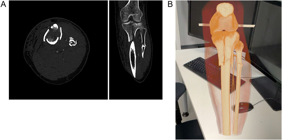
Figure 1. AR models used in musculoskeletal system radiology lecture. (A) Axial and coronal view of a 39 years old female after a skiing accident, with a complex fractured tibia and fibula in an arterial contrast phase and bone window. (B) AR model of the same patient.
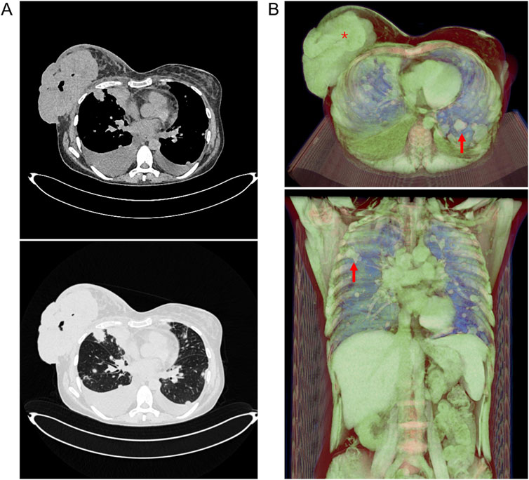
Figure 2. AR models used in gynecology radiology lecture. (A) Axial view of a 46 year old female with extensive breast cancer and multiple lung metastasis. CT with soft tissue (up) and lung (down) window. (B) AR model of the same patient. Arrows point at lung metastasis, * indicates primary tumor. The background was set black to allow for better visibility of the model.
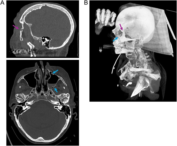
Figure 3. AR model used in the emergency medicine radiology lecture. 94 years old female after a fall with extensive head trauma including several cranial fractures. (A) CT in sagittal and axial view with bone window. (B) AR model of the same patient. Purple arrow points at the fracture in the left lateral orbital limitation and blue arrows indicate the osteodestruction of the ipsilateral maxillary sinus. The background was set black to allow for better visibility of the model.
Students were divided into seven different lecture groups and each group was exposed to MIXR in one of the five different topic lectures. Students were asked to give feedback in the form of a questionnaire after the lecture. Responding to the questionnaire was, however, voluntary and 47 students (32.6%) responded (Table 2). The higher number of replies came from the groups 4 and 5, with 25.5% and 27.7% of all replies, respectively. The lecture subject in which most students replied to the questionnaire was the Thorax, with 25 replies, corresponding to 53.2% of all replies (Table 2).
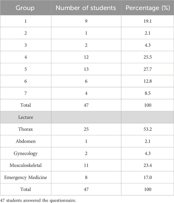
Table 2. Student distribution according to the answers to the questionnaire.
Students were asked to evaluate their experience with visualization of conventional and AR images. A greater proportion found AR images to be “very comprehensible” compared to conventional images (36.2% [17/47] vs. 21.3% [10/47], Table 3). However, a higher number of students also reported difficulty with AR imaging: 25.6% (12/47) found AR images “hardly comprehensible” or “incomprehensible,” compared to 10.6% (5/47) for conventional images (Table 3).
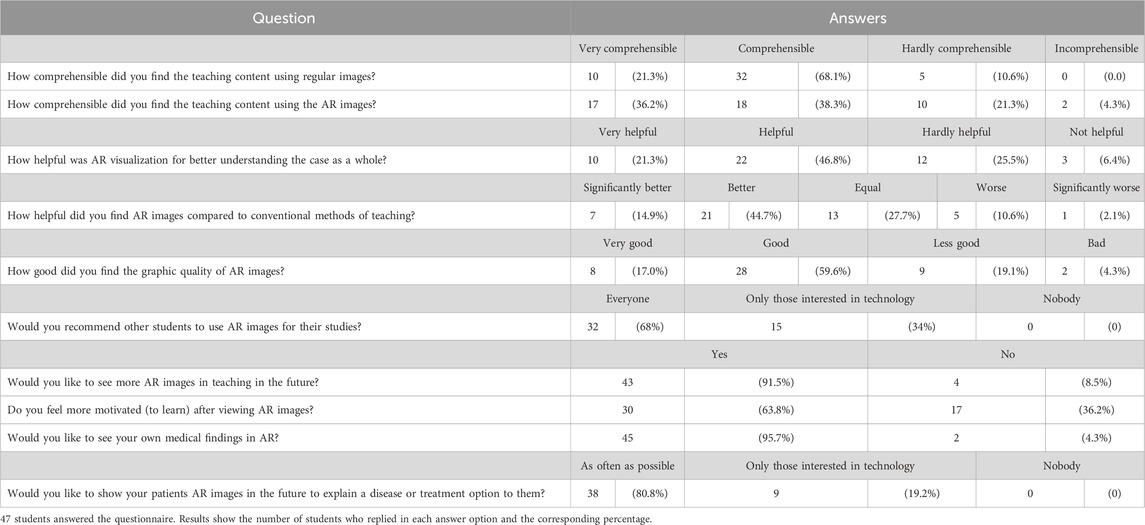
Table 3. Students answers to the questionnaire.
Regarding the usability of AR in medical education, 68.1% (32/47) of students reported that XR improved their understanding of the presented case (Table 3). Additionally, 59.5% (28/47) considered AR superior to standard teaching methodologies, with 14.9% (7/47) rating it as “significantly better” (Table 3). When assessing graphic quality, 76.6% (36/47) rated the image quality of MIXR as “Good” or “Very good” (Table 3). However, the remaining students (11/47) found the image quality of MIXR to be low, a similar ratio to the students that found AR difficult to understand (Table 3). Image quality might have contributed to the increased difficulty.
When asked about integrating AR into their studies, 91.8% of students expressed interest (Table 3). However, only 63.8% felt that AR use enhanced their motivation to learn. A majority (68.1%, 32/47) would recommend AR and 3D visualization to peers (Table 3). Additionally, over 95% expressed a desire to view their own medical findings using XR in the future (Table 3), while 80.9% (38/47) indicated interest in utilizing AR for patient interactions (Table 3).
Discussion
The findings of this study align with the growing body of evidence supporting the use of XR in medical education (Abundez Toledo et al., 2024; Saliba and Pather, 2025) but introduce a novel dimension by demonstrating the feasibility and effectiveness of using basic, widely available technologies like smartphones and tablets. This approach challenges the prevailing notion that XR-based educational tools require expensive equipment and technical expertise, opening the door for institutions with limited resources to explore similar interventions.
Only a small number of publications has explored the use of mobile device-based AR for medical education. Applications for smartphones have been developed, but not yet tested on a student population (Jain et al., 2017; Lee et al., 2018). Other applications were tested, but are no longer available on the market or were designed by the authors and not commercialized (Gonzalez et al., 2020; Moro et al., 2021; Sveinsson et al., 2021). MIXR, provided by Medicalholodeck™, is available for application in HMDs, but also in mobile devices. By utilizing devices that are already widely available in educational settings, such as iPhones and iPads, we eliminate many of the logistical and financial barriers typically associated with XR implementation (Qi et al., 2024). The ease of use for lecturers, who could seamlessly integrate AR demonstrations into their lectures without prior technical training, further enhances its practicality. This contrasts with studies involving HMDs, which often require specialized software, dedicated space, and significant preparation time (Qi et al., 2024; Fick et al., 2023). HMDs are also often source of discomfort among study participants, as shown in the works from Sánchez-Margallo et al. and Koger et al., using the Microsoft Headset HoloLens (Sánchez-Margallo et al., 2021; Koger et al., 2022; Tian et al., 2022). Indeed, a study comparing the benefits of HMDs and tablet based AR on a brain anatomy lecture concluded that, although the educational benefits for students are identical with both methods, tablet-based AR was preferred due to comfort (Moro et al., 2021).
The positive reception from students underscores the value of incorporating interactive and immersive elements into traditional teaching methods. The ability to visualize anatomical structures and clinical scenarios in CT images with AR, rather than relying on static images, likely played a significant role in improving comprehension and engagement. Different groups show similar result in terms of app based AR receptibility by students, using the app ARmedViewer. In this feasibility study, the app was compared to a computer-based alternative, and students overall preferred the app to the computer option (Sveinsson et al., 2021). Additionally, the live demonstration format, integrated into the existing lecture structure, provided a natural and effective way to highlight key concepts without disrupting the flow of the session. Whereas there was a general good response from students to the use of AR, there were students that did not find the AR information so easy to comprehend. This could be due to less contact with AR tools and could potentially be ameliorated with longer exposure times to the app. We also highlight that most students did not interact themselves with the app but rather were demonstrated during a video lecture. We hypothesize that giving the students the chance to directly interact with the app would improve their learning experience. Improving the means of interaction of students with the app, and therefore, potentially improving their comprehension of the AR images is a relevant point for optimization for further projects. Another point for optimization is the incorporation of other medical imaging modalities, such as magnetic resonance imaging (MRI), in the AR lectures, which is possible using MIXR. To our knowledge, there are no studies focusing on medical education with mobile device based AR generated from MRI DICOM series and the majority of works have been done using highly curated anatomical models (Gonzalez et al., 2020; Moro et al., 2021; Schneider et al., 2025).
Despite the strengths, there are limitations to consider to our work. First, the study relied on subjective evaluations from students, which, while informative, may not fully capture the extent of the educational benefits. Future research could explore objective measures, such as pre- and post-intervention assessments of knowledge retention and application. We can also not exclude a potential sampling bias in the questionnaire replies. The feedback questionnaire was not mandatory and only about 33% of students replied to the questionnaire and it is possible that the students that responded were the ones who had better experience, as the others might simply have not responded. The inclusion of open unstructured feedback options will, in future work, allow for a wider understanding of students’ experience with AR. Additionally, the scope of this study was limited to a single institution and specific lesson topics. Expanding the research to include a broader range of medical disciplines and institutions would provide a more comprehensive understanding of the intervention’s impact. The impact of AR in education of medical adjacent professionals such as nurses and physical therapists should also be address. Another limitation of our study design is the lack of a control group that did not experience AR. Future studies should aim at a better understanding of the effect of AR in education, including non-interventional groups and objective measures of learning improvement. These points have recently been addressed on a systematic review and their inclusion in future studies will contribute to advances in the field of AR-based medical education (Williams et al., 2025).
Another consideration is the potential for technological disparities among students. While smartphones and tablets are widely available, not all students may have access to devices with the same capabilities. Ensuring equity in access to these tools is essential for maximizing the benefits of AR implementation for educational purposes.
Conclusion
Our work demonstrates that integrating live AR demonstrations into medical lectures, using readily available mobile devices and open-source apps, can significantly enhance student engagement and understanding. By prioritizing simplicity and accessibility, using the intuitive MIXR, we could easily and conveniently integrate AR demonstrations during a normal lecture. Students acknowledge the benefits of using AR for education in Radiology and show interest in continuing using AR technologies in the future of their medical training and career.
Data availability statement
The raw data supporting the conclusion of this article will be made available by the authors, without undue reservation.
Ethics statement
The studies involving humans were approved by Ethics Commission of the Medical Faculty Bonn, Germany. The studies were conducted in accordance with the local legislation and institutional requirements. The participants provided their written informed consent to participate in this study.
Author contributions
AJ: Conceptualization, Data curation, Formal Analysis, Investigation, Methodology, Writing – original draft. AB: Conceptualization, Data curation, Investigation, Methodology, Writing – original draft. LB: Investigation, Methodology, Writing – review and editing. LO: Investigation, Methodology, Writing – review and editing. MB: Investigation, Methodology, Writing – review and editing. A-MO: Investigation, Methodology, Writing – review and editing. CE: Investigation, Methodology, Writing – review and editing. AH: Investigation, Methodology, Writing – review and editing. MR: Writing – review and editing, Data curation. SN: Formal analysis, Methodology, Writing – review and editing. NM: Writing – review and editing. AI: Writing – review and editing. CP: Writing – review and editing. JL: Funding acquisition, Resources, Writing – review and editing. DK: Conceptualization, Funding acquisition, Project administration, Resources, Supervision, Writing – original draft.
Funding
The author(s) declare that financial support was received for the research and/or publication of this article. This study was funded by the Clinic for Diagnostic and Interventional Radiology, University Hospital Bonn, and the Innovative Secure Medical Campus, supported by the Ministry of Economic Affairs, Innovation, Digitalization, and Energy of North Rhine-Westphalia, Germany.
Acknowledgments
We acknowledge the use of ChatGTP 3.5 for proofreading our manuscript.
Conflict of interest
The authors declare that the research was conducted in the absence of any commercial or financial relationships that could be construed as a potential conflict of interest.
Generative AI statement
The author(s) declare that Generative AI was used in the creation of this manuscript. ChatGTP 3.5 was used for proofreading our manuscript.
Publisher’s note
All claims expressed in this article are solely those of the authors and do not necessarily represent those of their affiliated organizations, or those of the publisher, the editors and the reviewers. Any product that may be evaluated in this article, or claim that may be made by its manufacturer, is not guaranteed or endorsed by the publisher.
Supplementary material
The Supplementary Material for this article can be found online at: https://www.frontiersin.org/articles/10.3389/frvir.2025.1583686/full#supplementary-material
References
Abundez Toledo, M., Ghanem, G., Fine, S., Weisman, D., Huang, Y. M., and Rouhani, A. A. (2024). Exploring the promise of virtual reality in enhancing anatomy education: a focus group study with medical students. Front. Virtual Real 5. doi:10.3389/frvir.2024.1369794
Asoodar, M., Janesarvatan, F., Yu, H., and de Jong, N. (2024). Theoretical foundations and implications of augmented reality, virtual reality, and mixed reality for immersive learning in health professions education. Adv. Simul. 9 (1), 36. doi:10.1186/s41077-024-00311-5
Chenais, N., and Görgen, A. (2024). Immersive interfaces for clinical applications: current status and future perspective. Front. Neurorobotics 18, 1362444. doi:10.3389/fnbot.2024.1362444
Fick, T., Meulstee, J. W., Köllen, M. H., Van Doormaal, J. a. M., Van Doormaal, T. P. C., and Hoving, E. W. (2023). Comparing the influence of mixed reality, a 3D viewer, and MRI on the spatial understanding of brain tumours. Front. Virtual Real 4. doi:10.3389/frvir.2023.1214520
Gonzalez, A. A., Lizana, P. A., Pino, S., Miller, B. G., and Merino, C. (2020). Augmented reality-based learning for the comprehension of cardiac physiology in undergraduate biomedical students. Adv. Physiol. Educ. 44 (3), 314–322. doi:10.1152/advan.00137.2019
Graf, L., Sykownik, P., Gradl-Dietsch, G., and Masuch, M. (2024). Towards believable and educational conversations with virtual patients. Front. Virtual Real 5. doi:10.3389/frvir.2024.1377210
Jain, N., Youngblood, P., Hasel, M., and Srivastava, S. (2017). An augmented reality tool for learning spatial anatomy on mobile devices. Clin. Anat. 30 (6), 736–741. doi:10.1002/ca.22943
Koger, C. R., Hassan, S. S., Yuan, J., and Ding, Y. (2022). Virtual reality for interactive medical Analysis. Front. Virtual Real 3, 782854. doi:10.3389/frvir.2022.782854
Lang, M., Ghandour, S., Rikard, B., Balasalle, E. K., Rouhezamin, M. R., Zhang, H., et al. (2024). Medical extended reality for radiology education and training. J. Am. Coll. Radiol. 21 (10), 1583–1594. doi:10.1016/j.jacr.2024.05.006
Lauinger, A. R., McNicholas, M., Bramlet, M., Bederson, M., Sutton, B. P., Cao, C. G. L., et al. (2024). Applications of mixed reality with medical imaging for training and clinical practice. J. Med. Imaging 11 (6), 062608. doi:10.1117/1.jmi.11.6.062608
Lee, D., Yi, J. W., Hong, J., Chai, Y. J., Kim, H. C., and Kong, H. J. (2018). Augmented reality to localize individual organ in surgical procedure. Healthc. Inf. Res. 24 (4), 394–401. doi:10.4258/hir.2018.24.4.394
Means, K., Kleiman, K., Ogdon, D., and Woodard, S. (2024). A review of virtual reality in radiology. Curr. Probl. Diagn Radiol. 53 (1), 17–21. doi:10.1067/j.cpradiol.2023.10.006
Moro, C., Phelps, C., Redmond, P., and Stromberga, Z. (2021). HoloLens and mobile augmented reality in medical and health science education: a randomised controlled trial. Br. J. Educ. Technol. 52 (2), 680–694. doi:10.1111/bjet.13049
Qi, Z., Corr, F., Grimm, D., Nimsky, C., and Bopp, M. H. A. (2024). Extended reality-based head-mounted displays for surgical education: a ten-year systematic review. Bioengineering 11 (8), 741. doi:10.3390/bioengineering11080741
Rahmat, RWOK, Anuar, CNSAC, Basri, N. A. H., Madzin, H., and Hod, R. (2023). A mobile augmented reality application for undergraduate medical students using A flipped classroom approach. J. Adv. Res. Appl. Sci. Eng. Technol. 33 (2), 151–159. doi:10.37934/araset.33.2.151159
Saliba, T., and Pather, S. (2025). The use of virtual reality and augmented reality in ultrasound education, a narrative review of the literature. J. Clin. Ultrasound 53, 315–324. doi:10.1002/jcu.23840
Sánchez-Margallo, J. A., Plaza de Miguel, C., Fernández Anzules, R. A., and Sánchez-Margallo, F. M. (2021). Application of mixed reality in medical training and surgical planning focused on minimally invasive surgery. Front. Virtual Real. doi:10.3389/frvir.2021.692641/full
Schneider, B., Ströbele, D. A., Grün, P., Mosch, R., Turhani, D., and von, S. C. (2025). Smartphone application-based augmented reality for pre-clinical dental implant placement training: a pilot study. Oral Maxillofac. Surg. 29 (1), 38. doi:10.1007/s10006-024-01317-z
Stauffer, T., Lohmeyer, Q., Melamed, S., Uhde, A., Hostettler, R., Wetzel, S., et al. (2024). Evaluation of augmented reality training for a navigation device used for CT-guided needle placement. Int. J. Comput. Assist. Radiol. Surg. 19 (12), 2411–2419. doi:10.1007/s11548-024-03112-3
Sveinsson, B., Koonjoo, N., and Rosen, M. S. (2021). ARmedViewer, an augmented-reality-based fast 3D reslicer for medical image data on mobile devices: a feasibility study. Comput. Methods Programs Biomed. 200, 105836. doi:10.1016/j.cmpb.2020.105836
Tian, N., Lopes, P., and Boulic, R. (2022). A review of cybersickness in head-mounted displays: raising attention to individual susceptibility. Virtual Real 26 (4), 1409–1441. doi:10.1007/s10055-022-00638-2
Williams, A., Sun, Z., and Vaccarezza, M. (2025). Comparison of augmented reality with other teaching methods in learning anatomy: a systematic review. Clin. Anat. 38 (2), 168–185. doi:10.1002/ca.24234
Keywords: augmented reality, medical education, radiology, smartphone, CT
Citation: Jacob AM, Böhner AMC, Beissel LD, Oelmeier L, Born M, Odenthal A-M, Endler C, Henkel A, Reinert M, Nowak S, Mesropyan N, Isaak A, Pieper CC, Luetkens JA and Kuetting D (2025) Augmented reality live demonstrations during traditional lectures improve understanding of computed tomography data sets by medical students. Front. Virtual Real. 6:1583686. doi: 10.3389/frvir.2025.1583686
Received: 26 February 2025; Accepted: 13 June 2025;
Published: 25 June 2025.
Edited by:
Irene Fondon, Sevilla University, SpainReviewed by:
Marco Parillo, Rovereto Hospital, ItalyVivek Parameswara Sarma, Kerala University of Health Sciences, India
Copyright © 2025 Jacob, Böhner, Beissel, Oelmeier, Born, Odenthal, Endler, Henkel, Reinert, Nowak, Mesropyan, Isaak, Pieper, Luetkens and Kuetting. This is an open-access article distributed under the terms of the Creative Commons Attribution License (CC BY). The use, distribution or reproduction in other forums is permitted, provided the original author(s) and the copyright owner(s) are credited and that the original publication in this journal is cited, in accordance with accepted academic practice. No use, distribution or reproduction is permitted which does not comply with these terms.
*Correspondence: Daniel Kuetting, RGFuaWVsLkt1ZXR0aW5nQHVrYm9ubi5kZQ==
†These authors have contributed equally to this work and share first authorship