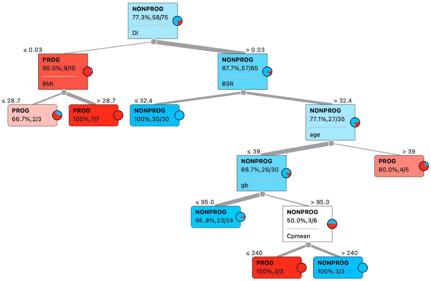
Four articles greatly showcase the application of AI towards the solution of previously known diagnostic limitations of the cardiovascular system. In the field of ischemic myocardial disease, Zhao et al. presents several models based on support vector machine, using calculated sample entropy, spatial heterogeneity index and temporal heterogeneity index of ST-T segments of electrocardiograms (ECGs) and vectorcardiograms, as input features, with the combined model having the best classifier performance as a non-invasive tool for detection of myocardial ischemia. When applying AI in the interpretation of structural heart disease, Bailoor et al. trained a linear discriminant classifier using "acoustic signatures" of healthy and stenotic aortic valves based on principal components of heart sounds and valve status in order to detect aortic valvular anomalies. Along the path of electrocardiographic diagnostics and arrhythmia interpretation, Brisk et al. demonstrated how wave segmentation can be a useful form of ECG representation learning, which improves model performance on downstream tasks. Finally, Cámara-Vázquez et al. discussed the potential of deep convolutional neural networks and body surface potential mapping to identify target regions for ablation in patients with atrial fibrillation.The collection includes two articles about the respiratory system. Giri et al. wrote a perspective article about how data from pulmonary function tests (PFTs) have potential to be used for both supervised learning and deep learning, notwithstanding the challenges the authors thoughtfully laid out. Meanwhile, Chen et al proposed an objective measure to quantify the late-expiratory flattening of the flow-volume loop, which is currently visually inspected by clinicians. They found that an angle made by two selected linear fitted lines improved inter-rater agreement on the presence of the late expiratory flattening and may help in the assessment of small airway disease in PFT interpretation.Finally, in the endocrine system, Zulfiqar et al. showed results of a pilot study, applying a remote monitoring platform to a small cohort of COVID-19 patients, using data from multiple non-intrusive medical sensors combined with an inference engine with evidence-based rules, to monitor dysglycemia via generated alerts. Ilari et al. meanwhile, used three supervised machine learning algorithms to determine factors that determine progression of patients with gestational diabetes mellitus to type 2 diabetes mellitus.These models have great potential in assisting physicians, especially in busy clinical settings, such as emergency departments where timing, in terms of seconds, is of the essence in addressing acute myocardial ischemia, and human interpretation alone may miss important diagnoses. It can also be used in less urgent settings, such as inpatient or outpatient settings to assist cardiologists in making diagnoses of aortic valve disease and guide elective atrial fibrillation ablation strategies, or pulmonologists in diagnosing small airway disease and other respiratory diseases. Such AI models also find application in remote settings, for monitoring via telemedicine of patients with difficult blood glucose control or used to predict disease progression in gestational diabetes. It is desirable that these models can be implemented and investigated as novel devices in randomized clinical trial settings, to assess their actual impact in clinical care.AI is also scalable and can screen large amounts of structured tabular data such as blood glucose level readings, and unstructured data such as ECGs, recorded auscultated heart sounds, or pulmonary function tests, in a high-throughput fashion, to alert physicians of possible underlying anomalies in the circulatory, respiratory, or endocrine systems. In these cases, each AI-generated interpretation should be generated before the physician signs off on the investigation, so that it is available at hand before committing to diagnostic conclusions. Caution needs to always apply, as trained models can also bias physicians and lead to undesirable heuristic shortcuts. Thus, it is largely advisable that a physician continues to interpret diagnostics at hand without AI support first, before gaining access to the above proposed AI interpretation. We must also be mindful that if AI-generated interpretations are frequently inaccurate or uninterpretable, or if AI-generated alerts are frequently inaccurate or excessive, this can lead to alert fatigue which can desensitize healthcare providers and lead to ignored interpretations or missed alerts, which will subsequently lead to delayed action or non-response. More importantly, it should always be remembered that human bedside physician-patient personal interaction remains crucial and must not be neglected due to the availability of AI model results [3].In conclusion, we hope this collection has demonstrated thematically to readers, over multiple biological systems in human physiology, how AI can integrate with traditional diagnostic tests in medicine, and how AI can sometimes determine additional findings missed by traditional diagnostic tests. AI continues to be an exciting area of development in the field of medicine and has great potential to change the way medicine is practiced and the understanding of human physiology, in the next decade and beyond.
Editorial18 November 2022
Editorial: Artificial intelligence in human physiology
ORIGINAL RESEARCH
30 May 2022
Xiaoye Zhao, 9 more and Ling Xia
11 Authors
- XZ
Xiaoye Zhao
School of Instrument Science and Opto-Electronic Engineering, China
- JZ
Jucheng Zhang
Department of Clinical Engineering, China
- YG
Yinglan Gong
Hangzhou Maixin Technology Co., Ltd., China
- LX
Lihua Xu
Hangzhou Linghua Biotech Ltd., China
- HL
Haipeng Liu
Research Centre for Intelligent Healthcare, United Kingdom
- SW
Shujun Wei
Department of Cardiology, China
- YW
Yuan Wu
Department of Cardiology, China
- GC
Ganhua Cha
School of Electrical and Information Engineering, China
- HW
Haicheng Wei
School of Electrical and Information Engineering, China
- JM
Jiandong Mao
School of Instrument Science and Opto-Electronic Engineering, China
- LX
Ling Xia
Key Laboratory for Biomedical Engineering of Ministry of Education, China
Background: Myocardial ischemia is a common early symptom of cardiovascular disease (CVD). Reliable detection of myocardial ischemia using computer-aided analysis of electrocardiogram (ECG) provides important reference for early diagnosis of CVD. The vectorcardiogram (VCG) could improve the performance of ECG-based myocardial ischemia detection by affording temporal-spatial characteristics related to myocardial ischemia and capturing subtle changes of ST-T segment in continuous cardiac cycles. We aim to investigate if the combination of ECG and VCG could improve the performance of machine learning algorithms in automatic myocardial ischemia detection.
Methods: The ST-T segments of 20-second, 12-lead ECGs and VCGs are extracted from 377 patients with myocardial ischemia and 52 healthy controls. Then, sample entropy (SampEn, of 12 ECG leads, and of 3 VCG leads), spatial heterogeneity index (SHI, of VCG) and temporal heterogeneity index (THI, of VCG) are calculated. Using a grid search, four SampEn and two features are selected as input signal features for ECG-only and VCG-only models based on support vector machine (SVM), respectively. Similarly, three features (SI, THI, and SHI, where SI is the SampEn of lead I) are further selected for the ECG+VCG model. The performances of ECG-only, VCG-only, and ECG+VCG models are evaluated using 5-fold cross validation. To fully evaluate the algorithmic generalization ability, the model with the best performance was selected and tested on a third independent dataset of 148 patients with myocardial ischemia and 52 healthy controls.
Results: The ECG+VCG model employing three features (SI,THI, and SHI) yields better classifying results than ECG-only and VCG-only models with the average accuracy of 0.903, 0.903 sensitivity, 0.905 specificity, 0.942 F1 score, and 0.904 AUC, which shows better performance with fewer features compared with existing works. On the third independent dataset, the testing showed AUC of 0.814.
Conclusion: The SVM algorithm based on ECG+VCG model could reliably detect myocardial ischemia, providing a potential tool to assist cardiologists in early diagnosis of CVD in routine screening during primary care services.