 Ruben Manuel Luciano Colunga Biancatelli
 Max Berrill4
 Paul E. Marik
- 1Division of Pulmonary and Critical Care Medicine, Eastern Virginia Medical School, Norfolk, VA, United States
- 2Frank Reidy Research Center for Bioelectrics, Old Dominion University, Norfolk, VA, United States
- 3Policlinico Umberto I, La Sapienza University of Rome, Rome, Italy
- 4Department of Respiratory Medicine, St. Peter's Hospital, Surrey, United Kingdom
- 5School of Medical Diagnostic & Translational Sciences, College of Health Sciences, Old Dominion University, Norfolk, VA, United States
Severe Acute Respiratory Syndrome Coronavirus-2 (SARS-CoV-2) represents an emergent global threat which is straining worldwide healthcare capacity. As of May 27th, the disease caused by SARS-CoV-2 (COVID-19) has resulted in more than 340,000 deaths worldwide, with 100,000 deaths in the US alone. It is imperative to study and develop pharmacological treatments suitable for the prevention and treatment of COVID-19. Ascorbic acid is a crucial vitamin necessary for the correct functioning of the immune system. It plays a role in stress response and has shown promising results when administered to the critically ill. Quercetin is a well-known flavonoid whose antiviral properties have been investigated in numerous studies. There is evidence that vitamin C and quercetin co-administration exerts a synergistic antiviral action due to overlapping antiviral and immunomodulatory properties and the capacity of ascorbate to recycle quercetin, increasing its efficacy. Safe, cheap interventions which have a sound biological rationale should be prioritized for experimental use in the current context of a global health pandemic. We present the current evidence for the use of vitamin C and quercetin both for prophylaxis in high-risk populations and for the treatment of COVID-19 patients as an adjunct to promising pharmacological agents such as Remdesivir or convalescent plasma.
Introduction
It is serendipitous (or perhaps indicative of hard work) that the Nobel prize winner Szent-Gyorgyi discovered both ascorbic acid (vitamin C) and the flavonoid quercetin (at the time labeled vitamin P) (1). Ascorbic acid is an essential vitamin with known antiviral properties (2) which is under investigation for its beneficial effects during the stress response in sepsis and critically ill patients (3).
Vitamin C exerts its antiviral properties by supporting lymphocyte activity, increasing interferon-α production, modulating cytokines, reducing inflammation, improving endothelial dysfunction, and restoring mitochondrial function (4–6). There are also suggestions that vitamin C may be directly viricidal (7). These in vitro effects, as we previously discussed (2), constitute a reflection of both the supra-physiological concentrations of ascorbate and the interaction between vitamin C and metal-containing culture media—both of which are pro-oxidant, generating reactive oxygen species.
Quercetin (also known as 3,3′,4′5,7-pentahydroxyflavone) is a widely distributed plant flavonoid, found in several vegetables, leaves, seeds, and grains, where it is conjugated with residual sugars to form quercetin glycosides (8). Studies suggest that quercetin supplementation may promote antioxidant (9), anti-inflammatory, antiviral (10), and immunoprotective effects (11). Quercetin has been studied in various types and models of viral infection due to its promising antiviral effects in inhibiting polymerases (12), proteases (13), reverse transcriptase (14), suppressing DNA gyrase, and binding viral capsid proteins (15, 16).
In this review we collate the evidence of the antiviral properties of quercetin, describe its biologic action and pharmacokinetics profile, expand on our previous review of vitamin C, discuss their synergistic actions, and propose this experimental multi-drug approach for the prevention and treatment of SARS-CoV-2/COVID-19 pandemic.
Chemistry of Quercetin
In plants, quercetin is produced from the phenylpropanoid pathway and is ultimately derived from phenylalanine. It is converted to 4-coumaroyl-CoA, via phenylalanine ammonia-lysate, to cinnamate-4-hydroxylase and 4-coumaroyl-CoA-ligase. This is combined with malonyl-CoA in a 1:3 ratio by 7,2′-dihydroxy-4′methoxyisoflavanol synthase to form tetrahydroxy chalcone. This in turn is converted to naringenin and to eriodyctiol through flavonoid 3′-hydroxylase. Finally, eriodyctiol is hydroxylated and converted to quercetin (Figure 1) using flavanol synthase (17).
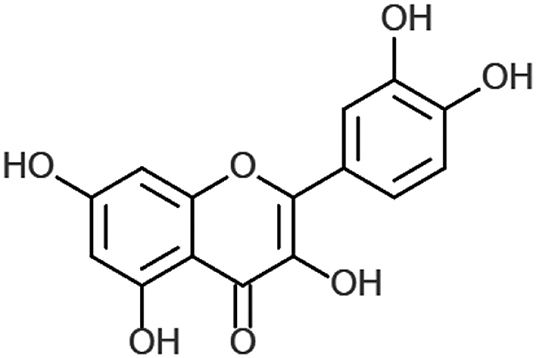
Figure 1. Chemical structure of quercetin. Created with ChemDoodle Web with permission (18).
Biology of Quercetin
Flavonoid compounds, such as quercetin, were initially studied for their biological activity in affecting capillary wall resistance (19) and continue to be investigated for their effects on vascular tension (20). Dietary supplements differ, but often contain the free form of quercetin—quercetin aglycone—under the FDA national drug code numbers 65448-3085, 65448-3005 (21). Once consumed, quercetin passes predominantly unaltered into the large intestine (22). Quercetin acts as a free radical scavenger, donating two electrons via o-quinone/quinone methide (23); both in vitro and in vivo (24, 25) studies implicate quercetin as a potent antioxidant. This antioxidant activity may also be potentiated by vitamin C (26), as will be discussed below. There is also significant longstanding interest in the anti-inflammatory activity of quercetin, as it has been suggested to be a key mediator in the cardiovascular protective element of the “Mediterranean” diet (27). This biological rationale is secondary to quercetin's free radical scavenging capacity, alongside diverse roles identified in in vitro and in vivo models including: inhibition of platelet aggregation (28), inhibition of lipid peroxidation (29), and its inhibitory effects on pro-inflammatory mediators such as lipoxygenase (30) and phospholipase A2 (31). This anti-inflammatory effect is primarily mediated by flavonoid activity on arachidonic acid metabolism and the associated leukotriene/prostaglandin pathways. Furthermore, 3-methyl-quercetin, a quercetin metabolite, displays stimulatory effects on nasal epithelial cell ciliary beat frequency, both in vitro and in vivo, when administered either alone or with absorption enhancer HP-β-CD (32). Quercetin also affects the function of several lipids, protein tyrosine, and serine/threonine kinases (33, 34), such as phosphatidylinositol (PI)-3-kinase and inducible nitric oxide synthase (NOS2) (35, 36).
Beneficial Effects of Vitamin C and Quercetin in Viral Infections
There is a tremendous amount of literature supporting the antiviral properties of quercetin, in both in vitro and in vivo experiments. Quercetin inhibits several respiratory viruses in cultured cells (16, 37). It inhibits the cytopathic effects provoked by many serotypes of rhinovirus, echovirus (type 7, 11, 12, and 19), coxsackievirus (A21 and B1), and poliovirus (type 1 Sabin) at a minimal inhibitory concentration of 0.03 to 0.5 μg/ml in Hela or WI-38 cells (38). Quercetin also significantly reduces plaque formation by RNA and DNA viruses [Respiratory Syncytial Virus (RSV), Polio type 1, parainfluenza type 3, and Herpes Simplex Virus-1(HSV-1)] displaying anti-infective and anti-replicative properties (39). It inhibits the replication of cytomegalovirus (CMV) inoculated HeLa cells at a half inhibitory concentration (IC50) of 3.2 ± 0.8 μM and with a selectivity index (SI) of 22 (40). Dengue virus type 2 (DENV-2) replication in Vero cells is inhibited by quercetin at an IC50 of 35.7 μg/mL, causing a DENV-2 RNA reduction of 67%. This is attributed to quercetin's ability to either block virus entry or inhibit viral replication enzymes such as viral polymerases (41).
In vivo studies indicate that mice inoculated with meningoencephalitis virus are protected from lethal infection by quercetin (30 or 40 mg/Kg BID, po, for 4 days) in a dose dependent manner (42). These beneficial effects are abolished if the compound is administered for <3 days, once per day or via subcutaneous injection. This may suggest that the antiviral effects may be dependent on a minimum inhibitory concentration or from some form of metabolic drug conversion (42). Quercetin treatment also displayed a beneficial effect in immunocompetent mice infected with Mengo virus, where it lessened the severity of organ damage (43). Athletes supplemented with quercetin are protected from stress-induced susceptibility to upper respiratory tract infection (44)—which was not related to immunomodulation (45, 46).
Vitamin C is an essential nutrient involved in a diverse array of immune functions; its supplementation has demonstrated beneficial effects in different types of viral infections. Reduced levels of ascorbate have been found in patients with viral infections (47), sepsis (48), sepsis-related ARDS (49), and other critical illness (50). During infection, vitamin C is necessary for neutrophil killing (51), is concentrated within macrophages (52), is responsible of T cell maturation (53), and promotes phagocytosis and apoptosis of spent neutrophils (4). It is not surprising, therefore, that viral infections, depending on their severity, are associated with an increased metabolism and reduced circulating ascorbate.
Vitamin C has improved survival in different murine models of lethal infection. Mice infected with Venezuelan encephalitis virus and treated with vitamin C (50 mg/kg) exhibit half the mortality of controls with associated reductions in viral titers, lipid peroxidation products, and NO content (54). Mice incapable of synthetizing vitamin C (L-Gulono-gamma-lactone oxidase nulls) were infected with influenza; mice not receiving supplemental vitamin C exhibited greater lung pathology scores despite no differences in viral titers (55). In restraint-stressed mice with H1N1 viral-induced pneumonia, vitamin C reduced mortality dose-dependently (100% vs. 80% vs. 50% at 0, 125, and 250 mg/kg/day) and reduced capillary-alveolar structural damage (56). Mice inoculated with Rabies+ mouse brain cells and treated with daily 100 mg/kg IM vitamin C exhibited nearly half the mortality of controls (57).
The only human study of vitamin C has been in USSR soldiers with severe viral infection indicated vitamin C supplementation (300 mg/day) protected from influenza-associated pneumonia and was associated with shorter hospital stays (58).
Vitamin C administration (i.v. 5 g/day twice/week) in patients with herpes zoster exhibited a lower incidence of postherpetic neuralgia (31.1% vs. 57.1%) and at study end (week 16) there was a lower pain score in the treatment group (0.64+/−0.9 vs. 1.98 +/−0.7) (59). Vitamin C administered at 1 g BID to 133 patients, reduced the risk (OR 0.25) of herpes simplex keratitis (HSK) recurrence (60), in accordance with previous studies indicating reduced ascorbate availability in the eye (61). It is noteworthy that a growing number of case reports of virus-related acute respiratory distress syndromes (ARDS) indicate successful treatment with intravenous high doses of Vitamin C (62, 63).
Co-administration of quercetin (12.5 mg/kg/week) and vitamin C and B3 in a murine model of exercise-induced susceptibility to influenza H1N1 prolonged time-to-death (median time to death: placebo 9.0 ± 0.33 vs. quercetin 16.5 ± 1.2) and improved survival (mortality: placebo 74% vs. quercetin 52%) when compared to mice receiving only vitamins B3 and C (64). An older, small clinical trial identified the combination of flavonoids and ascorbic acid (1:1 ratio) as beneficial for respiratory infection (200 mg TID) (65).
Inhibiting Virus Entry
Cell entry is a crucial step during viral infection and has been studied as a potential target of antiviral treatments (66–68). In an in vitro model of H1N1 and H3N2 influenza infection of MDCK cells, quercetin demonstrated reduced cytopathic effect 48 h post-infection (69). This effect was observed when quercetin was administered during viral entry (0–2 h), was maximal with quercetin pretreatment, and was dependent on quercetin's ability to bind hemagglutinin proteins (HA). Specifically, quercetin bound (dose-dependently) the HA subunit responsible for membrane fusion during virus entry and virus-mediated hemolysis (69). In vitro, quercetin pre-treatment (10 μM) inhibited Rhinovirus (RV) virulence, entry, and replication into BEAS-2B cells via multiple mechanisms: it impeded RV endocytosis though misdirecting EEA1 localization -an early endosomal marker- and inhibiting AKT phosphorylation with subsequent 3-fold viral load reduction at 24 h, lowering negative-strand RNA and modulating interferon (IFN) and IL-8 expression (70). These results were confirmed in vivo, with an estimated lower plasmatic concentration of quercetin (nM) (similarly to other studies (71–73)) during which quercetin reduced RV-RNA at 1 day post-infection, modulated KC, MIP-2, TNF-a, and MCP-2, decreased virus-induced airway hyper-responsiveness, and modulated IFNs (IFN-α and IFN-λ2) (70).
Interfering With DNA and RNA Polymerases
The in vitro antiviral effects of quercetin on herpesviruses (HSV-1, 2) and adenoviruses (ADV-3,−8,−11) suggest inhibition of early stage viral replication in a dose dependent manner (for HSV-1 100% inhibition at 60 mg/L) (16, 74), as well as inhibition of viral DNA and RNA polymerase (12, 75, 76). In human embryonic kidney cells (HEK), inoculated with polio, 3-methyl-quercetin disrupted plaque formation while quercetin itself demonstrated these effects when administered together with vitamin C (77). In fact, Vitamin C (either D- or L-ascorbate but not dehydroascorbate), prevented quercetin spontaneous degradation suggesting necessary co-administration with ascorbate to exert its antiviral effect. The beneficial effects of 3-methyl-quercetin (10 μM) were exerted primarily when the compound was administered 1–2 h post-poliovirus infection in Hela cells, inhibiting viral proteins and RNA synthesis in a dose dependent manner (78). In fact, 3-methyl-quercetin was identified as a molecule able to bind essential proteins required during the transcription from minus-strand RNA into positive polarity RNAs, thus interfering with cytoplasmic viral RNA replication (79).
In an in vivo study, a quercetin metabolite (4′,5-diacetyl-3,Y,7-trimethyl-quercetin), administered orally BID for 4 days protected mice against lethal infection by Coxsackie virus, promoting survival in a dose-response scale: 10, 20, and 40 mg/kg increased survival by 30, 40, and 50%, respectively (38). These beneficial effects were ascribed to a complete inhibition of virus replication when the compound was added within 2 h after virus absorption and related to the blockade of the RNA polymerase complex, as demonstrated in vitro (38).
Inhibition of Reverse Transcriptase
Quercetin has been investigated in vitro as an antiviral agent for HIV due to its ability to inhibit crucial enzymes: reverse transcriptase (RT), integrase (IN), and protease (PR) (80). Quercetin significantly reduces HIV viral replication (81) and, when added to peripheral blood mononuclear cells (PBMNc) infected with HIV and compared to HIV infected controls, quercetin reduced the levels of p24, Long Terminal Repeat (LTR) gene expression, and viral infectivity together with an inhibition of TNF-α and upregulation of IL-13 (11).
Quercetin has also been shown to inhibit non-HIV RT activity in vitro, including avian myeloblastosis reverse transcriptase (AMV-RT), Rous-associated virus-2 (RAV-2-RT), and Maloney murine leukemia virus (MMLV-RT). Quercetin displayed a dose-dependent inhibitory action: at 50 μM, 23% inhibition of both AMV-RT and RAV-2-RT, and at 10 μM inhibition of mammalian MMLV-RT of almost 60% were reached (14). HIV-RT was inhibited completely at 2 μg/ml quercetin in a partially-competitive mode (76). These antiviral effects of quercetin are believed to be related to the five hydroxyl groups on 3, 3′, 4′, 5, and 7 as the inhibitory activity is lower for baicalein, quercetagetin, or luteolin which lack these groups (75).
Interestingly, Harakeh et al. studied the dose-dependent effect of ascorbic acid (0–150 μg/ml) on HIV-infected T-lymphocytes in vitro and reported that >99% reverse transcriptase and nearly >90% p24 antigen suppression and a 93% inhibition of syncytia formation, a marker that correlates with viral infectivity and cytopathic effects (82).
Inhibition of Proteases
Quercetin is a potent HIV protease inhibitor in vitro, with an IC50 of 58.8 μM (83). Hepatitis C virus (HCV) NS3 serine protease catalytic activity was directly inhibited by quercetin treatment in a dose dependent manner (95% NS3 inhibition at 1.25 mg/ml); in this study quercetin blocked virus RNA production and impeded virus replication by 70% at 72 h without affecting cell viability (13).
Blocking Virus Assembly
Quercetin treatment inhibits HCV replication (84). This effect is attributed to its ability to modulate Heat Shock Protein expression (HSPs), thus impeding the crucial binding between heat shock factor and elements (HSF-HSE) necessary for the stress-induced transcription of stress genes (85, 86). Quercetin reduced HSP70 and HSP40, thereby impeding the formation of Non-Structural protein 5A complexes (NS5A-HSP70 and NS5A-HSP40) necessary for HCV genome replication apparatus through the internal ribosome entry site (IRES). Despite unaltered HCV titer, the production of infectious particles was decreased, interestingly more by quercetin treatment than by HSP knockdown, displaying a dose-dependent relationship: at 0.5 μM quercetin reduced viral production by 29%, at 5 μM by 90%, and at 50 μM by nearly 100% (84).
Immunomodulatory Properties
Quercetin stimulates T-helper cells to produce (Th-1)-derived Interferon-γ (IFN- γ) and downregulates Th2-derived IL-4 when added to cultured blood peripheral mononuclear cells (11). Immunonutrition studies in mice with supplementary polyphenols, including quercetin, showed enhanced NK cell lytic activity, neutrophil chemotaxis, and lymphocyte proliferation (87, 88).
Human foreskin fibroblast (HFF) and endothelial cells (EC) pretreated with 2-phospho-ascorbate (ASC-2P) resisted CMV infection; they displayed a reduction in immediate and late antigens and viral yield was inhibited 50–100-fold in ECs and 100–1,000-fold in HFF (89). This effect was not dependent on a sustained ASC-2P presence but was abolished if the ASC2-P was added after the virus infection, indicating an immunomodulatory effect, rather than directly antiviral. Animal models with gulo (–/–) mice insufficient in vitamin C, when infected with 20 hemagglutination units (HAU) of H3N2 influenza exhibited worse outcomes than wild type and Gulo (–/–) sufficient in vitamin C (90). Gulo (–/–) showed a reduction in IFN-α/β while displaying higher levels of IL-1α, TNF-α, and IL-1B. When Gulo (–/–) mice received supplemental Vitamin C, these cytokine expression profiles were lost.
Patients with acute Epstein-Barr infection (EBV) treated with high doses of intravenous vitamin C (7.5–50 g) displayed lower EBV-IgG (levels, while EBV VCA IgM antibody levels were negatively correlated to increasing plasma ascorbate concentration (91). Patients with HTLV-1-associated myelopathy/tropical spastic paraparesis HAM/TSP were all successfully treated with 35–40 mg/kg oral vitamin C for 3–5 days despite no changes in serum HTLV-1 or CSF HTLV-1 antibody titer, indicating an immunomodulative effect (92). Of these patients, 4 underwent a vitamin C on-off study which demonstrated a “positive dose response relationship with neurological symptoms.” A separate prospective trial into a diverse number of therapies indicated that vitamin C improved motor disability grades in HAM/TSP in 20% of patients (93). High dose ascorbic acid was then shown to display antiproliferative (95% decrease in lymphoproliferation) and immunomodulatory effects (via reduction of TNF-α, IFN-γ, IL-6, and p19) in peripheral blood mononuclear cells (PBMCs) extracted from HAM+ patients and T helper cell lines.
Vitamin C administration has been related to enhanced interferon production and was studied for its possible use for the prevention of vaccine failure. Rabies vaccination, when supplemented with 2 g oral vitamin C for each of the 3 injections provoked, at 24 h, increased serum IFN-α levels, indicating that “vitamin C is an effective stimulator of interferon production” (94). Mice on an ad libitum diet containing vitamin C increased induction of interferon (62–145%) depending on the viral titer of inoculation (95), and L-ascorbate added to stimulated mouse cell lines increases interferon synthesis (96). Low levels of vitamin C, in fact, have been related to insufficient phosphorylation of signal transducers and activation of transcription (STATs), which represent a crucial signaling process of IFNs (97). Specifically, T cells of mice deficient in vitamin C display defects in STAT3 phosphorylation (90).
Focus on SARS-CoV-2
Quercetin has been investigated for its possible antiviral effect on several members of the Coronaviridae family and, as mentioned by Ling Yi and colleagues, “quercetin offers great promise as a potential drug in the clinical treatment of SARS” (98). SARS-Coronavirus, described in 2003 (99), is a single-stranded RNA virus of ~29,700 nucleotides, which uses ribosome sites to encode two replicase glycoproteins, PP1a and PP1b, that mediate viral replication (99, 100). Once these precursor glycoproteins are synthesized, 3C-like protease (3CLpro) plays a critical role in the lytic release of its replicates (101). Quercetin-3β-galactoside binds SARS-Cov 3CL protease and inhibits its proteolytic activity with an IC50 of 42.79 ± 4.95 μM (102). This inhibitory action on 3CLpro is dependent on the hydroxyl group of quercetin which, as shown through molecular modeling and Q189A mutation, recognizes Gln189 as a crucial site on 3CLpro responsible for the binding of quercetin (102). Quercetin was also identified as a compound able to block SARS-Coronavirus entry into Vero E6 cells with a half-effective concentration (EC50) of 83.4 μM and with low cytotoxicity (CC50 3.32 mM) (98).
SARS-CoV-2, the virus responsible for the 2020 COVID-19 pandemic (103), belongs to the genus Betacoronavirus and subgenus Sarbecovirus and, due to its similar receptor-binding domain, it is assumed, similarly to SARS-CoV, to infect type II pneumocytes entering via the angiotensin-converting enzyme II receptor (104). SARS-Cov-2 protease 3CL maintains the same Gln189 site (105) of SARS-Cov 3CLpro, which previously was identified as the binding site for the hydroxyl groups of quercetin and its derivates (102).
Interestingly, an in vitro study of ascorbic acid treatment on chick-embryo ciliated tracheal organ cells (CETO) promoted resistance to Coronavirus infection but did not show any effect on orthomyxovirus or paramyxovirus (106).
Despite the breadth and depth of anti-viral in vitro and in vivo studies into the immunomodulatory effects of quercetin and vitamin C administration, further studies are absolutely necessary to confirm quercetin inhibitory activities on SARS-Cov-2 virus entry, RNA polymerase, and on other necessary viral life-cycle enzymes.
Pharmacokinetics of Quercetin
Orally administered quercetin glycosides are hydroxylated by β-glucosidases in the gut (107, 108). Aglycone quercetin passively permeate the intestinal epithelial barrier while quercetin glycosides are absorbed via the intestinal sodium/glucose cotransporter-1 (109). The bioavailability of oral quercetin is extremely variable, achieving values from 0 to 50% (110). Quercetin can be metabolized either in the enterocytes or in the hepatocytes forming glucuronidated, sulfated, or/and methylated compounds (111). Indeed, four out of five hydroxyl groups of quercetin can be glucuronidated by UDP-glucuronosyltransferase, forming its major metabolites: quercetin-3-glucuronide, 3'-methylquercetin-3-glucuronide, and quercetin-3'-sulfate (112). Rat tissue distribution of orally, long-term administered quercetin (12 weeks) shows the highest concentration in the lungs while pigs display the highest concentrations in the liver and kidneys (113). In contrast, short-term administration exhibits no marked distribution, implying that the beneficial effects of quercetin in preventing lung respiratory viral infection could be maximized by long-term administration. Following 500 mg oral quercetin, maximum plasma concentration of ~15 μg/L of aglycone quercetin (~50 nM, Tmax of 3 h) and 450 μg/L of quercetin non-methylated conjugates (Tmax of 4 h) were found (114). Intravenous administration results in an elimination half-life of 0.7–2.4 h with a distribution volume at steady-state of 6.2 to 92.6 L and with a total body clearance of 30 h (110).
Safe Profile and Optimal Dosing
Oral supplementation with quercetin up to 1 g/day for 3 months has not resulted in significant adverse effects (111). In a randomized placebo-controlled study, 30 patients with chronic prostatitis were supplemented with oral quercetin (1 g/day) and reported only two mild adverse reactions (headache and temporary peripheral paresthesia) (115). Intravenous administration of quercetin in a phase I clinical trial for cancer patients resulted in nausea, vomiting, sweating, flushing, and dyspnea at doses >10.5 mg/Kg (756 mg per 70 Kg individual) (116). Only higher intravenously administered doses up to 51.3 mg/Kg (around 3,591 mg per individual) were associated with renal toxicity (111). The safety of quercetin-based oral supplementation during pregnancy and breastfeeding has not been established.
We have previously described the safety profile and dosing strategies of vitamin C (117). According to the data presented above, we propose the following optimal dosing (Table 1). Further studies are needed to examine and discuss the possible administration of quercetin for prolonged periods of time (>1 year).
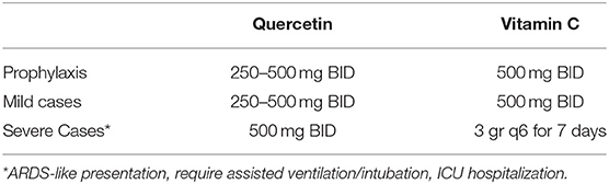
Table 1. Proposed multi-drug approach for either the prophylaxis for high risk population, and treatment of mild and severe cases.
Synergistic Antiviral Action
Quercetin spontaneously oxidizes to form O-semiquinone and O-quinone/quinone methide (QQ), which can bind protein thiols forming toxic compounds (118). This process of both anti- and pro-oxidant effects has been named the “quercetin paradox” (119). However, QQ can be recycled into quercetin by electron donors like NADH or ascorbate, or form together, with glutathione either 6-glutathionyl-quercetin or 8-glutathionyl-quercetin (GSQs) (107, 120). Importantly, if ascorbate or glutathione levels are insufficient, quercetin may be shunted to QQ and exert prooxidant effects. Therefore, we stress the importance for its co-administration with vitamin C (121, 122). However, even though QQ exhibits a higher affinity for glutathione than for vitamin C (121), the methylated metabolites of quercetin show a higher preference for ascorbate than for thiols, suggesting a cycling of activity which will exert anti-oxidant effects (Figure 2) (123). Furthermore, both GSQs (124) and QQ-protein thiols have been shown to be unstable and transient -lasting for minutes and hours instead of days- suggesting an overestimation of the proposed in vitro toxicity (125).
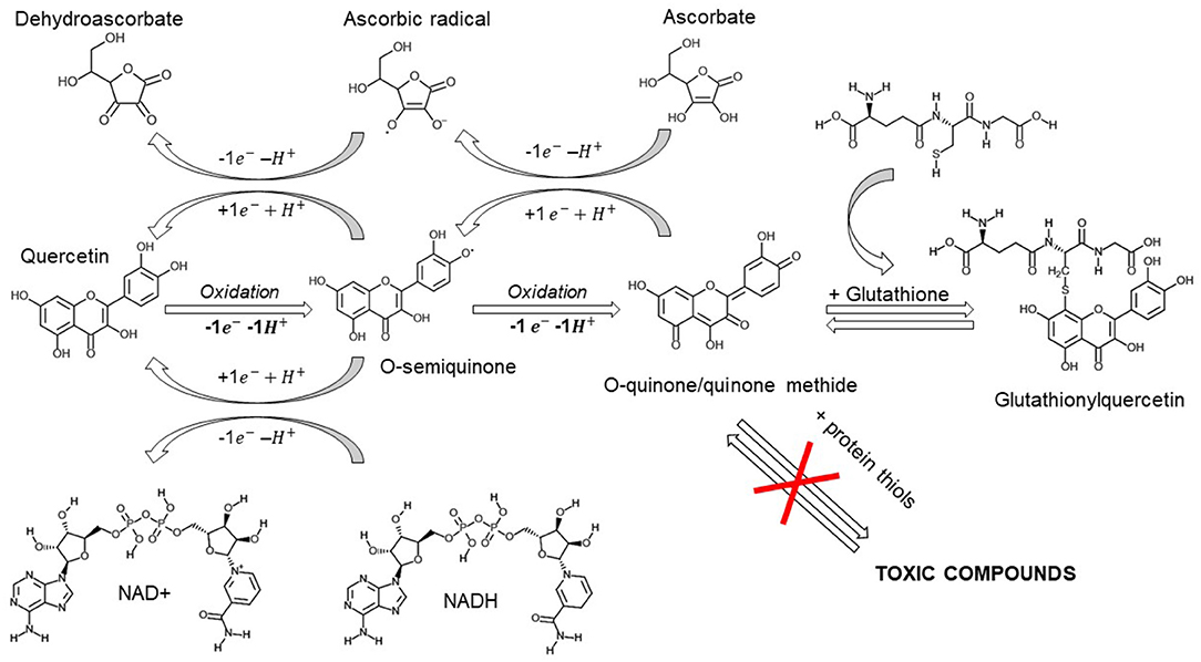
Figure 2. After exerting its scavenging properties, quercetin is oxidized into its reactive products o-semiquinone and o-quinone/quinone methide (QQ). These compounds can be recycled by antioxidants like ascorbate or NADH or removed by glutathione. If ascorbate or glutathione levels are reduced, QQ can bind protein thiols producing transient toxic compounds. Created with ChemDoodle Web with permission (18).
The supraphysiological concentrations of ascorbate achieved with intravenous administration (i.v. 3 gr q6) are capable of free radical scavenging and electron donation, preventing either quercetin or glutathione oxidation. In this scenario, ascorbate may exert antioxidant and immunoprotective effects, quercetin and its metabolites exert a concurrent antiviral response and, if quercetin-oxidized compounds are formed, they can be partially recycled by ascorbate and transported by glutathione, thus preventing their possible toxicity.
Discussion
A multi-drug approach with quercetin and vitamin C may disrupt virus entry, replication, enzyme activity and assembly, and concurrently fortify the immune response promoting early IFNs production, modulating interleukins, promoting T cell maturation, and phagocytic activity. Quercetin and ascorbic acid co-administration represents an experimental strategy for prophylaxis and treatment of several respiratory viruses, such as SARS-CoV-2. The blockage of virus entry represents a key strategy and quercetin impedes viral membrane fusion for both influenza and SARS-Cov in vitro (98). Quercetin also targets viral polymerases and may disrupt replication via the inhibition of reverse transcriptase enzymes. Quercetin further inhibits SARS 3CL protease by binding to its GLN189 site (102), which is expressed similarly by SARS-COV-2 (105) and provides a direct mechanistic rationale for its experimental clinical use—in addition to its immunoenhancing and anti-inflammatory actions. Despite the limitations of in vitro research, it is noteworthy that the few in vivo models reviewed here indicate increased survival from lethal viral infection when treated with quercetin (42, 64). Some studies suggest that oral administration and metabolic processing (methylation, conjugation, etc.) is necessary, and have identified quercetin derivates, which display variable Tmax, as responsible for a cooperative antiviral activity (126–128).
Vitamin C exerts immunomodulatory activity, enhancing interferon production through STAT3 phosphorylation (90), limiting cytokine-induced organ damage (55), promoting survival in lethal infections (54) and, importantly, is able to recycle oxidized quercetin (120), enhancing its antiviral effects. SARS-Cov-2 virus infection may initiate a strong inflammatory and dysregulated reaction in the lung with increased levels of IL-6 and a “cytokine-storm” (129) which has been shown to provoke either an asymptomatic, mild, or severe infections This cytokine dysregulation may be associated with neutrophil extracellular traps (130) and alterations in T cell activity (131). These immunological alterations which have characterized our current understanding of Covid-19 suggest that agents which target immune modulation, rather than direct viricidal activity, may present exciting targets for pharmacological intervention. In this scenario, Vitamin C and quercetin co-administration may represent a safe, effective, and inexpensive antiviral and immunomodulative approach for both the prophylaxis of high-risk populations and the treatment of both mild and severe cases.
They have also consistently been shown to display excellent safety profiles, and a consideration of risks and benefits in their therapeutic potential should be placed within this context. Vitamin C is a widely available supplement which many millions of people use already, and we have highlighted its antiviral properties in conjunction with quercetin. Due to its large-scale use, vitamin C in particular would be a cheap intervention with which to ascertain these compounds' efficacy as a prophylactic intervention. The prophylactic use of over-the-counter vitamin supplementation to combat infection is a behavior many people engage with already. Research into the potential prophylactic administration of vitamin C and quercetin in high-risk groups is therefore warranted.
The excellent side effect profile of these agents would also suggest that they may complement interventions which have displayed potential benefits in treating Covid-19, such as Remdesivir (132) and convalescent plasma (133, 134), which we believe warrants their experimental use in clinical trials.
There are potential limitations of their use in clinical studies. Both agents are present in varying degrees in individuals' diets and global recommendations for vitamin C intake vary extensively across the globe (135). Prophylactic interventions in general populations within the community will therefore be confounded by the quantity present in differing diets. Agents such as vitamin C also have well-characterized beneficial effects apart from the antiviral properties we have highlighted here. Supplementation with these agents may therefore promote general health and indirectly affect the capacity of individuals to combat viral infection. Although this would diminish the ability to identify the direct antiviral properties of vitamin C in clinical studies it may have ancillary benefits of promoting general health, which may be particularly pertinent if administered in communities with greater deprivation or from less economically developed countries.
Conclusion
Quercetin displays a broad range of antiviral properties which can interfere at multiple steps of pathogen virulence -virus entry, virus replication, protein assembly- and that these therapeutic effects can be augmented by the co-administration of vitamin C. Furthermore, due to their lack of severe side effects and low-costs, we strongly suggest the combined administration of these two compounds for both the prophylaxis and the early treatment of respiratory tract infections, especially including COVID-19 patients.
Author Contributions
All authors listed have made a substantial, direct and intellectual contribution to the work, and approved it for publication.
Funding
Supported by the CounterACT Program, National Institutes of Health Office of the Director (NIH OD) and the National Institute of Environmental Health Sciences (NIEHS) grant number R21ES030528.
Conflict of Interest
The authors declare that the research was conducted in the absence of any commercial or financial relationships that could be construed as a potential conflict of interest.
References
1. Formica JV, Regelson W. Review of the biology of Quercetin and related bioflavonoids. Food Chem Toxicol. (1995) 33:1061–80. doi: 10.1016/0278-6915(95)00077-1
2. Colunga Biancatelli RML, Berrill M, Marik PE. The antiviral properties of vitamin C. Expert Rev Anti Infect Ther. (2019) 18:99–101. doi: 10.1080/14787210.2020.1706483
3. Marik PE. Vitamin C: an essential “stress hormone” during sepsis. J Thorac Dis. (2020) 12(Suppl. 1):S84–8. doi: 10.21037/jtd.2019.12.64
4. Carr AC, Maggini S. Vitamin C and Immune Function. Nutrients. (2017) 9:1211. doi: 10.3390/nu9111211
5. Leibovitz B, Siegel BV. Ascorbic acid and the immune response. Adv Exp Med Biol. (1981) 135:1–25. doi: 10.1007/978-1-4615-9200-6_1
6. Dey S, Bishayi B. Killing of S. aureus in murine peritoneal macrophages by Ascorbic acid along with antibiotics Chloramphenicol or Ofloxacin: correlation with inflammation. Microb Pathog. (2018) 115:239–50. doi: 10.1016/j.micpath.2017.12.048
7. Furuya A, Uozaki M, Yamasaki H, Arakawa T, Arita Koyama MAH. Antiviral effects of ascorbic and dehydroascorbic acids in vitro. Int J Mol Med. (2008) 22:541–5. doi: 10.3892/ijmm_00000053
8. Li Y, Yao J, Han C, Yang J, Chaudhry MT, Wang S, et al. Quercetin, inflammation and immunity. Nutrients. (2016) 8:167. doi: 10.3390/nu8030167
9. Robaszkiewicz A, Balcerczyk A, Bartosz G. Antioxidative and prooxidative effects of quercetin on A549 cells. Cell Biol Int. (2007) 31:1245–50. doi: 10.1016/j.cellbi.2007.04.009
10. Uchide N, Toyoda H. Antioxidant therapy as a potential approach to severe influenza-associated complications. Molecules. (2011) 16:2032–52. doi: 10.3390/molecules16032032
11. Nair MP, Kandaswami C, Mahajan S, Chadha KC, Chawda R, NairandSchwartz SAH. The flavonoid, quercetin, differentially regulates Th-1 (IFNgamma) and Th-2 (IL4) cytokine gene expression by normal peripheral blood mononuclear cells. Biochim Biophys Acta. (2002) 1593:29–36. doi: 10.1016/S0167-4889(02)00328-2
12. Shinozuka K, Kikuchi Y, Nishino C, Mori A, Tawata S. Inhibitory effect of flavonoids on DNA-dependent DNA and RNA polymerases. Experientia. (1988) 44:882–5. doi: 10.1007/BF01941188
13. Bachmetov L, Gal-Tanamy M, Shapira A, Vorobeychik M, Giterman-Galam T, Sathiyamoorthy P, et al. Suppression of hepatitis C virus by the flavonoid quercetin is mediated by inhibition of NS3 protease activity. J Viral Hepat. (2012) 19:e81–8. doi: 10.1111/j.1365-2893.2011.01507.x
14. Spedding G, Ratty A, Middleton E Jr. Inhibition of reverse transcriptases by flavonoids. Antiviral Res. (1989) 12:99–110. doi: 10.1016/0166-3542(89)90073-9
15. Cushnie TP, Lamb AJ. Antimicrobial activity of flavonoids. Int J Antimicrob Agents. (2005) 26:343–56. doi: 10.1016/j.ijantimicag.2005.09.002
16. Debiaggi M, Tateo F, Pagani L, Luini M, Romero E. Effects of propolis flavonoids on virus infectivity and replication. Microbiologica. (1990) 13:207–13.
17. Winkel-Shirley B, Flavonoid Biosynthesis. A colorful model for genetics, biochemistry, cell biology, and biotechnology. Plant Physiol. (2001) 126:485. doi: 10.1104/pp.126.2.485
18. Burger MC. ChemDoodle web components: HTML5 toolkit for chemical graphics, interfaces, and informatics. J Cheminform. (2015) 7:35. doi: 10.1186/s13321-015-0085-3
19. Gábor M. Szent-Györgyi and the bioflavonoids: new results and perspectives of pharmacological research into benzo-pyrone derivatives. Commemoration on the 50th anniversary of the award of the Nobel Prize. Prog Clin Biol Res. (1988) 280:1–15.
20. Khoo NK, White CR, Pozzo-Miller L, Zhou F, Constance C, InoueandParks DAT. Dietary flavonoid quercetin stimulates vasorelaxation in aortic vessels. Free Radic Biol Med. (2010) 49:339–47. doi: 10.1016/j.freeradbiomed.2010.04.022
21. Andres S, Pevny S, Ziegenhagen R, Bakhiya N, Schäfer B, Hirsch-Ernst KI, et al. Safety aspects of the use of quercetin as a dietary supplement. Mol Nutr Food Res. (2018) 62:1700447. doi: 10.1002/mnfr.201700447
22. Brown JP. A review of the genetic effects of naturally occurring flavonoids, anthraquinones and related compounds. Mutat Res. (1980) 75:243–77. doi: 10.1016/0165-1110(80)90029-9
23. Awad HM, Boersma MG, Vervoort Rietjens JIM. Peroxidase-catalyzed formation of quercetin quinone methide-glutathione adducts. Arch Biochem Biophys. (2000) 378:224–33. doi: 10.1006/abbi.2000.1832
24. Terao J. Dietary flavonoids as antioxidants in vivo: conjugated metabolites of (-)-epicatechin and quercetin participate in antioxidative defense in blood plasma. J Med Invest. (1999) 46:159–68.
25. Abdelmoaty MA, Ibrahim MA, Ahmed NS, Abdelaziz MA. Confirmatory studies on the antioxidant and antidiabetic effect of quercetin in rats. Indian J Clin Biochem. (2010) 25:188–92. doi: 10.1007/s12291-010-0034-x
26. Nabavi SM, Nabavi SF, Eslami S, Moghaddam AH. In vivo protective effects of quercetin against sodium fluoride-induced oxidative stress in the hepatic tissue. Food Chem. (2012) 132:931–5. doi: 10.1016/j.foodchem.2011.11.070
27. Gormaz JG, Quintremil S, Rodrigo R. Cardiovascular disease: a target for the pharmacological effects of quercetin. Curr Top Med Chem. (2015) 15:1735–42. doi: 10.2174/1568026615666150427124357
28. Pace-Asciak CR, Hahn S, Diamandis EP, Soleas G, Goldberg DM. The red wine phenolics trans-resveratrol and quercetin block human platelet aggregation and eicosanoid synthesis: implications for protection against coronary heart disease. Clin Chim Acta. (1995) 235:207–19. doi: 10.1016/0009-8981(95)06045-1
29. Cai Q, Rahn RO, Zhang R. Dietary flavonoids, quercetin, luteolin and genistein, reduce oxidative DNA damage and lipid peroxidation and quench free radicals. Cancer Lett. (1997) 119:99–107. doi: 10.1016/S0304-3835(97)00261-9
30. Erden Inal M, Kahraman A. The protective effect of flavonol quercetin against ultraviolet a induced oxidative stress in rats. Toxicology. (2000) 154:21–9. doi: 10.1016/S0300-483X(00)00268-7
31. Morikawa K, Nonaka M, Narahara M, Torii I, Kawaguchi K, Yoshikawa T, et al. Inhibitory effect of quercetin on carrageenan-induced inflammation in rats. Life Sci. (2003) 74:709–21. doi: 10.1016/j.lfs.2003.06.036
32. Dimova S, Mugabowindekwe R, Willems T, Brewster ME, Noppe M, Ludwig A, et al. Safety-assessment of 3-methoxyquercetin as an antirhinoviral compound for nasal application: effect on ciliary beat frequency. Int J Pharm. (2003) 263:95–103. doi: 10.1016/S0378-5173(03)00363-6
33. Davies SP, Reddy H, Caivano M, Cohen P. Specificity and mechanism of action of some commonly used protein kinase inhibitors. Biochem J. (2000) 351(Pt. 1):95–105. doi: 10.1042/bj3510095
34. Huang YT, Hwang JJ, Lee PP, Ke FC, Huang JH, Huang CJ, et al. Effects of luteolin and quercetin, inhibitors of tyrosine kinase, on cell growth and metastasis-associated properties in A431 cells overexpressing epidermal growth factor receptor. Br J Pharmacol. (1999) 128:999–1010. doi: 10.1038/sj.bjp.0702879
35. Agullo G, Gamet-Payrastre L, Manenti S, Viala C, Rémésy C, Chap H, et al. Relationship between flavonoid structure and inhibition of phosphatidylinositol 3-kinase: a comparison with tyrosine kinase and protein kinase C inhibition. Biochem Pharmacol. (1997) 53:1649–57. doi: 10.1016/S0006-2952(97)82453-7
36. Peet GW, Li J. IkappaB kinases alpha and beta show a random sequential kinetic mechanism and are inhibited by staurosporine and quercetin. J Biol Chem. (1999) 274:32655–61. doi: 10.1074/jbc.274.46.32655
37. De Palma AM, Vliegen I, De Clercq E, Neyts J. Selective inhibitors of picornavirus replication. Med Res Rev. (2008) 28:823–84. doi: 10.1002/med.20125
38. Itsuka H, Ohsawa C, Ohiwa T, Umeda I, Suhara Y. Antipicornavirus flavone Ro 09-0179. Antimicrob Agents Chemother. (1982) 22:611–16. doi: 10.1128/AAC.22.4.611
39. Kaul TN, Middleton E Jr. Ogra PL. Antiviral effect of flavonoids on human viruses. J Med Virol. (1985) 15:71–9. doi: 10.1002/jmv.1890150110
40. Evers DL, Chao CF, Wang X, Zhang Z, Huong SM, Huang ES. Human cytomegalovirus-inhibitory flavonoids: studies on antiviral activity and mechanism of action. Antiviral Res. (2005) 68:124–34. doi: 10.1016/j.antiviral.2005.08.002
41. Zandi K, Teoh BT, Sam SS, Wong PF, Mustafa MR, Abubakar S. Antiviral activity of four types of bioflavonoid against dengue virus type-2. Virol J. (2011) 8:560. doi: 10.1186/1743-422X-8-560
42. Veckenstedt A, Béládi I, Mucsi I. Effect of treatment with certain flavonoids on Mengo virus-induced encephalitis in mice. Arch Virol. (1978) 57:255–60. doi: 10.1007/BF01315089
43. Güttner J, Veckenstedt A, Heinecke H, Pusztai R. Effect of quercetin on the course of mengo virus infection in immunodeficient and normal mice. A histologic study. Acta Virol. (1982) 26:148–55.
44. Nieman DC, Henson DA, Gross SJ, Jenkins DP, Davis JM, Murphy EA, et al. Quercetin reduces illness but not immune perturbations after intensive exercise. Med Sci Sports Exerc. (2007) 39:1561–9. doi: 10.1249/mss.0b013e318076b566
45. Nieman DC, Henson DA, Davis JM, Angela Murphy E, Jenkins DP, Gross SJ, et al. Quercetin's influence on exercise-induced changes in plasma cytokines and muscle and leukocyte cytokine mRNA. J Appl Physiol. (2007) 103:1728–35. doi: 10.1152/japplphysiol.00707.2007
46. Nieman DC, Henson DA, Davis JM, Dumke CL, Gross SJ, Jenkins DP, et al. Quercetin ingestion does not alter cytokine changes in athletes competing in the Western States Endurance Run. J Interferon Cytokine Res. (2007) 27:1003–11. doi: 10.1089/jir.2007.0050
47. Chen JY, Chang C-Y, Feng P-H, Chu C-C, So EC, Hu M-L. Plasma vitamin C is lower in postherpetic neuralgia patients and administration of vitamin C reduces spontaneous pain but not brush-evoked pain. Clin J Pain. (2009) 25:562–9. doi: 10.1097/AJP.0b013e318193cf32
48. Marik PE, Hooper MH. Doctor-your septic patients have scurvy! Crit Care. (2018) 22:23. doi: 10.1186/s13054-018-1950-z
49. Fowler AA III, Truwit JD, Hite RD, Morris PE, DeWilde C, Priday A, et al. Effect of vitamin C infusion on organ failure and biomarkers of inflammation and vascular injury in patients with sepsis and severe acute respiratory failure: the CITRIS-ALI randomized clinical trial. JAMA. (2019) 322:1261–70. doi: 10.1001/jama.2019.11825
50. Carr AC, Rosengrave PC, Bayer S, Chambers S, Mehrtens J, Shaw GM. Hypovitaminosis C and vitamin C deficiency in critically ill patients despite recommended enteral and parenteral intakes. Crit Care. (2017) 21:300. doi: 10.1186/s13054-017-1891-y
51. Anderson R, Smit MJ, Joone GK, Van Staden AM. Vitamin C and cellular immune functions. Protection against hypochlorous acid-mediated inactivation of glyceraldehyde-3-phosphate dehydrogenase and ATP generation in human leukocytes as a possible mechanism of ascorbate-mediated immunostimulation. Ann N Y Acad Sci. (1990) 587:34–48. doi: 10.1111/j.1749-6632.1990.tb00131.x
52. Lahiri S, Lloyd BB. The effect of stress and corticotrophin on the concentrations of vitamin C in blood and tissues of the rat. Biochem. J. (1962) 84:478–83. doi: 10.1042/bj0840478
53. Manning J, Mitchell B, Appadurai DA, Shakya A, Pierce LJ, Wang H, et al. Vitamin C promotes maturation of T-cells. Antioxid Redox Signal. (2013) 19:2054–67. doi: 10.1089/ars.2012.4988
54. Valero N, Mosquera J, Alcocer S, Bonilla E, Salazar J, Álvarez-Mon M. Melatonin, minocycline and ascorbic acid reduce oxidative stress and viral titers and increase survival rate in experimental Venezuelan equine encephalitis. Brain Res. (2015) 1622:368–76. doi: 10.1016/j.brainres.2015.06.034
55. Li W, Maeda N, Beck MA. Vitamin C deficiency increases the lung pathology of influenza virus-infected gulo-/- mice. J Nutr. (2006) 136:2611–16. doi: 10.1093/jn/136.10.2611
56. Cai Y, Li Y-F, Tang L-P, Tsoi B, Chen M, Chen H, et al. A new mechanism of vitamin C effects on A/FM/1/47(H1N1) virus-induced pneumonia in restraint-stressed mice. BioMed Res Int. (2015) 2015:675149. doi: 10.1155/2015/675149
58. Kimbarowski JA, Mokrow NJ. Colored precipitation reaction of the urine according to Kimbarowski (FARK) as an index of the effect of ascorbic acid during treatment of viral influenza. Das Deutsche Gesundheitswesen. (1967) 22:2413–18.
59. Kim MS, Kim DJ, Na CH, Shin BS. A study of intravenous administration of vitamin c in the treatment of acute herpetic pain and postherpetic neuralgia. Ann Dermatol. (2016) 28:677–83. doi: 10.5021/ad.2016.28.6.677
60. Kim GN, Yoo WS, Park MH, Chung JK, Han YS, Chung IY, et al. Clinical features of herpes simplex keratitis in a Korean tertiary referral center: efficacy of oral antiviral and ascorbic acid on recurrence. Kor J Ophthalmol. (2018) 32:353–60. doi: 10.3341/kjo.2017.0131
61. Hah YS, Chung HJ, Sontakke SB, Chung I-Y, Ju S. Ascorbic acid concentrations in aqueous humor after systemic vitamin C supplementation in patients with cataract: pilot study. BMC Ophthalmol. (2017) 17:121. doi: 10.1186/s12886-017-0515-2
62. Gonzalez MJ, Berdiel MJ, Duconge J, Levy T, Alfaro I, Morales R, et al. High dose intravenous vitamin C and influenza: a case report. J Orthomol Med. (2018) 33:1–3.
63. Fowler Iii AA, Kim C, Lepler L, Malhotra R, Debesa O, Natarajan R., et al. Intravenous vitamin C as adjunctive therapy for enterovirus/rhinovirus induced acute respiratory distress syndrome. World J Crit Care Med. (2017) 6:85–90. doi: 10.5492/wjccm.v6.i1.85
64. Davis JM, Murphy EA, McClellan JL, Carmichael MD, Gangemi JD. Quercetin reduces susceptibility to influenza infection following stressful exercise. Am J Physiol Regul Integr Comp Physiol. (2008) 295:R505–9. doi: 10.1152/ajpregu.90319.2008
65. Biskind MS, Martin WC. The use of citrus flavonoids in respiratory infections. Am J Dig Dis. (1954) 21:177. doi: 10.1007/BF02886384
66. Liu S, Wu S, Jiang S. HIV entry inhibitors targeting gp41: from polypeptides to small-molecule compounds. Curr Pharm Des. (2007) 13:143–62. doi: 10.2174/138161207779313722
67. Yang J, Li M, Shen X, Liu S. Influenza A virus entry inhibitors targeting the hemagglutinin. Viruses. (2013) 5:352–73. doi: 10.3390/v5010352
68. Xia S, Liu Q, Wang Q, Sun Z, Su S, Duand L, et al. Middle East respiratory syndrome coronavirus (MERS-CoV) entry inhibitors targeting spike protein. Virus Res. (2014) 194:200–10. doi: 10.1016/j.virusres.2014.10.007
69. Wu W, Li R, Li X, He J, Jiang S, Liu S, et al. Quercetin as an antiviral agent inhibits Influenza A Virus (IAV) entry. Viruses. (2015) 8:6. doi: 10.3390/v8010006
70. Ganesan S, Faris AN, Comstock AT, Wang Q, Nanua S, Hershenson MB, et al. Quercetin inhibits rhinovirus replication in vitro and in vivo. Antiviral Res. (2012) 94:258–71. doi: 10.1016/j.antiviral.2012.03.005
71. Ganesan S, Faris AN, Comstock AT, Chattoraj SS, Chattoraj A, Burgess JR, et al. Quercetin prevents progression of disease in elastase/LPS-exposed mice by negatively regulating MMP expression. Respir Res. (2010) 11:131. doi: 10.1186/1465-9921-11-131
72. Nanua S, Zick SM, Andrade JE, Sajjan US, Burgess JR, Lukacs NW, et al. Quercetin blocks airway epithelial cell chemokine expression. Am J Respir Cell Mol Biol. (2006) 35:602–10. doi: 10.1165/rcmb.2006-0149OC
73. Rogerio AP, Kanashiro A, Fontanari C, da Silva EV, Lucisano-Valim YM, Soares EG, et al. Anti-inflammatory activity of quercetin and isoquercitrin in experimental murine allergic asthma. Inflamm Res. (2007) 56:402–8. doi: 10.1007/s00011-007-7005-6
74. Chiang LC, Chiang W, Liu MC, Lin CC. In vitro antiviral activities of Caesalpinia pulcherrima and its related flavonoids. J Antimicrob Chemother. (2003) 52:194–8. doi: 10.1093/jac/dkg291
75. Ono K, Nakane H. Mechanisms of inhibition of various cellular DNA and RNA polymerases by several flavonoids. J Biochem. (1990) 108:609–13. doi: 10.1093/oxfordjournals.jbchem.a123251
76. Ono K, Nakane H, Fukushima M, Chermann JC, Barré-Sinoussi F. Differential inhibitory effects of various flavonoids on the activities of reverse transcriptase and cellular DNA and RNA polymerases. Eur J Biochem. (1990) 190:469–76. doi: 10.1111/j.1432-1033.1990.tb15597.x
77. Vrijsen R, Everaert L, Boeyé A. Antiviral activity of flavones and potentiation by ascorbate. J Gen Virol. (1988) 69:1749–51. doi: 10.1099/0022-1317-69-7-1749
78. Vrijsen R, Everaert L, Van Hoof LM, Vlietinck AJ, Vanden Berghe DA, Boeyé A., et al. The poliovirus-induced shut-off of cellular protein synthesis persists in the presence of 3-methylquercetin, a flavonoid which blocks viral protein and RNA synthesis. Antiviral Res. (1987) 7:35–42. doi: 10.1016/0166-3542(87)90037-4
79. Castrillo JL, Carrasco L. Action of 3-methylquercetin on poliovirus RNA replication. J Virol. (1987) 61:3319–21. doi: 10.1128/JVI.61.10.3319-3321.1987
80. Li BW, Zhang FH, Serrao E, Chen H, Sanchez TW, Yang LM, et al. Design and discovery of flavonoid-based HIV-1 integrase inhibitors targeting both the active site and the interaction with LEDGF/p75. Bioorg Med Chem. (2014) 22:3146–58. doi: 10.1016/j.bmc.2014.04.016
81. Áy É, Hunyadi A, Mezei M, Minárovits J, Hohmann J. Flavonol 7-O-glucoside herbacitrin inhibits HIV-1 replication through simultaneous integrase and reverse transcriptase inhibition. Evid Based Complement Alternat Med. (2019) 2019:1064793. doi: 10.1155/2019/1064793
82. Harakeh S, Jariwalla RJ, Pauling L. Suppression of human immunodeficiency virus replication by ascorbate in chronically and acutely infected cells. Proc Natl Acad Sci USA. (1990) 87:7245–9. doi: 10.1073/pnas.87.18.7245
83. Xu HX, Wan M, Dong H, But PP, Foo LY. Inhibitory activity of flavonoids and tannins against HIV-1 protease. Biol Pharm Bull. (2000) 23:1072–6. doi: 10.1248/bpb.23.1072
84. Gonzalez O, Fontanes V, Raychaudhuri S, Loo R, Loo J, Arumugaswami V, et al. The heat shock protein inhibitor Quercetin attenuates hepatitis C virus production. Hepatology. (2009) 50:1756–64. doi: 10.1002/hep.23232
85. Hosokawa N, Hirayoshi K, Kudo H, Takechi H, Aoike A, Kawai K, et al. Inhibition of the activation of heat shock factor in vivo and in vitro by flavonoids. Mol Cell Biol. (1992) 12:3490–8. doi: 10.1128/MCB.12.8.3490
86. Hosokawa N, Hirayoshi K, Nakai A, Hosokawa Y, Marui N, Yoshidaand M, et al. Flavonoids inhibit the expression of heat shock proteins. Cell Struct Funct. (1990) 15:393–401. doi: 10.1247/csf.15.393
87. Alvarez P, Alvarado C, Puerto M, Schlumberger A, Jiménez L, De la Fuente M., et al. Improvement of leukocyte functions in prematurely aging mice after five weeks of diet supplementation with polyphenol-rich cereals. Nutrition. (2006) 22:913–21. doi: 10.1016/j.nut.2005.12.012
88. Exon JH, Magnuson BA, South EH, Hendrix K. Effect of dietary chlorogenic acid on multiple immune functions and formation of aberrant crypt foci in rats. J Toxicol Environ Health A. (1998) 53:375–84. doi: 10.1080/009841098159231
89. Cinatl J, Cinatl J, Weber B, Rabenau H, Gumbel H, Chenot J, et al. In vitro inhibition of human cytomegalovirus replication in human foreskin fibroblasts and endothelial cells by ascorbic acid 2-phosphate. Antiviral Res. (1995) 27:405–18. doi: 10.1016/0166-3542(95)00024-G
90. Kim Y, Kim H, Bae S, Choi J, Lim SY, Lee N, et al. Vitamin C is an essential factor on the anti-viral immune responses through the production of interferon-α/β at the initial stage of influenza A virus (H3N2) infection. Immune Netw. (2013) 13:70–74. doi: 10.4110/in.2013.13.2.70
91. Mikirova N. Hunninghake R. Effect of high dose vitamin C on Epstein-Barr viral infection. Med Sci Monit. (2014) 20:725–32. doi: 10.12659/MSM.890423
92. Kataoka A, Imai H, Inayoshi S, Tsuda T. Intermittent high-dose vitamin C therapy in patients with HTLV-I associated myelopathy. J Neurol Neurosurg Psychiatry. (1993) 56:1213–16. doi: 10.1136/jnnp.56.11.1213
93. Nakagawa M, Nakahara K, Maruyama Y, Kawabata M, Higuchi I, Kubota H, et al. Therapeutic trials in 200 patients with HTLV-I-associated myelopathy/ tropical spastic paraparesis. J Neurovirol. (1996) 2:345–55. doi: 10.3109/13550289609146899
94. Stantic-Pavlinic M, Banic S, Marin J, Klemenc P. Vitamin C–a challenge in management of rabies. Swiss Med Weekly. (2004) 134:326–7.
95. Siegel BV. Enhanced interferon response to murine leukemia virus by ascorbic acid. Infect Immun. (1974) 10:409–10. doi: 10.1128/IAI.10.2.409-410.1974
96. Siegel BV. Enhancement of interferon production by poly(rI)-poly(rC) in mouse cell cultures by ascorbic acid. Nature. (1975) 254:531–2. doi: 10.1038/254531a0
97. Horvath CM. The Jak-STAT pathway stimulated by interferon gamma. Sci STKE. (2004) 2004:tr8. doi: 10.1126/stke.2602004tr8
98. Yi L, Li Z, Yuan K, Qu X, Chen J, Wang G, et al. Small molecules blocking the entry of severe acute respiratory syndrome coronavirus into host cells. J Virol. (2004) 78:11334. doi: 10.1128/JVI.78.20.11334-11339.2004
99. Rota PA, Oberste MS, Monroe SS, Nix WA, Campagnoli R, Icenogle JP, et al. Characterization of a novel coronavirus associated with severe acute respiratory syndrome. Science. (2003) 300:1394–9. doi: 10.1126/science.1085952
100. Marra MA, Jones SJ, Astell CR, Holt RA, Brooks-Wilson A, Butterfield YS, et al. The genome sequence of the SARS-associated coronavirus. Science. (2003) 300:1399–404. doi: 10.1126/science.1085953
101. Snijder EJ, Bredenbeek PJ, Dobbe JC, Thiel V, Ziebuhr J, Poon LLM, et al. Unique and conserved features of genome and proteome of SARS-coronavirus, an early split-off from the coronavirus group 2 lineage. J Mol Biol. (2003) 331:991–1004. doi: 10.1016/S0022-2836(03)00865-9
102. Chen L, Li J, Luo C, Liu H, Xu W, Chen G, et al. Binding interaction of quercetin-3-beta-galactoside and its synthetic derivatives with SARS-CoV 3CL(pro): structure-activity relationship studies reveal salient pharmacophore features. Bioorg Med Chem. (2006) 14:8295–306. doi: 10.1016/j.bmc.2006.09.014
103. Zhou P, Yang XL, Wang XG, Hu B, Zhang L, Zhang W, et al. A pneumonia outbreak associated with a new coronavirus of probable bat origin. Nature. (2020) 579:270–3. doi: 10.1038/s41586-020-2012-7
104. Lu R, Zhao X, Li J, Niu P, Yang B, Wu H, et al. Genomic characterisation and epidemiology of 2019 novel coronavirus: implications for virus origins and receptor binding. Lancet. (2020) 395:565–74. doi: 10.1016/S0140-6736(20)30251-8
105. Zhang L, Lin D, Sun X, Curth U, Drosten C, Sauerhering L, et al. Crystal structure of SARS-CoV-2 main protease provides a basis for design of improved α-ketoamide inhibitors. Science. (2020) 368:409–12. doi: 10.3410/f.737592020.793572879
106. Atherton JG, Kratzing CC, Fisher A. The effect of ascorbic acid on infection chick-embryo ciliated tracheal organ cultures by coronavirus. Arch Virol. (1978) 56:195–9. doi: 10.1007/BF01317848
107. Boots AW, Haenen GR, Bast A. Health effects of quercetin: from antioxidant to nutraceutical. Eur J Pharmacol. (2008) 585:325–37. doi: 10.1016/j.ejphar.2008.03.008
108. Guo Y, Bruno RS. Endogenous and exogenous mediators of quercetin bioavailability. J Nutr Biochem. (2015) 26:201–10. doi: 10.1016/j.jnutbio.2014.10.008
109. Murota K, Terao J. Antioxidative flavonoid quercetin: implication of its intestinal absorption and metabolism. Arch Biochem Biophys. (2003) 417:12–7. doi: 10.1016/S0003-9861(03)00284-4
110. Graefe EU, Derendorf H, Veit M. Pharmacokinetics and bioavailability of the flavonol quercetin in humans. Int J Clin Pharmacol Ther. (1999) 37:219–33.
111. Harwood M, Danielewska-Nikiel B, Borzelleca JF, Flamm GW, Williams GM, Lines TC. A critical review of the data related to the safety of quercetin and lack of evidence of in vivo toxicity, including lack of genotoxic/carcinogenic properties. Food Chem Toxicol. (2007) 45:2179–205. doi: 10.1016/j.fct.2007.05.015
112. Andrea J, Day JAR, Morgan RA. Characterization of polyphenols metabolites. In: Bao Y and Fenwick R editors. Phytochemicals in Health and Disease. New York, NY: Marcel Dekker, Inc. (2005). p. 50.
113. de Boer VC, Dihal AA, van der Woude H, Arts IC, Wolffram S. Tissue distribution of quercetin in rats and pigs. J Nutr. (2005) 135:1718–25. doi: 10.1093/jn/135.7.1718
114. Moon YJ, Wang L, DiCenzo R, Morris ME. Quercetin pharmacokinetics in humans. Biopharm Drug Dispos. (2008) 29:205–17. doi: 10.1002/bdd.605
115. Shoskes DA, Zeitlin SI, Shahed A, Rajfer J. Quercetin in men with category III chronic prostatitis: a preliminary prospective, double-blind, placebo-controlled trial. Urology. (1999) 54:960–3. doi: 10.1016/S0090-4295(99)00358-1
116. Ferry DR, Smith A, Malkhandi J, Fyfe DW, deTakats PG, Anderson D, et al. Phase I clinical trial of the flavonoid quercetin: pharmacokinetics and evidence for in vivo tyrosine kinase inhibition. Clin Cancer Res. (1996) 2:659–68.
117. Marik PE. Vitamin C for the treatment of sepsis: the scientific rationale. Pharmacol Ther. (2018) 189:63–70. doi: 10.1016/j.pharmthera.2018.04.007
118. Awad HM, Boersma MG, Boeren S, van der Woude H, van Zanden J, van Bladeren PJ, et al. Identification of o-quinone/quinone methide metabolites of quercetin in a cellular in vitro system. FEBS Lett. (2002) 520:30–34. doi: 10.1016/S0014-5793(02)02754-0
119. Boots AW, Li H, Schins RP, Duffin R, Heemskerk JW, Bast A, et al. The quercetin paradox. Toxicol Appl Pharmacol. (2007) 222:89–96. doi: 10.1016/j.taap.2007.04.004
120. Askari G, Ghiasvand R, Feizi A, Ghanadian SM, Karimian J. The effect of quercetin supplementation on selected markers of inflammation and oxidative stress. J Res Med Sci. (2012) 17:637–41.
121. Boots AW, Kubben N, Haenen GR, Bast A. Oxidized quercetin reacts with thiols rather than with ascorbate: implication for quercetin supplementation. Biochem Biophys Res Commun. (2003) 308:560–5. doi: 10.1016/S0006-291X(03)01438-4
122. Bors W, Michel C, Schikora S. Interaction of flavonoids with ascorbate and determination of their univalent redox potentials: a pulse radiolysis study. Free Radic Biol Med. (1995) 19:45–52. doi: 10.1016/0891-5849(95)00011-L
123. Moalin M, van Strijdonck GPF, Bast A, Haenen GRMM. Competition between ascorbate and glutathione for the oxidized form of methylated quercetin metabolites and analogues: tamarixetin, 4′o-methylquercetin, has the lowest thiol reactivity. J Agric Food Chem. (2012) 60:9292–7. doi: 10.1021/jf302068v
124. Boots AW, Balk JM, Bast A, Haenen GR. The reversibility of the glutathionyl-quercetin adduct spreads oxidized quercetin-induced toxicity. Biochem Biophys Res Commun. (2005) 338:923–9. doi: 10.1016/j.bbrc.2005.10.031
125. Awad HM, Boersma MG, Boeren S, Van Bladeren PJ, Vervoort J, Rietjens IM. Quenching of quercetin quinone/quinone methides by different thiolate scavengers: stability and reversibility of conjugate formation. Chem Res Toxicol. (2003) 16:822–31. doi: 10.1021/tx020079g
126. Roubalová L, Purchartová K, Papoušková B, Vacek J, Kren V, Ulrichová J, et al. Sulfation modulates the cell uptake, antiradical activity and biological effects of flavonoids in vitro: an examination of quercetin, isoquercitrin and taxifolin. Bioorg Med Chem. (2015) 23:5402–9. doi: 10.1016/j.bmc.2015.07.055
127. Ruotolo R, Calani L, Brighenti F, Crozier A, Ottonello S, Del Rio, D. Glucuronidation does not suppress the estrogenic activity of quercetin in yeast and human breast cancer cell model systems. Arch Biochem Biophys. (2014) 559:62–7. doi: 10.1016/j.abb.2014.03.003
128. Terao J, Murota K, Kawai Y. Conjugated quercetin glucuronides as bioactive metabolites and precursors of aglycone in vivo. Food Funct. (2011) 2:11–7. doi: 10.1039/C0FO00106F
129. Conti P, Ronconi G, Caraffa A, Gallenga CE, Ross R, Frydas I, et al. Induction of pro-inflammatory cytokines (IL-1 and IL-6) and lung inflammation by Coronavirus-19 (COVI-19 or SARS-CoV-2): anti-inflammatory strategies. J Biol Regul Homeost Agents. (2020) 34:1. doi: 10.23812/CONTI-E
130. Zuo Y, Yalavarthi S, Shi H, Gockman K, Zuo M, Madison JA, et al. Neutrophil extracellular traps in COVID-19. JCI Insight. (2020) 5. doi: 10.1172/jci.insight.138999
131. Wang F, Nie J, Wang H, Zhao Q, Xiong Y, Deng L, et al. Characteristics of peripheral lymphocyte subset alteration in COVID-19 pneumonia. J Infect Dis. (2020) 221:1762–9. doi: 10.1093/infdis/jiaa150
132. Beigel JH, Tomashek KM, Dodd LE, Mehta AK, Zingman BS, Kalil AC, et al. Remdesivir for the treatment of Covid-19 — preliminary report. N Engl J Med. (2020) doi: 10.1056/NEJMoa2007764
133. Shen C, Wang Z, Zhao F, Yang Y, Li J, Yuan J, et al. Treatment of 5 critically Ill patients with COVID-19 with convalescent plasma. JAMA. (2020) 323:1582–9. doi: 10.1001/jama.2020.4783
134. Ye M, Fu D, Ren Y, Wang F, Wang D, Zhang F, et al. Treatment with convalescent plasma for COVID-19 patients in Wuhan, China. J Med Virol. (2020). doi: 10.1002/jmv.25882. [Epub ahead of print].
Keywords: SARS-Cov-2, COVID-19, vitamin C, quercetin, flavonoids, antiviral, Coronavirus, immunonutrition
Citation: Colunga Biancatelli RML, Berrill M, Catravas JD and Marik PE (2020) Quercetin and Vitamin C: An Experimental, Synergistic Therapy for the Prevention and Treatment of SARS-CoV-2 Related Disease (COVID-19). Front. Immunol. 11:1451. doi: 10.3389/fimmu.2020.01451
Received: 09 April 2020; Accepted: 04 June 2020;
Published: 19 June 2020.
Edited by:
Xulin Chen, Jinan University, ChinaReviewed by:
Vincent T. Chow, National University of Singapore, SingaporeZhenlong Liu, McGill University, Canada
Copyright © 2020 Colunga Biancatelli, Berrill, Catravas and Marik. This is an open-access article distributed under the terms of the Creative Commons Attribution License (CC BY). The use, distribution or reproduction in other forums is permitted, provided the original author(s) and the copyright owner(s) are credited and that the original publication in this journal is cited, in accordance with accepted academic practice. No use, distribution or reproduction is permitted which does not comply with these terms.
*Correspondence: Ruben Manuel Luciano Colunga Biancatelli, cnViZW4uY29sdW5nYWJpYW5jYXRlbGxpQGdtYWlsLmNvbQ==