 Rodrigo Rojas
 Claudio D. Miranda
 Jaime Romero
 Juan L. Barja
 Javier Dubert
- 1Laboratorio de Patobiología Acuática, Departamento de Acuicultura, Universidad Católica del Norte, Coquimbo, Chile
- 2Laboratorio de Biotecnología, Instituto de Nutrición y Tecnología de los Alimentos, Universidad de Chile, Santiago, Chile
- 3Centro AquaPacífico, Coquimbo, Chile
- 4Departamento de Microbiología y Parasitología, CIBUS – Facultad de Biología, Universidad de Santiago de Compostela, Santiago de Compostela, Spain
The VPAP30 strain was isolated as the highly predominant bacteria from an episode of massive larval mortality occurring in a commercial culture of the Chilean scallop Argopecten purpuratus. The main aims of this study were, to characterize and identify the pathogenic strain using biochemical and molecular methods, to demonstrate its pathogenic activity on scallop larvae, to characterize its pathogenic properties and to describe the chronology of the pathology. The pathogenic strain was identified as Vibrio bivalvicida based on its phenotypic properties, the multilocus sequence analysis (MLSA) of eight housekeeping genes (ftsZ, gapA, gyrB, mreB, pyrH, recA, rpoA, and topA) and different in silico genome-to-genome comparisons. When triplicate cultures of healthy 10 days old scallop larvae were challenged with 1 × 10⁵ colony forming units (CFU) mL-1 of the VPAP30 strain, percentages of larval survival of 78.9 ± 3.3%, 34.3 ± 4.9%, and 0% were observed at 12, 2,4 and 36 h, respectively, whereas uninfected larval cultures showed survival rates of 97.4 ± 1.2% after of 48 h. Clinical symptoms exhibited by the scallop larvae infected with the VPAP30 strain include the accumulation of bacteria around the scallop larvae, velum disruption and necrosis of digestive gland. The 50% lethal dose (LD50) of VPAP30 strain at 24 and 48 h was 1.3 × 10⁴ and 1.2 × 10³ CFU mL-1, respectively. The invasive pathogenic activity of the VPAP30 strain was investigated with staining of the bacterial pathogen with 5-DTAF and analyzing bacterial invasion using epifluorescence, and a complete bacterial dissemination inside the larvae at 24 h post-infection was observed. When scallop larvae were inoculated with cell-free extracellular products (ECPs) of VPAP30, the larval survival rate was 59.5 ± 1.7%, significantly (P < 0.001) lower than the control group (97.4 ± 1.2%) whereas larvae treated with heat-treated ECPs exhibited a survival rate of 61.6 ± 1.8% after 48 h of exposure. V. bivalvicida VPAP30 exhibits high pathogenic activity on scallop larvae, mediated both by bacterial invasion and the production of toxigenic heat-stable compounds. This report constitutes the first isolation of V. bivalvicida out of Europe and extends the host range of this species, having demonstrated its pathogenic activity on the Chilean scallop larvae (A. purpuratus). These results supporting the pathogenic potential of V. bivalvicida to kill the larvae of a broad range of bivalve species reared in hatcheries located in the Atlantic and the Pacific coasts.
Introduction
The culture of the Chilean scallop Argopecten purpuratus is the second most important industry in Chilean mariculture and is primarily concentrated in the north region of the country (von Brand et al., 2006). However, recurrent episodes of larval mortalities mainly due to bacterial infections have been observed, causing high economical losses and consequently precluding the sustainability of this industry. These bacterial infections are commonly characterized by a sudden cessation of larval motility leading to massive mortalities of reared larvae. Previous studies demonstrated the pathogenic activity on scallop larvae of bacterial strains identified as Halomonas sp. (Rojas et al., 2009), Vibrio anguillarum-related (Riquelme et al., 1995), V. splendidus (Rojas et al., 2015a) and the association of Aeromonas hydrophila and V. alginolyticus (Riquelme et al., 1996).
Vibrio species have been described worldwide as the main aetiological agents of bacterial pathologies affecting reared larvae of various shellfish species (Waechter et al., 2002; Estes et al., 2004; Gómez-León et al., 2005; Prado et al., 2014, 2015; Rojas et al., 2015a; Dubert et al., 2016b, 2017). Clinical symptoms commonly exhibited by reared shellfish larvae affected by vibriosis include the reduction of larval motility, erratic swimming, closing of valves, velum detachment and bacterial swarming inside and around the larvae (Beaz-Hidalgo et al., 2010; Rojas et al., 2015a). Most of these clinical signs were described in larval cultures of the clam species Ruditapes decussatus (Gómez-León et al., 2005) and R. philippinarum (Dubert et al., 2016a), oyster species Crassostrea virginica (Gómez-León et al., 2008) and C. gigas (Estes et al., 2004; Elston et al., 2008), and scallop species Pecten maximus (Nicolas et al., 1996; Torkildsen et al., 2005), Argopecten ventricosus (Sainz et al., 1998; Luna-González et al., 2002) and Patinopecten yessoensis (Liu et al., 2013). The pathogenicity of Vibrio strains causing vibriosis outbreaks is mediated by bacterial invasion (Rojas et al., 2015a; Dubert et al., 2016a) as well as the production of toxigenic extracellular products (ECPs) (Binesse et al., 2008; Hasegawa et al., 2008; Labreuche et al., 2010; Rojas et al., 2015a). Within the genus Vibrio, the Orientalis clade includes the species V. crosai, V. orientalis,V. brasiliensis, V. sinaloensis, V. hepatarius and the pathogenic species for bivalves V. tubiashii, V. bivalvicida, and V. europaeus (Dubert et al., 2016a). Recently, V. europaeus and V. bivalvicida have gained a relevant significance in Europe, since they were identified as the aetiological agents responsible of outbreaks of vibriosis that affected recurrently to Spanish and French hatcheries (Travers et al., 2014; Dubert et al., 2016b). Interestingly, pathogenicity of these species has been demonstrated in larvae of R. decussatus, R. philippinarum, O. edulis, C. gigas, or Donax trunculus, some of the most important bivalve species reared in the European hatcheries (Dubert et al., 2017).
In general, 16S rRNA gene has a rather low interspecies resolution and is not useful for species differentiation but may provide a reliable identification at genus level. In genomic era, techniques as multilocus sequence analysis (MLSA) or whole genome sequencing (WGS) are essential to provide a better understanding of the taxonomic position of the pathogenic Vibrio isolates. Sawabe et al. (2013) proposed that a MLSA based on the eight housekeeping genes gapA, gyrB, ftsZ, mreB, pyrH, recA, rpoA, and topA is a powerful method for delineating a species of the genus Vibrio and a monophyletic group or clade. In addition, WGS enabled the possibility of establishing systematics on the basis of complete genomes by means of genome to genome comparisons using for example the average nucleotide identity (ANI) algorithm to identify bacterial strains accurately (Richter et al., 2016).
Despite that efficient rearing techniques for scallop larvae production that have been developed, Chilean commercial hatcheries are currently suffering recurrent episodes of high mortalities of reared larvae, mainly associated with high levels of vibrios (Miranda et al., 2014; Rojas et al., 2015a). The identification of bacterial strains causing epizootics in larval cultures and the understanding of their pathogenic activity are essential for the development of adequate and efficient protocols of larval management, as well as for the implementation of proper bacteriologic monitoring strategies to prevent and control scallop larvae mortality outbreaks occurring in commercial hatcheries. Considering that knowledge of the identity and pathogenic mechanisms of bacterial causing massive mortalities of scallop larvae reared in commercial hatcheries in Chile remains scarce, the aims of this study were to characterize and identify a highly pathogenic Vibrio strain recovered from a massive larval mortality event occurred in a commercial hatchery, to characterize its pathogenic properties and to describe the chronology of the pathology.
Materials and Methods
Bacterial Isolation
The pathogenic strain VPAP30 was recovered from a massive mortality event of reared-larvae of the scallop A. purpuratus occurring in a commercial hatchery located in Tongoy Bay in the north of Chile. Triplicate samples of settled dead and moribund larvae were aseptically collected from the bottom of the rearing tank during its water exchange using a sterile glass flask and were transported to the laboratory for immediate processing. Larval samples were centrifuged at 960 g for 2 min using an Eppendorf 5415D centrifuge (Hamburg, Germany) and the water excess was discarded. Settled larvae were ground by hand using a sterile glass digester containing 2 mL of sterile physiological saline (0.85% NaCl; PS) to obtain a homogenate according to the method of Nicolas et al. (1996). The homogenate was inoculated in triplicate onto Tryptic Soy Agar (Difco, NJ, United States) with 2% of NaCl (Oxoid, Hants, United Kingdom) (TSA2), and plates were incubated at 20°C for 48 h. The predominant colony, which grew almost as a pure culture on TSA2 plates, was isolated and preserved at -85°C in CryoBank (Mast Diagnostic, Merseyside, United Kingdom) vials prior use.
Biochemical and Physiological Characterization
Phenotypic characterization to identify Vibrio species were performed with different tests following Noguerola and Blanch (2008) such as cell morphology, Gram stain, oxidation/fermentation of glucose, and resistance to the vibriostatic agent O129 (2,4-diamino-6,7-diisopropylpteridine) (10 and 150 μg per disc) were determined according to the protocols described in Barrow and Feltham (1993). In addition, other phenotypic properties of VPAP30 strain were determined. Production of luminescence was evaluated in absence of light by using Marine agar 2216 (Difco, NJ, United States), whereas β-haemolysis of red cells was determined by using Columbia Blood agar (Oxoid, Hants, United Kingdom). Production of Møller’s lysine and ornithine decarboxylases and Thornley’s arginine dihydrolase were detected according to Hansen and Sörheim (1991). Growth at 4, 20, 30, 35, and 40°C was tested on Tryptic Soy broth supplemented with 2% NaCl (TSB2) and growth at 0, 3, 6, 8, and 10% of NaCl was assayed using peptone broth (BD, Sparks, United States). Additional phenotypic characteristics of VPAP30 strain were determined by using the API 20E (bioMérieux, Marcy-l´Etoile, France) and the Biolog (Biolog Inc., Hayward, CA, United States) systems. For the API system the VPAP30 strain was inoculated according to the manufacturer’s instructions with the modifications suggested by MacDonnell et al. (1982) and the API strip was incubated at 20°C for 48 h. For the Biolog system the strain was inoculated by using a solution containing 2.5% NaCl, 0.8% MgCl2, and 0.05% KCl, according to the instructions of the manufacturer and the microplate was aerobically incubated in the dark at 20°C for 72 h.
Enzymatic Analysis
The enzymatic activities of VPAP30 strain were determined by using the API ZYM system (bioMérieux, Marcy-l´Etoile, France) according to the manufacturer’s guidelines. Briefly, VPAP30 strain was cultured overnight in TSB2, centrifuged at 4,200 g at 4°C and resuspended in a NaCl 0.85% solution (bioMérieux, Marcy-l´Etoile, France) to obtain a turbidity of 5 McFarland (1.5 × 10⁹ bacteria mL-1), and 65 μL of this suspension were added to each cupule. Test strips were incubated for 4 h at 20°C and following incubation, 1 drop of ZYM A (API; tris-hydroxymethyl-aminomethane, hydrochloric acid, sodium laurel sulfate, H2O) and ZYM B (API; fast blue BB, 2-methoxyethanol) were added to each cupule. Test strips were read after 5 min and the results were scored using the following classification: 0, negative reaction; 1–2 weak activity; 3–5 strong activity. The assay was performed twice to ensure reproducibility.
Phylogenetic Analysis
DNA was extracted and purified from a pure culture using the Wizard Genomic DNA Purification Kit (Promega, Madison, WI, United States). PCR was performed as described in Romero and Navarrete (2006) with a reaction mixture (30 μL) containing 0.25 mM of each deoxynucleoside triphosphate, 0.05 U μL-1 Platinum Taq DNA polymerase (Invitrogen, San Diego, CA, United States), 1 × polymerase reaction buffer, 2 mM MgCl2, and 0.25 pmol μL-1 of each primer. To identify the bacterial strain, amplification of the 16S rRNA gene from positions 28 to 1,492 was performed using the primer pair 27F and 1492R as previously described (Navarrete et al., 2010). The housekeeping genes encoding for cell-division protein (ftsZ), glyceraldehyde-3-phosphate dehydrogenase (gapA), gyrase beta subunit (gyrB), rod shape-determining protein (mreB), uridine monophosphate kinase (pyrH), recombinase A (recA), RNA polymerase alpha subunit (rpoA), and topoisomerase I (topA) were used to perform a MLSA. Amplification of these genes was performed as previously described (Sawabe et al., 2013). PCR mixtures were identical to those previously used for the 16S rRNA gene, and the specific primers are listed in Supplementary Table 1. The thermal programme consisted of 5 min at 95°C, 25 cycles of 30 s at 95°C, 30 s at 55°C, and 30 s at 72°C, and a final 5 min extension at 72°C. All PCR products were verified, sequenced by Sanger and analyzed as described in Romero et al. (2002) using the Ribosomal Database Project II (Cole et al., 2007) or were compared to those available in the National Center for Biotechnology Information (NCBI) Reference Sequence database by using a BLAST search to ascertain their closest relatives. Previously, the genome sequence data of the strain VPAP30 has been deposited at DDBJ/EMBL/GenBank under the accession number LBLS00000000. Sequences obtained for each housekeeping gene were identical to those retrieved from the genome of the strain VPAP30 and they were included as a (Supplementary Material 1). The sequences of the closest Vibrio species were obtained from GenBank and their accession numbers are listed in Supplementary Table 1. Phylogenetic analysis based on the individual and concatenated sequences were performed using the MEGA 6.0 software, after multiple alignments of data by ClustalW tool (BioEdit software). Distances and clustering with the Neighbour Joining (NJ), Maximum Likelihood (ML), and Maximum Parsimony (MP) algorithms were determined using bootstrap values based on 1,000 replications.
Availability in the GenBank/EMBL/DDBJ of the genomes corresponding to the species of the Orientalis clade (V. tubiashii, V. bivalvicida, V. crosai, V. orientalis,V. brasiliensis, V. sinaloensis, V. hepatarius, and V. europaeus) allowed us to establish a complete taxonomic study based on comparisons among genome assemblies with the strain VPAP30. ANI calculations were performed according to BLAST (ANIb) algorithm, using JSpeciesWS (Richter et al., 2016), whereas OrthoANI percentages were calculated as described Lee et al. (2016). In addition, DNA-DNA Hydridizations (DDH) were also calculated in silico by the Genome-to-Genome Distance Calculator (GGDC 2) using the BLAST+ method (Meier–Kolthoff et al., 2013). Results were based on recommended formula 2 (identities/HSP length), which is independent of genome length and is thus robust against the use of incomplete draft genomes.
Time Course of the Infection
The pathogenic activity time course for the VPAP30 strain was studied using an in vitro challenge assay. Healthy 10 days old scallop larvae were added to each well of a 12-well tissue culture plate (Orange Scientific, Braine-l’Alleud, Belgium) containing 4 mL of 0.22 μm-filter sterilized seawater to obtain a final concentration of 20 larvae mL-1 and were challenged with a final approximate concentration of 8.0 ± 1.0 × 10⁵ CFU mL-1 of the VPAP30 strain. The pathogen V. pectenicida A365 (Lambert et al., 1998) was included as a positive control using identical conditions. Plates were incubated at 18°C for 48 h in the dark. The proportion of live and dead larvae was determined at 6, 12, 18, 24, 30, 36, 42, and 48 h, and the symptoms of the pathology were recorded using the inverted microscope Olympus CKX41 (Tokyo, Japan). Larvae were considered dead when no movement was observed within the valves. Larvae not inoculated with bacteria were use as negative control. The pathogenic activity of the VPAP30 strain on scallop larvae was demonstrated by re-isolating the VPAP30 strain from moribund experimentally infected larvae, thereby fulfilling Koch’s postulates.
Estimation of LD50 (50% Lethal Dose)
The virulence of the VPAP30 strain was estimated by determining its 50% lethal dose (LD50) values after 24 and 48 h of exposure, according to Reed and Muench (1938). The LD50 was defined as the dose of the VPAP30 strain required to kill 50% of infected scallop larvae. The VPAP30 strain was tested for its pathogenicity in triplicate using 12-well tissue culture plates (Orange Scientific, Braine-l’Alleud, Belgium). Scallop larvae were added to each well of the tissue culture plate containing 4 mL of 0.22 μm filter sterilized seawater at a concentration of 20 larvae mL-1, and the VPAP30 strain was added to the wells to obtain final concentrations of 1.4 ± 0.4 × 10², 1.4 ± 0.4 × 10³, 1.4 ± 0.4 × 10⁴, and 1.4 ± 0.4 × 10⁵ CFU ml-1, using six wells per plate for each concentration. The inverted microscope Olympus CKX41 (Tokyo, Japan) was used to determine the numbers of live and dead larvae at 24 and 48 h post-inoculation. A group of larvae were also inoculated with filtered seawater as negative control.
Pathogenic Activity of Extracellular Products (ECPs)
The ECPs produced by the VPAP30 and V. pectenicida A365 strains were obtained using the cellophane overlay plate method (Liu et al., 2001). Briefly, a volume of 0.2 mL of a 36 h culture of each bacterial strain grown in TSB2 was spread onto sterile cellophane films placed onto TSA2 plates and incubated at 20°C for 36 h. Cellophane overlays were transferred to empty Petri dishes and bacterial cells were washed off from the cellophane sheet using phosphate buffered saline (PBS, pH 7.4) and removed by centrifugation at 13,250 g for 20 min at 4°C. Supernatants were sterilized by filtration through a 0.22 μm filter (Sartorious Stedim Biotech, Germany) and stored at -85°C until use. Total protein concentrations of supernatants were measured using the Pierce BCA Protein Assay Kit (Thermo Scientific, Rockford, United States) and were read at 562 nm using a Microplate Reader Asys UVM 340 (biochrom, Cambridge, United Kingdom). Ten days old scallop larvae were added at a concentration of 20 larvae mL-1 to each well of a 12-well microplate (Orange Scientific, Braine-l’Alleud, Belgium) containing 3.8 mL of microfiltered seawater and then inoculated in triplicate with 0.2 mL of the cell-free supernatant to obtain a final concentration of 4 μg protein mL-1. Larval cultures inoculated with 0.2 mL of PBS were used as controls. Microplates were incubated at 18°C for 48 h in the dark and the proportion of dead larvae was determined at 12, 24, 36, and 48 h using the inverted microscope Olympus model CKX41 (Tokyo, Japan). In addition, supernatant samples of VPAP30 and V. pectenicida A365 strains were heated at 125°C for 15 min, and the pathogenic activity of treated supernatants was assayed in triplicate, as previously described.
Invasive Pathogenic Activity
The methodology of Sherr et al. (1987) to label bacteria with 5-([4,6-dichlorotriazin-2-yl]amino) fluorescein hydrochloride (5-DTAF, Sigma–Aldrich, D-0531, St. Louis, MO, United States) was modified to obtain the best labeling conditions for the Vibrio strain. The pathogenic strain was cultured in TSB2 (Difco) at 20°C for 24 h with shaking at 100 rpm using an orbital shaker (WiseShake SHO- 2D, Daihan Scientific, Gangwon-do, Korea). The broth was centrifuged at 5,725 g for 8 min, then the bacterial pellet was resuspended in 10 mL of sterile seawater and the optical density was adjusted to 0.8–1.3 at 610 nm in a spectrophotometer (PG Instruments T70, Leicestershire, United Kingdom) under aseptic conditions. The 5-DTAF was dissolved in 0.22 μm-filtered PBS (pH 7.4) to obtain a final concentration of 0.5 mg mL-1. A 0.5 mL aliquot of the 5-DTAF solution was added to 9.5 mL of the bacterial suspension, and the mixture was incubated at 20°C for 1 h in total darkness with shaking at 90 rpm. After incubation, the bacterial culture was pelleted by centrifugation (5,725 g for 6 min) and resuspended in 0.22 μm-filtered seawater and the procedure was repeated until an unstained suspension was observed. Healthy 10 days old scallop larvae of A. purpuratus maintained in 12-well microplates (Orange Scientific, Braine-l’Alleud, Belgium) at a density of 20 larvae mL-1 were inoculated in triplicate with the stained VPAP30 strain to obtain a final concentration of 1 × 10⁵ CFU mL-1 and were observed at 0.5, 1, 4, 6, 12, 18, and 24 h using the Nikon fluorescence microscope Eclipse 50i. Bacterial concentrations were confirmed by a standard dilution plating technique as previously described (Rojas et al., 2015a). Larval cultures inoculated with unstained pathogenic strain as well as larval cultures not inoculated with the assayed strain were included as controls. The bioassay was performed twice to confirm reproducibility.
Production of Virulence Factors
Production of the virulence factors caseinase, gelatinase, lipase, β-haemolysin and phospholipase were determined as described by Natrah et al. (2011). For the lipase and phospholipase assays, marine agar 2216 (Difco, NJ, United States) (MA) plates were supplemented with 1% Tween 80 (Sigma-Aldrich, St. Louis, MO, United States) or 1% egg yolk emulsion (Oxoid, Hants, United Kingdom), respectively. The development of opalescent zones around the colonies after 2 days of incubation at 20°C was considered a positive result. The caseinase assay plate was prepared by mixing double strength MA with a 4% skim milk powder suspension (Oxoid, Hants, United Kingdom), and sterilized separately at 121°C for 5 min. Clearing zones around the bacterial colonies grown after 2 days of incubation at 25°C were considered a positive result. Gelatinase assay plates were prepared by mixing 0.5% gelatine (Sigma-Aldrich, St. Louis, MO, United States) into MA. After incubation for 4 days, saturated ammonium sulfate (80%) in distilled water was poured over the plates and after 2 min, clearing zones around the colonies were considered a positive result. β-haemolytic activity was determined using Columbia Blood agar (Oxoid, Hants, United Kingdom), and clearing of the agar around the colony after 2 days of incubation at 25°C was considered a positive result. All assays were performed in triplicate.
Statistical Analysis
Larval survival percentages were transformed to arcsin [square root (survival rate ration)] and were compared using one-way ANOVA. When overall differences were significant, a posteriori Tukey’s multiple range test was used to determine significant differences (P < 0.05). Furthermore, the log-rank test was used to compare the survival rates of larval groups not infected, infected with VPAP30 and infected with V. pectenicida A365 using the Kaplan-Meier procedure. All statistical analyses were performed using SigmaStat 3.1 (Systat Software Inc.).
Biological Safety Procedures
All material contaminated with microorganisms, as well as all used bacterial cultures were discarded after sterilization by autoclaving.
Results
Phenotypic Characterization
The pathogenic strain showed phenotypic properties characteristic of the genus Vibrio (Thompson et al., 2004). The VPAP30 strain was a Gram-negative, motile short rod, positive for oxidase and catalase tests, susceptible to O/129 and unable to grow in the absence of NaCl (Table 1). The VPAP30 strain was able to produce arginine dihydrolase, indole and gelatinase, acid and gas from glucose, and degradation of amygdalin, whereas it was unable to produce the enzymes tryptophan deaminase, lysine decarboxylase and ornithine decarboxylase, as well as acetoin, H2S from glucose, and acid from sugars such as arabinose, inositol, mannitol, mannose, melibiose, rhamnose, and sorbitol. Additionally, the VPAP30 strain was positive for citrate production, growing as yellow colonies on TCBS medium and acid production from sucrose, whereas it was unable to grow at 4, 35, and 40°C. The API ZYM profile of the VPAP30 strain is presented in Table 2 showing the capacity to produce the enzymes alkaline phosphatase, leucine arylamidase, trypsin and naphthol-AS-BI-phosphohydrolase, as well as weak production of valine arylamidase. Several phenotypic tests differentiated the VPAP30 strain from the closest related Vibrio species belonging to the Orientalis clade, such as being negative for lysine decarboxylase activity and absence of growth at 8% NaCl.
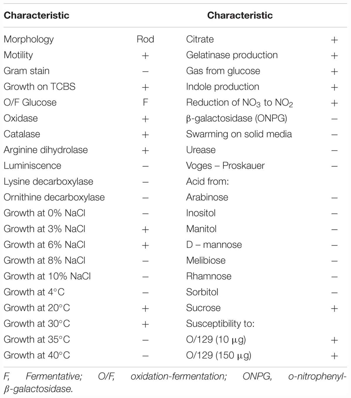
Table 1. Phenotypic characteristics of Vibrio bivalvicida VPAP30.
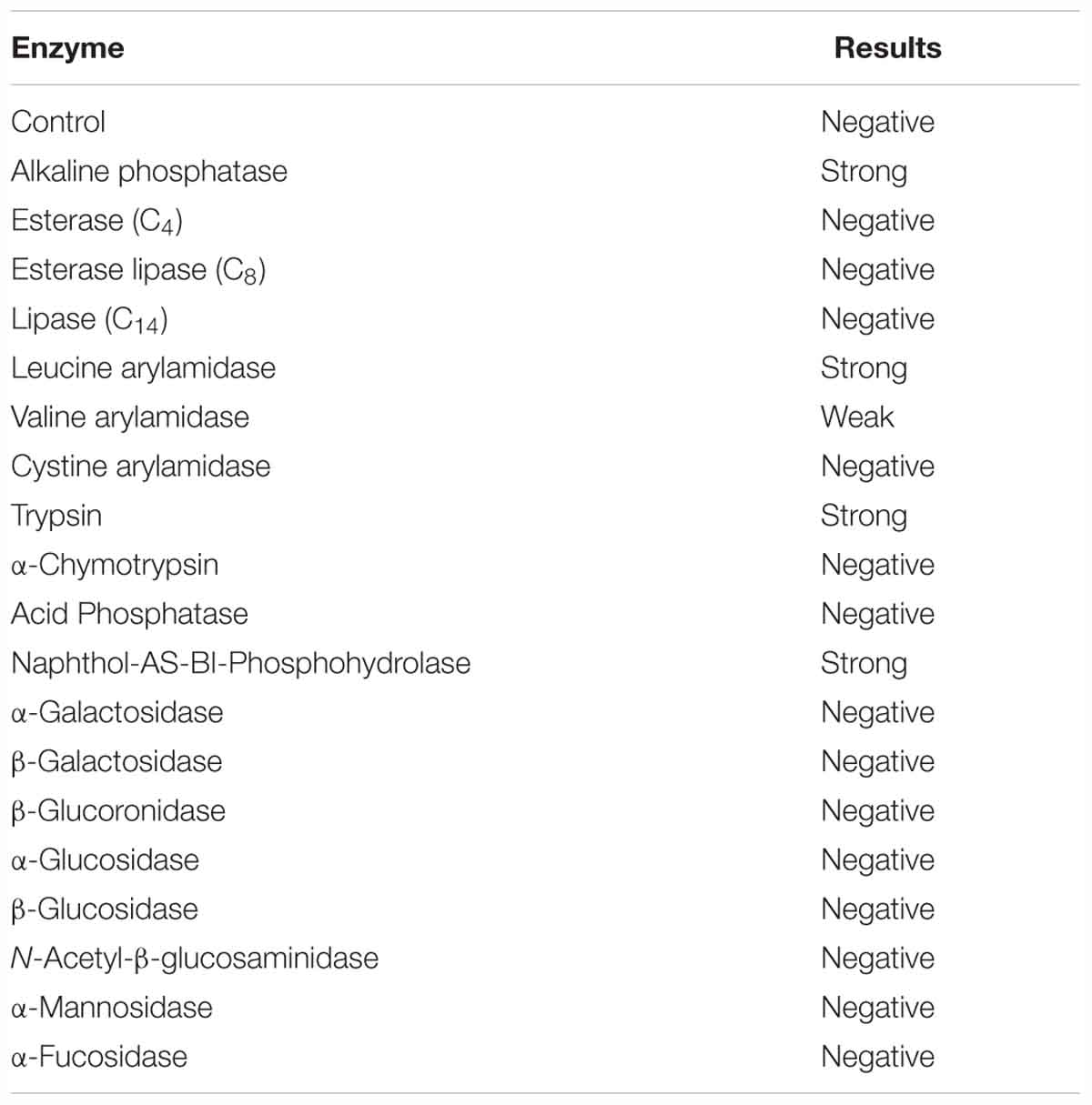
Table 2. Enzymatic properties of V. bivalvicida VPAP30 strain by using the API ZYM system (Biomerieux).
Further phenotypical characterization by using the Biolog system, demonstrated that VPAP30 strain was able to use as a sole carbon source, dextrin, glycogen, tween 40, tween 80, N-acetil-D-glucosamine, D-cellobiose, D-fructose, D-galactose, α-D-glucose, maltose, D-mannose, D-melibiose, sucrose, acetic acid, β-hydroxy butyric acid, α-keto butyric acid, D,L-lactic acid, succinic acid, bromo succinic acid, L-alanine, L-alanyl glycine, L-asparagine, L-aspartic acid, L-glutamic acid, glycyl-L-aspartic acid, glycyl-L-glutamic acid, L-histidine, L-ornithine, L-proline, D-serine, L-threonine, inosine, uridine, thymidine, and glycerol (Supplementary Table 2).
Taxonomic Affiliation of the Strain VPAP30
Molecular identification of the VPAP30 strain was determined by16S rDNA sequence analysis (1,350 bp). The phylogenetic tree constructed from evolutionary distances of 15 representative strains using the NJ algorithm method is shown in Supplementary Figure 1, showing that the amplified sequence of VPAP30 strain was identical to V. bivalvicida (100% of similarity). Furthermore, the 16S rRNA sequence of VPAP30 strain was aligned with reference using the public database of EzBio Cloud1 website and confirmed V. bivalvicida 605T (Accession number LLEI01000012) as the closest relative for the strain VPAP30.
In addition, we have performed an MLSA using the concatenation of (5,480 bp) eight housekeeping genes for encoding various functions (ftsZ, gapA, gyrB, mreB, pyrH, recA, rpoA, and topA) to improve the taxonomic resolution (Sawabe et al., 2013). For this, we have included in the phylogenetic analysis all type strains of the species belonging to Orientalis clade as well as other representative strains such as V. coralliilyticus RE98, strain isolated from diseased oyster larvae (Richards et al., 2014), and previously miss-classified as V. tubiashii (Elston et al., 2008). The resulting phylogenetic tree is shown in Figure 1, in which the Orientalis clade, updated recently by Dubert et al. (2016a) is highlighted. Within Orientalis clade, the VPAP30 strain and V. bivalvicida formed clearly a cluster separated from the other species. Percentages released from comparisons of the concatenated gene sequences revealed that the VPAP30 shared a 99.5% similarity with V. bivalvicida, whereas V. europaeus was the second nearest species with 92.7% identity.
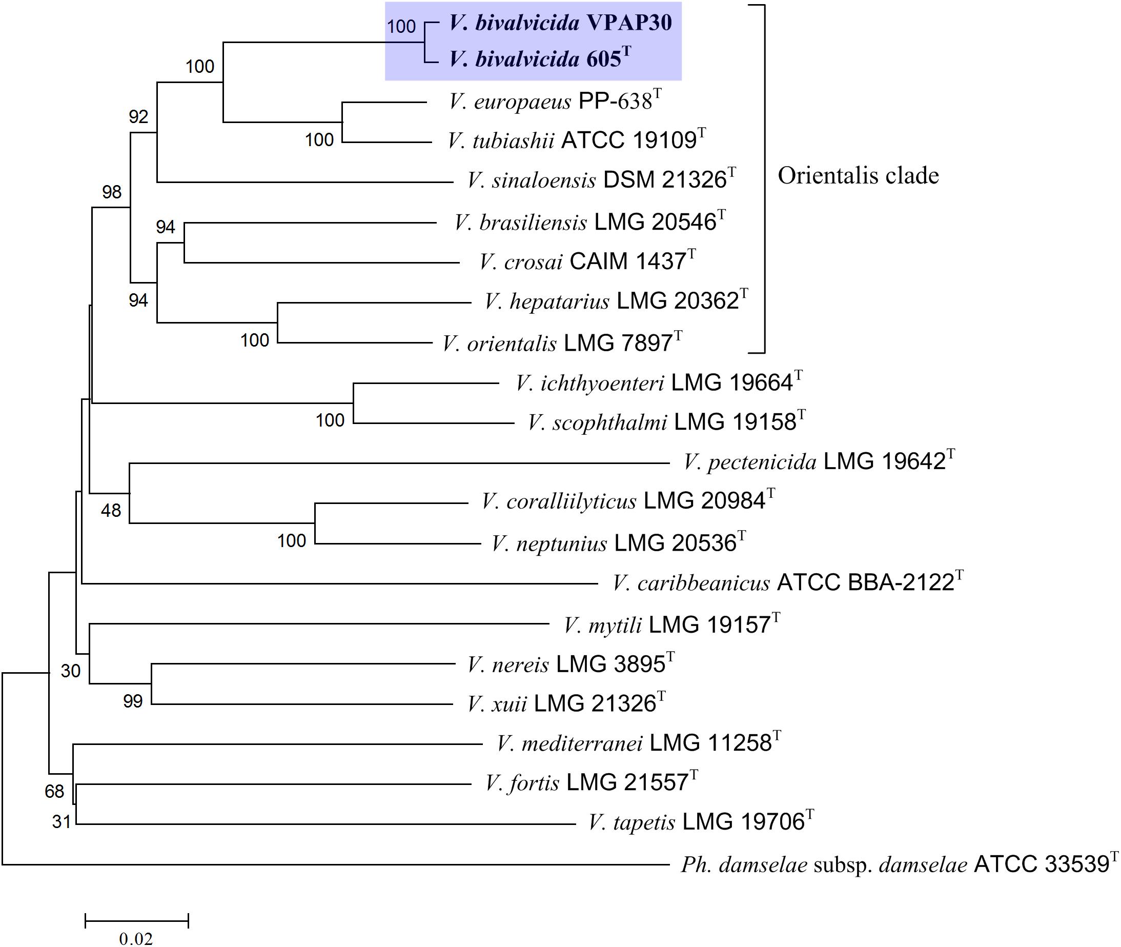
Figure 1. Phylogenetic tree based on concatenated sequences of the housekeeping genes ftsZ, gapA, gyrB, mreB, pyrH, recA, rpoA, and topA obtained by the neighbor-joining method. Horizontal branch lengths are proportional to evolutionary divergences. Bootstrap from 1,000 replicates appears next to the corresponding branch. Photobacterium damselae was used as an outgroup.
Genome to genome comparisons between the strain VPAP30 and the closest relatives confirmed definitely its taxonomic position within the Orientalis clade. ANIb and OrthoANI percentages between the VPAP30 strain and the closest relatives were 97.9 and 98.2%, respectively, to V. bivalvicida 605T and were 86.0 and 86.5%, respectively, to V. tubiashii ATCC 19109T (Table 3). Results supported the affiliation of the VPAP30 strain as V. bivalvicida since ANI values were clearly above than the proposed cut-off values for species boundary (95∼96%) (Richter and Rosselló-Móra, 2009).
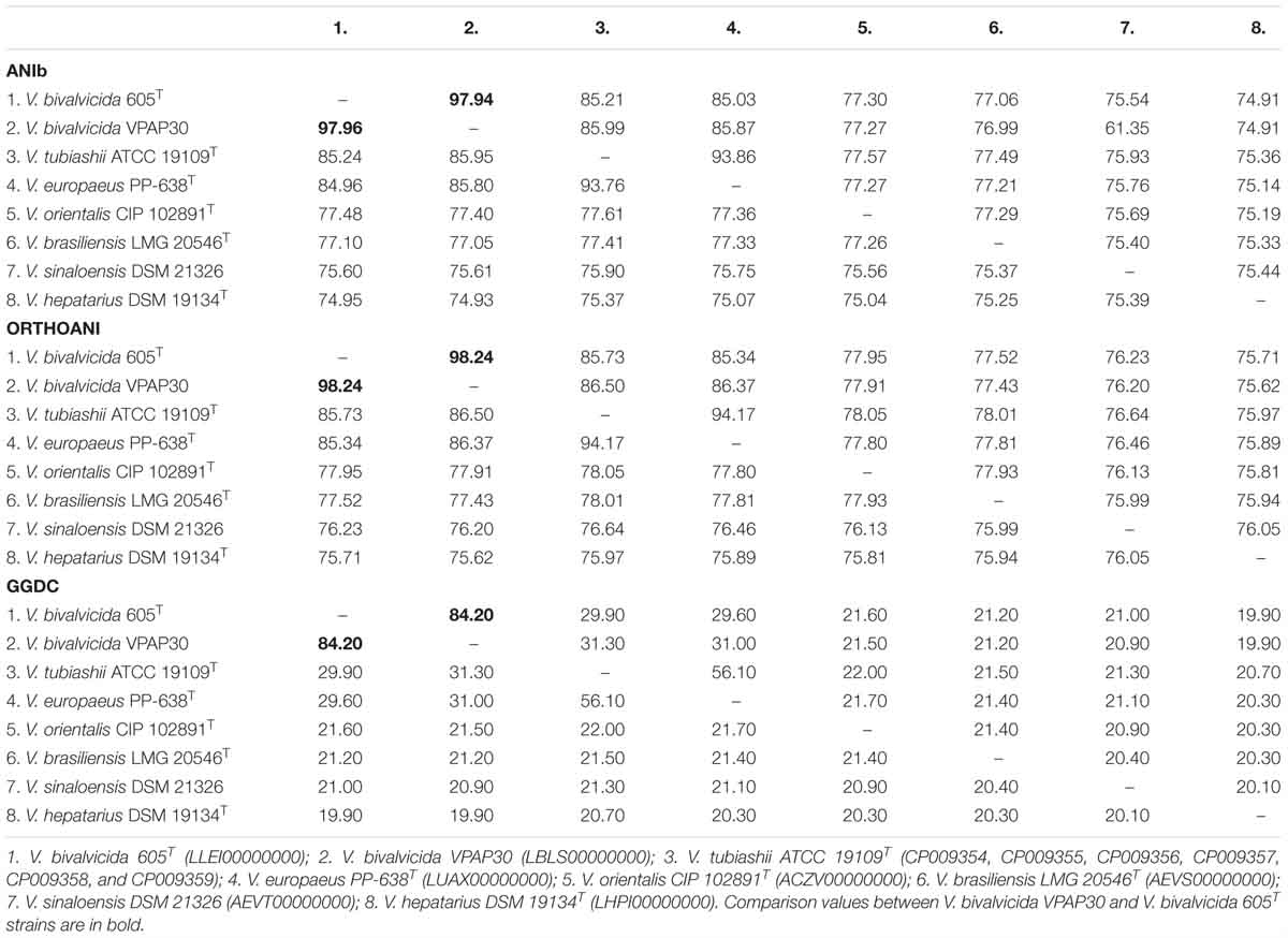
Table 3. Genomic comparisons between the species belonging to the Orientalis clade, including ANI calculations (ANIb and OrthoANI) and in silico DNA–DNA hybridizations (GGDC).
As expected, genomic comparison between the draft genome of the VPAP30 strain and V. bivalvicida 605T yielded 84.2% DDH similarity, clearly higher than the limit (70%) for delineation of prokaryotic species (Meier–Kolthoff et al., 2013), contrasting with the phylogenetic distance of the VPAP30 strain with V. tubiashii ATCC 19109T (31.5% DDH). Overall, the results obtained from the genomic analyses, including MLSA, ANI and DDH, supported the taxonomic affiliation of the strain VPAP30 as V. bivalvicida. Interestingly, the phenotypic profile of the VPAP30 strain is consistent with the characteristics described for V. bivalvicida (Dubert et al., 2016b), except for the use of D-Glucosamine as carbon source and fermentation of Melibiose (Table 4).
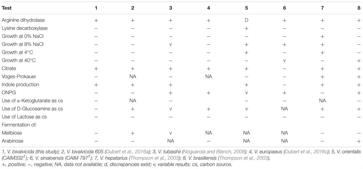
Table 4. Comparative phenotypic characteristics of V. bivalvicida (VPAP30) and Vibrio species belonging to the Orientalis Clade.
Bacterial Pathogenic Activity
Pathogenic activity of the VPAP30 strain was demonstrated by infecting healthy scallop larvae with VPAP30 and V. pectenicida A365 strains, demonstrating that both pathogenic strains produced high levels of larval mortality (Figure 2). However, the VPAP30 strain produced significantly (P > 0.001) higher levels of larval mortality than those produced by the V. pectenicida strain during all the challenge assays. After 24 h of exposure, larval survival of larvae challenged with the VPAP30 strain was 34.3 ± 4.9%, significantly (P < 0.001) lower than that observed in larvae challenged with V. pectenicida A365 (77.8 ± 4.6%) and not challenged larvae (100%). Larval survival at 36 h post-inoculation with the VPAP30 strain was 0%, significantly (P < 0.001) lower than that observed in larvae challenged with V. pectenicida (61.0 ± 3.6%). Not challenged larvae exhibiting a survival of 97.4 ± 1.2% after a period of 48 h. The LD50 for the VPAP30 strain at 24 and 48 h was 1.3 × 10⁴ and 1.2 × 10³ CFU mL-1, respectively.
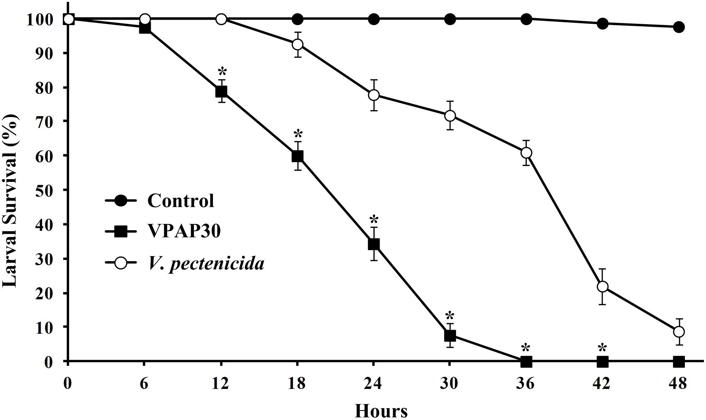
Figure 2. Survival of 10 days old scallop larvae not challenged (control) and challenged with 1 × 10⁵ CFU mL-1 of Vibrio bivalvicida VPAP30 and Vibrio pectenicida A365. Values are a mean (±SD) of three replicates. Asterisks indicate significant differences.
The VPAP30 strain produced on challenged scallop larvae, the classical signs of vibriosis affecting mollusc larvae. These signs were identical to those observed in the larval culture suffering a vibriosis outbreak that developed in the commercial hatchery when the VPAP30 strain was recovered. The main clinical symptoms exhibited by larvae infected with the VPAP30 strain were disruption of the velum, ciliary cells detached from the velum and necrosis of the digestive gland tissue (Figure 3). Erratic swimming was the first clinical sign and appeared at 6 h post-infection. At 12 h post-infection, the majority of challenged larvae showed destruction of the velum and necrosis of the digestive gland, whereas bacterial swarms around the larvae were observed after 24 h.
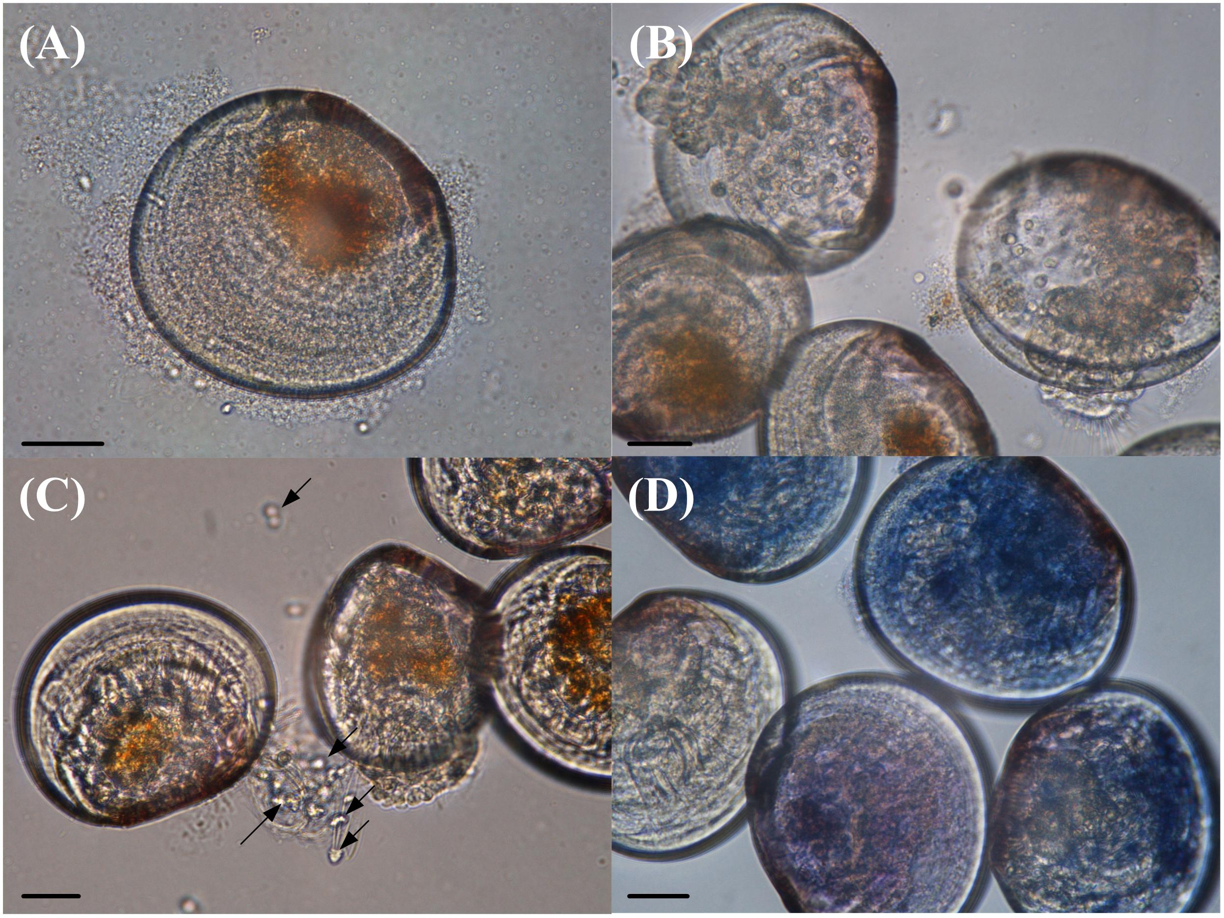
Figure 3. Main clinical signs exhibited by Argopecten purpuratus larvae infected with Vibrio bivalvicida VPAP30 after 24 h of exposure. (A) Bacterial swarms of bacteria on the margins of the larvae, (B) velum disruption, (C) detachment of ciliary cells of the velum (black arrows), and (D) necrosis of digestive tissue of scallop larvae stained with trypan blue. Scale bars: 30 μm.
Invasive Pathogenic Activity
The VPAP30 strain was efficiently stained with 5-DTAF and fluorescence was maintained for at least 36 h (Figure 4A), permitting the use of stained bacterial cells to visualize the invasive ability of the pathogenic strain along the time. The stained VPAP30 strain was detected at a low concentration in the digestive gland of challenged scallop larvae after 30 min of infection (Figure 4B), increasing to high levels after 1 h of infection (Figure 4C). Later, at 24 h post-infection, cells of the VPAP30 strain were detected at high concentrations in all larval tissues as well as around the larval shells (Figure 4D).
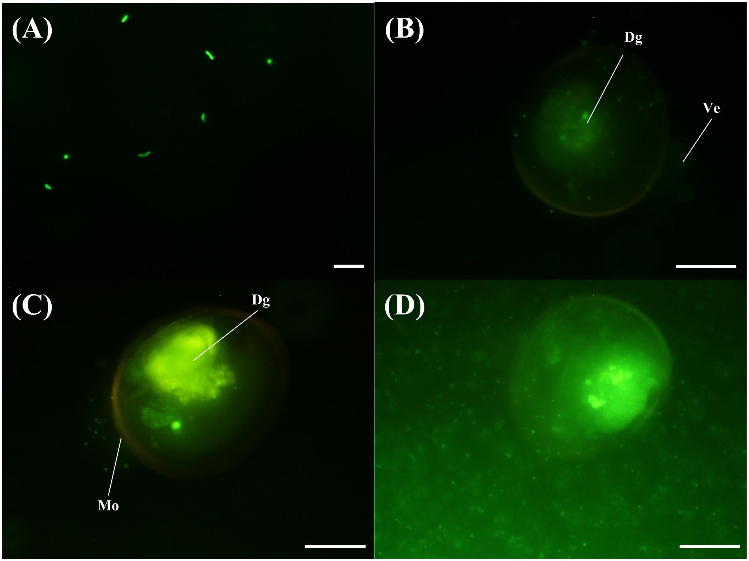
Figure 4. Invasive activity of V. bivalvicida VPAP30 strain on larvae of scallop Argopecten purpuratus determined by epifluorescence microscopy. (A) Bacterial cells of V. bivalvicida VPAP30 stained with 5-DTAF (A); Stained V. bivalvicida in the digestive gland at 30 min post-infection (B); Bacterial cells of V. bivalvicida in the digestive gland at 1 h post-infection (C); Bacterial cells invading completely the larval body cavity and surrounding shell at 24 h post-infection. (Dg) Digestive gland; (Ve) Velum; (Mo) Mouth. Scale bars: 10 μm (A); 50 μm (B–D).
Pathogenic Activity of ECPs
When scallop larvae were exposed to ECPs produced by VPAP30 and V. pectenicida A365 strains, they exhibited identical symptoms to those observed during bacterial challenges. At 12 h post-inoculation with the ECPs of VPAP30 and V. pectenicida strains, percentages of larval survival were 88.6 ± 2.7% and 90.3 ± 2.5%, respectively. Survival rates of larvae infected with VPAP30 and V. pectenicida strains remained at levels not significantly different (P < 0.05) until 36 h post-inoculation (69.0 ± 0.7% and 67.0 ± 5.0%, respectively). However, at 48 h post-inoculation, ECPs of V. pectenicida produced a larval survival of 41.9 ± 5.4%, significantly (P < 0.05) lower than that produced by the ECPs of the VPAP30 strain (59.5 ± 1.7%), whereas larval survival of the control group was 97.4 ± 1.2% (Figure 5). When heat-treated ECPs of the VPAP30 strain were assayed, their pathogenic activity remained present, and treated larvae exhibited survival rates of 74.3 ± 2.0% and 61.6 ± 1.8% after 36 and 48 h of exposure, respectively. Otherwise, the enzymatic activity of the untreated and heat-treated ECPs showed that only naphthol-AS-BI-phosphohydrolase activity remains in the heat-treated ECPs of V. bivalvicida and V. pectenicida strains (Table 5).
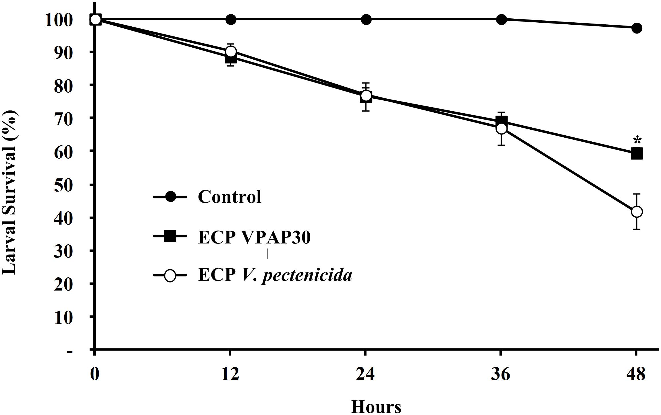
Figure 5. Survival of 10 days old scallop larvae not challenged (control) and challenged with extracellular products of V. bivalvicida VPAP30 and V. pectenicida A365. Values are a mean (±SD) of three replicates. Asterisks indicate significant differences.
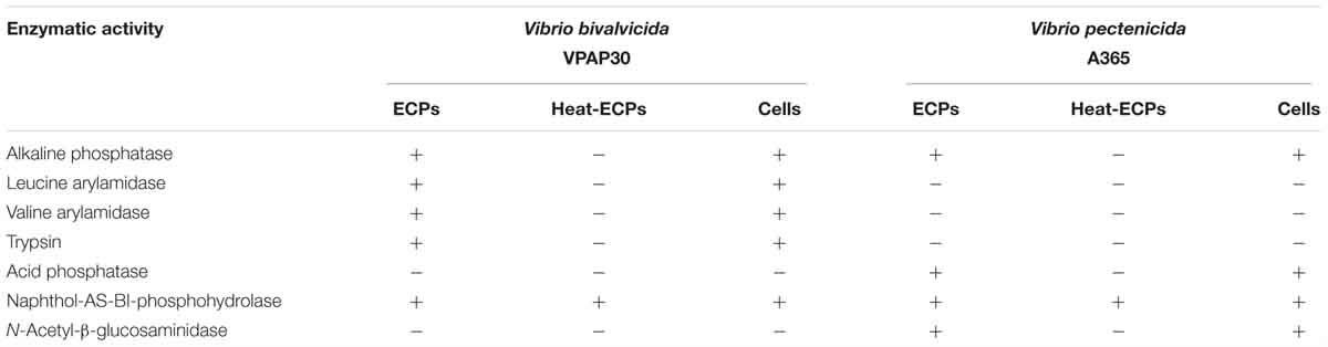
Table 5. Comparative enzymatic activities displayed by whole cells and Extracellular Products (ECPs) of Vibrio bivalvicida VPAP30 and V. pectenicida A365.
Discussion
One of the most important problems to define the current Vibrio species pathogenic to bivalve larvae and spat is related with their misleading taxonomic affiliation. In genomic era, techniques as MLSA or WGS are essential to provide a better understanding of the taxonomic position of the pathogenic Vibrio isolates and then to define them accurately (Sawabe et al., 2007, 2013). Results obtained from the taxonomic analyses, including MLSA, DDH, and ANI, as well as phenotypic tests supported accurately the taxonomic affiliation of the strain VPAP30 as V. bivalvicida. Dubert et al. (2016a) described this species for the first time through the characterization of three strains isolated in a shellfish hatchery located in Galicia (North West of Spain) from healthy broodstock, and moribund reared larvae and larval tank water of a culture of the carpet shell clam, R. decussatus. Virulence of this species was demonstrated in larvae of various clam species (R. decussatus, R. philippinarum, Donax trunculus) and flat oyster (Ostrea edulis), some of the most important bivalve species reared in the European hatcheries (Dubert et al., 2017). Present study constitutes the first report of the isolation of V. bivalvicida out of Europe and extend the host range of this species to the Chilean scallop larvae (A. purpuratus), supporting the potential threat for the shellfish hatcheries in the Atlantic and Pacific coasts.
The high concentration of V. bivalvicida VPAP30 strain in the dead and moribund scallop larvae samples, recovered almost as a pure culture in a non-selective bacteriological medium, the high pathogenic activity on experimentally infected scallop larvae, resemblance of the chronology of the pathology as well as the capacity to produce the same pathological symptoms exhibited by the sampled scallop larvae, strongly support the hypothesis that the mortality event that occurred in the commercial scallop hatchery in Chile was primarily caused by this strain. Notably, the high density and predominance of the colony morphotype of the VPAP30 strain was not observed in agar plates seeded with rearing tank water and swimming larvae samples. Furthermore, in a similar study, the primary pathogenic role of three V. splendidus strains in hatchery-reared scallop larvae was previously demonstrated in a similar manner, fulfilling the Koch’s postulates (Rojas et al., 2015a). These recent studies suggest that despite good sanitary conditions and preventive measures, such as efficient influent water treatment strategies, a high diversity of Vibrio species exhibiting important virulence factors frequently enter to larval rearing tanks, thus exposing Chilean scallop hatcheries to recurrent vibriosis outbreaks.
This study demonstrated the high virulence of strain VPAP30, identified as V. bivalvicida, which produced the mortality of the entire challenged population of healthy scallop larvae after a period of 36 h. The clinical signs caused by the VPAP30 strain resembled those previously described for larval vibriosis occurring in various bivalve species, such as clams, oysters and scallops (Gómez-León et al., 2005; Torkildsen et al., 2005; Elston et al., 2008; Prado et al., 2014; Rojas et al., 2015a; Dubert et al., 2016a). Furthermore, the LD50 value of the VPAP30 strain was similar to the values reported for three highly pathogenic Vibrio strains pathogenic to Pacific oyster larvae, with values of approximately 104 CFU mL-1 and 103 CFU mL-1 after 24 and 48 h, respectively (Estes et al., 2004), but it must be noted that some of these strains have been re-classified as V. coralliilyticus (Wilson et al., 2013; Richards et al., 2014). The V. bivalvicida VPAP30 strain showed to be more virulent that V. bivalvicida 605 and V. bivalvicida 194 strains (Dubert et al., 2016b), since the larvae of the O. edulis, R. philipinarum, and D. trunculus showed the typical signs of vibriosis after 48 h when were inoculated at a final concentration of 106 CFU mL-1.
The observed pathogenic activity of the VPAP30 strain is remarkably higher than that produced by other Vibrio species pathogenic to mollusc larvae. For example, Yue et al. (2010) estimated a LD50 of ∼6 × 10⁶ CFU mL-1 for a Vibrio parahaemolyticus-related strain pathogenic to larvae of Meretrix meretrix, 100-fold higher than the LD50 value of the VPAP30 strain. In another study, V. parahaemolyticus-related strain caused 100% mortality only after 6 days of challenge (Sainz et al., 1998), contrasting with the high virulence exhibited by the VPAP30 strain, which killed all challenged larvae after 36 h of exposure.
The V. bivalvicida VPAP30 strain was able to invade the scallop larvae entering through the mouth to the digestive system and colonizing the body cavity and surface of the shell. These results of invasive activity are consistent with the observed by Dubert et al. (2016a) for the pathogenic species tagged with GFP V. neptunius PP-145.98, V. europaeus PP-638T, formerly V. tubiashii subsp. europaeus (Dubert et al., 2016c), and V. bivalvicida 605T on Manila clam larvae. These authors defined three stages in the infective process caused by these pathogenic species as follows: (1) during the first 2 h of infection, Vibrio strains were filtered by larval vellum and entered the digestive system through the esophagus and stomach colonizing the digestive gland and intestine; (2) then, Vibrio strains spread and proliferated to the surrounding organs in the body cavity (6–8 h post infection), and (3) after 14 h of challenge, the body cavity was completely colonized by Vibrio strains. Interestingly, DTAF-stained cells of the VPAP30 strain maintained their virulence on scallop larvae producing high mortality as well as the clinical signs typical of vibriosis. Therefore, the use of bacterial cell staining with 5-DTAF to study the chronology of invasive processes caused by Vibrio species is a time-saving protocol, highly recommended to follow the vibriosis on mollusc larvae.
Additionally, the pathogenic activity of V. bivalvicida VPAP30 strain mediated by the production of ECPs was evaluated. Our results demonstrated that cell-free ECPs of the VPAP30 strain are involved in the pathogenic action on scallop larvae, causing a mortality of 40% of challenged larvae after 48 h of exposure. The results of this study demonstrate that the extracellular toxigenic activity exhibited by this strain is mainly mediated by the production of heat stable compounds, causing larval necrosis and the detachment of ciliary cells, consistent with previous reports, which demonstrated that Vibrio strains can produce heat stable ciliostatic toxins and proteinases that degrade larval tissue (DiSalvo et al., 1978; Nottage et al., 1989). Furthermore, Travers et al. (2014) demonstrated the toxicity of ECPs produced by the French V. europaeus 07/118 T2, formerly V. tubiashii (Dubert et al., 2016c), on the oyster C. gigas, producing a mortality of 41% after 2 days of challenge. In addition, the enzymatic activities of ECPs released by strains belonging to the V. europaeus, V. neptunius, and V. bivalvicida species were determined by Dubert et al. (2016a) using API ZYM, primarily describing a protease activity. In contrast to the results obtained by Dubert et al. (2016a), the pathogenic activity of ECPs produced by the VPAP30 strain remained intact after heat treatment, indicating the presence of thermo-resistant toxins. Otherwise, only naphthol-AS-Bi-phosphohydrolase activity remained in the heat treated ECPs, suggesting that enzymatic activities detected by the API ZYM system are not involved in the pathogenic activity of this strain. Riquelme et al. (1995) reported an episode of larval mortality in reared larvae of Argopecten purpuratus identifying the causal agent as V. anguillarum-like and causing 30% mortality at 24 h post-infection, mainly mediated by the production of an extracellular toxin. Rojas et al. (2015a) isolated 3 pathogenic strains identified as V. splendidus, which were recovered from different episodes of massive mortalities occurring in a commercial culture of the scallop larvae A. purpuratus in Chile, causing larval mortalities of approximately the 80% of challenged larvae at 48 h post-inoculation, and characterized by bacterial invasion of larval tissue as well as the production of ECPs toxigenic to A. purpuratus larvae.
Several proteins are secreted by V. tubiashii strains, including a low molecular weight ciliostatic toxin, which is a very important virulence factor in shellfish larval vibriosis (Nottage et al., 1989). As previously described, a major trait of vibriosis is extensive necrosis followed by sudden death, which is consistent with the involvement of proteinases and haemolysins (Nottage and Birkbeck, 1986), as exhibited by scallop larvae inoculated with ECPs released by the VPAP30 strain.
A mollusc larvae pathogenic strain reclassified by Wilson et al. (2013) as V. coralliilyticus (formerly V. tubiashii ATCC 19105) produces some extracellular compounds, including a cytolysin/haemolysin (Kothary et al., 2001) and a protease (Delston et al., 2003). More recently, Mersni-Achour et al. (2015) remarked on the importance of the production of metalloproteases in the pathogenic activity by this V. europaeus strain (formerly V. tubiashii ATCC 19105) on oyster larvae, detecting the production of a metalloprotease encoded by the vtpA gene. It must be noted that a metalloprotease encoding gene similar to vtpA was detected in the genome of this pathogenic strain (formerly V. tubiashii VPAP30) (Rojas et al., 2015b). Moreover, Hasegawa et al. (2008) demonstrated that metalloprotease inhibitors severely reduce the toxicity of ECPs produced by 2 strains recently identified as V. coralliilyticus (Wilson et al., 2013; Richards et al., 2014), but previously miss-identified as V. tubiashii, on Pacific oyster larvae. In addition, the authors reported an inhibitory activity on its extracellular haemolysin of the extracellular metalloprotease produced by these strains (Hasegawa and Häse, 2009).
Conclusion
In conclusion, this is the first isolation of a V. bivalvicida strain outside Europe, being recovered from a massive mortality episode in a commercial larval culture of the scallop A. purpuratus in Chile. Our results demonstrated that V. bivalvicida VPAP30 is highly pathogenic to scallop larvae exhibiting an invasive activity as well as a production of toxigenic heat-stable ECPs and producing the typical clinical signs of vibriosis. In addition, this strain produced on experimentally infected scallop larval cultures very similar symptoms to those observed during the massive mortality event in the Chilean commercial hatchery strongly suggesting that this strain was the causative agent of the mass mortalities occurring in the referred event. Then, we have demonstrated for the first time the occurrence of a highly pathogenic strain of V. bivalvicida in a commercial hatchery of scallop A. purpuratus, suggesting this species can become in a pathogen of major concern for the Chilean pectinid industry. The proper identification of this bacterial pathogen causing epizootics in larval cultures is essential for developing efficient epidemiological management strategies to prevent and control outbreaks in Chilean intensive scallop larvae husbandry.
Author Contributions
RR designed the study, isolated the bacterial strain, performed all challenge assays, drafted the manuscript, and is the corresponding author and the primary contact. CM contributed significantly to the design, drafting, revisions, and interpretation of data. JR designed the study together with RR, supervised the study and advised molecular analysis and interpretation. JD and JB helped to analyze the MLSA analysis and genomic comparisons and in silico DNA-DNA hybridizations. All authors have made intellectual contributions to the work and approved it for publication.
Funding
This study was financially supported by the Science and Technology National Council (CONICYT) of Chile by the Postdoctoral Project Grant No. 3150395 and FONDECYT grant No. 1140734 and FONDEF ID16I10291.
Conflict of Interest Statement
The authors declare that Invertec Ostimar company only contributed to permitting the scallop larvae collection, and had no role in the study.
Acknowledgments
We thank staff of commercial hatchery Invertec Ostimar for good collaboration with scallop larval sampling and also are grateful to Dr. Jean Louis Nicolas for providing the V. pectenicida A365 strain.
Supplementary Material
The Supplementary Material for this article can be found online at: https://www.frontiersin.org/articles/10.3389/fmicb.2019.00855/full#supplementary-material
Footnotes
References
Barrow, G. I., and Feltham, R. K. (1993). Cowan and Steel’s Manual for the Identification of Medical Bacteria, 3rd Edn. Cambridge: Cambridge University Press, 331.
Beaz-Hidalgo, R., Balboa, S., Romalde, J. L., and Figueras, M. J. (2010). Diversity and pathogenecity of Vibrio species in cultured bivalve molluscs. Environ. Microbiol. Rep. 2, 34–43. doi: 10.1111/j.1758-2229.2010.00135.x
Binesse, J., Delsert, C., Saulnier, D., Champomier, M. C., Zagorec, M., Munier, H., et al. (2008). Metalloprotease Vsm is the mayor determinant of toxicity for extracellular products of Vibrio splendidus. Appl. Environ. Microbiol. 74, 7108–7117. doi: 10.1128/AEM.01261-08
Cole, J. R., Chai, B., Farris, R. J., Wang, Q., Kulam-Syed-Mohideen, A. S., McGarrell, D. M., et al. (2007). The ribosomal database project (RDP-II): introducing myRDP space and quality controlled public data. Nucleic Acids Res. 35, D169–D172. doi: 10.1093/nar/gkl889
Delston, R. B., Kothary, M. H., Shangraw, K. A., and Tall, B. D. (2003). Isolation and characterization of zinc-containing metalloprotease expressed by Vibrio tubiashii. Can. J. Microbiol. 49, 525–529. doi: 10.1139/w03-067
DiSalvo, L. H., Blecka, J., and Zebal, R. (1978). Vibrio anguillarum and larval mortality in a California coastal shellfish hatchery. Appl. Environ. Microbiol. 35, 219–221.
Dubert, J., Barja, J. L., and Romalde, L. (2017). New insights into pathogenic vibrios affecting bivalves in hatcheries: present and future prospects. Front. Microbiol. 8:762. doi: 10.3389/fmicb.2017.00762
Dubert, J., Nelson, N., Spinard, E., Kessner, L., Gomez-Chiarri, M., da Costa, F., et al. (2016a). Following the infection process of vibriosis in Manila clam (Ruditapes philippinarum) larvae through GFP-tagged pathogenic Vibrio species. J. Invertebr. Pathol. 133, 27–33. doi: 10.1016/j.jip.2015.11.008
Dubert, J., Romalde, J. L., Prado, S., and Barja, J. L. (2016b). Vibrio bivalvicida sp. nov., a novel larval pathogen for bivalve molluscs reared in a hatchery. Syst. Appl. Microbiol. 39, 8–13. doi: 10.1016/j.syapm.2015.10.006
Dubert, J., Romalde, J. L., Spinard, E. J., Nelson, D. R., Gomez-Chiarri, M., and Barja, J. L. (2016c). Reclassification of the larval pathogen for marine bivalves Vibrio tubiashii subsp. europaeus as Vibrio europaeus sp. nov. Int. J. Syst. Evol. Microbiol. 66, 4791–4796. doi: 10.1099/ijsem.0.001431
Elston, R., Hasegawa, H., Humphrey, K. L., Polyak, I. K., and Häse, C. C. (2008). Re-emergence of Vibrio tubiashii in bivalve shellfish aquaculture: severity, environmental drivers, geographic extent and management. Dis. Aquat. Org. 82, 119–134. doi: 10.1139/f80-126
Estes, R., Friedman, C., Elston, R., and Herwig, R. (2004). Pathogenicity testing of shellfish hatchery bacterial isolates on Pacific oyster Crassostrea gigas larvae. Dis. Aquat. Org. 58, 223–230. doi: 10.3354/dao058223
Gómez-León, J., Villamil, L., Lemos, M. L., Novoa, B., and Figueras, A. (2005). Isolation of Vibrio alginolyticus and Vibrio splendidus from aquacultured carpet shell clam (Ruditapes decussatus) larvae associated with mass mortalities. Appl. Environ. Microbiol. 71, 98–104. doi: 10.1128/AEM.71.1.98-104.2005
Gómez-León, J., Villamil, L., Salger, S., Sallum, R., Remacha-Triviño, A., Leavitt, D., et al. (2008). Survival of eastern oyster Crassostrea virginica from three lines following experimental challenge with bacterial pathogens. Dis. Aquat. Org. 79, 95–105. doi: 10.3354/dao01902
Hansen, G. H., and Sörheim, R. (1991). Improved method for phenotypical characterization of marine bacteria. J. Microbiol. Meth. 13, 231–241. doi: 10.1016/0167-7012(91)90049-V
Hasegawa, H., and Häse, C. C. (2009). The extracellular metalloprotease of Vibrio tubiashii directly inhibits its extracellular haemolysin. Microbiology 155, 2296–2305. doi: 10.1099/mic.0.028605-0
Hasegawa, H., Lind, E., Boin, M., and Häse, C. C. (2008). The extracellular metalloprotease of Vibrio tubiashii is a major virulence factor for Pacific oyster (Crassostrea gigas) larvae. Appl. Environ. Microbiol. 74, 4101–4110. doi: 10.1128/AEM.00061-08
Kothary, M. H., Delston, R. B., Curtis, S. K., McCardell, B. A., and Tall, B. D. (2001). Purification and characterization of vulnificolysin-like cytolysin produced by Vibrio tubiashii. Appl. Environ. Microbiol. 67, 3707–3711. doi: 10.1128/AEM.67.8.3707-3711.2001
Labreuche, Y., Le Roux, F., Henry, J., Zatylny, C., Huvet, A., Lambert, C., et al. (2010). Vibrio aestuarianus zinc metalloprotease caused lethality in the Pacific oyster Crassostrea gigas and impairs the host cellular immune defenses. Fish Shellfish Immunol. 29, 753–758. doi: 10.1016/j.fsi.2010.07.007
Lambert, C., Nicolas, J. L., Cilia, V., and Corre, S. (1998). Vibrio pectenicida sp. nov., a pathogen of scallop (Pecten maximus) larvae. Int. J. Syst. Bacteriol. 48, 481–487. doi: 10.1099/00207713-48-2-481
Lee, I., Kim, Y. O., Park, S. C., and Chun, J. (2016). OrthoANI: an improved algorithm and software for calculating average nucleotide identity. Int. J. Syst. Evol. Microbiol. 66, 1100–1103. doi: 10.1099/ijsem.0.000760
Liu, P. C., Chen, Y. C., and Lee, K. K. (2001). Pathogenicity of Vibrio alginolyticus isolated from diseased small abalone Haliotis diversicolor supertexta. Microbios 104, 71–77.
Liu, R., Qiu, L., Yu, Z., Zi, J., Yue, F., Wang, L., et al. (2013). Identification and characterisation of pathogenic Vibrio splendidus from Yesso scallop (Patinopecten yessoensis) cultured in a low temperature environment. J. Invertebr. Pathol. 114, 144–150. doi: 10.1016/j.jip.2013.07.005
Luna-González, A., Maeda-Martínez, A. N., Sainz, J. C., and Ascencio-Valle, F. (2002). Comparative susceptibility of veliger larvae of four bivalve mollusks to a Vibrio alginolyticus strain. Dis. Aquat. Org. 49, 221–226. doi: 10.3354/dao049221
MacDonnell, M. T., Singleton, F. L., and Hood, M. A. (1982). Diluent composition for use of API 20E in characterizing marine and estuarine bacteria. Appl. Environ. Microbiol. 44, 423–427.
Meier–Kolthoff, J. P., Auch, A. F., Klenk, H. P., and Göker, M. (2013). Genome sequence–based species delimitation with confidence intervals and improved distance functions. BMC Bioinformatics 14:60. doi: 10.1186/1471-2105-14-60
Mersni-Achour, R., Ben, Y., Pichereau, V., Doghri, I., Etien, C., Dégremont, L., et al. (2015). Factors other than metalloprotease are required for full virulence of French Vibrio tubiashii isolates in oyster larvae. Microbiology 161, 997–1007. doi: 10.1099/mic.0.000058
Miranda, C. D., Rojas, R., Abarca, A., and Hurtado, L. (2014). Effect of florfenicol and oxytetracycline treatments on the intensive larval culture of the Chilean scallop Argopecten purpuratus (Lamarck, 1819). Aquac. Res. 45, 16–30. doi: 10.1111/j.1365-2109.2012.03200.x
Natrah, F. M. I., Ruwandeepika, H. A. D., Pawar, S. D., Karunasagar, I., Sorgeloos, P., Bossier, P., et al. (2011). Regulation of virulence factors by quorum sensing in Vibrio harveyi. Vet. Microbiol. 154, 124–129. doi: 10.1016/j.vetmic.2011.06.024
Navarrete, P., Magne, F., Mardones, P., Riveros, M., Opazo, R., Suau, A., et al. (2010). Molecular analysis of intestinal microbiota of rainbow trout (Oncorhynchus mykiss). FEMS Microb. Ecol. 71, 148–156. doi: 10.1111/j.1574-6941.2009.00769.x
Nicolas, J. L., Corre, S., Gauthier, G., Robert, R., and Ansquer, D. (1996). Bacterial problems associated with scallop Pecten maximus larval culture. Dis. Aquat. Org. 27, 67–76. doi: 10.3354/dao027067
Noguerola, I., and Blanch, A. R. (2008). Identification of Vibrio spp. with a set dichotomous keys. J. Appl. Microbiol. 105, 175–185. doi: 10.1111/j.1365-2672.2008.03730.x
Nottage, A. S., and Birkbeck, T. H. (1986). Toxicity to marine bivalves of culture supernatant fluids of the bivalve pathogenic Vibrio strain NCMB 1338 and other marine vibrios. J. Fish Dis. 9, 249–256. doi: 10.1111/j.1365-2761.1986.tb01009.x
Nottage, A. S., Sinclair, P. D., and Birkbeck, T. H. (1989). Role of low-molecular weight ciliostatic toxins in vibriosis of bivalve mollusks. J. Aquat. Anim. Health 1, 180–186. doi: 10.1577/1548-8667(1989)001<0180:ROLMWC>2.3.CO;2
Prado, S., Dubert, J., and Barja, J. L. (2015). Characterization of pathogenic vibrios isolated from bivalve hatcheries in Galicia, NW Atlantic coast of Spain. Description of Vibrio tubiashii subsp. europaensis subsp. nov. Syst. Appl. Microbiol. 38, 26–29. doi: 10.1016/j.syapm.2014.11.005
Prado, S., Dubert, J., Romalde, J. L., Toranzo, A. E., and Barja, J. L. (2014). Vibrio ostreicida sp. nov., a new pathogen of bivalve larvae. Int. J. Syst. Evol. Microbiol. 64, 1641–1646. doi: 10.1099/ijs.0.051417-0
Reed, L. J., and Muench, H. (1938). A simple method of estimating fifty percent endpoints. Am. J. Hyg. 27, 493–497. doi: 10.1093/oxfordjournals.aje.a118408
Richards, G. P., Bono, J. L., Watson, M. A., and Needleman, S. (2014). Complete genome sequence for the shellfish pathogen Vibrio coralliilyticus RE98 isolated from a shellfish hatchery. Genome Announc. 2, 1–2. doi: 10.1128/genomeA.01253-14
Richter, M., and Rosselló-Móra, R. (2009). Shifting the genomic gold standard for the prokaryotic species definition. Proc. Natl. Acad. Sci. U.S.A. 106, 19126–19131. doi: 10.1073/pnas.0906412106
Richter, M., Rosselló-Móra, R., Glöckner, F. O., and Peplies, J. (2016). JSpeciesWS: a web server for prokaryotic species circumscription based on pairwise genome comparison. Bioinformatics 32, 929–931. doi: 10.1093/bioinformatics/btv681
Riquelme, C., Hayashida, G., Toranzo, A., Vilches, J., and Chavez, P. (1995). Pathogenicity studies on a Vibrio anguillarum-related (VAR) strain causing an epizootic in Argopecten purpuratus larvae cultured in Chile. Dis. Aquat. Org. 22, 135–141. doi: 10.3354/dao022135
Riquelme, C., Toranzo, A., Barja, J. L., Vergara, N., and Araya, R. (1996). Association of Aeromonas hydrophila and Vibrio alginolyticus with larval mortalities of scallop (Argopecten purpuratus). J. Invertebr. Pathol. 67, 213–218. doi: 10.1006/jipa.1996.0035
Rojas, R., Miranda, C. D., and Amaro, A. M. (2009). Pathogenicity of a highly exopolysaccharide-producing Halomonas strain causing epizootics in larval cultures of the Chilean scallop Argopecten purpuratus (Lamarck, 1819). Microb. Ecol. 57, 129–139. doi: 10.1007/s00248-008-9401-z
Rojas, R., Miranda, C. D., Opazo, R., and Romero, J. (2015a). Characterization and pathogenicity of Vibrio splendidus strains associated with massive mortalities of commercial hatchery-reared larvae of scallop Argopecten purpuratus (Lamarck, 1819). J. Invertebr. Pathol. 124, 61–69. doi: 10.1016/j.jip.2014.10.009
Rojas, R., Miranda, C. D., Romero, J., Asenjo, F., Valderrama, K., Segovia, C., et al. (2015b). Genome sequence of Vibrio VPAP30, isolated from an episode of massive mortality of reared larvae of the scallop Argopecten purpuratus. Genome Announc. 3, 1–2. doi: 10.1128/genomeA.00745-15
Romero, J., Garcia-Varela, M., Laclette, J. P., and Espejo, R. T. (2002). Bacterial 16S rRNA gene analysis revealed that bacteria related to Arcobacter spp. constitute an abundant and common component of the oyster microflora (Tiostrea chilensis). Microb. Ecol. 44, 365–371. doi: 10.1007/s00248-002-1063-7
Romero, J., and Navarrete, P. (2006). 16S rDNA-based analysis of dominant bacterial populations associated with early life stages of Coho salmon (Oncorhynchus kisutch). Microb. Ecol. 51, 422–430. doi: 10.1007/s00248-006-9037-9
Sainz, J., Maeda-Martínez, A., and Ascencio, F. (1998). Experimental vibriosis induction with Vibrio alginolyticus of larval of the Caterina scallop (Argopecten ventricosis = circularis) (Sowerby II, 1842). Microb. Ecol. 35, 188–192. doi: 10.1007/s002489900073
Sawabe, T., Kita-Tsukamoto, K., and Thompson, F. L. (2007). Inferring the evolutionary history of vibrios by means of multilocus sequence analysis. J. Bacteriol. 189, 7932–7936. doi: 10.1128/JB.00693-07
Sawabe, T., Ogura, Y., Matsumura, Y., Feng, G., Amin, A. R., Mino, S., et al. (2013). Updating the Vibrio clades defined by multilocus sequence phylogeny: proposal of eight new clades, and the description of Vibrio tritonius sp. nov. Front. Microbiol. 4:414. doi: 10.3389/fmicb.2013.00414
Sherr, B., Sherr, E., and Fallon, R. (1987). Use of monodispersed, fluorescently labeled bacteria to estimate in situ protozoan bacterivory. Appl. Environ. Microbiol. 53, 958–965. doi: 10.1128/MMBR.68.3.403-431.2004
Thompson, F. L., Iida, T., and Swings, J. (2004). Biodiversity of vibrios. Microbiol. Mol. Biol. Rev. 68, 403–431. doi: 10.1128/mmbr.68.3.403-431.2004
Thompson, F. L., Thompson, C. C., Hoste, B., Vandemeulebroecke, K., Gullian, M., and Swings, J. (2003). Vibrio fortis sp. nov. and Vibrio hepatarius sp. nov., isolated from aquatic animals and the marine environment. Int. J. Syst. Evol. Microbiol. 53, 1495–1501. doi: 10.1099/ijs.0.02658-0
Torkildsen, L., Lambert, C., Nylund, A., Magnesen, T., and Bergh, Ø (2005). Bacteria associated with life stages of the great scallop, Pecten maximus: impact on larval survival. Aquacult. Int. 13, 575–592. doi: 10.1007/s10499-005-9002-5
Travers, M. A., Achour, R. M., Haffner, P., Tourbiez, D., Cassone, A. L., and Morga, B. (2014). First description of French V. tubiashii strains pathogenic to mollusk: I. Characterization of isolates and detection during mortality events. J. Invertebr. Pathol. 123, 38–48. doi: 10.1016/j.jip.2014.04.009
von Brand, E., Merino, G. E., Abarca, A., and Stotz, W. (2006). “Scallop fishery and aquaculture in Chile,” in Scallops: Biology, Ecology and Aquaculture Developments in Aquaculture and Fisheries Science, Vol. 35, eds S. E. Shumway and J. Parsons (Amsterdam: Elsevier), 1293–1311.
Waechter, M., Le Roux, F., Nicolas, J. L., Marissal, E., and Berthe, F. (2002). Characterization of pathogenic bacteria of the cupped oyster Crassostrea gigas. C. R. Biol. 325, 231–238. doi: 10.1016/S1631-0691(02)01428-2
Wilson, B., Muirhead, A., Bazanella, M., Huete-Stauffer, C., Vezzulli, L., and Bourne, D. G. (2013). An improved detection and quantification method for the coral pathogen Vibrio coralliilyticus. PLoS One 8:e81800. doi: 10.1371/journal.pone.0081800
Keywords: Vibrio bivalvicida, vibriosis, scallop larvae, hatchery, shellfish pathology, Argopecten purpuratus, Chile
Citation: Rojas R, Miranda CD, Romero J, Barja JL and Dubert J (2019) Isolation and Pathogenic Characterization of Vibrio bivalvicida Associated With a Massive Larval Mortality Event in a Commercial Hatchery of Scallop Argopecten purpuratus in Chile. Front. Microbiol. 10:855. doi: 10.3389/fmicb.2019.00855
Received: 16 January 2019; Accepted: 03 April 2019;
Published: 10 May 2019.
Edited by:
Hongyue Dang, Xiamen University, ChinaReviewed by:
Melissa Garren, California State University, Monterey Bay, United StatesMarie-Agnès Travers, Institut Français de Recherche pour l’Exploitation de la Mer (IFREMER), France
Copyright © 2019 Rojas, Miranda, Romero, Barja and Dubert. This is an open-access article distributed under the terms of the Creative Commons Attribution License (CC BY). The use, distribution or reproduction in other forums is permitted, provided the original author(s) and the copyright owner(s) are credited and that the original publication in this journal is cited, in accordance with accepted academic practice. No use, distribution or reproduction is permitted which does not comply with these terms.
*Correspondence: Rodrigo Rojas, cnJvamFzQHVjbi5jbA== Jaime Romero, anJvbWVyb0BpbnRhLnVjaGlsZS5jbA==; amFpbWUubS5yb21lcm9AZ21haWwuY29t