- 1Department of Biomedical Engineering, Case Western Reserve University, Cleveland, OH, United States
- 2Medical Scientist Training Program, Case Western Reserve University, Cleveland, OH, United States
- 3Department of Chemical and Biomedical Engineering, Cleveland State University, Cleveland, OH, United States
Therapeutic tissue regeneration remains a significant unmet need in heart failure and cardiovascular disease treatment, which are among the leading causes of death globally. Decellularized heart matrix (DHM) offer promising advantages for tissue engineering, including low immunogenicity and seamless integration into biological processes, facilitating biocompatibility. However, DHM is challenged by weak mechanical properties that limit its utility to biomedical applications like tissue engineering. To address this limitation, we functionalized DHM with methacryloyl functional groups (DHMMA) that support UV-induced crosslinking to enhance mechanical properties. By modulating the degree of methacryloyl substitution, a broad range of stiffness was achieved while maintaining cell viability on crosslinked DHMMA. Additionally, we show that increasing UV exposure time and pH increases DHMMA stiffness. Furthermore, topographical features transferred on DHMMA via soft lithography facilitated physical orientation of cells in culture. We demonstrate DHMMA as a scaffold with tunable stiffness and matrix-degradation properties suitable for cell survival and microfabrication for cardiac tissue engineering applications.
1 Introduction
Currently, there are no clinical therapies to repair the failing heart, motivating the need for advancements in cardiac tissue engineering strategies aimed at regenerating damaged myocardial tissue. The cardiac patch approach considers cell therapy on support scaffolds to rebuild parts of tissue (Wang Lu et al., 2021; Zhang et al., 2022). Synthetic biomaterials, such as poly (ε-caprolactone) and poly (lactic-co-glycolic acid), are used due to their tunable properties and facile synthesis (McMahan et al., 2020; Zhang Dongshan et al., 2024). Despite their versatility, synthetic biomaterials lack the complex biological cues for tissue integration and directed tissue repair (Kafili et al., 2023). Natural biomaterials comprised of gelatin, collagen, or decellularized ECM emerged as promising alternatives due to their biocompatibility (You et al., 2024; Xu et al., 2024). Specifically, there is a growing interest in exploring decellularized heart matrix (DHM) hydrogels as an alternative substrate for cardiac tissue engineering applications (Kafili et al., 2023; Vu et al., 2022; Zhe et al., 2023).
DHM offers a promising regenerative therapy for the heart due to the intrinsic properties of the native tissue microenvironment. DHM is comprised primarily of extracellular matrix proteins and provides insoluble building blocks such as collagen which suppress adverse tissue remodeling (McLaughlin et al., 2019; Baehr et al., 2020). DHM retains bioactive and soluble factors that are enriched in the cardiac tissue origin that facilitate crucial repair processes (Hamsho et al., 2024; Wu et al., 2023). The presence of cardiac tissue-specific ECM components facilitates programming of multicellular processes. When delivered to the ischemic heart, DHM promotes cardiomyocyte cell survival and proliferation, angiogenesis, suppresses fibroblast activation and potentiates immunogenic responses that support tissue repair (Jin et al., 2022). These collective effects help preserve cardiac function and reduce fibrosis (Wang et al., 2020; Wang et al. 2021a; Wang et al., 2021b; Wang et al. 2022; Diaz et al., 2021; Wassenaar Jean W. et al., 2016). We, along with others, have begun to elucidate the biofactors and responsive cellular players involved (Wang et al., 2021a; Wang et al., 2021b). Additionally, DHM has been explored as a delivery vehicle for matrix proteins and enriched soluble factors such as VEGF for vascularization (X. Wang et al., 2022). For cardiac therapy, DHM is delivered as a liquid hydrogel precursor. At physiological temperature, DHM undergoes gelation over several minutes (Wang et al., 2020; Wang et al. 2021a; Wang et al. 2021b; Wang et al., 2022). However, the slow gelation process presents a challenge for retention in the beating heart, with observable dispersion from the injection site. The spreading of DHM further accelerates degradation due to enzymatic remodeling processes. We previously developed solid DHM microparticles via electrospray and emulsification methods to increase retention after injection. The solid microparticles extended DHM tissue retention, increased resistance to enzymatic digestion, and promoted microvascularization compared to liquid DHM (Wang et al., 2022). Strategies to further improve long-term stability and control the physical properties of DHM will be critical for advancing both in vitro tissue modeling and clinical in vivo therapy.
Crosslinking and chemical functionalization are used to expand the limited material properties of natural biomaterials. Methacrylation has emerged as a pivotal technique to impart tunable physical properties to meet specific tissue engineering requirements. Methacrylation of ECM-derived biomaterials, such as gelatin (Shirahama et al., 2016; S. Chen et al., 2023; Yin et al., 2018), kidney (Ali et al., 2019) and liver decellularized matrix (Ravichandran et al., 2021), undergo ultra-violet (UV)-induced crosslinking, which introduces covalent bonds between polymer chains. This strategy allows for precise control over scaffold stiffness and degradation kinetics. In this context, exploring tunable crosslinking parameters, such as methacrylation buffer (Shirahama et al., 2016; S. Chen et al., 2023), UV exposure time (Choi et al., 2019), and pH (Shirahama et al., 2016) is essential for developing adaptable materials for biomedical applications. It has been demonstrated that increasing the degree of substitution of methacryloyl in gelatin (GelMA) increases its stiffness (Shirahama et al., 2016; Chen et al., 2023; Zhu et al., 2019). Methacrylation has yet to be explored with DHM hydrogels which provides a possibility for tuning the stiffness beyond simply changing the gel concentration.
In this study, we fabricated and characterized UV-crosslinkable DHM methacryloyl (DHMMA) with tunable mechanical and matrix degradation properties for cardiac tissue engineering applications. We demonstrated that the degree of methacryloyl substitution (DS) is dependent on the reaction buffer pH range. We showed that crosslinked DHMMA formed stable hydrogels that achieved a broad range of stiffness, controlled release of matrix proteins, and increased resistance to enzymatic digestion. Furthermore, we showed that the UV exposure time and pH of DHMMA suspension further tuned these properties. Additionally, we demonstrated that cardiomyoblasts remained viable and responded to physical alignment cues on crosslinked DHMMA. The tunable physical properties and biocompatibility of DHMMA makes it a promising candidate for in vitro modelling and in vivo therapeutic applications.
2 Materials and methods
2.1 Materials
Left ventricles came from adult Yorkshire/landrace porcine hearts (12–16 weeks). The following materials were used: Sodium dodecyl sulfate (SDS; Sigma Aldrich, Inc., United States), Triton X-100 (TX-100; Sigma Aldrich, Inc., United States), Pepsin (Sigma Aldrich, Inc., United States), Phosphate buffered saline (PBS; Research Products International, United States), Pepsin from porcine gastric mucosa (Sigma Aldrich, Inc., United States), 8N hydrochloric acid (HCl; Sigma Aldrich, Inc., United States), 8N sodium hydroxide (NaOH; Sigma Aldrich, Inc., United States), Methacrylate anhydride (MAA; Sigma Aldrich, Inc., United States), Irgacure 2959 (I2959; Sigma Aldrich, Inc., United States), Pure Methanol (Sigma Aldrich, Inc., United States), Polydimethylsiloxane (PDMS; Karyden, United States), High-glucose DMEM (ThermoFisher Scientific, Inc., United States), Penicillin-Streptomycin (P/S; ThermoFisher Scientific, Inc., United States), Fetal Bovine Serum (FBS; ThermoFisher Scientific, Inc., United States), TNBS (Sigma Aldrich, Inc., United States), Sodium bicarbonate (Sigma Aldrich, Inc., United States), and Gelatin Type A 300 bloom (Sigma Aldrich, Inc., United States).
2.2 Porcine heart decellularization and solubilization
Decellularized heart matrix (DHM) was generated based on our previous methods (Wang et al., 2019; Wang et al. 2021a; Wang et al. 2021a; Wang et al., 2022). Briefly, porcine cardiac tissue was harvested and prepared using protocols approved by Case Western Reserve University Institutional Animal Care and Use Committee (IACUC). First, an intramuscular injection of Telazol was used to anesthetize the pigs. The pigs were euthanized by an overdosage administration of Fatal-Plus, pentobarbital sodium (>100 mg kg-1). This method is based on recommendations by the 2000 Panel on Euthanasia of the American Veterinary Medical Association. Left ventricles, from 4 different pigs, were pulsed chopped in a food processor, washed with deionized water, and suspended in 1% SDS solution for 48 h with full decellularization indicated by a white color. The decellularized tissue pieces were then immersed in 1% TX-100 solution for 4 h. Tissues were subsequently washed 3 times in deionized water for 24 h then lyophilized. For solubilization, lyophilized DHM was cryo-pulverized and mixed with 0.01 M HCl, at 10 mg/mL, containing pepsin (1 mg/mL). The pH was lowered to 2-3 and the reaction proceeded for 48 h. The suspension was neutralized to pH 7.4, lyophilized, then stored at −80°C until further use. The decellularization protocol was adapted from previous protocol which confirmed removal of chromosomal DNA (Behmer et al., 2021; Williams, Sullivan, and Black, 2015; Singelyn et al., 2012).
2.3 Decellularized heart matrix-methacryloyl synthesis and chemical characterization
2.3.1 Methacrylation
The lyophilized DHM was dissolved in either PBS or carbonate-bicarbonate (CB) buffer, both at pH 9.4, for 1 h using a modified protocol (Table 1) described elsewhere (Ali et al., 2019; Ravichandran et al., 2021). For DHM functionalization, methacrylic anhydride (MAA) was added dropwise (0.1 mL/min) to the DHM solution to achieve a final concentration of 2.5 mL/g MAA:DHM (in PBS or CB). The methacrylation reaction results in methacrylic acid as a byproduct, which lowers the pH of the DHM methacryloyl (DHMMA) suspension. To maintain basic pH, 1/6 of the total MAA volume required was added every 8 h after adjusting the reaction suspension to basic pH. All reactions proceeded for 48 h at 4°C under constant stirring. Then the suspension was neutralized (pH 7.4) and dialyzed against deionized water for 7 days using 5–7 K MWCO dialysis tubing, followed by lyophilization. Samples were stored dry at −80°C before use. Collectively, we achieved four DHMMA formulations: DHMMA synthesized in PBS or carbonate-bicarbonate buffer, with or without maintaining pH 9 (
GelMA was generated using the protocol detailed by Shirahama et al. (Shirahama et al., 2016; Luo et al., 2019). Briefly, gelatin was dissolved, under continuous stirring, in PBS or CB buffer at 50°C to a final concentration of 10% w/v. MAA was added to the gelatin solution dropwise (0.1 mL/min) to a final MAA concentration of 0.8 mL/g of MAA:Gelatin. A pH of 9 was maintained as 1/6 of the total MAA volume was added every 30 min. The reaction proceeded for 3 h at 50°C. GelMA was dialyzed, lyophilized, and stored dry at −80°C. Methacrylation was confirmed using TNBS assay (Supplementary Figure S1).
2.3.2 Degree of methacryloyl substitution
The degree of substitution was determined using TNBS assay as described by Ali et al. (2019). Briefly, the four formulations of lyophilized DHMMA were resuspended in sodium bicarbonate solution at 1.6 mg/mL. Then 500 µL of 0.1% TNBS solution was added and allowed to react for 2 h at 37°C. The reaction was stopped by adding 500 µL of 10% SDS and 250 µL of 1N HCl. Optical density (OD) was measured at 340 nm on a plate reader (Bio-Tek Synergy H1 Hybrid Reader). The degree of methacrylation was calculated as follows:
Chemical characterization of DHMMA was done by Fourier-Transform Infrared Spectroscopy (FTIR; Agilent 630). Approximately 5 mg of lyophilized DHM or DHMMA was placed in between the indenter and crystal of the instrument. The spectrum was set to 4000–600 cm-1 with step measurements of 4 cm-1. The background was calibrated before reading each group.
2.3.3 Zeta potential
Zeta potential was measured in deionized water. Suspension of different formulations of DHMMA were diluted to approximately 1 mg/mL and injected into folded capillary zeta cell cuvettes. Measurements were taken using an Anton Paar Litesizer 500.
2.4 DHMMA crosslinking and physical characterization
2.4.1 Crosslinking DHMMA hydrogel
DHMMA crosslinking was evaluated for all four formulations. Lyophilized DHMMA was suspended in deionized water at 20 mg/mL. To ensure full solubilization, the suspension was homogenized using Omni International TH-01 Homogenizer for 10 s. The pH was then adjusted to ∼7.4. After neutralization, photoinitiator (10% Irgacure 2959 stock in pure methanol) was mixed into the DHMMA suspension to achieve a final concentration of 0.5%. The mixture was then pipetted into PDMS molds (5 mm or 8 mm diameter discs). The molds were placed approximately 1 mm under the UV lamp (Spectroline Model ENF-240C) and exposed to 365 nm UV. The UV crosslinking time was 10 min unless otherwise indicated. Concentrations were based on values from the literature for ECM-based biomaterials (Ali et al., 2019; Ravichandran et al., 2021). The UV time was based on the intensity of the UV lamp which was approximately 2 mW/cm2 (Karl Suss, UV Intensity Meter Model 1,000).
The degree of DHMMA crosslinking was varied using two additional parameters: UV exposure time (30 s, 60 s, and 300 s) and pH (5, 7, 8, 9). The pH of the DHMMA suspension was adjusted before photoinitiator addition and UV crosslinking.
2.4.2 DHMMA swelling
DHMMA (50 μL, pH 7.4) was crosslinked into 8 mm discs for swelling analysis. After UV exposure, the hydrogels were frozen at −80°C then lyophilized to normalize to the dry mass. The mass of the lyophilized samples was recorded, and the samples were immersed in 500 µL of deionized water. The swelling mass was measured after 24 h incubation at 37°C. The degree of swelling was calculated as follows:
2.4.3 Mechanical compression of DHMMA
Mechanical compressive stress was measured using the Biomomentum MACH-1™ Mechanical Testing System (MA056-v500c) equipped with a 70 N load cell (MA235) and 8 mm diameter flat indenter. The measurements were performed on 50 µL of crosslinked DHMMA hydrogel with 5 mm diameter. Briefly, crosslinked DHMMA was loaded onto the sample stage. The indenter was initially lowered and upon contact with the surface of the gel started measuring the compressive stress as it displaced a total of 0.6 mm from the height of initial contact at a rate of 0.2 mm/s. The stress was obtained by retrieving the maximum force produced to compress crosslinked DHMMA and dividing it by the hydrogel cross-sectional area (5 mm diameter).
2.4.4 Atomic force microscopy (AFM)
The DHMMA stiffness measurements were obtained with a MFP-3D-Bio Atomic Force Microscope (AFM; Oxford Instruments, Santa Barbara, CA, United States) at room temperature. Samples were generated as hydrogel discs (20 μL, 5 mm diameter) and were tested in triplicate. Modified tip-less AFM cantilevers (TL-CONT, Nanosensors; nominal spring constant: 0.02–0.77 N/m) with an 80-μm polystyrene bead were used and the actual spring constant was determined with a thermal calibration method before each experiment. Samples were placed on a Petri dish and force-distance curves were collected from random positions with a speed of 0.1 μm/s up to a setpoint of 2 nN in air. Four different sets of samples were analyzed, and Young’s Modulus (EY) was calculated using the Hertzian model.
2.4.5 Scanning electron microscope (SEM)
SEM (ThermoFisher Apreo2) was used to image the different formulations. All DHMMA formulations, at 20 mg/mL, were crosslinked for 10 min under UV light. The crosslinked DHMMA was cut in half prior to lyophilization to evaluate internal structure. Then, lyophilized DHMMA formulations were mounted on SEM stubs with carbon tape and sputter coated to 8 nm with iridium (Denton Desk V). To reduce charging effects and improve image quality, copper tape was used to ground samples to their respective stubs. Pore size distribution was analyzed using FIJI (v 2.16.0/1.54p). After cleaning text information from SEM micrographs, thresholds were applied using the Huang algorithm, and the “Analyze Particles” function was used. Thresholding and particle size limitations were adjusted to isolate surface layer pores or interior pores. Identified pore data was constructed into histograms using MATLAB (v 23a).
2.4.6 Matrix-derived passive protein release
Protein release kinetics for all DHMMA formulations was determined by 30 days of incubation in neutral water. The four DHMMA formulations (50 µL) were crosslinked as described. The samples were lyophilized then weighed. The lyophilized DHMMA was placed in 500 µL of deionized water at 37°C. After the first day, the entire buffer was removed and stored, with fresh buffer replenishment every 3 days up to 30 days (infinite dilution). The aliquoted sample buffers were stored at −20°C. The Pierce™ BCA Protein Assay (ThermoFisher Scientific) was used to determine protein concentration by measuring absorbance at 540 nm using a plate reader (Bio-Tek Synergy H1 Hybrid Reader). Standard concentration curves were generated with all four DHMMA formulations to account for deviations in protein sensitivity due to the methacryloyl functionalization. Additionally, each gel was normalized to its initial dried mass.
2.4.7 Collagenase digestion
DHMMA was subjected to enzymatic digestion for 24 h. The four DHMMA formulations (50 µL each) were crosslinked as described above. The crosslinked DHMMA was lyophilized, and the dry weight recorded. The digestion buffer was prepared by adding 1 mg/mL Collagenase Type I (Fisher Scientific) to 0.36 mM CaCl2. The lyophilized DHMMA was placed in 700 µL of digestion buffer and incubated at 37°C. Aliquots (45 µL) were removed, with replenishment, from the digestion buffer of the incubated gels at 5 min, 30 min, 1 h, 2 h, 4 h, 8 h, and 24 h. The aliquoted samples were stored at −20°C prior to analysis. The Pierce™ BCA Protein Assay (ThermoFisher Scientific) was used to determine protein concentration. Standard concentration curves were also generated for all four DHMMA formulations. Finally, each gel was normalized to its initial lyophilized mass.
2.5 Biocompatibility and biofabrication of DHMMA
2.5.1 H9C2 cell culture
Crosslinked DHMMA was sterilized by immersion in 70% ethanol for 1 h followed by three washes with 1× PBS prior to cell seeding. H9C2 cells were cultured on crosslinked DHMMA or tissue culture plastic at 20,000 cells/well in a 96-well plate. Cells were maintained in high glucose DMEM containing 1% P/S, and 10% FBS. H9C2 cells were incubated for 72 h prior to analysis.
2.5.2 Cell viability with live staining
Cell viability on crosslinked DHMMA was assessed using the Live/Dead Assay (ThermoFisher R37601). The four DHMMA formulations at 20 mg/mL were individually mixed with 0.5% Irgacure, poured into 5 mm diameter molds (15 µL), and exposed to UV. This generated crosslinked DHMMA hydrogels with 5 mm diameter and ∼1 mm thickness. For each DHMMA formulation, three biological replicates were produced, each with three technical replicates (averaged for each biological replicate). Cultured H9C2 cells were given fresh media mixed with Live/Dead solution (Calcein AM: BOBO-3 Iodide 1:1) and Hoechst (1 μg/mL) followed by 15 min incubation at room temperature. The hydrogels were removed from the wells to image the adhered H9C2 cells. Multispectral fluorescent images were captured using an Olympus IX81 fluorescence microscope, with three images taken per technical replicate to ensure representative sampling. Live cells (Calcein AM: green), dead cells (BOBO-3 Iodide: red), and total cell count (Hoechst: blue) were quantified using CellProfiler (version 4.25). Due to the high background intensity of DHMMA in the red channel, only the green channel for live cells was used for quantification of viability. Total cell count was determined from the Hoechst channel, while live cell counts were obtained by identifying overlapping objects in the Live and Hoechst channels. The percentage of live cells was calculated as the number of live cells divided by the total cell count.
2.5.3 MTT assay
Metabolic activity of H9C2 cells on crosslinked DHMMA was assessed using the MTT assay (ThermoFisher). H9C2 cells cultured on crosslinked DHMMA were exposed to MTT for 4 h and the formazan crystals were dissolved with 10% SDS for 4 h. The neutralized solution was carefully removed and placed into a blank well to read the absorbance. Absorbance was read at 540 nm using a plate reader (Bio-Tek Synergy H1 Hybrid Reader).
2.5.4 DHMMA micropatterning using soft-lithography
The master silicon and PDSM molds were fabricated following a previously described method (Watson et al., 2022; Liu et al., 2024). Briefly, a 100 mm silicon wafer (University Wafer, test-grade silicon) was spin-coated with SU-8 2025 photoresist (Kayaku Advanced Materials) to a thickness of 20 μm and patterned using a transparent glass photomask (Photo Sciences Inc.). After photolithography, the wafer was coated with a 500 nm layer of parylene C to facilitate PDMS demolding. PDMS molds were then cast from the silicon wafer, which featured grooves with four different widths (10, 20, 50, and 100 μm), using a standard single layer soft lithography technique. PDMS (Sylgard 184) base and curing agent (Dow 1317318) were mixed at a 10:1 (base-to-crosslinker) ratio, poured onto the wafer, and cured on a hot plate at 80 °C for 2 h. The resulting micropatterned PDMS mold was subsequently employed to create a negative template for the DHMMA micropatterned gel.
To imprint patterns on crosslinked DHMMA, two PDMS molds were utilized: one featuring micropatterns or a flat surface, and another open cylindrical mold with a 5 mm diameter placed atop to prevent spillover and ensure uniform gel height. The DHMMA suspension was carefully added onto the micropatterned or flat PDMS within the inner mold and exposed to UV light for crosslinking. Subsequently, solid gels were gently removed and transferred to tissue culture wells. For sterilization, the gels were immersed in 70% ethanol for 1 h. Following sterilization, micropatterned crosslinked DHMMA gels were rinsed 3 times with sterile PBS to eliminate residual ethanol.
2.5.5 Optical profilometry
The topography of the micropatterned DHMMA, PDMS mold, and casting wafer surfaces was imaged using an optical profilometer (Zygo NewView 7300). The profilometer operated at ×5 and ×10 magnification, with measurement ranges of 1.04 mm × 1.05 mm and 0.71 mm × 0.53 mm, and resolutions of 2.19 μm and 1.10 μm, respectively. Data files were processed in Gwyddion (version 2.6.1) to measure the width and height of microgrooves and to generate 3D surface views.
2.5.6 DHMMA microparticle generation using microfluidic chip
The PDMS microfluidic chips were fabricated using standard soft lithography techniques from a silicon wafer. The wafer contains eight replicates of three different nozzle sizes (10, 15, and 30 µm width), with one unit shown in Supplementary Figure S2. The PDMS base was mixed with a curing agent at a 10:1 ratio, poured onto the wafer, and baked at 80°C for 30 min. The inlets and outlets of the PDMS chip were then punched using a 1 mm biopsy punch (Electron Microscopy Sciences, United States, 69,039-10). Glass slides were spin-coated with PDMS mixed at a 20:1 ratio and partially cured for 12 min at 80°C. The PDMS chips were subsequently bonded to the PDMS-coated glass slides and baked for an additional hour.
The microfluidic chip was mounted on a light microscope and recorded using a camera connected to a computer, controlled via a microfluidic control system (Watson and Senyo, 2019). Tygon (McMaster-Carr, United States) tubing was inserted into the chip inlets and outlets using a bent stainless-steel needle after the material was loaded into the tubing with a syringe. Fluorinated oil containing 5% surfactant was introduced through the oil input. DHMMA was prepared at a concentration of 5 mg/mL and filtered to remove large aggregates, preventing channel clogging. To this solution, 0.5% Irgacure photo-initiator was added, and the solution was flowed through the water input. The DHMMA droplets were collected from the output and crosslinked under UV light using a UV lamp. The collected GelMA or DHMMA microparticles were dried in air, to evaporate the fluorinated oil, washed several times with ethanol, and stored in 1x PBS at 2°C–8°C.
2.5.7 Immunostaining
Cells were fixed with 4% paraformaldehyde for 10 min at room temperature. The cells were permeabilized for 10 min (0.1% TX-100 in 1× PBS) and blocked for another 10 min (0.1% TX-100 + 5% Goat Serum in 1× PBS). Phalloidin-iFluor™ 555 (Cayman Chemical Company) was diluted 1:1,000 in the blocking buffer. The H9C2s were incubated in phalloidin for 45 min at room temperature. DAPI was added for 10 min. The wells were washed with 1× PBS and H9C2s imaged using Olympus BX60 Microscope with ×10 objective (Olympus UPlanFl 4/0.13).
2.6 Statistics
Graphs were generated using GraphPad Prism 8.0.1, and statistical analysis was conducted using one-way ANOVA with Tukey’s post hoc test. Statistical analysis for time dependent release curves was conducting using Two-Way ANOVA with repeated measurements. Error bars show the mean ± standard deviation (SD) for each group or mean ± standard error of the mean (SEM) for AFM analysis (significance shown by *p < 0.05, **p < 0.01, and ***p < 0.001, ****p < 0.0001).
3 Results
3.1 DHMMA synthesis and varying methacryloyl substitution
The left ventricles of adult porcine hearts were decellularized using established protocols which we have previously shown to preserve core ECM components (Figure 1A) (Wang et al., 2021a; Wang et al., 2021a; Wang et al., 2022). The degree of substitution (DS) was varied by reacting methacrylic anhydride with DHM in PBS or CB buffer (
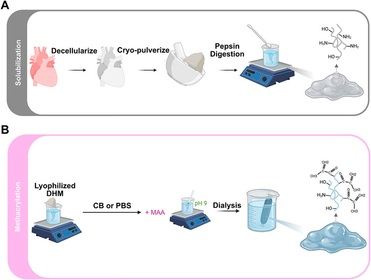
Figure 1. Schematic illustration of DHMMA fabrication process: (A) Decellularization, solubilization, and (B) Methacrylation.
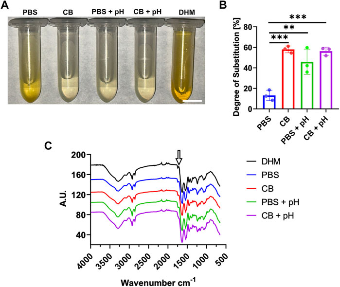
Figure 2. Chemical characterization of DHMMA. (A) Solutions of DHM/DHMMA from TNBS reaction to determine (B) degree of substitution. (C) FTIR spectra comparing DHM and DHMMA formulations. Arrow indicating the peak between 1720–1740 cm-1 increasing for all DHMMA formulations. Bar graph data represented as mean ± standard deviation, n = 3. Significance level: **p < 0.01, and ***p < 0.001. Scale bar = 10 mm.
Methacryloyl substitution in DHMMA was further evaluated via Fourier-Transform Infrared Spectroscopy (FTIR) analysis (Figure 2C). FTIR spectra for DHMMA showed characteristic amide absorptions bands at 1,640 (C=C stretching) and 1,540 (N-H) cm-1, suggesting a retention of DHM structure with methacrylation. The increase in peak intensity at 1720–1740 cm-1 (C=O), relative to DHM spectra, suggests incorporation of ester and vinyl groups during methacrylation (Martineau, Peng, and Shek, 2005). Furthermore, a slight shift in the 1,540 cm-1 region suggests protein backbone modifications caused by methacrylate moieties. The data further supports that buffer choice and maintaining a basic pH influence methacryloyl substitution.
3.2 DHMMA crosslinking and physical characterization
The 20 mg/mL DHMMA concentration was used for subsequent experiments after it was determined in pilot studies that it retained the shape of the mold better than 10 and 5 mg/mL after crosslinking (Supplementary Figure S3).
To determine the effect of reaction buffer conditions on DHMMA crosslinking, 50 µL of all four formulations of DHMMA was pipetted into 5 mm diameter PDMS molds and crosslinked. Visual inspection confirmed the crosslinking of all four formulations (Figure 3A). Crosslinked DHMMA was lyophilized, cut in half, and stored at −80°C. SEM imaging of the internal microstructure showed distinct differences across reaction conditions. Porosity analysis of the SEM images showed that DHMMA methacrylation in PBS resulted in more porous microstructure compared to methacrylation in CB buffer. More sheet-like structures were observed with
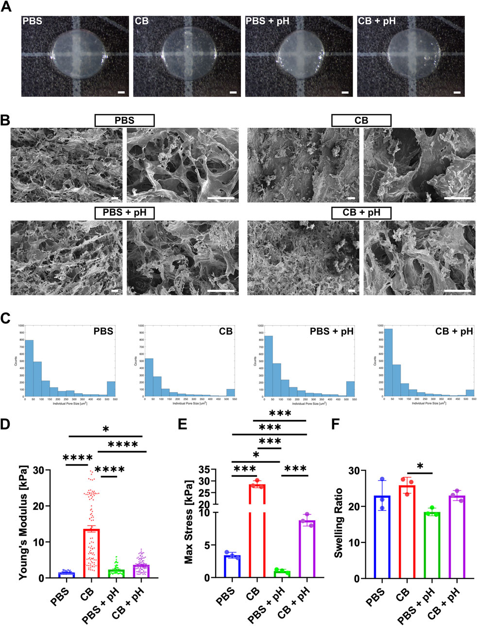
Figure 3. Physical characterization of crosslinked DHMMA. (A) Crosslinked DHMMA after 10 min of UV exposure (scale bar = 1 mm). (B) Representative SEM images at ×500 (left) and ×2000 (right) magnification (scale bar = 50 µm). (C) Histogram representation of changes in DHMMA cross-section with methacrylation reaction conditions. (D) Young’s Modulus of DHMMA from AFM analysis (mean ± standard error mean). (E) DHMMA compressed by 0.6 mm to determine the maximum stress. (F) Swelling ratio after immersion in DI water for 24 h at 37°C. All data represented as mean ± standard deviation, n = 3 gels. Significance level: *p < 0.05, **p < 0.01, ***p < 0.001, and ****p < 0.0001.
Mechanical properties were determined through AFM (local stiffness) and compression analysis (bulk stiffness). Measurement of local stiffness via AFM showed that methacrylation in CB buffer produced stiffer gels compared to PBS (
Swelling is an intrinsic property of hydrogels that affect shape, mechanical properties, and small molecule diffusion. DHMMA swelling in water for 24 h was assessed by measuring the difference between the wet and dry mass, reflecting near-equilibrium water uptake. A significant increase in swelling ratio was observed for
3.3 Matrix protein release and digestion
DHM contains cardiac-specific matrix protein that drive heart repair signaling (Hamsho et al., 2024; Jin et al., 2022; Bejleri and Davis, 2019). However, matrix protein release is challenged by rapid degradation in vitro and in vivo (X. Wang et al., 2022). To evaluate methacrylation effects on total protein release kinetics, crosslinked DHMMA was incubated in neutral buffer for 30 days at 37°C. All DHMMA formulations were crosslinked into 5 mm discs. A decrease in the percentage of matrix protein released with

Figure 4. Passive and enzymatically induced matrix protein release. (A) Protein release from crosslinked DHMMA immersed in DI water at 37°C for 30 days with sampling every 3 days (B) DHMMA exposed to digestion buffer for 24 h at 37°C with sample aliquots taken at 5 min, 30 min, 1 h, 2 h, 4 h, 8 h, 16 h, and 24 h. Data represented as mean ± standard deviation, n = 3 gels. Statistics: Two-way ANOVA.
Furthermore, all four formulations of crosslinked DHMMA were subjected to a 24 h collagenase (0.1 mg/mL) digestion. The mass of matrix protein released was significantly reduced for
3.4 Varying UV crosslinking time
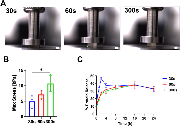
Figure 5. Physical characterization of DHMMA crosslinked for 30 s, 60 s, and 300 s. (A) Crosslinked
3.5 Varying DHMMA suspension pH
To determine the effect of suspension pH on crosslinking, DHMMA pH was adjusted to 5, 7, 8, and 9 prior to UV exposure.
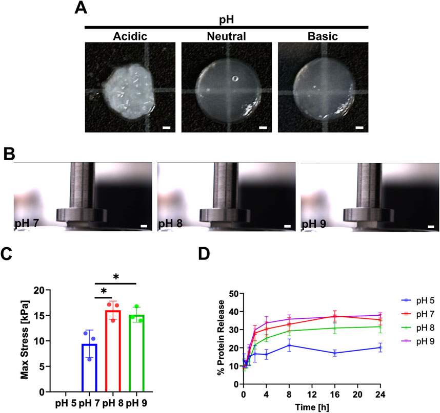
Figure 6. Varying DHMMA suspension pH prior to UV crosslinking. (A) Crosslinked hydrogels after adjusting
To determine the effect of suspension pH on mechanical properties,
3.6 Cell behavior on crosslinked DHMMA
Initial staining of H9C2 cytoskeleton on crosslinked
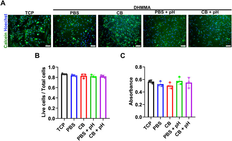
Figure 7. Live staining of H9C2 cells on crosslinked DHMMA. (A,B) H9C2 cells adhere and spread on crosslinked DHMMA and remain viable 72 h post-seeding. (C) MTT assay of H9C2 cells on crosslinked DHMMA after 72 h post-seeding. All data represented as mean ± standard deviation, n = 3. Significance level: ns (p > 0.05) for all conditions. Scale bar = 100 µm. TCP = tissue culture plastic.
Since methacrylation chemically modified DHMMA, we evaluated bioactivity after functionalization. H9C2 cells were exposed to soluble DHM or DHMMA (100 μg/mL) in growth media. Firstly, H9C2 cells cultured with soluble DHM or DHMMA for 24 h were labeled with BrdU for the last 4 h to determine proliferation frequency. No significant difference in H9C2 proliferation was observed across all groups (Supplementary Figures S5B, C).
3.7 DHMMA bio-fabrication using soft-lithography
The cardiomyocytes in the heart are highly organized. To support the use of DHMMA as a substrate for cell alignment, the softest formulation (based on AFM) was used to show the feasibility of surface micropatterning to promote anisotropic cellular organization.
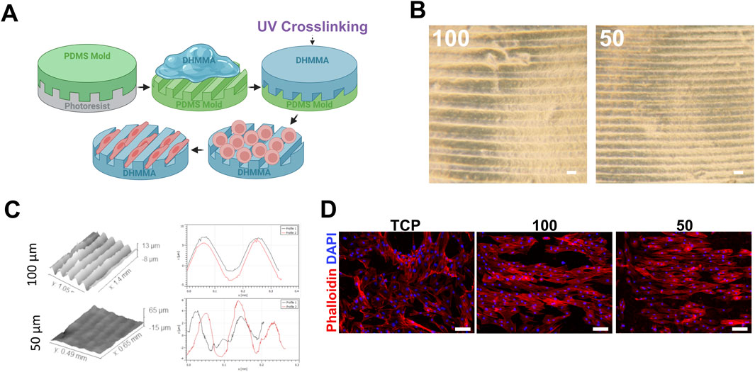
Figure 8. Crosslinked DHMMA for soft-lithography applications. (A) Fabrication process of micropatterned
Furthermore, the ability of micropatterned
Other fabrication methods were employed including microparticle generation. Crosslinked
4 Discussion
DHM therapy for cardiac tissue repair faces challenges with maintaining its stability (resistance to mechanical forces) and durability (resistance to degradation) throughout the critical injury repair period in mammalian models of ischemic heart disease. While DHM hydrogels show promise in promoting cardiac repair, their rapid degradation and mechanical instability may limit their therapeutic efficacy (Shin et al., 2021). This research aims to address these limitations in cardiac tissue engineering by developing methacrylated DHM (DHMMA) with a broad range of stiffness and matrix protein release profiles. We and others have shown that injection of DHM in injured mice hearts induces cardiac repair (X. Wang et al., 2020; Wang et al. 2021a; Wang et al., 2021b; X. Wang et al., 2022; Diaz et al., 2021; Wassenaar Jean W. et al., 2016). Furthermore, previous attempts to enhance DHM stability through solid microparticle generation demonstrated increased tissue retention up to 2 weeks. However, since the repair period lasts up to 4–5 weeks (in murine models), there is still a need to increase the longevity of DHM (X. Wang et al., 2022).
While synthetic and composite hydrogels offer precise control over degradation and mechanical properties, they lack the complex protein milieu present in native cardiac ECM (Pisheh et al., 2024). In the heart, ECM composition changes with age (fetal to adult) and influence cardiomyocyte maturation (Cui et al., 2019; Derrick and Noël, 2021; Johnson et al., 2024). In vitro, delivery of ECM molecules promote cardiomyocyte proliferation (Bigotti et al., 2020; Sorbini et al., 2023), angiogenesis (Wang et al., 2021a; X. Wang et al., 2022), and fibroblast quiescence (Wang et al., 2021b). It is challenging for synthetic hydrogels to mimic the multicellular effects observed with native ECM. Therefore, there is a need to develop biomaterials that mimic the cardiac molecular composition. To match tissue stiffness, crosslinking using genipin, transglutaminase, or glutaraldehyde can stabilize ECM-derived biomaterials (Williams et al., 2015; You et al., 2024), but their rapid reaction kinetics severely limit fabrication methods such as 3D printing. Furthermore, challenges with toxicity limits practical use of some chemical crosslinkers (Jayachandran et al., 2022). Recent advances in ECM functionalization with methacryloyl functional groups suggest a promising direction for combining the advantages of natural and synthetic systems while maintaining biological complexity (Ali et al., 2019; Ravichandran et al., 2021; Behan et al., 2022).
4.1 Material properties and tunability
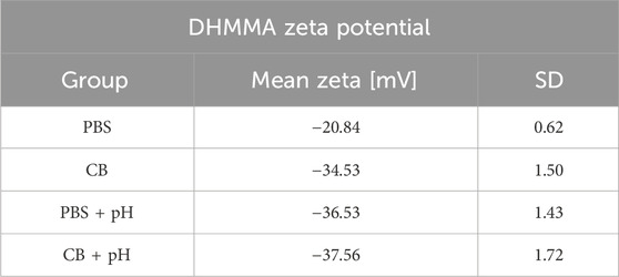
Here we demonstrate degree of substitution (DS) as a driver of DHMMA mechanical properties. A significantly greater DS was achieved in DHM methacrylated in CB buffer compared to PBS. Maintaining a basic pH (around 9) increases methacryloyl substitution in DHM. This is supported by results from the TNBS assay and FTIR analysis. There was no significant difference in DS across
We measured total protein release because matrix-derived proteins are hypothesized to be the key drivers of cardiac repair with injectable DHM therapy. The decellularization process preserves ECM components which stimulates heart repair. Using DHMMA, we evaluated how methacrylation affects total matrix protein release under both neutral and enzymatic buffer conditions. We found that maintaining a basic pH during methacrylation reduces total protein release in CB-buffered samples. For therapeutic applications, DHMMA thus enables a range of controlled release profiles. When combined with mechanical analysis, these findings provide insight into how best to engineer DHMMA to balance mechanical stability with the delivery of bioactive cues essential for cardiac repair.
Swelling capacity influences shape, mechanical properties, and molecule diffusion. We measured swelling over 24 h based on established findings that DHM swelling typically stabilizes within this timeframe (X. Wang et al., 2022). The lack of trend observed with DHMMA swelling may be due to counteracting factors such as protein heterogeneity, size, conformation, DS, charge (Panahi and Baghban-Salehi, 2019; Nichol et al., 2010). Only some of these factors are measured in this study. Further investigation is needed to understand the degree of contribution to matrix swelling. The increase in DHMMA zeta potential with methacrylation is consistent with what is observed with collagen (Yang et al., 2020) and may promote cell survival (Y. M. Chen et al., 2009). Although lyophilizing samples before rehydration differs from fully hydrated physiological conditions, it reflects the scenario of prepackaged, dried scaffolds that subsequently swell upon implantation to conform to the target site. From a tissue engineering perspective, the ability to affect swelling behavior offers a strategy to balance mechanical strength with sufficient porosity and diffusivity for cells and bioactive molecules. Therefore, optimizing methacrylation may provide a practical avenue to tailor structural and functional properties of DHMMA for cardiac repair applications.
UV exposure time emerged as a critical factor in fine-tuning the final mechanical properties of crosslinked DHMMA at constant concentration. Typically, the stiffness of a hydrogel is enhanced by increasing protein concentration. Varying protein concentration can achieve a wide range of stiffness for hydrogels such as GelMA (around 1–80 kPa, 2.5%–10% w/v) (O’Connell et al., 2018). However, the increased protein density may hamper cell functionality, especially in 3D where they adopt a spherical morphology due to decrease rate of stress relaxation (Shin et al., 2021; Chaudhuri et al., 2015; Shie et al., 2020; He et al., 2023). Building on the mechanical control achieved through methacrylation, we confirmed UV exposure time as an additional parameter for tuning mechanical properties at constant concentration. We observed enhanced
We observed that the DHMMA suspension pH, prior to UV exposure, offered an additional layer of tunability. First, we observed increase in
This tunable approach addresses a key limitation of ECM-hydrogels from decellularized tissue: stiffness less than 1 kPa. Achieving a broad range of mechanical properties at a constant protein concentration represents a significant advancement.
4.2 Structural characteristics
The fabrication parameters we investigated not only influenced mechanical properties but dramatically impacted the microscale architecture of crosslinked DHMMA. Varying the methacrylation reaction conditions resulted in distinct structural features suited for different applications.
Hydrogel porosity plays an important role in facilitating bulk erosion, cell, and nutrient infiltration (Xiang and Cui, 2021). Controlling the crosslinked DHMMA porous microstructure by varying fabrication parameters has significant implications for material function. SEM analysis revealed that PBS-mediated methacrylation produced highly porous structures ideal for cell infiltration and tissue integration, while CB buffer yields sheet-like structures better suited for controlled drug delivery. This structural versatility provides unique control over microstructure eliminating the need of a sacrificial layer. The degree through which microstructure can be tuned by varying other crosslinking parameters remains to be determined. The ability to control both bulk properties and microscale features represents a significant advantage over traditional DHM crosslinking methods, where structural control is often limited.
4.3 Biological performance
While methacrylation expands the physical properties of a biomaterial, the impact on cell viability is crucial for tissue engineering adoption. DHMMA demonstrates excellent biocompatibility across all four formulations, with H9C2 viability greater than 80%. Furthermore, no significant difference in metabolic activity was observed between all groups. These results are comparable to cell viability on other crosslinked ECM-derived hydrogels and suggest minimal impact of methacrylation on cellular biocompatibility with no visual evidence that suggest cell death (Ferchichi et al., 2024). While 3D cell encapsulation remains to be evaluated, the initial data suggests minimal impact of crosslinked DHMMA on cellular compatibility. This is likely due to the retention of sufficient cell adhesion sites available since the highest DS achieved is ∼58%. For 3D cell encapsulation, cell viability is negatively impacted by UV exposure time (Pepelanova et al., 2018; Ding, Illsley, and Chang, 2019). This can be mitigated by using higher UV intensity with shorter crosslinking time (<60 s) or a visible light-activated photoinitiator (Choi et al., 2019). Soluble DHMMA had no significant impact on H9C2 proliferation in vitro suggesting maintained viability and comparable response to previously established DHM (Wang et al., 2020). This suggests that methacrylation of DHM has minimal impact on biocompatibility. Future work will evaluate different DHMMA dosages on cardiac cell behavior.
4.4 Applications
Micropatterning allows for precise control over substrate topography, which is essential for guiding cell behavior (e.g., maturation) (Zhang Bin et al., 2024) and promoting tissue organization in cardiac tissue engineering (Y. Zhang et al., 2022). The successful transfer of micropatterns and subsequent cellular alignment illustrates the potential of DHMMA to facilitate biomimetic microenvironments conducive to cell and tissue organization. We demonstrated successful pattern transfer on the softest DHMMA formulation determined by AFM analysis (
We previously showed that generated solid DHM microparticles resulted in increased tissue retention post-injection. Furthermore, microparticles were stiffer and showed greater resistance to enzymatic stress (Wang et al., 2022). While DHM microparticles were beneficial to heart repair, electrospray microparticles had relatively high polydispersity. Furthermore, thermal gelation only achieved particle retention up to 2 weeks. We hypothesized that crosslinking will further enhance the longevity of DHM and a more homogenous population can be achieved using droplet microfluidics (Moreira et al., 2021). Our results show that generated DHMMA microparticles via water-in-oil emulsion (bulk and microfluidics methods) had homogenous morphology. Moreover, we demonstrate that droplet size can be controlled by varying flow parameters. The ability to generate microparticles with distinct size and tunable physical properties potentiates DHMMA as a drug delivery biomaterial for cardiac tissue engineering applications.
4.5 Limitations and future directions
While UV-induced crosslinking offers numerous advantages, including rapid processing and spatiotemporal control over hydrogel properties, several limitations warrant consideration. The cytotoxicity of UV radiation and the potential for photoinitiator residues pose challenges to embedded-cell viability and scaffold biocompatibility for future 3D culture studies. Moreover, achieving homogeneous crosslinking throughout the heterogeneous DHMMA remains a critical aspect to ensure consistent stability and durability. The heterogenous protein composition of DHMMA makes it challenging to characterize due to varying DS for different proteins. Furthermore, different matrix proteins have different charges, isoelectric points, and solubility amongst other properties. Chemical characterization such as NMR is challenged by insoluble DHMMA components. Despite this, it is likely that we will observe similar but not identical behavior between GelMA, collagen, and DHMMA.
Moving forward, addressing these limitations requires a comprehensive understanding of ECM-derived methacrylate hydrogel fabrication, including chemical characterization, optimizing crosslinking parameters, and evaluating biocompatibility profiles. Additionally, elucidating the influence of methacrylation on ECM-derived hydrogel bioactivity and its impact on cellular behavior is paramount for advancing cardiac tissue engineering strategies. These limitations, while significant, point to clear directions for future development that could further enhance DHMMA utility in cardiac tissue engineering.
5 Conclusion
The development of DHMMA represents a significant advancement in cardiac tissue engineering biomaterials, providing control over physical properties by tuning DS (indirectly), UV exposure time, and pH. This versatility enables a broad range of mechanical properties matching native cardiac tissue, from developmental to disease states, while preserving the inherent biological complexity of the ECM. Furthermore, DHMMA fabrication parameters allow regulation of microscale architecture achieving varying degrees of porosity suitable for different applications. Cell viability and alignment on crosslinked DHMMA provides a rationale for investigating DHMMA as a viable option for cardiac tissue engineering applications.
Data availability statement
The original contributions presented in the study are included in the article/Supplementary Material, further inquiries can be directed to the corresponding author.
Ethics statement
The animal study was approved by Case Western Reserve University, Institutional Animal Care and Use Committee. The study was conducted in accordance with the local legislation and institutional requirements.
Author contributions
VP: Conceptualization, Data curation, Formal Analysis, Investigation, Methodology, Project administration, Validation, Visualization, Writing – original draft, Writing – review and editing. DW: Data curation, Formal Analysis, Methodology, Visualization, Writing – review and editing. CL: Data curation, Formal Analysis, Methodology, Writing – review and editing, Visualization. EE: Formal Analysis, Methodology, Visualization, Writing – review and editing. CK: Funding acquisition, Methodology, Project administration, Resources, Validation, Writing – review and editing. SS: Conceptualization, Data curation, Funding acquisition, Methodology, Project administration, Resources, Supervision, Validation, Writing – review and editing.
Funding
The author(s) declare that financial support was received for the research and/or publication of this article. This work was supported by R25 HL145817, Taipei Medical University/CWRU Translational Collaborative Award to SS, partial support from National Science Foundation (NSF) grants 1927602 and 1337859 to CK, CWRU Biomedical Imaging T32 5T32EB007509, and Medical Scientist Training Program 5T32GM007250, 5TL1TR002549 to DW.
Acknowledgments
We would like to acknowledge Steve Schomisch for harvesting pig hearts, Agata Exner granting access to Litesizer 500 for zeta potential measurements, Ozan Akkus and Phillip McClellan for training and granting access to Biomomentum for compression analysis, the SDLE Research Center at Case Western Reserve University for access to FTIR instrument, Yiwen Gao for generating preliminary data for the microfluidic droplet generator. Created in BioRender.com: Figure 1A https://BioRender.com/m93p918, Figure 1B https://BioRender.com/sq2ub1h, and Figure 8A https://BioRender.com/t05a780.
Conflict of interest
The authors declare that the research was conducted in the absence of any commercial or financial relationships that could be construed as a potential conflict of interest.
The author(s) declared that they were an editorial board member of Frontiers, at the time of submission. This had no impact on the peer review process and the final decision.
Generative AI statement
The author(s) declare that no Generative AI was used in the creation of this manuscript.
Publisher’s note
All claims expressed in this article are solely those of the authors and do not necessarily represent those of their affiliated organizations, or those of the publisher, the editors and the reviewers. Any product that may be evaluated in this article, or claim that may be made by its manufacturer, is not guaranteed or endorsed by the publisher.
Supplementary material
The Supplementary Material for this article can be found online at: https://www.frontiersin.org/articles/10.3389/fbioe.2025.1579246/full#supplementary-material
References
Ali, M., Anil Kumar, P. R., Yoo, J. J., Zahran, F., Atala, A., and Lee, S. J. (2019). A photo-crosslinkable kidney ECM-derived bioink accelerates renal tissue formation. Adv. Healthc. Mater. 8 (7), e1800992. doi:10.1002/adhm.201800992
Baehr, A., Baruch Umansky, K., Bassat, E., Jurisch, V., Klett, K., Bozoglu, T., et al. (2020). Agrin promotes coordinated therapeutic processes leading to improved cardiac repair in pigs. Circulation 142 (9), 868–881. doi:10.1161/CIRCULATIONAHA.119.045116
Basara, G., Gulberk Ozcebe, S., Ellis, B. W., and Zorlutuna, P. (2021). Tunable human myocardium derived decellularized extracellular matrix for 3D bioprinting and cardiac tissue engineering. Gels 7 (2), 70. doi:10.3390/gels7020070
Behan, K., Dufour, A., Garcia, O., and Kelly, D. (2022). Methacrylated cartilage ECM-based hydrogels as injectables and bioinks for cartilage tissue engineering. Biomolecules 12 (2), 216. doi:10.3390/biom12020216
Behmer, H., Ryan, A., Wang, X., Kaw, G., Pierre, V., and Senyo, S. E. (2021). Accounting for material changes in decellularized tissue with underutilized methodologies. BioMed Res. Int. 2021 (June), e6696295. doi:10.1155/2021/6696295
Bejleri, D., and Davis, M. E. (2019). Decellularized extracellular matrix materials for cardiac repair and regeneration. Adv. Healthc. Mater. 8 (5), e1801217. doi:10.1002/adhm.201801217
Bigotti, M. G., Skeffington, K. L., Jones, F. P., Caputo, M., and Brancaccio, A. (2020). Agrin-mediated cardiac regeneration: some open questions. Front. Bioeng. Biotechnol. 8 (June), 594. doi:10.3389/fbioe.2020.00594
Chaudhuri, O., Gu, L., Klumpers, D., Darnell, M., Bencherif, S. A., Weaver, J. C., et al. (2015). Hydrogels with tunable stress relaxation regulate stem cell fate and activity. Nat. Mater. 15 (3), 326–334. doi:10.1038/nmat4489
Chen, S., Wang, Y., Lai, J., Tan, S., and Wang, M. (2023). Structure and properties of gelatin methacryloyl (GelMA) synthesized in different reaction systems. Biomacromolecules 24 (6), 2928–2941. doi:10.1021/acs.biomac.3c00302
Chen, Y., Li, C., Li, C., Chen, J., Li, Y., Xie, H., et al. (2020). Tailorable hydrogel improves retention and cardioprotection of intramyocardial transplanted mesenchymal stem cells for the treatment of acute myocardial infarction in mice. J. Am. Heart Assoc. Cardiovasc. Cerebrovasc. Dis. 9 (2), e013784. doi:10.1161/JAHA.119.013784
Chen, Y. M., Ogawa, R., Kakugo, A., Osada, Y., and Gong, J. P. (2009). Dynamic cell behavior on synthetic hydrogels with different charge densities. Soft Matter 5 (9), 1804–1811. doi:10.1039/B818586G
Choi, J.Ru, Yong, K. W., Jean, Yu C., and Cowie, A. C. (2019). Recent advances in photo-crosslinkable hydrogels for biomedical applications. BioTechniques 66 (1), 40–53. doi:10.2144/btn-2018-0083
Cui, Y., Zheng, Y., Liu, X., Yan, L., Fan, X., Yong, J., et al. (2019). Single-cell transcriptome analysis maps the developmental track of the human heart. Cell Rep. 26 (7), 1934–1950.e5. doi:10.1016/j.celrep.2019.01.079
Derrick, C. J., and Noël, E. S. (2021). The ECM as a driver of heart development and repair. Development 148 (5), dev191320. doi:10.1242/dev.191320
Diamantides, N., Wang, L., Pruiksma, T., Siemiatkoski, J., Dugopolski, C., Shortkroff, S., et al. (2017). Correlating rheological properties and printability of collagen bioinks: the effects of riboflavin photocrosslinking and pH. Biofabrication 9 (3), 034102. doi:10.1088/1758-5090/aa780f
Diaz, M. D., Tran, E., Spang, M., Wang, R., Roberto, G., Luo, C. G., et al. (2021). Injectable myocardial matrix hydrogel mitigates negative left ventricular remodeling in a chronic myocardial infarction model. JACC Basic Transl. Sci. 6 (4), 350–361. doi:10.1016/j.jacbts.2021.01.003
Ding, H., Illsley, N. P., and Chang, R. C. (2019). 3D bioprinted GelMA based models for the study of trophoblast cell invasion. Sci. Rep. 9 (1), 18854. doi:10.1038/s41598-019-55052-7
English, E. J., Samolyk, B. L., Gaudette, G. R., and Pins, G. D. (2023). Micropatterned fibrin scaffolds increase cardiomyocyte alignment and contractility for the fabrication of engineered myocardial tissue. J. Biomed. Mater. Res. Part A 111 (9), 1309–1321. doi:10.1002/jbm.a.37530
Ferchichi, E., Stealey, S., Bogert, P., and Zustiak, S. P. (2024). Tunable gelatin methacrylate polyethylene glycol diacrylate hydrogels for cell mechanosensing applications. Front. Biomaterials Sci. 3 (July). doi:10.3389/fbiom.2024.1408748
Goudie, K. J., McCreath, S. J., Parkinson, J. A., Davidson, C. M., and Liggat, J. J. (2023). Investigation of the influence of pH on the properties and morphology of gelatin hydrogels. J. Polym. Sci. 61 (19), 2316–2332. doi:10.1002/pol.20230141
Hamsho, K., Broadwin, M., Stone, C. R., Sellke, F. W., and Ruhul Abid, M. (2024). The current state of extracellular matrix therapy for ischemic heart disease. Med. Sci. 12 (1), 8. doi:10.3390/medsci12010008
He, J., Sun, Y., Gao, Q., He, C., Yao, Ke, Wang, T., et al. (2023). Gelatin methacryloyl hydrogel, from standardization, performance, to biomedical application. Adv. Healthc. Mater. 12 (23), 2300395. doi:10.1002/adhm.202300395
Jayachandran, B., Parvin, T. N., Mujahid Alam, M., Chanda, K., and Mm, B. (2022). Insights on chemical crosslinking strategies for proteins. Molecules 27 (23), 8124. doi:10.3390/molecules27238124
Jin, Y., Kim, H., Min, S., Sun Choi, Yi, Seo, S.Ju, Jeong, E., et al. (2022). Three-dimensional heart extracellular matrix enhances chemically induced direct cardiac reprogramming. Sci. Adv. 8 (50), eabn5768. doi:10.1126/sciadv.abn5768
Johnson, B. B., Cosson, M.-V., Tsansizi, L. I., Holmes, T. L., Gilmore, T., Hampton, K., et al. (2024). Perlecan (HSPG2) promotes structural, contractile, and metabolic development of human cardiomyocytes. Cell Rep. 43 (1), 113668. doi:10.1016/j.celrep.2023.113668
Kafili, G., Kabir, H., Jalali Kandeloos, A., Golafshan, E., Ghasemi, S., Mashayekhan, S., et al. (2023). Recent advances in soluble decellularized extracellular matrix for heart tissue engineering and organ modeling. J. Biomaterials Appl. 38 (5), 577–604. doi:10.1177/08853282231207216
Kalkunte, N. G., Delambre, T. E., Sohn, S., Pickett, M., Parekh, S., and Zoldan, J. (2023). Engineering alignment has mixed effects on human induced pluripotent stem cell differentiated cardiomyocyte maturation. Tissue Eng. Part A 29 (February), 322–332. doi:10.1089/ten.tea.2022.0172
Kim, J., and Bonassar, L. J. (2023). Controlling collagen gelation pH to enhance biochemical, structural, and biomechanical properties of tissue-engineered menisci. J. Biomed. Mater. Res. Part A 111 (4), 478–487. doi:10.1002/jbm.a.37464
Liu, C., Feng, X., Jeong, S., Carr, M. L., Gao, Y., Atit, R. P., et al. (2024). Lamellipodia-mediated osteoblast haptotaxis guided by fibronectin ligand concentrations on a multiplex chip. Small 20 (49), 2401717. doi:10.1002/smll.202401717
Luo, Z., Sun, W., Fang, J., Lee, K.Ju, Song, Li, Gu, Z., et al. (2019). Biodegradable gelatin methacryloyl microneedles for transdermal drug delivery. Adv. Healthc. Mater. 8 (3), 1801054. doi:10.1002/adhm.201801054
Martineau, L., Peng, H., and Pang, S. (2005). “Development of a novel biomaterial: Part II,” in Evaluation of a photo cross-linking method. Toronto, ON: Defence Research & Development Canada.
McLaughlin, S., McNeill, B., Podrebarac, J., Hosoyama, K., Sedlakova, V., Cron, G., et al. (2019). Injectable human recombinant collagen matrices limit adverse remodeling and improve cardiac function after myocardial infarction. Nat. Commun. 10 (1), 4866. doi:10.1038/s41467-019-12748-8
McMahan, S., Taylor, A., Copeland, K. M., Pan, Z., Liao, J., and Yi, H. (2020). Current advances in biodegradable synthetic polymer based cardiac patches. J. Biomed. Mater. Res. Part A 108 (4), 972–983. doi:10.1002/jbm.a.36874
Moreira, A., Carneiro, J., Campos, J. B. L. M., and Miranda, J. M. (2021). Production of hydrogel microparticles in microfluidic devices: a review. Microfluid. Nanofluidics 25 (2), 10. doi:10.1007/s10404-020-02413-8
Nichol, J. W., Koshy, S. T., Bae, H., Hwang, C. M., Yamanlar, S., and Ali, K. (2010). Cell-laden microengineered gelatin methacrylate hydrogels. Biomaterials 31 (21), 5536–5544. doi:10.1016/j.biomaterials.2010.03.064
O’Connell, C. D., Zhang, B., Onofrillo, C., Duchi, S., Blanchard, R., Quigley, A., et al. (2018). Tailoring the mechanical properties of gelatin methacryloyl hydrogels through manipulation of the photocrosslinking conditions. Soft Matter 14 (11), 2142–2151. doi:10.1039/C7SM02187A
Panahi, R., and Baghban-Salehi, M. (2019). “Protein-based hydrogels,” in Cellulose-based superabsorbent hydrogels. Editors Md. Ibrahim, and H. Mondal (Cham: Springer International Publishing), 1561–1600. doi:10.1007/978-3-319-77830-3_52
Pepelanova, I., Kruppa, K., Scheper, T., and Lavrentieva, A. (2018). Gelatin-methacryloyl (GelMA) hydrogels with defined degree of functionalization as a versatile toolkit for 3D cell culture and extrusion bioprinting. Bioengineering 5 (3), 55. doi:10.3390/bioengineering5030055
Pisheh, R., Darvishi, A., Ali, R. P., Sani, M., and Sani, M. (2024). Cardiac tissue engineering: an emerging approach to the treatment of heart failure. Front. Bioeng. Biotechnol. 12 (August), 1441933. doi:10.3389/fbioe.2024.1441933
Ravichandran, A., Murekatete, B., Moedder, D., Meinert, C., and Bray, L. J. (2021). Photocrosslinkable liver extracellular matrix hydrogels for the generation of 3D liver microenvironment models. Sci. Rep. 11 (1), 15566. doi:10.1038/s41598-021-94990-z
Roeder, B. A., Kokini, K., Sturgis, J. E., Paul Robinson, J., and Voytik-Harbin, S. L. (2002). Tensile mechanical properties of three-dimensional Type I collagen extracellular matrices with varied microstructure. J. Biomechanical Eng. 124 (2), 214–222. doi:10.1115/1.1449904
Shie, M.-Y., Lee, J.-Jr, Ho, C.-C., Yen, S.-Y., Ng, H. Y., and Chen, Y.-W. (2020). Effects of gelatin methacrylate bio-ink concentration on mechano-physical properties and human dermal fibroblast behavior. Polymers 12 (9), 1930. doi:10.3390/polym12091930
Shin, Yu J., Shafranek, R. T., Tsui, J. H., Walcott, J., Nelson, A., and Kim, D.-Ho (2021). 3D bioprinting of mechanically tuned bioinks derived from cardiac decellularized extracellular matrix. Acta Biomater. 119 (January), 75–88. doi:10.1016/j.actbio.2020.11.006
Shirahama, H., Lee, B. H., Tan, L. P., and Cho, N.-J. (2016). Precise tuning of facile one-pot gelatin methacryloyl (GelMA) synthesis. Sci. Rep. 6 (August), 31036. doi:10.1038/srep31036
Singelyn, J. M., Sundaramurthy, P., Johnson, T. D., Schup-Magoffin, P. J., Hu, D. P., Faulk, D. M., et al. (2012). Catheter-deliverable hydrogel derived from decellularized ventricular extracellular matrix increases endogenous cardiomyocytes and preserves cardiac function post-myocardial infarction. J. Am. Coll. Cardiol. 59 (8), 751–763. doi:10.1016/j.jacc.2011.10.888
Sorbini, M., Arab, S., Soni, T., Frisiras, A., and Mehta, S. (2023). How can the adult zebrafish and neonatal mice teach us about stimulating cardiac regeneration in the human heart? Regen. Med. 18 (1), 85–99. doi:10.2217/rme-2022-0161
Vu, T. V. A., Lorizio, D., Vuerich, R., Lippi, M., Nascimento, D. S., and Zacchigna, S. (2022). Extracellular matrix-based approaches in cardiac regeneration: challenges and opportunities. Int. J. Mol. Sci. 23 (24), 15783. doi:10.3390/ijms232415783
Wang, Lu, Serpooshan, V., and Zhang, J. (2021c). Engineering human cardiac muscle patch constructs for prevention of post-infarction LV remodeling. Front. Cardiovasc. Med. 8 (February), 621781. doi:10.3389/fcvm.2021.621781
Wang, X., Ali, A., Pierre, V., Young, K., Kothapalli, C. R., von Recum, H. A., et al. (2022). Injectable extracellular matrix microparticles promote heart regeneration in mice with post-ischemic heart injury. Adv. Healthc. Mater. 11 (8), 2102265. doi:10.1002/adhm.202102265
Wang, X., Pierre, V., Liu, C., Senapati, S., Park, P. S.-H., and Senyo, S. E. (2021a). Exogenous extracellular matrix proteins decrease cardiac fibroblast activation in stiffening microenvironment through CAPG. J. Mol. Cell. Cardiol. 159 (October), 105–119. doi:10.1016/j.yjmcc.2021.06.001
Wang, X., Pierre, V., Senapati, S., Park, P. S.-H., and Senyo, S. E. (2021b). Microenvironment stiffness amplifies post-ischemia heart regeneration in response to exogenous extracellular matrix proteins in neonatal mice. Front. Cardiovasc. Med. 8 (November), 773978. doi:10.3389/fcvm.2021.773978
Wang, X., Senapati, S., Akinbote, A., Gnanasambandam, B., Park, P. S.-H., and Senyo, S. E. (2020). Microenvironment stiffness requires decellularized cardiac extracellular matrix to promote heart regeneration in the neonatal mouse heart. Acta Biomater. 113 (September), 380–392. doi:10.1016/j.actbio.2020.06.032
Wang, Z., Long, D. W., Huang, Y., Chen, W. C. W., Kim, K., and Wang, Y. (2019). Decellularized neonatal cardiac extracellular matrix prevents widespread ventricular remodeling in adult mammals after myocardial infarction. Acta Biomater. 87 (March), 140–151. doi:10.1016/j.actbio.2019.01.062
Wassenaar Jean, W., Gaetani, R., Garcia, J. J., Braden, R. L., Luo, C. G., Huang, D., et al. (2016). Evidence for mechanisms underlying the functional benefits of a myocardial matrix hydrogel for post-MI treatment. J. Am. Coll. Cardiol. 67 (9), 1074–1086. doi:10.1016/j.jacc.2015.12.035
Watson, C., Liu, C., Ali, A., Miranda, H. C., Somoza, R. A., and Senyo, S. E. (2022). Multiplexed microfluidic chip for cell Co-culture. Analyst 147 (23), 5409–5418. doi:10.1039/D2AN01344D
Watson, C., and Senyo, S. (2019). All-in-One automated microfluidics control system. HardwareX 5, e00063. doi:10.1016/j.ohx.2019.e00063
Williams, C., Budina, E., Stoppel, W. L., Sullivan, K. E., Emani, S., Emani, S. M., et al. (2015). Cardiac extracellular matrix–fibrin Hybrid scaffolds with tunable properties for cardiovascular tissue engineering. Acta Biomater. 14 (March), 84–95. doi:10.1016/j.actbio.2014.11.035
Williams, C., Kelly, S., and Black, L. D. (2015). Partially digested adult cardiac extracellular matrix promotes cardiomyocyte proliferation in vitro. Adv. Healthc. Mater. 4 (10), 1545–1554. doi:10.1002/adhm.201500035
Wu, K., Wang, Y., Yang, H., Chen, Y., Lu, K., Wu, Y., et al. (2023). Injectable decellularized extracellular matrix hydrogel containing stromal cell-derived factor 1 promotes transplanted cardiomyocyte engraftment and functional regeneration after myocardial infarction. ACS Appl. Mater. and Interfaces 15 (2), 2578–2589. doi:10.1021/acsami.2c16682
Xiang, L., and Cui, W. (2021). Biomedical application of photo-crosslinked gelatin hydrogels. J. Leather Sci. Eng. 3 (1), 3. doi:10.1186/s42825-020-00043-y
Xu, Y., Yu, Y., and Guo, Z. (2024). Hydrogels in cardiac tissue engineering: application and challenges. Mol. Cell. Biochem. 480, 2201–2222. doi:10.1007/s11010-024-05145-3
Yang, J., Xiao, Y., Tang, Z., Luo, Z., Li, D., Wang, Q., et al. (2020). The negatively charged microenvironment of collagen hydrogels regulates the chondrogenic differentiation of bone marrow mesenchymal stem cells in vitro and in vivo. J. Mater. Chem. B 8 (21), 4680–4693. doi:10.1039/D0TB00172D
Yin, J., Yan, M., Wang, Y., Fu, J., and Suo, H. (2018). 3D bioprinting of low-concentration cell-laden gelatin methacrylate (GelMA) bioinks with a two-step cross-linking strategy. ACS Appl. Mater. and Interfaces 10 (8), 6849–6857. doi:10.1021/acsami.7b16059
You, C., Zhang, Z., Guo, Y., Liu, S., Hu, K., Zhan, Y., et al. (2024). Application of extracellular matrix cross-linked by microbial transglutaminase to promote wound healing. Int. J. Biol. Macromol. 266 (May), 131384. doi:10.1016/j.ijbiomac.2024.131384
Zhang, B., Luo, Y., Zhou, X., Gao, L., Yin, X., and Yang, H. (2024a). GelMA micropattern enhances cardiomyocyte organization, maturation, and contraction via contact guidance. Apl. Bioeng. 8 (2), 026108. doi:10.1063/5.0182585
Zhang, D., He, R., Qu, Y., He, C., and Chu, B. (2024b). Application of biomaterials in cardiac tissue engineering: current status and prospects. MedComm – Biomaterials Appl. 3 (4), e103. doi:10.1002/mba2.103
Zhang, Yi, Mu, W., Zhang, Y., He, X., Wang, Y., Ma, H., et al. (2022). Recent advances in cardiac patches: materials, preparations, and properties. ACS Biomaterials Sci. and Eng. 8 (9), 3659–3675. doi:10.1021/acsbiomaterials.2c00348
Zhe, M., Wu, X., Yu, P., Xu, J., Liu, M., Yang, G., et al. (2023). Recent advances in decellularized extracellular matrix-based bioinks for 3D bioprinting in tissue engineering. Materials 16 (8), 3197. doi:10.3390/ma16083197
Keywords: decellularized heart matrix, biomaterials, ultra-violet crosslinking, methacrylation, matrix protein release
Citation: Pierre V, Wu DH, Liu C, Ertugral E, Kothapalli C and Senyo SE (2025) Tunable methacrylated decellularized heart matrix: a versatile scaffold for cardiac tissue engineering. Front. Bioeng. Biotechnol. 13:1579246. doi: 10.3389/fbioe.2025.1579246
Received: 18 February 2025; Accepted: 12 May 2025;
Published: 12 June 2025.
Edited by:
Chaenyung Cha, Ulsan National Institute of Science and Technology, Republic of KoreaReviewed by:
Dylan Mostert, Eindhoven University of Technology, NetherlandsSuntae Kim, Boston University, United States
Copyright © 2025 Pierre, Wu, Liu, Ertugral, Kothapalli and Senyo. This is an open-access article distributed under the terms of the Creative Commons Attribution License (CC BY). The use, distribution or reproduction in other forums is permitted, provided the original author(s) and the copyright owner(s) are credited and that the original publication in this journal is cited, in accordance with accepted academic practice. No use, distribution or reproduction is permitted which does not comply with these terms.
*Correspondence: Samuel E. Senyo, c3NlbnlvQGNhc2UuZWR1
 Valinteshley Pierre
 Douglas H. Wu
 Chao Liu
 Elif Ertugral
 Chandrasekhar Kothapalli
 Samuel E. Senyo
