 Azar D. Khosravi
 Ameneh Alami
 Hossein Meghdadi
 Atta A. Hosseini3
- 1Department of Microbiology, School of Medicine, Ahvaz Jundishapur University of Medical Sciences, Ahvaz, Iran
- 2Health Research Institute, Infectious and Tropical Diseases Research Center, Ahvaz Jundishapur University of Medical Sciences, Ahvaz, Iran
- 3Aria Hospital, Ahvaz, Iran
Definitive and rapid diagnosis of extrapulmonary tuberculosis (EPTB) is challenging since conventional techniques have limitations due to the paucibacillary nature of the disease. To increase the sensitivity of detection of Mycobacterium tuberculosis (MTB) in EPTB specimens, we performed a nested PCR assay targeting several genes of MTB on EPTB specimens. A total of 100 clinical specimens from suspected cases of EPTB were processed. Standard staining for acid fast bacilli (AFB) was performed as the preliminary screening test. Extracted DNAs from specimens were subjected to Nested PCR technique for the detection of five different MTB target genes of IS6110, IS1081, hsp65kd, mbp64, and mtp40. On performing AFB staining, only 13% of specimens were positive, of which ascites fluid (33.3%), followed by pleural effusion (30.8%) showed the greatest AFB positivity rate. We demonstrated slight improvement in yields in lymph node which comprised the majority of specimens in this study, by employing PCR targeted to IS6110- and hsp65-genes in comparison to AFB staining. However, the yields in ascites fluid and pleural effusion were not substantially improved by PCR, but those from bone and wound were, as in nested PCR employing either gene, the same positivity rate were obtained for ascites fluid (33.3%), while for pleural effusion specimens only IS1081 based PCR showed identical positivity rate with AFB stain (30.8%). The results for bone and wound specimens, however, demonstrated an improved yield mainly by employing IS1081 gene. Here, we report higher detection rate of EPTB in clinical specimens using five different targeted MTB genes. This nested PCR approach facilitates the comparison and the selection of the most frequently detected genes. Of course this study demonstrated the priority of IS1081 followed by mtp40 and IS6110, among the five tested genes and indicates the effectiveness of any of the three genes in the design of an efficient nested-PCR test that facilitates an early diagnosis of paucibacillary EPTB cases, which are difficult to diagnose with the available standard.
Introduction
Tuberculosis (TB) is a serious disease which accounts a major global public health problem worldwide (Ates Guler et al., 2014). According to the (World Health Organization, 2014), Mycobacterium tuberculosis complex (MTBC) has infected nearly one-third of the world's population, and TB remains as one of the top ten leading cause of death in the world. Although the lung remains the common site of TB infection, there are increasing reports of extrapulmonary tuberculosis (EPTB) from all over the world, showing the potential spread capacity of M. tuberculosis (MTB) in many organs in the body (Hillemann et al., 2011; da Cruz Furini et al., 2013). EPTB comprises about 15–20% of TB cases and can comprise up to 50% of TB cases in human immunodeficiency virus (HIV)-infected individuals (Mehta et al., 2012). Therefore, early action to control the disease and intervention in the transmission chain, necessitates accurate diagnosis and appropriate treatment (Bollela et al., 1999; Lee, 2015).
Currently, nucleic acid-based technologies are used for the detection of MTB with very high sensitivity and specificity (Eisenach et al., 1990), and it can overcome some of the problems associated with the classical standard laboratory methods. Microscopic examination of acid-fast bacilli (AFB) although is rapid and cost-effective, have low sensitivity and specificity particularly in paucibacillary specimens, and culture, even though it is considered the gold standard due to its high sensitivity, requires several weeks to produce a result, and also comprises lower sensitivity for the detection of EPTB (Therese et al., 2005; da Cruz et al., 2011). Since there are many problems associated with performance of conventional diagnostic methods, various molecular biological tools like polymerase chain reaction (PCR)-based assays are developed for the reliable, early detection and speciation of mycobacteria in clinical specimens of EPTB. PCR is currently the preferred method for identification of EPTB, as this method is rapid and proved to be sensitive for the detection of bacteria in paucibacillary specimens (Chakravorty et al., 2005). The PCR technique, which can detect < 10 bacilli per milliliter of different biological specimens, is an useful tool for the diagnosis of MTB (da Cruz et al., 2011). However, a serious problem of detecting mycobacteria by PCR techniques, is the presence of PCR inhibitory substances which are found to be more associated with the EPTB in comparison to the pulmonary specimens. The presence of inhibitors is largely due to the carryover of substances in the sample preparation that interfere with the activity of the polymerase. Other investigators recommended the re-amplification after diluting out the inhibitors to improve the yield (Alli et al., 2011). Variation in sensitivity is one of the obstacles that prevent the full standardization of PCR technique in the laboratories of clinical analysis centers. To overcome this problem, PCR can be improved by a series of modifications to be able to detect the low number of MTB bacilli (Meghdadi et al., 2015). Alternatively, combination of two PCR techniques in the form of nested-PCR will increases its sensitivity and specificity in comparison to the conventional single PCR (da Cruz et al., 2011). The sensitivity of nested PCR compared to conventional single PCR, has already been well-documented in several recent and previous studies (Mishra et al., 2005; da Cruz et al., 2011). In the latter study, the sensitivity of nested PCR for the diagnosis of MTB, was increased in comparison to the conventional single PCR. In general, due to serious challenges associated with the diagnosis of EPTB, a careful selection of several target genes, is essential for increasing the chance of detection of MTBC in EPTB paucibacillary specimens (Nisha et al., 2012). Therefore, the present study was designed with the aim to detect MTBC in clinical specimens of patients suspected of having EPTB. For higher improvement of detection rate, nested PCR technique targeting five different genes of MTBC, was used to find out the frequency of EPTB in Ahvaz, Iran.
Materials and Methods
Sampling and Specimen Processing
A total of 100 specimens were obtained from clinically suspected EPTB cases on the basis of imaging, clinical findings, histological and cytological observations, admitted to the university teaching hospitals, Ahvaz Jundishapur University of Medical Sciences, Iran, during 1 year period from April 2014 to April 2015. The initial proposal on the work was approved in the University high research and ethics combined committee and necessary permission was obtained for specimen collection (Ethics code.1615 dated 21th April 2014), and specimens were appropriately de-identified. The specimens type were: 26 lymph nodes, 19 bone biopsies, 19 wound specimens, 13 pleural effusions, seven urine specimens, four breast biopsies, three CSF, three synovial fluids, three ascites fluids, two colon biopsies, and one para thyroid biopsy. Standard staining for acid fast bacilli (AFB) was performed for the specimens as the preliminary screening test after the necessary specimens processing.
DNAs were extracted from the specimens by High pure PCR Template Preparation Kit (Roche-Germany). The extraction protocol for fluids and tissue specimens was the same except for performance of a pre-treatment step for tissues. In brief, 200 μl tissue lysis buffer and 40 μl proteinase K were added to completely squelched tissue section and incubated for 1 h at 55°C for fully digestion. The processed tissue specimens were then underwent DNA extraction according to kit provider instructions. The extracted DNA was stored at −20°C until PCR amplification.
Nested Polymerase Chain Reaction
Nested PCR targeting five different genes of IS1081 (Taylor et al., 2007), IS6110 (Marchetti et al., 1998), Hsp65 (Telenti et al., 1993; Velayati et al., 2014), Mtp40 (Marchetti et al., 1998), and Mpb64 (Therese et al., 2005), of MTB was performed in two steps using the primers listed in Table 1. All primers were obtained from Bioneer Co., South Korea. In the first reaction a DNA fragment of desired base pair (bp) size for each gene was amplified using a pair of external primers; and in the second reaction, the first PCR amplicon served as template for the nested amplification of a fragment of certain bp within the initial sequence of the target gene using a pair of internal primers. The amplicon size of each gene in both reactions of nested PCR are listed in Table 1 adjacent to each target gene.
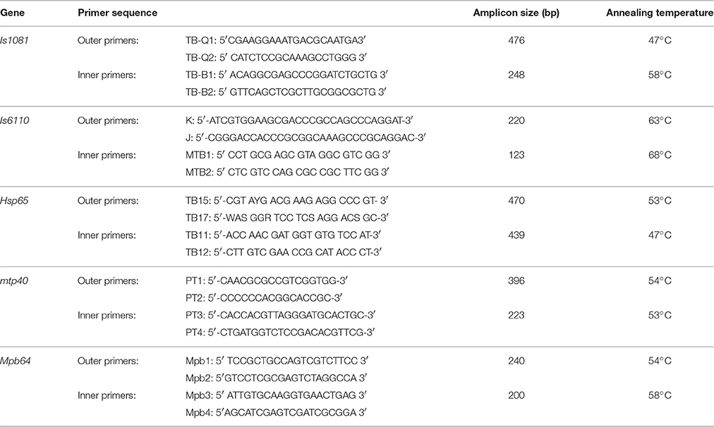
Table 1. The primers used for five genes target of M. tuberculosis in nested PCR.
The final volume of amplification reaction for the first and second round of individual gene targets, was 25 μl and consisted of 0.2 mM each dNTP, 1.5 mM MgCl2, 0.5 μM of each primer, 1.5 U of Super Taq™ DNA polymerase (Roche, Germany), and 5 μl of template DNA. The PCR conditions were as: initial denaturation at 95°C for 5 min, followed by 30 cycles of denaturation at 95°C for 30 s, annealing of respective primers at an appropriate temperature (Table 1), for 30 s, extension at 72°C for 30 s, and final extension at 72°C for 4 min. In the second reaction (nested), the PCR product of the first round was diluted (100-fold for IS6110, mpb64 genes and 200-fold for IS1081, hsp65, and mtp40 genes) and used as a template in the second reaction (nested). The cycling program for nested reaction was the same as the first, except for the number of cycles which was increased to 35. The PCR products were analyzed by gel electrophoresis on 2% agarose in 1X TAE buffer containing 0.5 μg/ml ethidium bromide, and visualized by using the Gel Documentation System (Proteinsimp, USA).
SPSS software (SPSS Inc., no. 13) was used for data analysis. The generalized estimating equation (GEE) test was applied for investigating of correlation and statistical relationships between the applied methods.
Results
In the present study, the analyzed clinical specimens were obtained from 46 male and 54 female patients, with the majority in the age group of 41–60 years. The lymph nodes were the most frequent specimens (26%), followed by bone biopsies and wound discharges, each 19%. Thirteen specimens (13%), were positive by AFB staining, while by application of nested PCR targeting 5 genes of MTB, 26 specimens (26%), showed positive results.
For setting up and optimizing the nested PCR, 10 specimens (pleural effusions, sputum, and CSF) from clinically TB cases confirmed by culture and biochemical tests and real time PCR, and 10 specimens from confirmed non-TB cases (colonies from non-mycobacterial bacteria including Pseudomonas, E. coli, and Staphylococci) were included in the study. These control specimens underwent the same processing steps as the tested specimens. All positive control samples showed positive results for all tested genes, while none of the negative controls showed any positive results in nested PCR with either gene. The overall positivity rate for tested genes were as: IS6110 (22%), IS1081 (24%), hsp65 (21%), mbp64 (12%), and mtp40 (22%) (Table 2).
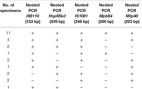
Table 2. Comparison of nested PCR results among EPTB specimens with different gene targets.
The results from AFB staining and nested PCR in relation to clinical specimens are presented in Table 3, and as it shows the highest positive rate for lymph node and CSF specimens, was obtained by IS6110 (23.1%) and hsp65 (33.3%) genes respectively. For bone (26.3%), and urine (14.3%) specimens, IS1081 and mtp40 genes showed more positive results, and in wound (36.8%), and plural effusion (30.8%) specimens, greatest positive rate was demonstrated with IS1081 gene. Thus, the highest positive rate in nested PCR was achieved by IS1081 gene (24%), while the least was reported for the mpb64 gene (12%). For urine and CSF specimens, the positive results were achieved by only two out of five tested genes, and none of the breast, synovial fluid or colon specimens showed positive results by AFB staining or any of tested genes in nested PCR. Figure 1 represents the positive amplification results for target genes.
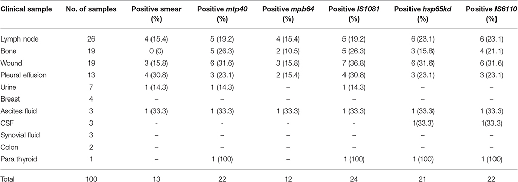
Table 3. The overall positive rate of smear and nested PCR using five different gene targets of M. tuberculosis according to the type of extrapulmonary specimens.
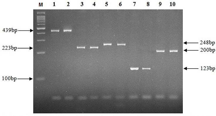
Figure 1. Electrophoresis of PCR products on a 2% agarose gel. M, Molecular size marker; Lanes 1, 2, 439 bp fragment of hsp65 gene; Lanes 3, 4, 223 bp fragment of mtp40 gene; Lanes 5, 6, 248 bp fragment of IS1081 gene; Lanes 7, 8, 123 bp fragment of IS6110 gene; Lanes 9, 10, 200 bp fragment of mpb64 gene.
In this study, a comparison made between the AFB staining and nested PCR results (Table 3). We demonstrated slight improvement in yields in lymph node, by employing PCR targeted IS6110- and hsp65 genes in comparison to AFB staining. However, the yields in ascites fluid and pleural effusion were not substantially improved by PCR, but those from bone and wound were, as in nested PCR employing either gene, the same positivity rate were obtained for ascites fluid (33.3%). While for pleural effusion specimens only IS1081 -based PCR showed identical positivity rate with AFB staining (30.8%). The results for bone and wound specimens, however, demonstrated an improved yield mainly by employing IS1081 gene.
Statistical analysis using GEE test on the results of nested PCR, revealed no significant differences in positivity rate for IS6110, hsp65, IS1081, and mtp40 genes (P > 0.05), while significant differences were found between the positive rate achieved by mbp64 gene in contrast to other tested genes (Table 4), and AFB staining results (Table 5).
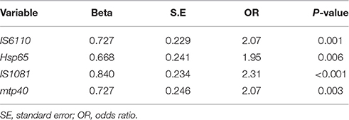
Table 4. Correlation significance between mbp64 gene with other target genes in nested PCR.
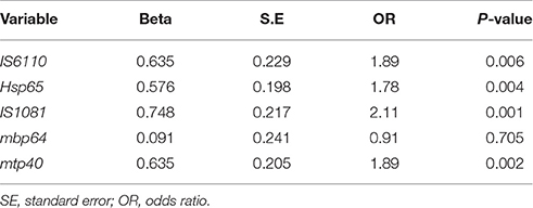
Table 5. Correlation significance between AFB smear results with results from nested PCR targeted to five different genes.
Discussion
The diagnosis of EPTB is always faced with challenges associated with sampling, as the collection of some of specimens such as plural effusion or bone biopsies requires invasive and laborious process. This usually leads to the lack of adequate specimen collection needed for processing. For this reason, a specimen should simultaneously undergoes various diagnostic tests such as histology, microbiology, biochemical analysis, and PCR. Sometimes, even in case of microbiologic analysis, the number of microorganisms in the specimen is inadequate or not distributed uniformly enough for robust detection. In the present work, for DNA extraction we used the high pure extraction and purification kit to minimize the inhibitors in EPTB. Moreover, a dilution of the product of the first reaction of PCR was used in nested PCR, which effectively reduced the concentration of the remaining inhibitory substances. However, since both false negative and positive results have also been reported for nested PCR (Sinha et al., 2016), employing several targets in PCR will improve the yields and eliminates the false positive or negative results which is more common in single target gene PCR performances. In the present study for elimination false results, we used the controls of confirmed TB and non-TB cases simultaneously. We obtained positive nested PCR results for all TB confirmed cases with all tested genes.
Several factors could influence the relative sensitivity of the target genes. The paucibacillary nature of EPTB specimens is a problem affecting the yield in PCR. The copy number of target genes in the genome of MTB is another matter. Although in molecular investigations of MTB, IS6110, and IS1081 are commonly preferred target due to a higher copy number of these genes in bacterial genome, however in case of IS6110, there are reports of strains with low copy number (Sankar et al., 2011). On the other hand, in certain circumstances, a gene with under-limit copy number, could not be detected by nested PCR in spite of improvement of the technique.
Thus, in this study, by employing five various target genes of IS6110, IS1081, hsp65, mbp64, and mtp40, we tried to get more positive results and demonstrated 22, 24, 21, 12, and 22% positivity for EPTB specimens respectively. Nested PCR targeting IS1081 gene, showed more sensitivity in comparison to other tested genes. Previously Fatolahzadeh et al. (2007), from Iran, used IS1081–PCR for the detection of pulmonary tuberculosis successfully and 78.2% of their isolates yielded positive results.
For CSF specimens, we obtained 33.3% positive rate by using either genes of IS6110 or hsp65. Although the study of Sastry and Sandhya Bhat (2013) in India which was carried out on CSF specimens using IS6110-nested PCR, 40.3% positive rate was achieved which was higher than our findings, probably due to their larger sample size, as the number of CSF specimens in this study was too few to make any conclusion. Also Alfonso et al. (2002), in Colombia, reported a positivity rate of 74% by applying nested PCR targeting mtp40 on clinical specimens (urine, sputum, CSF, etc.), while in present study 22% of the specimens were positive using mtp40 genes. The reason may be due to inclusion of both pulmonary and extrapulmonary specimens in their work, rises the positive rate as work with pulmonary is much easier compared to extrapulmonary specimens.
Regarding mpb64 gene as target in Nested PCR, we obtained 12% positivity rate. Therese et al. (2005), in India, reported 43.4% positive results using the same gene in nested PCR on various specimens suspected to EPTB, and they reported this gene as a sensitive target for the diagnosis of MTB in EPTB specimens. However, our findings, demonstrated the lowest positive rate by this gene representing lower sensitivity, compared to other target genes.
A comparison between the first and second reactions of nested PCR showed that results of the first reaction which is actually a representative of classic PCR, demonstrated lower positivity in comparison to second reaction for all tested genes, with the greatest positivity rate difference for mtp40, improving from 16 to 22%. Therefore, the results from this study demonstrated that nested PCR technique has higher sensitivity compared to traditional PCR method for the detection of MTB genome in specimens from EPTB. Our findings were concordant with other studies have emphasized on the superiority of nested PCR to conventional PCR in diagnosis of MTB (Marchetti et al., 1998; da Cruz et al., 2011). In this study, from total specimens of suspected EPTB, 26% of specimens were positive for at least one of the tested genes in comparison to confirmed TB and non-TB control specimens. Therefore, molecularly, the frequency of EPTB in our setting is determined as 26% in contract to lower frequency obtained by AFB staining (13%).
In conclusion, here we report higher detection rate of EPTB in clinical specimens using five different targeted MTB genes. This nested PCR approach facilitates the comparison and the selection of the most frequently detected genes. Of course this study demonstrated the priority of IS1081 followed by mtp40 and IS6110, among the five tested genes and indicates the effectiveness of any of the three genes in the design of an efficient nested-PCR test that facilitates an early diagnosis of paucibacillary EPTB cases, which are difficult to diagnose with the available standard methods. However, since for some specimen types such as CSF, ascites, or synovial fluids, the number of samples analyzed was low, more specimens in future studies will be needed to investigate the efficacy of target genes in the diagnosis. Moreover, a better comparison could be made with the results of phenotypic AFB staining.
Author Contributions
AK and AH: Substantial contributions to the conception, design of the work; Final approval of the version to be published; Agreement to be accountable for all aspects of the work in ensuring that questions related to the accuracy or integrity of any part of the work are appropriately investigated and resolved. AA: Acquisition, analysis, interpretation of data for the work; Agreement to be accountable for all aspects of the work in ensuring that questions related to the accuracy or integrity of any part of the work are appropriately investigated and resolved. HM: Acquisition, analysis, interpretation of data for the work; Final approval of the version to be published.
Conflict of Interest Statement
The authors declare that the research was conducted in the absence of any commercial or financial relationships that could be construed as a potential conflict of interest.
Acknowledgments
This work is part of MSc. thesis of Ameneh Alami, which was approved in Infectious and Tropical Diseases Research Center, and was financially supported by a grant (No. A-1615) from Research affairs, Ahvaz Jundishapur University of Medical Sciences, Ahvaz, Iran.
References
Alfonso, R., Romero, R. E., Patarroyo, M. E., and Murillo, L. A. (2002). Mtp-40 and alpha antigen gene fragment amplification for the detection of Mycobacterium tuberculosis in Colombian clinical specimens. Mem. Inst. Oswaldo Cruz 97, 1157–1163. doi: 10.1590/S0074-02762002000800017
Alli, O. A., Ogbolu, O. D., and Alaka, O. O. (2011). Direct molecular detection of Mycobacterium tuberculosis complex from clinical samples -an adjunct to cultural method of laboratory diagnosis of tuberculosis. N. Am. J. Med. Sci. 3, 281–288. doi: 10.4297/najms.2011.3281
Ates Guler, S., Bozkus, F., Inci, M. F., Kokoglu, O. F., Ucmak, H., Ozden, S., et al. (2014). Evaluation of pulmonary and extrapulmonary tuberculosis in immunocompetent adults: a retrospective case series analysis. Med. Princ. Pract. 24, 75–79. doi: 10.1159/000365511
Bollela, V. R., Sato, D. N., and Fonseca, B. A. (1999). Problems in the standardization of the polymerase chain reaction for the diagnosis of pulmonary tuberculosis. Rev. Saude Publica. 33, 281–286. doi: 10.1590/S0034-89101999000300009
Chakravorty, S., Sen, M. K., and Tyagi, J. S. (2005). Diagnosis of extrapulmonary tuberculosis by smear, culture, and PCR using universal specimen processing technology. J. Clin. Microbiol. 43, 4357–4362. doi: 10.1128/JCM.43.9.4357-4362.2005
da Cruz, H. L. A. D., Montenegro, R. D. A., Lima, J. F. D. A., Poroca, D. D. R., Lima, J. F. D. C., Montenegro, L. M. L., et al. (2011). Evaluation of a nested-PCR for Mycobacterium tuberculosis detection in blood and urine samples. Braz. J. Microbiol. 42, 321–329. doi: 10.1590/S1517-83822011000100041
da Cruz Furini, A. A., Pedro, H. D. S. P., Rodrigues, J. F., Montenegro, L. M. L., Machado, R. L. D., Franco, C., et al. (2013). Detection of Mycobacterium tuberculosis complex by nested polymerase chain reaction in pulmonary and extrapulmonary specimens. J. Bras. Pneumol. 39, 711–718. doi: 10.1590/S1806-37132013000600010
Eisenach, K. D., Cave, M. D., Bates, J. H., and Crawford, J. T. (1990). Polymerase chain reaction amplification of a repetitive DNA sequence specific for Mycobacterium tuberculosis. J. Infect. Dis. 161, 977–981. doi: 10.1093/infdis/161.5.977
Fatolahzadeh, B., Maleknejad, P., Bahador, A., Peeri-Dogaheh, H., Alikhani, M. Y., and Radmanesh-Ahsani, R. (2007). Evaluation of different primer sets for the rapid diagnosis of tuberculosis. Pak. J. Biol. Sci. 10, 107–111. doi: 10.3923/pjbs.2007.107.111
Hillemann, D., Rüsch-Gerdes, S., Boehme, C., and Richter, E. (2011). Rapid molecular detection of extrapulmonary tuberculosis by the automated GeneXpert MTB/RIF system. J. Clin. Microbiol. 49, 1202–1205. doi: 10.1128/JCM.02268-10
Lee, J. Y. (2015). Diagnosis and treatment of extrapulmonary tuberculosis. Tuberc. Respir. Dis. 78, 47–55. doi: 10.4046/trd.2015.78.2.47
Marchetti, G., Gori, A., Catozzi, L., Vago, L., Nebuloni, M., Rossi, M. C., et al. (1998). Evaluation of PCR in detection of Mycobacterium tuberculosis from formalin-fixed, paraffin-embedded tissues: comparison of four amplification assays. J. Clin. Microbiol. 36, 1512–1517.
Meghdadi, H., Khosravi, A. D., Ghadiri, A. A., Sina, A. H., and Alami, A. (2015). Detection of Mycobacterium tuberculosis in extrapulmonary biopsy specimens using PCR targeting IS6110, rpoB, and nested-rpoB PCR Cloning. Front. Microbiol. 6:675. doi: 10.3389/fmicb.2015.00675
Mehta, P. K., Raj, A., Singh, N., and Khuller, G. K. (2012). Diagnosis of extrapulmonary tuberculosis by PCR. FEMS Immunol. Med. Microbiol. 66, 20–36. doi: 10.1111/j.1574-695X.2012.00987.x
Mishra, A., Singhal, A., Chauhan, D. S., Katoch, V. M., Srivastava, K., Thakral, S. S., et al. (2005). Direct detection and identification of Mycobacterium tuberculosis and Mycobacterium bovis in bovine specimens by a novel nested PCR assay: correlation with conventional techniques. J. Clin. Microbiol. 43, 5670–5678. doi: 10.1128/JCM.43.11.5670-5678.2005
Nisha, A., Govindarajan, S., Muthuraj, M., Manupriya, S., Usharani, B., Kamatchyammal, S., et al. (2012). Molecular characterization of rpoB gene encoding the RNA polymerase b subunit in rifampin-resistant Mycobacterium tuberculosis strains from south India. Afr. J. Biotechnol. 11, 3160–3168. doi: 10.5897/AJB10.449
Sankar, S., Kuppanan, S., Balakrishnan, B., and Nandagopal, B. (2011). Analysis of sequence diversity among IS6110 sequence of Mycobacterium tuberculosis: possible implications for PCR based detection. Bioinformation 6, 283–285. doi: 10.6026/97320630006283
Sastry, A. S., and Sandhya Bhat, K. K. (2013). The diagnostic utility of Bact/ALERT and nested PCR in the diagnosis of tuberculous meningitis. J. Clin. Diagn. Res. 7, 74–78. doi: 10.7860/jcdr/2012/5098.2674
Sinha, P., Gupta, A., Prakash, P., Anupurba, S., Tripathi, R., and Srivastava, G. N. (2016). Differentiation of Mycobacterium tuberculosis complex from non-tubercular mycobacteria by nested multiplex PCR targeting IS6110, MTP40 and 32kD alpha antigen encoding gene fragments. BMC Infect. Dis. 16:123. doi: 10.1186/s12879-016-1450-1
Taylor, G. M., Worth, D. R., Palmer, S., Jahans, K., and Hewinson, R. G. (2007). Rapid detection of Mycobacterium bovis DNA in cattle lymph nodes with visible lesions using PCR. BMC Vet. Res. 3:12. doi: 10.1186/1746-6148-3-12
Telenti, A., Marchesi, F., Balz, M., Frank, B., Botrger, E. C., and Bodmer, T. (1993). Rapid identification of mycobacteria to the species level by polymerase chain reaction and restriction enzyme analysis. J. Clin. Microbiol. 31, 175–178.
Therese, K. L., Jayanthi, U., and Madhavan, H. N. (2005). Application of nested polymerase chain reaction (nPCR) using MPB 64 gene primers to detect Mycobacterium tuberculosis DNA in clinical specimens from extrapulmonary tuberculosis patients. Indian J. Med. Res. 122, 165–170.
Keywords: extrapulmonary tuberculosis, Mycobacterium tuberculosis, nested PCR, clinical specimens, gene
Citation: Khosravi AD, Alami A, Meghdadi H and Hosseini AA (2017) Identification of Mycobacterium tuberculosis in Clinical Specimens of Patients Suspected of Having Extrapulmonary Tuberculosis by Application of Nested PCR on Five Different Genes. Front. Cell. Infect. Microbiol. 7:3. doi: 10.3389/fcimb.2017.00003
Received: 07 October 2016; Accepted: 03 January 2017;
Published: 17 January 2017.
Edited by:
Saleh A. Naser, University of Central Florida, USAReviewed by:
Susu M. Zughaier, Emory University, USAYusuke Minato, University of Minnesota, USA
Brian Weinrick, Albert Einstein College of Medicine, USA
Copyright © 2017 Khosravi, Alami, Meghdadi and Hosseini. This is an open-access article distributed under the terms of the Creative Commons Attribution License (CC BY). The use, distribution or reproduction in other forums is permitted, provided the original author(s) or licensor are credited and that the original publication in this journal is cited, in accordance with accepted academic practice. No use, distribution or reproduction is permitted which does not comply with these terms.
*Correspondence: Ameneh Alami, YW1lbmUuYWxhbWkyMEB5YWhvby5jb20=