- 1Department of Infection, Immunity and Cardiovascular Disease, University of Sheffield, Sheffield, United Kingdom
- 2Department of Cardiology, Sheffield Teaching Hospitals NHS Foundation Trust, Sheffield, United Kingdom
- 3Insigneo Institute for in Silico Medicine, University of Sheffield, Sheffield, United Kingdom
Background: Increased coronary microvascular resistance (CMVR) is associated with coronary microvascular dysfunction (CMD). Although CMD is more common in women, sex-specific differences in CMVR have not been demonstrated previously.
Aim: To compare CMVR between men and women being investigated for chest pain.
Methods and results: We used a computational fluid dynamics (CFD) model of human coronary physiology to calculate absolute CMVR based on invasive coronary angiographic images and pressures in 203 coronary arteries from 144 individual patients. CMVR was significantly higher in women than men (860 [650–1,205] vs. 680 [520–865] WU, Z = −2.24, p = 0.025). None of the other major subgroup comparisons yielded any differences in CMVR.
Conclusion: CMVR was significantly higher in women compared with men. These sex-specific differences may help to explain the increased prevalence of CMD in women.
1. Introduction
In health, the epicardial coronary arteries act as low resistance conductance vessels, whereas the distal microvessels exhibit dynamic resistance, variation in which matches coronary blood flow (CBF) closely to the prevailing metabolic demands of the myocardium. Pathological increases in the resistance of either compartment can reduce maximal CBF, resulting in ischaemia. Unlike epicardial disease, the investigation and treatment of coronary microvascular dysfunction (CMD) is less well established. In many cases, CMD is associated with increased coronary microvascular resistance (CMVR) (1). CMD is common in patients with epicardial coronary artery disease (CAD) and in those with angina with no obstructive epicardial disease (ANOCA) (2), with a recent meta-analysis suggesting a prevalence of 41% in the latter group (3). Furthermore, In the CE-MARC2 coronary physiology sub-study, Corcoran et al. found that, in patients undergoing invasive assessment for suspected CAD, 68% had some evidence of impaired coronary microvascular physiology, with similarly high rates in those with obstructive CAD (4). When CMD reduces the maximum vasodilatory reserve of the coronary circulation, which may be measured using coronary flow reserve (CFR), it is associated with an increased likelihood of major adverse cardiac events (5). Similarly, microvascular assessment has prognostic value in the assessment of patients with both acute coronary syndrome (ACS) (6) and chronic coronary syndrome (CCS) (7). CMD can be treated with guideline-indicated therapy, with improvements in angina, quality of life and illness perception (8). Studies show CMD is more common in women than men (9–11). However, there are no data showing sex differences in CMVR. The aim of this study was to compare hyperaemic CMVR in men vs. women and investigate other major subgroups in patients undergoing angiography for the investigation of chest pain.
2. Methods
2.1. Patient recruitment
Patients undergoing cardiac catheterisation for acute and chronic coronary syndromes at Sheffield Teaching Hospitals NHS Foundation Trust were considered eligible. For acute cases, only non-culprit arteries were considered. Further exclusion criteria for all cases included ST-segment elevation myocardial infarction within the preceding 60 days, any contraindication to adenosine or contrast media, previous coronary artery bypass surgery, patient age below 18 years, chronic total occlusion, severe valvular disease and an inability to consent. This was a post hoc analysis of the Complementary Value of Absolute Coronary Flow in the Assessment of Patients with Ischaemic Heart Disease (the COMPAC-Flow) study (12), in which computational fluid dynamics- (CFD-) derived absolute flow reduction in CAD was assessed using the virtuQ™ software package (13). The study was approved by Regional Ethics Committees (16/NW/0897 and 08/H1308/193) and informed consent was obtained.
2.2. Clinical data collection
Coronary angiography and FFR assessment was performed using standard techniques. During angiography, operators were encouraged to acquire clear images of the vessel of interest, with minimal overlap, panning and foreshortening, to optimise computational arterial reconstruction (14). Translesional pressure measurements under hyperaemic and baseline conditions were taken with either the PressureWire X (Abbott Laboratories) or PrimeWire Prestige (Philips Volcano). Hyperaemia was achieved with an intravenous infusion of adenosine 140 µg/kg/min. Pseudoanonymised angiography (DICOM), physiological (pressure) and other clinical data were exported to the University of Sheffield for computational processing and analysis.
2.3. Simulating coronary flow and CMVR
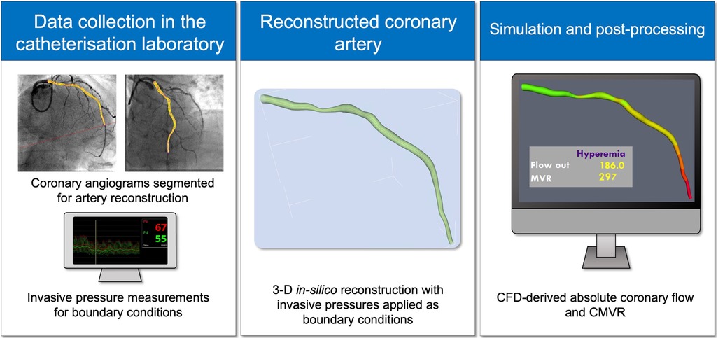
A full description of the virtuQ workflow, including arterial reconstruction, has previously been published (13, 15). In summary, two angiographic projections taken at least 30° apart were used to produce a 3D, axisymmetric, rigid reconstruction of the coronary artery of interest from an epipolar line method. Arteries with no appreciable stenosis were excluded, because the CFD method requires an epicardial pressure gradient to derive the flow and resistance values (13, 16). The quality of arterial reconstructions was assessed by three cardiologists who were also expert users of the virtuQ software, all of whom were blinded to the CFD results. Invasive pressure measurements, corresponding to proximal (Pa) and distal (Pd) measurements were prescribed at the reconstruction inlet and outlet respectively to define boundary conditions. A CFD simulation was then performed, resolving the Navier-Stokes and continuity equations to yield absolute coronary blood flow (QCFD), in ml/min, at the outlet of the reconstructed artery under both hyperaemic and baseline conditions. CFD simulations used standard blood parameters (density 1,056 kg/m3; viscosity 0.0035 Pa s) and modelled steady, laminar flow of a Newtonian fluid, the suitability of which has previously been demonstrated (17–19). Computed CMVR (CMVRCFD) was calculated using the hydraulic equivalent of Ohm's law:
A conversion factor of 1,000 was applied to yield CMVRCFD results in Woods units (WU) (Figure 1). Computed CFR (CFRCFD) was calculated as the ratio of hyperaemic and baseline QCFD:
2.4. Statistical analysis
Categorical variables are presented as frequency (percentage). The Shapiro–Wilk test was used to assess the spread of data. Normally distributed continuous variables are presented as mean ± standard deviation, while skewed data are presented as median [interquartile range]. Continuous values of haemodynamic parameters were compared using the unpaired t-test, Mann–Whitney U, one-way ANOVA and Kruskal–Wallis tests where appropriate, categorical variables were compared with Chi Square. Cohen's d and Hedges' g were used to compare effect size between two samples as indicated. Correlation was quantified using Pearson's correlation coefficient (r). A statistical threshold of p = 0.05 was considered significant and all statistical tests were two-tailed. The primary endpoint was a comparison of the CMVRCFD between men and women. The secondary endpoints were comparisons of other major subgroups.
3. Results
3.1. Patient characteristics
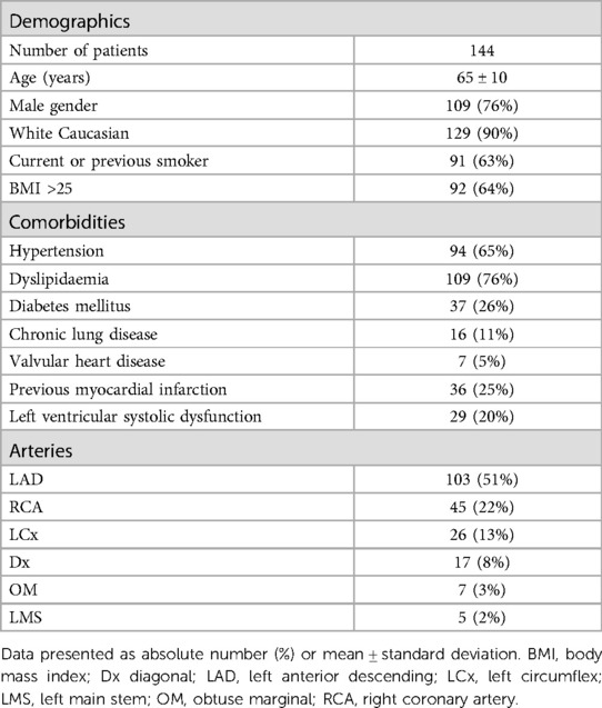
From a potential 169 patients, 144 were included. Of these, 109 were male (76%), mean age was 65 ± 10 years and 129 patients were white Caucasian. Ninety-two (64%) patients were overweight (BMI > 25) and 91 (63%) had a history of smoking. Further details of demographics and comorbidities shown in Table 1.
3.2. Artery characteristics and case exclusions
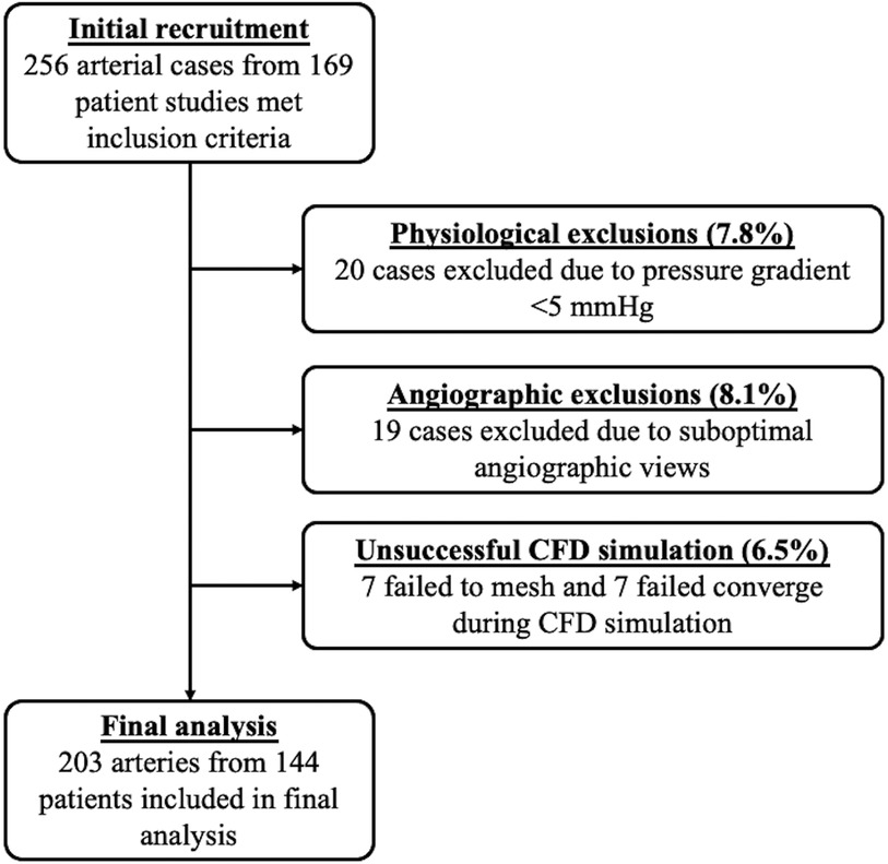
From a potential 256 arterial cases, 203 were included. These comprised 103 left anterior descending (LAD) arteries, 45 right coronary arteries (RCA), 26 left circumflex (LCx) arteries, 17 diagonal (Dx) arteries, seven obtuse marginal (OM) arteries and five left main stem (LMS) arteries. Median CMVRCFD for all cases was 710 [515–980] WU. The median FFR was 0.80 [0.72–0.87] and median visually assessed lesion stenosis was 60 [50%–70%]. Cases were excluded due to inadequate pressure gradients for CFD simulation (n = 20), inadequate angiographic views for arterial reconstruction (n = 19), failure of volumetric meshing (pre-requisite for CFD simulation, n = 7), failure of CFD simulation convergence (n = 7). All 203 included cases yielded CMVRCFD results. See Figure 2 for a full consort diagram.
3.3. Comparison of CMVRCFD between key patient groups
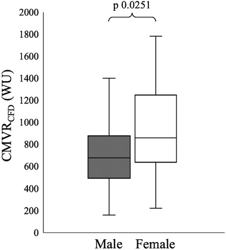
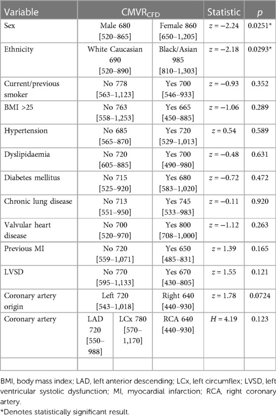
Hyperaemic CMVRCFD was significantly higher in women 860 [650–1,205] WU vs. men 680 [520–865] WU (Z = −2.24, p = 0.02). The effect of this difference was small (Hedges' g = 0.35) (Figure 3). There were no significant differences between male and female patients for any demographic or comorbidity variables, with comparable FFR (women 0.80 [0.72–0.87], men 0.81 [0.72–0.89], Z = −0.85, p = 0.40) and percentage lesion stenosis (women 60% [50%–70%], men 60% [50%–70%], Z = 0.05, p = 0.96). Further analysis using baseline conditions, revealed resting CMVRCFD was also significantly higher in women 1,765 [1,260–2,713] WU vs. men 1,370 [990–2,020] (Z = −2.46, p = 0.01, Hedges’ g = 0.46), but CFRCFD did not vary between the sexes (women 1.74 [1.35–2.30] vs. men 1.61 [1.32–1.98], Z = −0.48, p = 0.63) (Table 2).
CMVRCFD was not influenced by smoking status (Z = −0.93, p = 0.35), body mass index (BMI) > 25 (Z = −1.06, p = 0.30), hypertension (Z = 0.54, p = 0.59), dyslipidaemia (Z = −0.48, p = 0.63), diabetes (Z = −0.72, p = 0.47), chronic lung disease (Z = −0.11, p = 0.92), valvular heart disease (Z = −1.12, p = 0.26), previous myocardial infarction (Z = 1.39, p = 0.16) or left ventricular systolic dysfunction (Z = 1.55, p = 0.12) (Table 3). No significant correlations were identified between CMVRCFD with age (r = −0.08, p = 0.25), estimated glomerular filtration rate (r = 0.12, p = 0.10), haemoglobin concentration (r = −0.07, p = 0.32) or haematocrit (r = −0.06, p = 0.45). CMVRCFD was also higher in patients of black and Asian ethnicity 985 [810–1,303] WU vs. white Caucasian patients 690 [520–890] WU (Z = −2.18, p = 0.03) (Supplementary Material). However, the number of patients within the black and Asian group was only eight.
3.4. Inter-artery comparison of CMVRCFD
CMVRCFD did not differ between arteries originating from, and including, the LMS 720 [543–1,018] WU vs. the RCA 640 [440–930] WU (Z = 1.80, p = 0.07). Inter-artery comparison did not show a significant difference in CMVRCFD between the LAD and main diagonal branch 720 [550–988] WU vs. the LCx and obtuse marginal branch 780 [570–1,170] WU vs. RCA 640 [440–930] WU (H = 4.19, p = 0.12) (Table 2).
4. Discussion
In this study, we analysed absolute CMVRCFD derived from invasive pressure measurements using CFD simulation in 203 coronary arteries from 144 patients (109 male, 35 female). CMRCFD was significantly higher in women when compared to men. There were no other significant differences comparing major sub-groups. CMVRCFD was higher in those of black and Asian vs. Caucasian ethnicity, but this group was very small (n = 8).
4.1. Subgroup differences in CMVR
Despite the well-established increased prevalence of CMD in women (9–11) and the numerous techniques for quantifying CMVR (13, 20–23), no previous study has demonstrated a significant sex-specific difference in CMVR. Prior studies have shown no difference in the index of microvascular resistance (IMR) between men and women (24, 25), with apparent discrepancies in microvascular function attributed to lower CFR in women as a result of elevated baseline coronary flow (26). Our study, therefore, provides the first observation of a sex-specific difference in CMVR, suggesting a true microvascular dysfunction may contribute to sex-specific differences in CMD. The reasons underpinning these discrepancies are currently unknown, with a lack of absolute flow results (ml/min) in previous studies hindering comparisons. Differences in enrollment may have contributed; prior studies predominantly included patients with ANOCA (mean lesion percentage stenosis ranged from 20% to 30%), while our study included a large proportion of patients with haemodynamically significant CAD. The hyperaemic flow values quoted in this study are lower than previously measured with the Rayflow catheter in patients with ANOCA (27) and it is possible the flow limiting effect of epicardial stenoses blunted any sex-specific differences in CFR. Furthermore, prior studies used the mean transit time (MTT) of an intracoronary saline bolus as a surrogate for coronary flow (IMR = distal pressure × MTT) (24, 25), a technique which is subject to significant error (28). The direction of this effect appears cogent with the fact that women have a higher prevalence of CMD and that CMVR and CMD are associated (9–11). The underlying mechanism(s) behind sex differences in microvascular function are largely unknown. Some data suggest changes in sex hormones, particularly in the peri- and post-menopausal periods may contribute to coronary endothelial dysfunction and abnormal vasomotor control (29) and this does appear to be consistent with clinical practice. In our study, the mean age of female participants was 67 ± 10 years old (the men were 64 ± 10 years old), making menopause-induced microvascular changes a plausible explanation for the observed difference between sexes. In the females, age was not correlated with CMVRCFD (r = −0.22, p = 0.20); but as only four patients were less than 55 years old, we could not determine whether CMVR differed between the peri- and post-menopausal groups. We also demonstrated a statistically significant difference in CMVRCFD between white Caucasian vs. black and Asian patients. This however, was based upon only eight patients and so these results are unreliable.
4.2. Clinical implications
Despite being described over thirty years ago (30), CMD continues to pose a clinical challenge. Angiography alone is good at excluding epicardial disease, but is unable to diagnose CMD. Both European and American guidelines now recommend invasive assessment of CFR or IMR to support diagnosis (31, 32). CFR alone does not discriminate between epicardial and microvascular compartments, whereas indices of microvascular resistance require combined pressure and flow measurements. While the measurement of intracoronary pressure is simple, accurate and reproducible, estimating coronary flow is more challenging. Traditionally, a surrogate of flow rate was inferred from either Doppler flow velocity or the MTT of in injected bolus of room temperature saline (thermodilution). Both these techniques for estimating coronary flow are subject to variability; Doppler readings are dependent upon sensor alignment with the direction of flow and proximity to the vessel wall (33), whilst bolus thermodilution is dependent upon injection quality and is unsuitable for some bradycardic patients and is affected by side branch flow (34). Recent work has demonstrated poor agreement between Doppler and thermodilution derived CFR (mean bias 0.59 ± 1.24; R2 = 0.36, p < 0.0001) and microvascular resistance (R2 = 0.19; p < 0.0001) even in expert hands (28). The continuous infusion thermodilution technique, using a the Rayflow™ catheter, provides an alternative method of invasively deriving absolute coronary blood flow and microvascular resistance that delivers better reproducibility (35). All invasive measurements add time and expense to a standard angiogram and this may affect widespread uptake. The current results are not entirely consistent with previous work suggesting CMD in women is a functional phenotype characterized by a decreased CFR with increased resting flow but maintained hyperaemic flow and resistance (24, 25). Given the observational nature of our study and the potential limitations of the methodology, further work is needed to corroborate the findings and evaluate prognostic significance. The method for quantifying CMVRCFD in this study does however, allow for real-time assessment of the coronary microcirculation from a simple angiography and a standard FFR assessment and may influence future approaches in coronary physiological assessments.
4.3. Study limitations
First, more men were recruited than women. However, this is not unusual in studies of CAD. Second, patients with completely normal epicardial arteries were excluded. This is likely to have reduced the numbers of patients with ANOCA. This is important because it may have reduced the magnitude of the observed differences. Future studies of sex-specific differences in CMVR should also include ANOCA patients and not exclude patients with unobstructed epicardial arteries. The computational method used in this study did not account for side-branch flow, subtended myocardial mass or collateral blood supply (36). This may also have influenced CMVRCFD results, but is unlikely to have influenced between-group differences. Although the CFD method has been validated in vitro, the physiological calculations may be subject to several sources of inaccuracies introduced from both invasive pressure measurements and the various stages of the CFD workflow, not least the in silico arterial reconstruction (15, 16). Model sensitivity to these various sources of error is yet to be fully quantified and is likely to be case specific. For example, in minimally-stenosed arteries, geometric error in the reference vessel is a dominant source of inaccuracy (16). In stenosed arteries, any error in the 3D reconstruction around the region of the stenosis will contribute significantly to overall model error (37). Gravitational error of invasively measured pressure was not corrected for, which will also contribute error (38, 39), albeit to a lesser extent in increasingly stenosed cases.
5. Conclusion
In this single center study, using a computational method, we have demonstrated sex-specific differences in calculated CMVR, in patients under invasive investigation for chest pain. These findings suggest hyperaemic CMVR may be higher in women than men and may help to explain the higher prevalence of CMD in women. Further investigation and studies are required to confirm these findings.
Data availability statement
The original contributions presented in the study are included in the article/Supplementary Material, further inquiries can be directed to the corresponding author.
Ethics statement
The studies involving human participants were reviewed and approved by Sheffield Regional Ethics Committees (16/NW/0897 and 08/H1308/193). The patients/participants provided their written informed consent to participate in this study.
Author contributions
PM and JG conceived the original idea for the study and collected the clinical data. DH, IH, PL and AN supported software development and modeling. LA-R led the physiological reconstruction and simulations, supported by DT and TN, DT led the statistical analysis. All authors contributed to the article and approved the submitted version.
Funding
PM was funded by the Wellcome Trust (214567/Z/18/Z). For the purpose of open access, the author has applied a CC BY public copyright license to any Author Accepted Manuscript version arising from this submission. RG was supported by a National Institute for Health and Care Research UK Clinical Lectureship. This independent research was carried out at the National Institute for Health and Care Research (NIHR) Sheffield Biomedical Research Centre (NIHR203321). The views expressed are those of the authors and not necessarily those of the NIHR or the Department of Health and Social Care.
Conflict of interest
PM, JG, PL and DH are named as an inventors on a University of Sheffield patent that describes elements of the CFD method.
The remaining authors declare that the research was conducted in the absence of any commercial or financial relationships that could be construed as a potential conflict of interest.
Publisher's note
All claims expressed in this article are solely those of the authors and do not necessarily represent those of their affiliated organizations, or those of the publisher, the editors and the reviewers. Any product that may be evaluated in this article, or claim that may be made by its manufacturer, is not guaranteed or endorsed by the publisher.
Supplementary material
The Supplementary Material for this article can be found online at: https://www.frontiersin.org/articles/10.3389/fcvm.2023.1159160/full#supplementary-material
References
1. Morris PD, Al-Lamee RK, Berry C. Coronary physiological assessment in the catheter laboratory: haemodynamics, clinical assessment and future perspectives. Heart. (2022) 108(21):1737–46. doi: 10.1136/heartjnl-2020-318743
2. Patel MR, Peterson ED, Dai D, Brennan JM, Redberg RF, Anderson HV, et al. Low diagnostic yield of elective coronary angiography. N Engl J Med. (2010) 362(10):886–95. doi: 10.1056/NEJMoa0907272
3. Mileva N, Nagumo S, Mizukami T, Sonck J, Berry C, Gallinoro E, et al. Prevalence of coronary microvascular disease and coronary vasospasm in patients with nonobstructive coronary artery disease: systematic review and meta-analysis. J Am Heart Assoc. (2022) 11(7):e023207. doi: 10.1161/JAHA.121.023207
4. Corcoran D, Young R, Adlam D, McConnachie A, Mangion K, Ripley D, et al. Coronary microvascular dysfunction in patients with stable coronary artery disease: the CE-MARC 2 coronary physiology sub-study. Int J Cardiol. (2018) 266:7–14. doi: 10.1016/j.ijcard.2018.04.061
5. Lee JM, Jung J-H, Hwang D, Park J, Fan Y, Na S-H, et al. Coronary flow reserve and microcirculatory resistance in patients with intermediate coronary stenosis. J Am Coll Cardiol. (2016) 67(10):1158–69. doi: 10.1016/j.jacc.2015.12.053
6. Jin X, Yoon MH, Seo KW, Tahk SJ, Lim HS, Yang HM, et al. Usefulness of hyperemic microvascular resistance index as a predictor of clinical outcomes in patients with ST-segment elevation myocardial infarction. Korean Circ J. (2015) 45(3):194–201. doi: 10.4070/kcj.2015.45.3.194
7. Nishi T, Murai T, Ciccarelli G, Shah SV, Kobayashi Y, Derimay F, et al. Prognostic value of coronary microvascular function measured immediately after percutaneous coronary intervention in stable coronary artery disease: an international multicenter study. Circ Cardiovasc Interv. (2019) 12(9):e007889. doi: 10.1161/CIRCINTERVENTIONS.119.007889
8. Ford TJ, Stanley B, Good R, Rocchiccioli P, McEntegart M, Watkins S, et al. Stratified medical therapy using invasive coronary function testing in angina: the CorMicA trial. J Am Coll Cardiol. (2018) 72(23 Pt A):2841–55. doi: 10.1016/j.jacc.2018.09.006
9. Daly C, Clemens F, Lopez Sendon JL, Tavazzi L, Boersma E, Danchin N, et al. Gender differences in the management and clinical outcome of stable angina. Circulation. (2006) 113(4):490–8. doi: 10.1161/CIRCULATIONAHA.105.561647
10. Bugiardini R, Bairey Merz CN. Angina with “normal” coronary ArteriesA changing philosophy. JAMA. (2005) 293(4):477–84. doi: 10.1001/jama.293.4.477
11. Humphries KH, Pu A, Gao M, Carere RG, Pilote L. Angina with “normal” coronary arteries: sex differences in outcomes. Am Heart J. (2008) 155(2):375–81. doi: 10.1016/j.ahj.2007.10.019
12. Aubiniere-Robb L, Gosling R, Taylor DJ, Newman T, Rodney D, Ian Halliday H, et al. The complementary value of absolute coronary flow in the assessment of patients with ischaemic heart disease (the COMPAC-flow study). Nat Cardiovasc Res. (2022) 1(7):611–6. doi: 10.1038/s44161-022-00091-z
13. Morris PD, Gosling R, Zwierzak I, Evans H, Aubiniere-Robb L, Czechowicz K, et al. A novel method for measuring absolute coronary blood flow & microvascular resistance in patients with ischaemic heart disease. Cardiovasc Res. (2020) 117(6):1567–77. doi: 10.1093/cvr/cvaa220
14. Ghobrial M, Haley HA, Gosling R, Rammohan V, Lawford PV, Hose DR, et al. The new role of diagnostic angiography in coronary physiological assessment. Heart. (2021) 107(10):783. doi: 10.1136/heartjnl-2020-318289
15. Solanki R, Gosling R, Rammohan V, Pederzani G, Garg P, Heppenstall J, et al. The importance of three dimensional coronary artery reconstruction accuracy when computing virtual fractional flow reserve from invasive angiography. Sci Rep. (2021) 11(1):19694. doi: 10.1038/s41598-021-99065-7
16. Taylor DJ, Feher J, Czechowicz K, Halliday I, Hose DR, Gosling R, et al. Validation of a novel numerical model to predict regionalized blood flow in the coronary arteries. Eur Heart J Digit Health. (2023) 4(2):81–9. doi: 10.1093/ehjdh/ztac077
17. Morris PD. Computational fluid dynamics modelling of coronary artery disease. Sheffield: University of Sheffield (2015). https://etheses.whiterose.ac.uk/11772/
18. Huckaba CE, Hahn AW. A generalized approach to the modeling of arterial blood flow. Bull Math Biophys. (1968) 30(4):645–62. doi: 10.1007/BF02476681
19. Brown AG, Shi Y, Marzo A, Staicu C, Valverde I, Beerbaum P, et al. Accuracy vs. computational time: translating aortic simulations to the clinic. J Biomech. (2012) 45(3):516–23. doi: 10.1016/j.jbiomech.2011.11.041
20. Fearon WF, Balsam LB, Farouque HM, Caffarelli AD, Robbins RC, Fitzgerald PJ, et al. Novel index for invasively assessing the coronary microcirculation. Circulation. (2003) 107(25):3129–32. doi: 10.1161/01.CIR.0000080700.98607.D1
21. Nolte F, van de Hoef TP, Meuwissen M, Voskuil M, Chamuleau SA, Henriques JP, et al. Increased hyperaemic coronary microvascular resistance adds to the presence of myocardial ischaemia. EuroIntervention. (2014) 9(12):1423–31. doi: 10.4244/EIJV9I12A240
22. De Bruyne B, Pijls NHJ, Gallinoro E, Candreva A, Fournier S, Keulards DCJ, et al. Microvascular resistance reserve for assessment of coronary microvascular function: JACC technology corner. J Am Coll Cardiol. (2021) 78(15):1541–9. doi: 10.1016/j.jacc.2021.08.017
23. van ‘t Veer M, Adjedj J, Wijnbergen I, Tóth GG, Rutten MC, Barbato E, et al. Novel monorail infusion catheter for volumetric coronary blood flow measurement in humans: in vitro validation. EuroIntervention. (2016) 12(6):701–7. doi: 10.4244/EIJV12I6A114
24. Kobayashi Y, Fearon WF, Honda Y, Tanaka S, Pargaonkar V, Fitzgerald PJ, et al. Effect of sex differences on invasive measures of coronary microvascular dysfunction in patients with angina in the absence of obstructive coronary artery disease. JACC: Cardiovascular Interventions. (2015) 8(11):1433–41. doi: 10.1016/j.jcin.2015.03.045
25. Chung J-H, Lee Kyung E, Lee Joo M, Her A-Y, Kim Chee H, Choi Ki H, et al. Effect of sex difference of coronary microvascular dysfunction on long-term outcomes in deferred lesions. JACC Cardiovasc Interv. (2020) 13(14):1669–79. doi: 10.1016/j.jcin.2020.04.002
26. Nardone M, McCarthy M, Ardern CI, Nield LE, Toleva O, Cantor WJ, et al. Concurrently low coronary flow reserve and low Index of microvascular resistance are associated with elevated resting coronary flow in patients with chest pain and nonobstructive coronary arteries. Circ Cardiovasc Interv. (2022) 15(3):e011323. doi: 10.1161/CIRCINTERVENTIONS.121.011323
27. Fournier S, Keulards DCJ, van ‘t Veer M, Colaiori I, Di Gioia G, Zimmermann FM, et al. Normal values of thermodilution-derived absolute coronary blood flow and microvascular resistance in humans. EuroIntervention. (2020) 17(4):e309–e16. doi: 10.4244/EIJ-D-20-00684
28. Demir OM, Boerhout CKM, de Waard GA, van de Hoef TP, Patel N, Beijk MAM, et al. Comparison of Doppler flow velocity and thermodilution derived indexes of coronary physiology. JACC Cardiovasc Interv. (2022) 15(10):1060–70. doi: 10.1016/j.jcin.2022.03.015
29. Gilligan DM, Quyyumi AA, Cannon R III. Effects of physiological levels of estrogen on coronary vasomotor function in postmenopausal women. Circulation. (1994) 89(6):2545–51. doi: 10.1161/01.CIR.89.6.2545
30. Cannon RO III, Epstein SE. “microvascular angina” as a cause of chest pain with angiographically normal coronary arteries. Am J Cardiol. (1988) 61(15):1338–43. doi: 10.1016/0002-9149(88)91180-0
31. Knuuti J, Wijns W, Saraste A, Capodanno D, Barbato E, Funck-Brentano C, et al. 2019 ESC guidelines for the diagnosis and management of chronic coronary syndromes: the task force for the diagnosis and management of chronic coronary syndromes of the European society of cardiology (ESC). Eur Heart J. (2019) 41(3):407–77. doi: 10.1093/eurheartj/ehz425
32. Gulati M, Levy PD, Mukherjee D, Amsterdam E, Bhatt DL, Birtcher KK, et al. 2021 AHA/ACC/ASE/CHEST/SAEM/SCCT/SCMR guideline for the evaluation and diagnosis of chest pain: a report of the American college of cardiology/American heart association joint committee on clinical practice guidelines. Circulation. (2021) 144(22):e368–454.
33. Doucette JW, Corl PD, Payne HM, Flynn AE, Goto M, Nassi M, et al. Validation of a Doppler guide wire for intravascular measurement of coronary artery flow velocity. Circulation. (1992) 85(5):1899–911. doi: 10.1161/01.CIR.85.5.1899
34. Pijls NHJ, De Bruyne B, Smith L, Aarnoudse W, Barbato E, Bartunek J, et al. Coronary thermodilution to assess flow reserve. Circulation. (2002) 105(21):2482–6. doi: 10.1161/01.CIR.0000017199.09457.3D
35. Gallinoro E, Bertolone Dario T, Fernandez-Peregrina E, Paolisso P, Bermpeis K, Esposito G, et al. Reproducibility of bolus versus continuous thermodilution for assessment of coronary microvascular function in patients with ANOCA. EuroIntervention. (2023) 19(2):e155–e66. doi: 10.4244/EIJ-D-22-00772
36. Taylor DJ, Feher J, Halliday I, Hose DR, Gosling R, Aubiniere-Robb L, et al. Refining our understanding of the flow through coronary artery branches; revisiting Murray’s law in human epicardial coronary arteries. Front Physiol. (2022) 13:871912. doi: 10.3389/fphys.2022.871912
37. Sturdy J, Kjernlie JK, Nydal HM, Eck VG, Hellevik LR. Uncertainty quantification of computational coronary stenosis assessment and model based mitigation of image resolution limitations. J Comput Sci. (2019) 31:137–50. doi: 10.1016/j.jocs.2019.01.004
38. Tar B, Ágoston A, Üveges Á, Szabó GT, Szűk T, Komócsi A, et al. Pressure- and 3D-derived coronary flow reserve with hydrostatic pressure correction: comparison with intracoronary Doppler measurements. J Pers Med. (2022) 12(5):780. doi: 10.3390/jpm12050780
Keywords: coronary microvascular resistance, sex, computational fluid dynamics, coronary microvascular dysfunction, coronary physiology
Citation: Taylor DJ, Aubiniere-Robb L, Gosling R, Newman T, Hose DR, Halliday I, Lawford PV, Narracott AJ, Gunn JP and Morris PD (2023) Sex differences in coronary microvascular resistance measured by a computational fluid dynamics model. Front. Cardiovasc. Med. 10:1159160. doi: 10.3389/fcvm.2023.1159160
Received: 5 February 2023; Accepted: 22 June 2023;
Published: 6 July 2023.
Edited by:
Giovanni Luigi De Maria, University of Oxford, United KingdomReviewed by:
Emanuele Gallinoro, OLV Aalst, BelgiumZsolt Kőszegi, University of Debrecen, Hungary
Maurizio Lodi Rizzini, Polytechnic University of Turin, Italy
Anna Corti, Polytechnic University of Milan, Italy
© 2023 Taylor, Aubiniere-Robb, Gosling, Newman, Hose, Halliday, Lawford, Narracott, Gunn and Morris. This is an open-access article distributed under the terms of the Creative Commons Attribution License (CC BY). The use, distribution or reproduction in other forums is permitted, provided the original author(s) and the copyright owner(s) are credited and that the original publication in this journal is cited, in accordance with accepted academic practice. No use, distribution or reproduction is permitted which does not comply with these terms.
*Correspondence: Daniel J. Taylor ZGFuaWVsLnRheWxvckBzaGVmZmllbGQuYWMudWs=
†These authors have contributed equally to this work and share first authorship
 Daniel J. Taylor
 Louise Aubiniere-Robb1,†
 Rebecca Gosling
 Tom Newman
 Patricia V. Lawford
 Andrew J. Narracott
 Julian P. Gunn
 Paul D. Morris