- 1School of Teacher Education, Hezhou University, Hezhou, Guangxi, China
- 2School of Tourism and Sports Health, Hezhou University, Hezhou, Guangxi, China
Background: Several small randomized trials have examined the effects of high-intensity interval training (HIIT) on hemostasis and vascular stiffness. However, a clear consensus regarding these effects has not been established. The study is to systematically review the evidence and quantify the impacts of HIIT compared with moderate-intensity continuous training (MICT) or usual care (UC) on hemostasis and vascular stiffness.
Methods: Randomized controlled trials (RCTs) exploring the impact of HIIT, MICT, or UC on hemostasis and vascular stiffness were retrieved from PubMed, Embase, Web of Science, and Cochrane databases up to June 10, 2025. A meta-analysis was performed to compare the standardized mean differences (SMD) of changes from baseline to post-intervention in platelet count (PLT), fibrinogen (FIB), D-dimer (D-D), carotid-femoral pulse wave velocity (cfPWV), augmentation index (AIx), AIx normalized to a heart rate of 75 beats·min−1 (AIx@75HR), flow-mediated dilation (FMD), and brachial flow-mediated dilation normalized (nFMD), which were used to evaluate hemostasis and vascular stiffness. The 95% confidence interval (95% CI) was calculated along with the SMD. All analyses were conducted in R (version 4.3.3).
Results: Overall, 68 RCTs involving 2,679 patients were included in the analysis. PLT [SMD (95% CI) = −0.26 (−0.51; −0.01)] and FIB [SMD (95% CI) = −0.60 (−1.18; −0.01)] in hemostasis were decreased. Decreased cfPWV [SMD (95% CI) = −0.22 (−0.38; −0.06)], AIx [SMD (95% CI) = −0.16 (−0.30; −0.02)], and AIx@75HR [SMD (95% CI) = −0.35 (−0.61; −0.10)], as well as increased FMD [SMD (95% CI) = 0.37 (0.02; 0.72)] were observed in vascular stiffness. However, there were no notable differences in the D-D and nFMD parameters.
Conclusion: HIIT notably improved FIB, cfPWV, AIx, and FMD compared to MICT, or UC. Under certain conditions, PLT and AIx@75HR can also benefit from HIIT. It may be particularly advantageous for patients with cardiovascular disease.
Systematic Review Registration: https://www.crd.york.ac.uk/PROSPERO/view/522614, identifier CRD42024522614.
1 Introduction
Cardiovascular disease (CVD) is the most prevalent disease globally, responsible for roughly one-third of all global deaths (1, 2). It accounts for 35% of total female deaths and 31% of total male deaths. Additionally, CVD has a serious impact on health losses and expenditure on health system costs (3–5). In 2021, nearly 612 million people worldwide suffered from CVD, with approximately 846 new cases per 100,000 people (6), resulting in approximately 19.4 million deaths and 428.3 million disability-adjusted life years (DALYs) (7). In addition to the health burden, CVD also imposes a significant economic burden on individuals and society. Between 2019 and 2020, the cost of heart disease in the United States was approximately $252.2 billion (8, 9). Globally, the economic burden of CVD is projected to increase from $957 billion in 2015 to $1.04 trillion in 2030 (10).Patients with CVD may exhibit chest pain, dyspnea, dizziness, exercise intolerance, and palpitations (11–13), which largely impair patient's quality of life (14, 15). Moreover, the risk of heart attack, stroke, and other serious cardiovascular events may be increased (16). There is evidence that thrombosis and atherosclerosis, which are largely attributed to hemostasis and vascular stiffness, are involved in the pathogenesis of CVD (17–19). Both the hypercoagulable state of the blood and the progression of atherosclerosis (20, 21) result in an elevated risk of cardiovascular events (16), which in turn causes the occurrence and progression of CVD.
Many studies have emphasized the importance of high-intensity interval training (HIIT) and moderate-intensity continuous training (MICT) for health (22, 23). HIIT is broadly defined as alternating between high-intensity bursts [>80% of maximum oxygen consumption (VO2max) or >85% of maximum heart rate (HRmax)] and low-intensity active recovery (24). MICT is defined as prolonged exercise within the moderate-intensity range (40%–60%) (25, 26). Some studies have shown that HIIT can achieve benefits equivalent to or higher than MICT in a shorter time (27). Compared to traditional MICT, HIIT alternates between short periods of high-intensity exercise and recuperation phases, leading to significant improvements in cardiorespiratory adaptations and metabolic health in a shorter time (28). Recent evidence suggests that HIIT (29, 30) and MICT (22, 31) exercise methods have an effective improvement in both CVD [coronary heart disease (32), heart failure (33–35), stroke (36, 37), and hypertension (38)] and non-CVD [diabetes (39), obesity (40), and healthy adults (41)] patients. These beneficiary groups are diverse, meaning that regardless of age or sex, there is a tendency to benefit from HIIT. Further research has shown that HIIT also has beneficial effects on various cardiovascular-related inflammatory factors (such as IL-6, TNF-α, and IL-10) (42) and biomarkers (such as hs-CRP) (43). Although several studies have been reported on the impacts of HIIT on hemostasis and vascular stiffness in the overall population, there is still controversy over their effectiveness (44, 45). For the effects on hemostasis, existing studies indicate conflicting results. Specifically, HIIT or MICT can improve hemostasis in some studies (40, 46). However, there is no improvement in hemostasis in other studies (47, 48). In addition, the effects on vascular stiffness are also contradictory in existing studies. Specifically, HIIT or MICT improves vascular stiffness in certain studies (49). However, there was no improvement in vascular stiffness in other studies (50, 51). Furthermore, limited studies compare the impacts of HIIT vs. MICT on hemostasis and vascular stiffness. This complicates the identification of the superior exercise modality in clinical settings (32, 52).
This study seeks to synthesize all available evidence using a systematic review and meta-analysis to address the effectiveness of HIIT and MICT in improving hemostasis [platelet count (PLT), fibrinogen (FIB), D-dimer (D-D)] and alleviating vascular stiffness (carotid-femoral pulse wave velocity [cfPWV], augmentation index [AIx], AIx normalized to a heart rate of 75 beats·min−1 [AIx@75HR], flow-mediated dilation [FMD], and brachial flow-mediated dilation normalized [nFMD]) in the overall population, including CVD and non-CVD populations. Additionally, it aims to determine the relative effectiveness of these two exercise approaches. Through this study, we aim to clarify the roles of HIIT and MICT in primary and secondary prevention of CVD, providing new insights for clinical practice.
2 Methods
2.1 Registration
The study was performed in compliance with the statement of the preferred reporting items for systematic reviews and meta-analyses (PRISMA) (53). The complete checklist can be found in Supplementary Appendix 1. Moreover, it was pre-registered on the international prospective register of systematic reviews (CRD42024522614) (54).
2.2 Literature search strategy
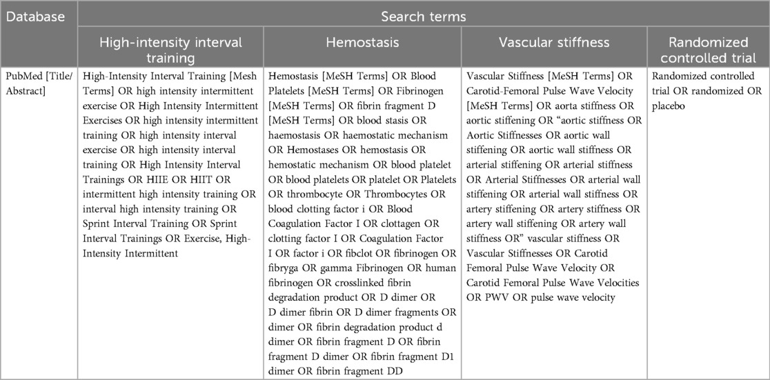
After a systematic search, relevant studies were searched from PubMed, Embase, Cochrane Library, and Web of Science. Searches in PubMed, Cochrane Library, and Embase employed a combination of subject and free words. The search strategy in PubMed is presented in the main text (Table 1). Supplementary Appendix 2 details the search strategy for each database. The main terms used to construct the search strategy include “HIIT” and “cardiovascular”. The retrieval strategy is reviewed and adjusted by experienced researchers. In addition, reference lists from chosen studies and reviews were also reviewed to find any pertinent studies that might have been missed in the electronic search. All randomized controlled trials (RCTs) that were written in the English language and published between the inception of the database and June 10, 2025 were included. To ensure the reliability of the study, all included RCTs should be published in peer-reviewed journals.
2.3 Eligibility criteria
Studies were included in the analysis according to the following criteria: (P) any subjects aged 18 years and older with or without CVD; (I) studies that used HIIT in the experimental group; (C) studies that used MICT or usual care (UC) in the control group; (O) studies that reported at least one outcome measure about hemostasis (PLT, FIB, D-D) and vascular stiffness (cfPWV, AIx, AIx@75HR, FMD, and nFMD), and (S) RCT. Studies were excluded according to: (i) patients who were unable to receive HIIT for any reason; or (ii) duplicate publications, literature reviews, letters to editors, abstracts presented at conferences, and animal studies. All relevant studies were individually evaluated by two investigators. In case of disagreement, a third investigator reassessed the studies. Only studies that received unanimous agreement from all reviewers were included.
2.4 Risk of bias
The risk of bias (ROB) of the included studies was evaluated by two investigators independently using Cochrane's risk-of-bias tool (55). The tool includes seven distinct domains: (a) random sequence generation, (b) allocation concealment, (c) blinding of participants and personnel, (d) blinding of outcome assessment, (e) incomplete outcome data, (f) selective reporting, and (g) other sources of bias.
2.5 Data extraction
Data used in the analysis were independently extracted by two investigators. Discrepancies were addressed either through discussion to achieve consensus or by involving a third investigator if necessary. The extracted information included: first author, publication year, country, subject characteristics (experimental and control groups, number, sex, and age), HIIT intervention information (intensity, duration, frequency, and period), and outcome measures. In instances where information was lacking, authors of the included studies were reached out to by email to ask for any missing values. The graphical data extraction software Engauge Digitizer (version 4.1) was used to extract data that were only available in image form.
2.6 Statistical analysis
The outcome measures were hemostasis (PLT, FIB, D-D) and vascular stiffness (cfPWV, AIx, AIx@75HR, FMD, nFMD). Meta-analysis was performed only for studies reporting at least one of the above measures. Data synthesis and analysis were carried out focusing on changes from baseline to post-intervention. Since all extracted data were continuous, the analysis employed the standardized mean difference (SMD) and 95% confidence interval (CI). In this study, effect sizes were combined using a fixed-effects model (FEM). If heterogeneity was significant (I2 > 50%, P < 0.05), a random-effects model (REM) was applied instead. Subgroup analyses were performed to assess the impact of various interventions in the control group, the repetition of HIIT intervention, and the presence of CVD on hemostasis and vascular stiffness. The extent of heterogeneity among the included studies was measured by the I2 test. Sensitivity analysis and meta-regression would be performed if substantial heterogeneity existed. The independent variables for the meta-regression were intensity and duration of HIIT and participant age. Publication bias was evaluated by examining funnel plots. If more than ten studies were included, Egger's test was used to evaluate publication bias. Results were considered statistically significantly different only if the two-sided P-value was below 0.05.
All statistical analyses were employed R [version 4.3.3, meta-package (version 4.18-1)].
3 Results
3.1 Literature selection
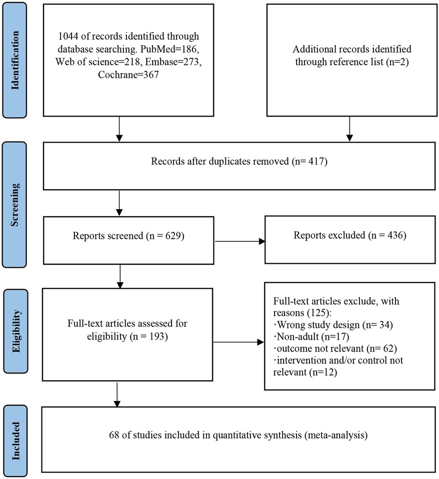
The flow diagram depicting the study selection is presented in Figure 1. After the selection process, 1,046 potentially eligible studies were identified, 1,044 from database searches, and two from reference lists. After removing 417 duplicates, 629 studies remained to be filtered. After screening titles and abstracts, 436 studies were deleted, and 125 were removed post full-text review. Finally, 68 studies were included in the meta-analysis.

Figure 1. Flow diagram of the preferred reporting items for systematic reviews and meta-analyses depicting the study selection process.
3.2 ROB analysis for the included studies
The overall ROB was rated as medium to low. The reduced quality of the evidence was primarily attributed to the difficulty in implementing HIIT regarding blinding of participants by implementers. Moreover, the significant intervention nature of HIIT itself made it difficult to adopt a blinded design. Therefore, 100% blinding of participants and personnel, and 2% blinding of outcome assessment were evaluated as high risk. Additionally, it was noted that the loss to follow-up rate reported in four studies exceeded 20%. These studies were rated as high risk unless the study was conducted using an intention-to-treat (ITT) analysis, and it was clearly stated in the paper that loss to follow-up did not affect the comparability between groups, or that the number and reasons for missing data between groups were similar (Supplementary Appendices 3 and 4).
3.3 Characteristics of the included studies
Supplementary Appendix 5 outlines the characteristics of the included studies. The list of the included studies is available in Supplementary Appendix 6. These studies were published between 2008 and 2025. 14 studies were performed in North America, one in South America, 28 in Asia, three in Europe, and 10 in Oceania. A total of 1,256 subjects (>18 years) were included in the experimental group and 1,423 in the control group (MICT: 763; UC: 660). In the HIIT group, 80% of subjects were aged 60 or younger, while 20% were older than 60. In the MICT group, 71.1% were aged 60 or younger, and 28.9% were older than 60. In the UC group, 73.8% were aged 60 or younger, and 26.2% were older than 60. About 1,433 (53.5%) subjects were female. Among the included studies, two focused exclusively on females, five on males only, 49 on both sexes, and 12 did not report sex distributions. Additionally, a total of 20 studies involved subjects with CVD.
As outlined in Supplementary Appendix 5, the exercise duration for the HIIT intervention included warm-up and relaxation phases. The mean duration of the exercise interventions was 1–52 weeks, with more than half (51.47%) of the studies reporting up to 8 weeks. Subjects typically engaged in an average of 3.5 sessions per week. A total of 13 studies involved acute exercise. A total of five studies involved two different HIIT intervention protocols, two of which used both short and prolonged HIIT.
High-intensity interval cycling and treadmills were the most commonly used interventions across the studies, with some employing high-intensity interval walking and one focusing on high-intensity interval respiratory muscle training. Of the studies, 51 exclusively used HIIT. In six studies, HIIT was combined with MICT, and resistance training, respectively.
There were significant differences in the intervention measures for the control group UC (Supplementary Appendix 5), which can be primarily categorized into five distinct features: no exercise training (26.3%, e.g., “no exercise training,” “maintaining a sedentary lifestyle”), maintaining daily activities (28.9%, e.g., “continuing routine physical activities,” “maintaining habitual activities”), seated rest (21.1%, e.g., “seated for 30 min,” “seated for 90 min”), standard care or medical management (13.2%, e.g., “standard heart failure management”, “medical care”), and other interventions (10.5%, e.g., green tea, intermittent fasting).
The control group MICT demonstrated moderate consistency in terms of exercise mode, intensity range, and duration (Supplementary Appendix 5). The primary exercise modes were cycling (43.4%) and treadmill/walking (31.2%), with a smaller proportion involving swimming, rowing, or mixed exercises (25.4%). Intensity metrics were primarily based on heart rate (HRpeak/HRmax, 60.9%) or oxygen consumption (VO2peak/VO2max, 34.8%), followed by peak power output (PPO, 10.9%) or the Rating of Perceived Exertion (RPE, 8.7%). Intensity ranges were concentrated between 60 and 75% HRpeak (71.4% of the heart rate group) or 50–70% VO2peak (87.5% of the oxygen uptake group). Duration was primarily 30–60 min (89.6%), with only 5 studies lasting less than 30 min or more than 60 min.
3.4 Synthesis of the results
3.4.1 PLT analysis
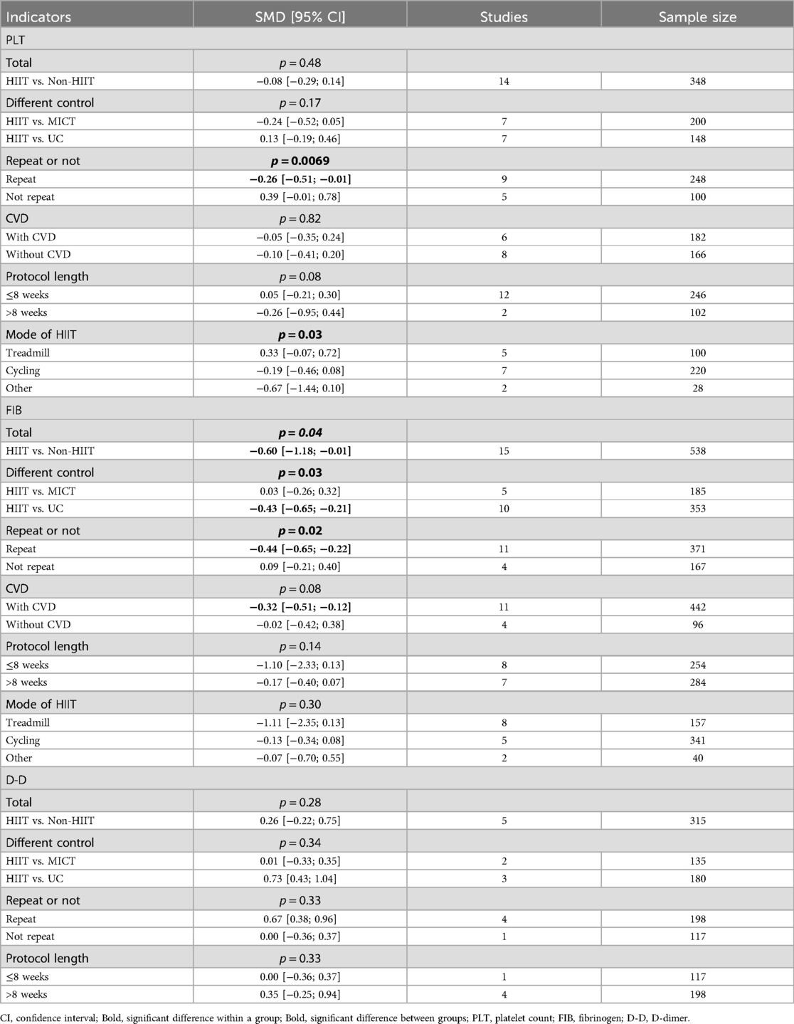
Subgroup analysis based on the repetition of HIIT intervention indicated different results. Nine studies (n = 248 subjects) reported that HIIT interventions lasting more than one week with a repetitive exercise pattern led to notable improvements in PLT levels compared to the control group [I2 = 0%; FEM; SMD (95% CI) = –0.26 (–0.51; −0.01)] (Table 2). In contrast, no significant effect was observed for a single HIIT intervention. A notable difference (P = 0.007) in the impact on PLT was observed between repeated HIIT and non-repeated HIIT. In addition, there were no notable differences in PLT levels according to subgroup analyses based on interventions (MICT or UC) in the control group, the presence of CVD, the protocol length of HIIT (≤8 weeks vs. >8 weeks), and exercise modes of HIIT interventions (treadmill, cycling, or others).
3.4.2 FIB analysis
Subgroup analysis based on interventions in the control group revealed different results. Ten studies (n = 353 subjects) reported a difference in FIB levels between before and after HIIT and UC [I2 = 83%; REM; SMD (95% CI) = −0.98 (−1.87; −0.09)] (Table 2). Additionally, no notable differences in FIB levels were observed between HIIT and MICT. However, the intergroup difference test for MICT and UC reached a significant level (P = 0.03).
Subgroup analyses were conducted according to the repetition of HIIT intervention. Eleven studies (n = 371 subjects) reported that compared to the control group, a notable difference in FIB was found in the repeated HIIT group when HIIT interventions lasted over one week and employed a repetitive exercise pattern [I2 = 81%; REM; SMD (95% CI) = −0.93 (−1.71; −0.14)] (Table 2). In addition, no improvement in FIB levels was observed in the single HIIT subgroup. However, subgroup analysis comparing repeated HIIT intervention with a single intervention indicated a notable difference between the two groups (P = 0.02).
In the subgroup analysis of the presence of CVD, eleven studies (n = 442 subjects) showed that HIIT treatment improved FIB levels in the group of CVD patients (C) [I2 = 83%; REM; SMD (95% CI) = −0.85 )−1.69; −0.02)] (Table 2).
Additionally, no significant differences were observed between subgroups based on HIIT protocol length (≤8 weeks vs. >8 weeks) and training methods (treadmill, cycling, or other methods).
3.4.3 D-D analysis
In the analysis of D-D, four studies were included, all of which involved CVD patients (n = 315 subjects). The results demonstrated that no notable difference was found between the HIIT group and the control group [I2 = 76%, P < 0.01; REM; SMD [95% CI] = 0.26 [−0.22, 0.75], P = 0.28] (Supplementary Appendix 7). Additionally, no notable differences were found in D-D levels according to subgroup analyses based on interventions (MICT or UC) in the control group, the repetition of HIIT intervention, and the protocol length of HIIT intervention (≤8 weeks vs. >8 weeks).
3.4.4 cfPWV analysis
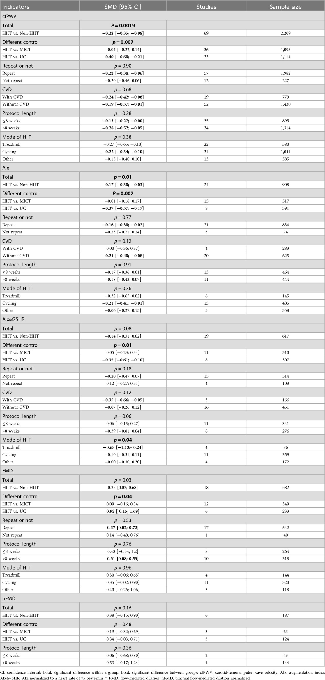
Subgroup analysis was conducted based on MICT or UC for the control group. A subgroup analysis containing 33 studies (n = 1,114 subjects) indicated a notable difference in cfPWV between HIIT and UC [I2 = 59.3%; REM; SMD (95% CI) = −0.40 (−0.60; −0.21)] (Table 3). The subgroup analysis based on MICT or UC as the control group showed significant differences between subgroups (P = 0.007).
Subgroup analysis was performed depending on the repetition of HIIT intervention. A subgroup analysis containing 57 studies (n = 1,982 subjects) revealed a notable difference in cfPWV between the repeated HIIT group and the control group [I2 = 64.5%; REM; SMD (95% CI) = −0.22 (−0.38; −0.06)] (Table 3).
Subgroup analysis depending on the presence of CVD revealed the same results. In subgroup C (n = 779 subjects), data from 19 studies showed that HIIT significantly reduced cfPWV levels [I2 = 32%; REM; SMD (95% CI) = −0.24 (−0.42; −0.06)] (Table 3). The subgroup analysis of non-CVD (NC) patients (n = 1,430 subjects) including 52 studies similarly revealed significant improvements in cfPWV after HIIT [I2 = 63.4%; REM; SMD (95% CI) = −0.19 (−0.37; −0.01)] (Table 3).
Subgroup analysis based on the HIIT protocol duration (≤8 weeks vs. >8 weeks) revealed homogeneous results. 34 studies (n = 1,314 participants) reported that HIIT protocol duration exceeded 8 weeks. Compared to the control group, participants showed significant improvements in cfPWV levels in the HIIT group [I²=76%; REM; SMD [95% CI] = −0.28 [−0.52; −0.05]; Table 3]. The analysis of the subgroup with ≤8 weeks (n = 895 participants) included 35 studies and similarly revealed a significant improvement in cfPWV after HIIT [I2 = 0%; REM; SMD [95% CI] = −0.13 [−0.27; −0.00]; Table 3].
Subgroup analyses based on HIIT training methods (treadmill, cycling, or other methods) yielded different results. 34 studies (n = 1,044 participants) reported that the training method was cycling. Compared with the control group, cfPWV levels showed significant improvement in the HIIT group [I²=0%; REM; SMD [95% CI] = −0.22 [−0.34; −0.10]; Table 3]. No significant effects were observed with treadmills or other methods.
3.4.5 Aix analysis
Subgroup analyses were conducted using MICT or UC as the grouping criterion. Specifically, 9 studies in the UC subgroup (n = 391 participants) reported AIx levels in response to HIIT and UC, revealing significant differences between the two groups [I²=0%; FEM; SMD [95% CI] = −0.37 [−0.57; −0.17]; Table 3]. The results of the subgroup analysis based on the control group being MICT or UC showed significant differences between subgroups (P = 0.03).
Subgroup analyses based on HIIT training methods (treadmill, cycling, or other methods) yielded different results. 13 studies (n = 405 participants) reported that the HIIT training method was cycling. Compared to the control group, participants showed a significant improvement in AIx levels [I²=30.8%; FEM; SMD [95% CI] = −0.21 [−0.41; −0.01]; Table 3]. However, no significant effects were observed for treadmill or other training methods.
Subgroup analysis depending on the presence of CVD showed a notable difference between HIIT in the NC subgroup (n = 625 subjects) and the control group [I2 = 30.6%; REM; SMD (95% CI) = −0.24 (−0.43; −0.05)] (Table 3). Additionally, no notable differences were found in AIx levels according to subgroup analyses based on the repetition of HIIT intervention, and HIIT protocol length (≤8 weeks vs. >8 weeks).
3.4.6 Aix@75hr analysis
Subgroup analysis was conducted depending on MICT or UC for the control group. Specifically, eight studies (n = 307 subjects) in the UC subgroup reported a notable difference in AIx@75HR levels between HIIT and UC [I2 = 34%; FEM; SMD (95% CI) = −0.36 (−0.59; −0.13)] (Table 3). The subgroup analysis based on MICT or UC as the control group demonstrated notable differences between subgroups (P = 0.01).
Subgroup analysis based on the presence or absence of CVD showed a significant difference between the HIIT group and the control group in the subgroup with CVD (n = 166 participants) [I2 = 37%; FEM; SMD [95% CI] = −0.35 [−0.66; −0.05]; Table 3].
Additionally, no notable differences were found in AIx@75HR levels according to subgroup analyses based on the repetition of HIIT intervention and HIIT protocol length (≤8 weeks vs. >8 weeks).
Subgroup analyses based on HIIT training methods (treadmill, cycling, others) revealed different results. Four studies (n = 86 participants) reported that the HIIT training method was treadmill. Compared to the control group, AIx@75HR levels showed significant improvement [I²=50%; FEM; SMD [95% CI] = −0.68 [−1.13; −0.24]; Table 3]. However, no significant effects were observed for cycling or other training methods. There were significant differences in the effects of treadmill, cycling, and other training methods on AIx@75HR (P = 0.04).
3.4.7 FMD analysis
In the analysis of FMD, 18 studies that exclusively focused on non-CVD patients were included (n = 582 subjects). Subgroup analysis was conducted depending on MICT or UC for the control group. Specifically, HIIT was compared with UC in a subgroup analysis containing 6 studies (n = 233 subjects). The results indicated a notable difference in FMD between the two groups [I2 = 82.3%; REM; SMD (95% CI) = 0.92 (0.15; 1.69)] (Table 3). In addition, in the subgroup analysis of the repetition of HIIT intervention containing 17 studies (n = 542 subjects), repeated HIIT significantly increased FMD levels [I2 = 70.4%; REM; SMD (95% CI) = 0.37 (0.02; 0.72)] (Table 3). Additionally, in a subgroup analysis of the HIIT protocol length (≤8 weeks vs. >8 weeks) involving 10 studies (n = 318 participants), the >8 weeks subgroup showed a significant improvement in FMD levels [I²=2%; REM; SMD [95% CI] = 0.31 [0.08; 0.53]; Table 3]. Finally, no significant differences were observed between subgroups based on the mode of HIIT training (treadmill, cycling, or other methods).
3.4.8 nFMD analysis
In the analysis of nFMD, six studies that exclusively focused on non-CVD patients were included (n = 187 subjects). The results demonstrated that there was no notable difference between the HIIT group and the control group [I2 = 67%, P = 0.01; REM; SMD [95% CI] = 0.38 [−0.15, 0.90]. P = 0.16] (Supplementary Appendix 7). Additionally, no notable differences were found in nFMD levels according to subgroup analysis based on interventions (MICT or UC) in the control group and HIIT protocol length (≤8 weeks vs. >8 weeks) (Supplementary Appendix 8).
3.5 Sensitivity analysis
To confirm the robustness of the results, sensitivity analyses were conducted for cfPWV, FMD, FIB, D-D, and nFMD, respectively, demonstrating that the synthesized results were reliable (Supplementary Appendices 9A–9E). In addition, sensitivity analyses were conducted for studies lacking sex information (Supplementary Appendices 9F–9P) and studies that combined other exercise interventions (Supplementary Appendices 9l–S9I). Most indicators were not affected by the lack of sex information or the combination of other exercise interventions.
3.6 Meta-regression
In this meta-regression analysis, three potential associations were explored: the relationship of training intensity, training duration, and mean age with PLT, FIB, D-D, cfPWV, AIx, AIx@75HR, FMD, and nFMD levels. The analysis results revealed that the P-values for the association between training duration and FIB, AIx, AIx@75HR, FMD, and nFMD levels were 0.15, 0.80, 0.22, 0.61, and 0.32, respectively, and did not reach statistical significance thresholds. It was worth noting that the P-value for the correlation between training duration and D-D level was <0.01, with a regression coefficient of −0.08. However, since only five studies were included, the results of this meta-regression should be interpreted with caution (Supplementary Appendix 10A). Additionally, the P-value for the correlation between training duration and AIx@75HR level was 0.04, with a regression coefficient of −0.02. This indicated that within the 20–50-min exercise duration, as time increased, the effect of HIIT on reducing AIx@75HR became greater (Supplementary Appendix 10B). Additionally, the p-value for the association between training duration and PLT levels was 0.02, with a regression coefficient of −0.07, within the 30–50 min exercise duration range. The effect of HIIT on reducing PLT levels increased as duration increased (Supplementary Appendix 10C). The P-value for the association between training duration and cfPWV levels was 0.02, with a regression coefficient of −0.03, within a 20–45-min exercise duration. The effect of HIIT on reducing cfPWV increased as duration increased (Supplementary Appendix 10D). Similarly, the P-values for the association between training intensity and PLT, FIB, D-D, AIx, AIx@75HR, FMD, and nFMD levels were 0.08, 0.10, 0.06, 0.44, 0.35, 0.95, and 0.96, respectively, and did not meet conventional statistical significance thresholds. This indicated that the current evidence cannot support a correlation between training intensity and various indicators. Notably, the P value for the association between training intensity and cfPWV level was 0.05, and the regression coefficient was 0.0001, within the intensity range of 80–10. The effect of HIIT on reducing cfPWV increased as intensity increased (Supplementary Appendix 10E). Finally, the P-values for the association between average age and FIB, D-D, cfPWV, AIx, FMD, and nFMD levels were 0.44, 0.14, 0.20, 0.38, 0.67, 0.09, and 0.44, respectively, and also did not reach statistical significance thresholds.
3.7 Publication bias analysis
In the funnel plots of PLT, FIB, cfPWV, AIx, AIx@75HR, and FMD levels, no significant asymmetric distribution was observed. Egger's test was employed to further evaluate publication bias. The results demonstrated that there was no notable publication bias in the analyses of the levels of PLT (P = 0.27), FIB (P = 0.06), cfPWV (P = 0.04), AIx (P = 0.29), AIx@75HR (P = 0.34) and FMD (P = 0.29) (Supplementary Appendices 11A–F). After performing an Egger publication bias test on cfPWV, we found significant publication bias in cfPWV. After adjustment using the trimming method, the results (Supplementary Appendix 11G) were inconsistent with previous findings, indicating that the overall results for cfPWV are unstable. Considering that the control groups in the included studies were MICT or UC, this may be an important factor for this phenomenon. Therefore, we conducted a subgroup analysis. We found that there was no significant difference in the effects of the two intervention methods on cfPWV compared to MICT, and the publication bias was not significant (Egger p = 0.18). Compared to the UC subgroup, HIIT significantly improved cfPWV, and the publication bias was not significant (Egger p > 0.05).
4 Discussion
The results of the meta-analysis suggest that HIIT has an ameliorative effect on cardiovascular risk in the overall population, mainly reflected in the following three aspects: (i) HIIT significantly improves PLT, FIB, cfPWV, AIx, AIx@75HR, and FMD and may be even more effective in patients with CVD; (ii) during HIIT exercise, repeated exercises are preferable, as the benefits from a single session are limited or even ineffective; (iii) Subgroup analysis found that long-term HIIT (>8 weeks) effectively improved atherosclerosis and endothelial function; (iv) based on the results of subgroup analyses, HIIT and MICT do not show any differences in all parameters; and (v) Subgroup analysis also found that training mode (treadmill/cycling) may affect vascular function-related indicators (cfPWV, AIx).
There is evidence that platelet overactivation (56), abnormally elevated FIB (57), cfPWV (58), and Aix (59), and abnormally reduced FMD (60) are associated with the incidence of CVD, such as the development of atherosclerotic plaques (61, 62). HIIT is found to notably improve PLT, FIB, cfPWV, AIx, AIx@75HR, and FMD. Additionally, the effects might be more significant in patients with CVD and may be achieved through multiple mechanisms. Firstly, HIIT reduces platelet activation and aggregation (63, 64) by increasing the levels of nitric oxide (NO) (65, 66) produced by endothelial cells and decreasing the levels of pro-inflammatory cytokines such as C-reactive protein (67, 68) and tumor necrosis factor α (69), thereby reducing the risk of thrombosis. Jia et al. demonstrate that strenuous exercise is associated with lower PLT. The reason for this decrease may be that the pattern and intensity of strenuous exercise are different from those of acute exercise (70). Secondly, in patients with CVD, HIIT may reduce plasma levels of fibrinogen by reducing chronic inflammation in the body and promoting the release of anti-inflammatory cytokines such as IL-10 (68, 71, 72). Furthermore, long duration (>8 weeks) and specific modes (treadmill/cycling) of HIIT can increase NO bioavailability and improve endothelial function through the endothelial nitric oxide synthase (eNOS) signaling pathway (73) and by eliciting greater blood flow and shear stress stimulation (74, 75), thereby improving indicators of vascular stiffness such as cfPWV, AIx, AIx@75HR, and FMD. AIx, and its heart rate-adjusted form AIx@75HR, serve as an indirect indicator of vascular stiffness (76). Our results also indicate that AIx@75HR after HIIT notably decreases with the increase in training time, which is similar to a previous study (77). Hence, HIIT is a potent stimulus for the release of NO induced by shear stress (78). Future studies could further explore the repetition of HIIT and its effectiveness in CVD patients.
Our results demonstrate that during HIIT exercise, engaging in repetitive exercises is preferable, as the benefits of a single exercise are limited or even ineffective. Repetitive exercise has been demonstrated to decrease the incidence of platelet overactivation (79). A study by Heber et al. indicates that 12 weeks of HIIT + MICT training can reduce platelet aggregation in patients with coronary heart disease (32). The possible mechanisms are that (i) repetitive release of cytokines, growth factors, or catecholamines may ultimately lead to fewer reactive platelets produced by megakaryocytes (80, 81); and (ii) the reduction of pro-inflammatory state may also have an effect 21. Further studies have shown that repetitive training also decreases plasma proteins, such as FIB and albumin, which results in a decrease in plasma viscosity and blood viscosity (82, 83). Notably, regular physical activity is inversely linked to plasma fibrinogen concentrations (83). A decrease in fibrinogen concentration observed following exercise training, especially HIIT, is attributed to the improvement of exercise-induced inflammation and anti-inflammatory properties (84). On the contrary, a single acute exercise may temporarily increase platelet reactivity, thereby promoting thrombosis (85, 86). Sobhani et al. indicate that PLT and FIB are further elevated after two single HIIT training regimens in patients who have undergone coronary artery bypass graft surgery (47), as evidenced by an increase in adenosine diphosphate-induced platelet aggregation. This is possibly due to the higher risk of inducing thrombosis with high-intensity exercise compared to MICT (52, 87). Wang et al. demonstrate the similar results (88). Single exercise-induced platelet aggregation may be linked to several mechanisms, including increased shear stress due to elevated blood flow during exercise (89), augmented catecholamine concentrations (particularly norepinephrine), activation of 2-adrenergic receptors on platelets (90, 91), and heightened oxidative stress (92). Moreover, the increase in platelet function after exercise can be ascribed to the activation of glycoprotein IIb/IIIa receptors and their agonists (93).
This subgroup analysis found that long-term HIIT (>8 weeks) effectively improved arterial stiffness and endothelial function but had no significant effect on coagulation markers (PLT, FIB, D-D). The improvements in cfPWV and FMD may be related to HIIT increasing endothelial NO bioavailability (73) and shear stress-mediated vascular remodeling (74, 75). In contrast, HIIT had no significant effect on coagulation markers (PLT, FIB, D-D) and AIx series markers, possibly because these markers are more significantly influenced by chronic inflammation (71, 72, 94) and genetics (95). Exercise interventions may have a higher threshold for their effects (70) or require combination with other interventions (32).
According to the results of the subgroup analyses in this study, there is no notable difference in the improvement of all parameters between HIIT and MICT, which can be explored from multiple perspectives. Firstly, a reason might cause the failure of detection of difference between HIIT and MICT is the relatively small trial number and sample size, which may disable sufficient narrowing of confidence interval. Secondly, we found that HIIT and MICT showed no significant differences in coagulation and vascular hardening indicators, which may be supported by the following mechanisms (1). The two exercise modes may exert similar effects on vascular stiffening and the coagulation system through common mechanisms [e.g., improving endothelial function (75, 96) and reducing systemic inflammation (97)], such as jointly upregulating NO synthesis, inhibiting vascular smooth muscle proliferation (74), or regulating the metabolic pathways of coagulation factors [e.g., FIB degradation (97)]. (2) Differences in exercise intensity may be offset by the balance of total exercise volume or energy expenditure, especially in long-term interventions. The convergence of the two modes on sympathetic activation or metabolic adaptation may weaken the differences (98, 99). Third, it may be due to the similar impact of the two exercise regimens on cardiovascular risk, such as improving cardiorespiratory function (29), endothelial function (23, 65, 100), left ventricular function (65), and overall myocardial function (101). This improvement has important clinical implications for the health, quality of life, and morbidity and mortality of CVD patients (102). Fourth, there may be differences in HIIT and MICT among different patient groups. However, due to limited existing evidence, it is not yet possible to confirm the existence of such a population group. Lastly, although HIIT is more efficient (103), the risk of injury and compliance issues cannot be ignored. Many articles have overlooked these issues, and we hope that future research will pay more attention to them.
Our subgroup analysis also found that the training mode (treadmill/cycling) influenced vascular function-related indicators (cfPWV, AIx) but had no significant effect on coagulation indicators (PLT, FIB, D-D). This may be related to the shear stress induced by lower limb cyclic exercise activating the eNOS pathway (73) and may also be associated with enhanced leg muscle strength to increase basal leg blood flow (104, 105). Additionally, the absence of changes in coagulation parameters (PLT, FIB, D-D) may be related to exercise intensity not reaching the procoagulant threshold or maintaining appropriate PLT and coagulation (70, 106). Clinically, exercise modes can be personalized based on the type of atherosclerosis.
Although the meta-regression results achieved significance (exercise duration and intensity), their clinical significance is limited due to their low regression coefficients. Further research may be needed to confirm these findings.
Recent studies have focused solely on meta-analyses of the effects of exercise on platelet function (107, 108). However, this study is the first systematic review and meta-analysis to gather comprehensive evidence. It aims to evaluate the impact of HIIT on two key cardiovascular risk factors, hemostasis levels and degree of vascular stiffness, in the overall population. PLT, FIB, cfPWV, AIx, AIx@75HR, and FMD are important biomarkers or functional testing indicators that affect the course of CVD. The study offers a thorough evaluation of how HIIT influences these factors. It further fills the current research gap and provides new evidence for optimizing cardiovascular health management and prevention strategies. There are inevitable limitations to this study. Firstly, due to the nature of HIIT itself, it is difficult to use a blinded design for all included studies. Secondly, HIIT intensity and duration are not consistent across the included studies, which may contribute to a degree of heterogeneity and bias. Factors such as exercise intensity need to be grouped for future assessment. Thirdly, there may be differences in HIIT and MICT among different patient groups. However, due to limited existing evidence, it is uncertain whether such differences can be found among subgroups of people with other diseases. Fourthly, different definitions of control groups across studies may compromise the reliability of results. Finally, publication bias tests were not feasible due to the limited sample size of data in some of the included studies. In addition, future studies can further screen for potential biomarkers closely associated with cardiovascular status, atherosclerosis progression, and aneurysm pathogenesis, such as the matrix metalloproteinase family (MMPs, including MMP-2, MMP-9), which mediate extracellular matrix remodeling.
Repeated training in HIIT might offer greater safety and effectiveness in enhancing cardiovascular health compared to just one exercise session. This finding suggests that medical and fitness professionals should encourage patients to adhere to repeated HIIT exercises within acceptable limits. In addition, future studies should investigate the long-term impacts of HIIT with different quantified exercise intensities and duration on cardiovascular health. The relative merits of HIIT vs. MICT should be further explored. In addition, studies should focus on the repetition of HIIT and its effectiveness in CVD patients.
5 Conclusion
In this systematic review and meta-analysis, the results demonstrate that HIIT can significantly improve PLT, FIB, cfPWV, AIx, AIx@75HR, and FMD in the overall population. Moreover, the effect may be even more prominent in CVD patients. Some characteristics, such as repetition, long duration, and specific patterns, are associated with greater benefits. There is no difference between HIIT and MICT in all parameters. It is crucial to interpret the results with caution due to the limited amount of available evidence, and further studies are needed to confirm these findings.
Data availability statement
The original contributions presented in the study are included in the article/Supplementary Material, further inquiries can be directed to the corresponding author.
Author contributions
MJ: Data curation, Formal analysis, Investigation, Writing – original draft, Writing – review & editing. QL: Data curation, Formal analysis, Investigation, Writing – review & editing. XX: Data curation, Formal analysis, Investigation, Writing – review & editing. ZQ: Data curation, Formal analysis, Investigation, Writing – review & editing.
Funding
The author(s) declare that financial support was received for the research and/or publication of this article. The study was supported by the school-level scientific research project of Hezhou University (Grant No.: 2023SHFW01).
Conflict of interest
The authors declare that the research was conducted in the absence of any commercial or financial relationships that could be construed as a potential conflict of interest.
Generative AI statement
The author(s) declare that no Generative AI was used in the creation of this manuscript.
Publisher's note
All claims expressed in this article are solely those of the authors and do not necessarily represent those of their affiliated organizations, or those of the publisher, the editors and the reviewers. Any product that may be evaluated in this article, or claim that may be made by its manufacturer, is not guaranteed or endorsed by the publisher.
Supplementary material
The Supplementary Material for this article can be found online at: https://www.frontiersin.org/articles/10.3389/fcvm.2025.1573100/full#supplementary-material
Abbreviations
CVD, cardiovascular disease; HIIT, high-intensity interval training; MICT, moderate-intensity continuous training; RCTs, randomized controlled trials; UC, usual care; PLT, platelet count; FIB, fibrinogen; cfPWV, carotid-femoral pulse wave velocity; D-D, D-dimer; AIx, augmentation index; AIx@75HR, AIx normalized to a heart rate of 75 beats·min−1; FMD, flow-mediated dilation; nFMD, brachial flow-mediated dilation normalized; SMD, standardized mean difference; CI, confidence interval; FEM, fixed-effects model; REM, random-effects model.
References
1. Joseph P, Leong D, McKee M, Anand SS, Schwalm JD, Teo K, et al. Reducing the global burden of cardiovascular disease, part 1: the epidemiology and risk factors. Circ Res. (2017) 121(6):677–94. doi: 10.1161/circresaha.117.308903
2. Global Burden of Disease Collaborative Network. Global Burden of Disease Study 2021 (GBD 2021) Demographics 1950-2021. Seattle, WA: Institute for Health Metrics and Evaluation (IHME) (2024). Available online at: https://ghdx.healthdata.org/gbd-2021 (Accessed October 23, 2024).
3. Mensah GA, Roth GA, Fuster V. The global burden of cardiovascular diseases and risk factors: 2020 and beyond. J Am Coll Cardiol. (2019) 74(20):2529–32. doi: 10.1016/j.jacc.2019.10.009
4. Roth GA, Mensah GA, Fuster V. The global burden of cardiovascular diseases and risks: a compass for global action. J Am Coll Cardiol. (2020) 76(25):2980–1. doi: 10.1016/j.jacc.2020.11.021
5. Mensah GA, Fuster V, Murray CJL, Roth GA. Global burden of cardiovascular diseases and risks, 1990–2022. J Am Coll Cardiol. (2023) 82(25):2350–473. doi: 10.1016/j.jacc.2023.11.007
6. Institute for Health Metrics and Evaluation (IHME). Global Burden of Disease Collaborative Network: Global Burden of Disease Study 2019. (GBD 2019) Results. Seattle, United States: Institute for Health Metrics and Evaluation (IHME) (2020). Available online at: https://vizhub.healthdata.org/gbd-results/
7. Vaduganathan M, Mensah GA, Turco JV, Fuster V, Roth GA. The global burden of cardiovascular diseases and risk: a compass for future health. J Am Coll Cardiol. (2022) 80(25):2361–71. doi: 10.1016/j.jacc.2022.11.005
8. Martin SS, Aday AW, Almarzooq ZI, Anderson CAM, Arora P, Avery CL, et al. 2024 Heart disease and stroke statistics: a report of US and global data from the American Heart Association. Circulation. (2024) 149(8):e347–913. doi: 10.1161/cir.0000000000001209
9. Centers for Disease Control and Prevention. Heart disease facts (2024). Available online at: https://www.cdc.gov/heart-disease/data-research/facts-stats/index.html (Accessed October 24, 2024).
10. World Heart Federation. White paper for circulatory health (2018). Available online at: https://world-heart-federation.org/wp-content/uploads/2018/11/White-Paper-for-Circulatory-Health.pdf (Accessed October 24, 2024).
11. Lichtman JH, Leifheit EC, Safdar B, Bao H, Krumholz HM, Lorenze NP, et al. Sex differences in the presentation and perception of symptoms among young patients with myocardial infarction: evidence from the VIRGO study (variation in recovery: role of gender on outcomes of young AMI patients). Circulation. (2018) 137(8):781–90. doi: 10.1161/circulationaha.117.031650
12. Jurgens CY, Lee CS, Aycock DM, Masterson Creber R, Denfeld QE, DeVon HA, et al. State of the science: the relevance of symptoms in cardiovascular disease and research: a scientific statement from the American Heart Association. Circulation. (2022) 146(12):e173–84. doi: 10.1161/cir.0000000000001089
13. Streur M, Ratcliffe SJ, Callans D, Shoemaker MB, Riegel B. Atrial fibrillation symptom clusters and associated clinical characteristics and outcomes: a cross-sectional secondary data analysis. Eur J Cardiovasc Nurs. (2018) 17(8):707–16. doi: 10.1177/1474515118778445
14. Roth GA, Abate D, Abate KH, Abay SM, Abbafati C, Abbasi N, et al. Global, regional, and national age-sex-specific mortality for 282 causes of death in 195 countries and territories, 1980–2017: a systematic analysis for the Global Burden of Disease Study 2017. Lancet. (2018) 392(10159):1736–88. doi: 10.1016/S0140-6736(18)32203-7
15. Kyu HH, Abate D, Abate KH, Abay SM, Abbafati C, Abbasi N. Global, regional, and national disability-adjusted life-years (DALYs) for 359 diseases and injuries and healthy life expectancy (HALE) for 195 countries and territories, 1990–2017: a systematic analysis for the Global Burden of Disease Study 2017. Lancet. (2018) 392(10159):1859–922. doi: 10.1016/S0140-6736(18)32335-3
16. Wald NJ, Law MR. A strategy to reduce cardiovascular disease by more than 80%. Br Med J. (2003) 326(7404):1419. doi: 10.1136/bmj.326.7404.1419
17. Furie B, Furie BC. Mechanisms of thrombus formation. N Engl J Med. (2008) 359(9):938–49. doi: 10.1056/NEJMra0801082
18. Lippi G, Franchini M, Targher G. Arterial thrombus formation in cardiovascular disease. Nat Rev Cardiol. (2011) 8(9):502–12. doi: 10.1038/nrcardio.2011.91
19. Zieman SJ, Melenovsky V, Kass DA. Mechanisms, pathophysiology, and therapy of arterial stiffness. Arterioscler Thromb Vasc Biol. (2005) 25(5):932–43. doi: 10.1161/01.Atv.0000160548.78317.29
20. Fatah K, Hamsten A, Blombäck B, Blombäck M. Fibrin gel network characteristics and coronary heart disease: relations to plasma fibrinogen concentration, acute phase protein, serum lipoproteins and coronary atherosclerosis. Thromb Haemost. (1992) 68(2):130–5. doi: 10.1055/s-0038-1656337
21. Ajjan RA, Ariëns RA. Cardiovascular disease and heritability of the prothrombotic state. Blood Rev. (2009) 23(2):67–78. doi: 10.1016/j.blre.2008.07.001
22. Mezzani A, Hamm LF, Jones AM, McBride PE, Moholdt T, Stone JA, et al. Aerobic exercise intensity assessment and prescription in cardiac rehabilitation: a joint position statement of the European association for cardiovascular prevention and rehabilitation, the American association of cardiovascular and pulmonary rehabilitation and the Canadian association of cardiac rehabilitation. Eur J Prev Cardiol. (2013) 20(3):442–67. doi: 10.1177/2047487312460484
23. Guimarães GV, Ciolac EG, Carvalho VO, D'Avila VM, Bortolotto LA, Bocchi EA. Effects of continuous vs. Interval exercise training on blood pressure and arterial stiffness in treated hypertension. Hypertens Res. (2010) 33(6):627–32. doi: 10.1038/hr.2010.42
24. Gibala MJ, Hawley JA. Sprinting toward fitness. Cell Metab. (2017) 25(5):988–90. doi: 10.1016/j.cmet.2017.04.030
25. Hofmann P, Tschakert G. Intensity- and duration-based options to regulate endurance training. Front Physiol. (2017) 8:337. doi: 10.3389/fphys.2017.00337
26. Iannetta D, Inglis EC, Mattu AT, Fontana FY, Pogliaghi S, Keir DA, et al. A critical evaluation of current methods for exercise prescription in women and men. Med Sci Sports Exerc. (2020) 52(2):466–73. doi: 10.1249/mss.0000000000002147
27. Mattioni Maturana F, Martus P, Zipfel S, Nieß AM. Effectiveness of HIIE versus MICT in improving cardiometabolic risk factors in health and disease: a meta-analysis. Med Sci Sports Exerc. (2021) 53(3):559–73. doi: 10.1249/mss.0000000000002506
28. Laursen PB, Jenkins DG. The scientific basis for high-intensity interval training: optimising training programmes and maximising performance in highly trained endurance athletes. Sports Med. (2002) 32(1):53–73. doi: 10.2165/00007256-200232010-00003
29. Ciolac EG, Bocchi EA, Bortolotto LA, Carvalho VO, Greve JM, Guimarães GV. Effects of high-intensity aerobic interval training vs. Moderate exercise on hemodynamic, metabolic and neuro-humoral abnormalities of young normotensive women at high familial risk for hypertension. Hypertens Res. (2010) 33(8):836–43. doi: 10.1038/hr.2010.72
30. Gjellesvik TI, Brurok B, Hoff J, Tørhaug T, Helgerud J. Effect of high aerobic intensity interval treadmill walking in people with chronic stroke: a pilot study with one year follow-up. Top Stroke Rehabil. (2012) 19(4):353–60. doi: 10.1310/tsr1904-353
31. Boutcher SH, Dunn SL. Factors that may impede the weight loss response to exercise-based interventions. Obes Rev. (2009) 10(6):671–80. doi: 10.1111/j.1467-789X.2009.00621.x
32. Heber S, Fischer B, Sallaberger-Lehner M, Hausharter M, Ocenasek H, Gleiss A, et al. Effects of high-intensity interval training on platelet function in cardiac rehabilitation: a randomised controlled trial. Heart. (2020) 106(1):69–79. doi: 10.1136/heartjnl-2019-315130
33. Koufaki P, Mercer TH, George KP, Nolan J. Low-volume high-intensity interval training vs continuous aerobic cycling in patients with chronic heart failure: a pragmatic randomised clinical trial of feasibility and effectiveness. J Rehabil Med. (2014) 46(4):348–56. doi: 10.2340/16501977-1278
34. Meyer P, Gayda M, Juneau M, Nigam A. High-intensity aerobic interval exercise in chronic heart failure. Curr Heart Fail Rep. (2013) 10(2):130–8. doi: 10.1007/s11897-013-0130-3
35. Taylor RS, Sagar VA, Davies EJ, Briscoe S, Coats AJ, Dalal H, et al. Exercise-based rehabilitation for heart failure. Cochrane Database Syst Rev. (2014) 2014(4):Cd003331. doi: 10.1002/14651858.CD003331.pub4
36. Askim T, Dahl AE, Aamot IL, Hokstad A, Helbostad J, Indredavik B. High-intensity aerobic interval training for patients 3–9 months after stroke: a feasibility study. Physiother Res Int. (2014) 19(3):129–39. doi: 10.1002/pri.1573
37. Boyne P, Dunning K, Carl D, Gerson M, Khoury J, Kissela B. Within-session responses to high-intensity interval training in chronic stroke. Med Sci Sports Exerc. (2015) 47(3):476–84. doi: 10.1249/mss.0000000000000427
38. Lamina S, Okoye GC. Therapeutic effect of a moderate intensity interval training program on the lipid profile in men with hypertension: a randomized controlled trial. Niger J Clin Pract. (2012) 15(1):42–7. doi: 10.4103/1119-3077.94096
39. Bellia A, Iellamo F, De Carli E, Andreadi A, Padua E, Lombardo M, et al. Exercise individualized by TRIMPi method reduces arterial stiffness in early onset type 2 diabetic patients: a randomized controlled trial with aerobic interval training. Int J Cardiol. (2017) 248:314–9. doi: 10.1016/j.ijcard.2017.06.065
40. Ghasemi E, Nayebifar S. Benefits of 10 weeks of high-intensity interval training and green tea supplementation on cardiovascular risk factors and VO(2max) in overweight women. J Res Med Sci. (2019) 24:79. doi: 10.4103/jrms.JRMS_499_18
41. Wu LH, Chang SC, Fu TC, Huang CH, Wang JS. High-intensity interval training improves mitochondrial function and suppresses thrombin generation in platelets undergoing hypoxic stress. Sci Rep. (2017) 7(1):4191. doi: 10.1038/s41598-017-04035-7
42. Chen C, Zhang D, Ye M, You Y, Song Y, Chen X. Effects of various exercise types on inflammatory response in individuals with overweight and obesity: a systematic review and network meta-analysis of randomized controlled trials. Int J Obes (Lond). (2025) 49(2):214–25. doi: 10.1038/s41366-024-01649-6
43. Allen NG, Higham SM, Mendham AE, Kastelein TE, Larsen PS, Duffield R. The effect of high-intensity aerobic interval training on markers of systemic inflammation in sedentary populations. Eur J Appl Physiol. (2017) 117(6):1249–56. doi: 10.1007/s00421-017-3613-1
44. Dall CH, Gustafsson F, Christensen SB, Dela F, Langberg H, Prescott E. Effect of moderate- versus high-intensity exercise on vascular function, biomarkers and quality of life in heart transplant recipients: a randomized, crossover trial. J Heart Lung Transplant. (2015) 34(8):1033–41. doi: 10.1016/j.healun.2015.02.001
45. Novaković M, Prokšelj K, Rajkovič U, Vižintin Cuderman T, Janša Trontelj K, Fras Z, et al. Exercise training in adults with repaired tetralogy of fallot: a randomized controlled pilot study of continuous versus interval training. Int J Cardiol. (2018) 255:37–44. doi: 10.1016/j.ijcard.2017.12.105
46. Soltani M, Aghaei Bahmanbeglou N, Ahmadizad S. High-intensity interval training irrespective of its intensity improves markers of blood fluidity in hypertensive patients. Clin Exp Hypertens. (2020) 42(4):309–14. doi: 10.1080/10641963.2019.1649687
47. Sobhani V, Taghizadeh M, Moshkani-Farahani M. Platelet indices and function response to two types of high intensity interval exercise and comparison with moderate intensity continuous exercise among men after coronary artery bypass graft: a randomized trial. ARYA Atheroscler. (2018) 14(5):188–95. doi: 10.22122/arya.v14i5.1780
48. Mora-Rodriguez R, Ramirez-Jimenez M, Fernandez-Elias VE, de Prada MV G, Morales-Palomo F, Pallares JG, et al. Effects of aerobic interval training on arterial stiffness and microvascular function in patients with metabolic syndrome. J Clin Hypertens (Greenwich). (2018) 20(1):11–8. doi: 10.1111/jch.13130
49. Taha MM, Aneis YM, Hasanin ME, Felaya EE, Aldhahi MI, Abdeen HAA. Effect of high intensity interval training on arterial stiffness in obese hypertensive women: a randomized controlled trial. Eur Rev Med Pharmacol Sci. (2023) 27(9):4069–79. doi: 10.26355/eurrev_202305_32314
50. Zhou H, Wang S, Zhao C, He H. Effect of exercise on vascular function in hypertension patients: a meta-analysis of randomized controlled trials. Front Cardiovasc Med. (2022) 9:1013490. doi: 10.3389/fcvm.2022.1013490
51. Clark T, Morey R, Jones MD, Marcos L, Ristov M, Ram A, et al. High-intensity interval training for reducing blood pressure: a randomized trial vs. Moderate-intensity continuous training in males with overweight or obesity. Hypertens Res. (2020) 43(5):396–403. doi: 10.1038/s41440-019-0392-6
52. Ahmadizad S, Nouri-Habashi A, Rahmani H, Maleki M, Naderi N, Lotfian S, et al. Platelet activation and function in response to high intensity interval exercise and moderate continuous exercise in CABG and PCI patients. Clin Hemorheol Microcirc. (2016) 64(4):911–9. doi: 10.3233/ch-168010
53. Liberati A, Altman DG, Tetzlaff J, Mulrow C, Gøtzsche PC, Ioannidis JP, et al. The PRISMA statement for reporting systematic reviews and meta-analyses of studies that evaluate healthcare interventions: explanation and elaboration. Br Med J. (2009) 339:b2700. doi: 10.1136/bmj.b2700
54. Jiao M, Meng C. Effect of HIIT on hemostasis and vascular stiffness in patients with cardiovascular disease: a systematic review and meta-analysis of randomized controlled trials (2024). Available online at: https://www.crd.york.ac.uk/prospero/display_record.php?ID=CRD42024522614 (Accessed March 20, 2024).
55. Higgins JP, Altman DG, Gøtzsche PC, Jüni P, Moher D, Oxman AD, et al. The cochrane collaboration’s tool for assessing risk of bias in randomised trials. Br Med J. (2011) 343:d5928. doi: 10.1136/bmj.d5928
56. Kumar A, Kar S, Fay WP. Thrombosis, physical activity, and acute coronary syndromes. J Appl Physiol (1985). (2011) 111(2):599–605. doi: 10.1152/japplphysiol.00017.2011
57. Shankar A, Wang JJ, Rochtchina E, Mitchell P. Positive association between plasma fibrinogen level and incident hypertension among men: population-based cohort study. Hypertension. (2006) 48(6):1043–9. doi: 10.1161/01.HYP.0000245700.13817.3c
58. Zhong Q, Hu MJ, Cui YJ, Liang L, Zhou MM, Yang YW, et al. Carotid-Femoral pulse wave velocity in the prediction of cardiovascular events and mortality: an updated systematic review and meta-analysis. Angiology. (2018) 69(7):617–29. doi: 10.1177/0003319717742544
59. Vlachopoulos C, Aznaouridis K, Stefanadis C. Prediction of cardiovascular events and all-cause mortality with arterial stiffness: a systematic review and meta-analysis. J Am Coll Cardiol. (2010) 55(13):1318–27. doi: 10.1016/j.jacc.2009.10.061
60. Simons PC, Algra A, Bots ML, Grobbee DE, van der Graaf Y. Common carotid intima-media thickness and arterial stiffness: indicators of cardiovascular risk in high-risk patients. The SMART study (second manifestations of ARTerial disease). Circulation. (1999) 100(9):951–7. doi: 10.1161/01.cir.100.9.951
61. Burger PC, Wagner DD. Platelet P-selectin facilitates atherosclerotic lesion development. Blood. (2003) 101(7):2661–6. doi: 10.1182/blood-2002-07-2209
62. Mauriello A, Sangiorgi G, Palmieri G, Virmani R, Holmes DR Jr, Schwartz RS, et al. Hyperfibrinogenemia is associated with specific histocytological composition and complications of atherosclerotic carotid plaques in patients affected by transient ischemic attacks. Circulation. (2000) 101(7):744–50. doi: 10.1161/01.cir.101.7.744
63. El-Sayed MS, Ali N, El-Sayed Ali Z. Aggregation and activation of blood platelets in exercise and training. Sports Med. (2005) 35(1):11–22. doi: 10.2165/00007256-200535010-00002
64. Barale C, Melchionda E, Tempesta G, Morotti A, Russo I. Impact of physical exercise on platelets: focus on its effects in metabolic chronic diseases. Antioxidants (Basel). (2023) 12(8):1609. doi: 10.3390/antiox12081609
65. Wisløff U, Støylen A, Loennechen JP, Bruvold M, Rognmo Ø, Haram PM, et al. Superior cardiovascular effect of aerobic interval training versus moderate continuous training in heart failure patients: a randomized study. Circulation. (2007) 115(24):3086–94. doi: 10.1161/circulationaha.106.675041
66. Pearson MJ, Smart NA. Effect of exercise training on endothelial function in heart failure patients: a systematic review meta-analysis. Int J Cardiol. (2017) 231:234–43. doi: 10.1016/j.ijcard.2016.12.145
67. Martland R, Mondelli V, Gaughran F, Stubbs B. Can high-intensity interval training improve physical and mental health outcomes? A meta-review of 33 systematic reviews across the lifespan. J Sports Sci. (2020) 38(4):430–69. doi: 10.1080/02640414.2019.1706829
68. Thompson G, Davison GW, Crawford J, Hughes CM. Exercise and inflammation in coronary artery disease: a systematic review and meta-analysis of randomised trials. J Sports Sci. (2020) 38(7):814–26. doi: 10.1080/02640414.2020.1735684
69. Khalafi M, Symonds ME. The impact of high-intensity interval training on inflammatory markers in metabolic disorders: a meta-analysis. Scand J Med Sci Sports. (2020) 30(11):2020–36. doi: 10.1111/sms.13754
70. Jia M, Wang Z, Hu F. Causal relationship between physical activity and platelet traits: a Mendelian randomization study. Front Physiol. (2024) 15:1371638. doi: 10.3389/fphys.2024.1371638
71. Feng L, Li G, An J, Liu C, Zhu X, Xu Y, et al. Exercise training protects against heart failure via expansion of myeloid-derived suppressor cells through regulating IL-10/STAT3/S100A9 pathway. Circ Heart Fail. (2022) 15(3):e008550. doi: 10.1161/circheartfailure.121.008550
72. Calegari L, Nunes RB, Mozzaquattro BB, Rossato DD, Dal Lago P. Exercise training improves the IL-10/TNF-α cytokine balance in the gastrocnemius of rats with heart failure. Braz J Phys Ther. (2018) 22(2):154–60. doi: 10.1016/j.bjpt.2017.09.004
73. Hasegawa N, Fujie S, Horii N, Miyamoto-Mikami E, Tsuji K, Uchida M, et al. Effects of different exercise modes on arterial stiffness and nitric oxide synthesis. Med Sci Sports Exerc. (2018) 50(6):1177–85. doi: 10.1249/mss.0000000000001567
74. Green DJ, Hopman MT, Padilla J, Laughlin MH, Thijssen DH. Vascular adaptation to exercise in humans: role of hemodynamic stimuli. Physiol Rev. (2017) 97(2):495–528. doi: 10.1152/physrev.00014.2016
75. Ramos JS, Dalleck LC, Tjonna AE, Beetham KS, Coombes JS. The impact of high-intensity interval training versus moderate-intensity continuous training on vascular function: a systematic review and meta-analysis. Sports Med. (2015) 45(5):679–92. doi: 10.1007/s40279-015-0321-z
76. Laurent S, Cockcroft J, Van Bortel L, Boutouyrie P, Giannattasio C, Hayoz D, et al. Expert consensus document on arterial stiffness: methodological issues and clinical applications. Eur Heart J. (2006) 27(21):2588–605. doi: 10.1093/eurheartj/ehl254
77. Hanssen H, Nussbaumer M, Moor C, Cordes M, Schindler C, Schmidt-Trucksäss A. Acute effects of interval versus continuous endurance training on pulse wave reflection in healthy young men. Atherosclerosis. (2015) 238(2):399–406. doi: 10.1016/j.atherosclerosis.2014.12.038
78. Adams V, Reich B, Uhlemann M, Niebauer J. Molecular effects of exercise training in patients with cardiovascular disease: focus on skeletal muscle, endothelium, and myocardium. Am J Physiol Heart Circ Physiol. (2017) 313(1):H72–h88. doi: 10.1152/ajpheart.00470.2016
79. Mittleman MA, Maclure M, Tofler GH, Sherwood JB, Goldberg RJ, Muller JE. Triggering of acute myocardial infarction by heavy physical exertion. Protection against triggering by regular exertion. Determinants of myocardial infarction onset study investigators. N Engl J Med. (1993) 329(23):1677–83. doi: 10.1056/nejm199312023292301
80. Kaushansky K. Thrombopoiesis. Semin Hematol. (2015) 52(1):4–11. doi: 10.1053/j.seminhematol.2014.10.003
81. Chen S, Du C, Shen M, Zhao G, Xu Y, Yang K, et al. Sympathetic stimulation facilitates thrombopoiesis by promoting megakaryocyte adhesion, migration, and proplatelet formation. Blood. (2016) 127(8):1024–35. doi: 10.1182/blood-2015-07-660746
82. Connes P, Simmonds MJ, Brun JF, Baskurt OK. Exercise hemorheology: classical data, recent findings and unresolved issues. Clin Hemorheol Microcirc. (2013) 53(1–2):187–99. doi: 10.3233/ch-2012-1643
83. Gomez-Marcos MA, Recio-Rodríguez JI, Patino-Alonso MC, Martinez-Vizcaino V, Martin-Borras C, de-la-Cal-Dela-Fuente A, et al. Relationship between physical activity and plasma fibrinogen concentrations in adults without chronic diseases. PLoS One. (2014) 9(2):e87954. doi: 10.1371/journal.pone.0087954
84. Steckling FM, Farinha JB, Santos DL, Bresciani G, Mortari JA, Stefanello ST, et al. High intensity interval training reduces the levels of Serum inflammatory cytokine on women with metabolic syndrome. Exp Clin Endocrinol Diabetes. (2016) 124(10):597–601. doi: 10.1055/s-0042-111044
85. Albert CM, Mittleman MA, Chae CU, Lee IM, Hennekens CH, Manson JE. Triggering of sudden death from cardiac causes by vigorous exertion. N Engl J Med. (2000) 343(19):1355–61. doi: 10.1056/nejm200011093431902
86. Wang JS. Exercise prescription and thrombogenesis. J Biomed Sci. (2006) 13(6):753–61. doi: 10.1007/s11373-006-9105-7
87. Hilberg T, Menzel K, Gläser D, Zimmermann S, Gabriel HH. Exercise intensity: platelet function and platelet-leukocyte conjugate formation in untrained subjects. Thromb Res. (2008) 122(1):77–84. doi: 10.1016/j.thromres.2007.08.018
88. Wang JS, Jen CJ, Kung HC, Lin LJ, Hsiue TR, Chen HI. Different effects of strenuous exercise and moderate exercise on platelet function in men. Circulation. (1994) 90(6):2877–85. doi: 10.1161/01.cir.90.6.2877
89. Wang JS. Intense exercise increases shear-induced platelet aggregation in men through enhancement of von willbrand factor binding, glycoprotein IIb/IIIa activation, and P-selectin expression on platelets. Eur J Appl Physiol. (2004) 91(5–6):741–7. doi: 10.1007/s00421-004-1050-4
90. Ikarugi H, Taka T, Nakajima S, Noguchi T, Watanabe S, Sasaki Y, et al. Norepinephrine, but not epinephrine, enhances platelet reactivity and coagulation after exercise in humans. J Appl Physiol (1985). (1999) 86(1):133–8. doi: 10.1152/jappl.1999.86.1.133
91. Wang JS, Cheng LJ. Effect of strenuous, acute exercise on alpha2-adrenergic agonist-potentiated platelet activation. Arterioscler Thromb Vasc Biol. (1999) 19(6):1559–65. doi: 10.1161/01.atv.19.6.1559
92. Whittaker JP, Linden MD, Coffey VG. Effect of aerobic interval training and caffeine on blood platelet function. Med Sci Sports Exerc. (2013) 45(2):342–50. doi: 10.1249/MSS.0b013e31827039db
93. Mongirdienė A, Kubilius R. Effect of physical training on indices of platelet aggregation and fibrinogen concentration in patients with chronic heart failure. Medicina (Kaunas). (2015) 51(6):343–50. doi: 10.1016/j.medici.2015.11.001
94. Sahin F, Yazar E, Yıldız P. Prominent features of platelet count, plateletcrit, mean platelet volume and platelet distribution width in pulmonary tuberculosis. Multidiscip Respir Med. (2012) 7(1):38. doi: 10.1186/2049-6958-7-38
95. Patell R, Hsu C, Shi M, Grosso MA, Duggal A, Buller HR, et al. Impact of mild thrombocytopenia on bleeding and recurrent thrombosis in cancer. Haematologica. (2024) 109(6):1849–56. doi: 10.3324/haematol.2023.284192
96. Biondi-Zoccai GG, Abbate A, Liuzzo G, Biasucci LM. Atherothrombosis, inflammation, and diabetes. J Am Coll Cardiol. (2003) 41(7):1071–7. doi: 10.1016/s0735-1097(03)00088-3
97. Malandish A, Karimi A, Naderi M, Ghadamyari N, Gulati M. Impacts of exercise interventions on inflammatory markers and vascular adhesion molecules in patients with heart failure: a meta-analysis of RCTs. CJC Open. (2023) 5(6):429–53. doi: 10.1016/j.cjco.2023.02.009
98. Kresnajati S, Lin YY, Mündel T, Bernard JR, Lin HF, Liao YH. Changes in arterial stiffness in response to Various types of exercise modalities: a narrative review on physiological and endothelial senescence perspectives. Cells. (2022) 11(22):3544. doi: 10.3390/cells11223544
99. Taylor JL, Keating SE, Holland DJ, Green DJ, Coombes JS, Bailey TG. Comparison of high intensity interval training with standard cardiac rehabilitation on vascular function. Scand J Med Sci Sports. (2022) 32(3):512–20. doi: 10.1111/sms.14106
100. Tang S, Huang W, Wang S, Wu Y, Guo L, Huang J, et al. Effects of aquatic high-intensity interval training and moderate-intensity continuous training on central hemodynamic parameters, endothelial function and aerobic fitness in inactive adults. J Exerc Sci Fit. (2022) 20(3):256–62. doi: 10.1016/j.jesf.2022.04.004
101. Molmen-Hansen HE, Stolen T, Tjonna AE, Aamot IL, Ekeberg IS, Tyldum GA, et al. Aerobic interval training reduces blood pressure and improves myocardial function in hypertensive patients. Eur J Prev Cardiol. (2012) 19(2):151–60. doi: 10.1177/1741826711400512
102. Hussain SR, Macaluso A, Pearson SJ. High-Intensity interval training versus moderate-intensity continuous training in the prevention/management of cardiovascular disease. Cardiol Rev. (2016) 24(6):273–81. doi: 10.1097/crd.0000000000000124
103. Sultana RN, Sabag A, Keating SE, Johnson NA. The effect of low-volume high-intensity interval training on body composition and cardiorespiratory fitness: a systematic review and meta-analysis. Sports Med. (2019) 49(11):1687–721. doi: 10.1007/s40279-019-01167-w
104. Dinenno FA, Seals DR, DeSouza CA, Tanaka H. Age-related decreases in basal limb blood flow in humans: time course, determinants and habitual exercise effects. J Physiol. (2001) 531(Pt 2):573–9. doi: 10.1111/j.1469-7793.2001.0573i.x
105. Ho TY, Redmayne GP, Tran A, Liu D, Butlin M, Avolio A, et al. The effect of interval sprinting exercise on vascular function and aerobic fitness of post-menopausal women. Scand J Med Sci Sports. (2020) 30(2):312–21. doi: 10.1111/sms.13574
106. De Ciuceis C, Rizzoni D, Palatini P. Microcirculation and physical exercise in hypertension. Hypertension. (2023) 80(4):730–9. doi: 10.1161/hypertensionaha.122.19465
107. Mo C, Wang Y, Yue Z, Hu D, Yin C. Influence of exercise test on platelet function in patients with coronary arterial disease: a systematic review. Medicine (Baltimore). (2021) 100(8):e24932. doi: 10.1097/md.0000000000024932
Keywords: HIIT, hemostasis, vascular stiffness, systematic review, meta-analysis
Citation: Jiao M, Li Q, Xie X and Qiu Z (2025) Effect of HIIT on hemostasis and vascular stiffness: a systematic review and meta-analysis of randomized controlled trials. Front. Cardiovasc. Med. 12:1573100. doi: 10.3389/fcvm.2025.1573100
Received: 8 February 2025; Accepted: 20 June 2025;
Published: 8 July 2025.
Edited by:
Giuseppe Caminiti, Università telematica San Raffaele, ItalyReviewed by:
Karuppasamy Govindasamy, Symbiosis International (Deemed University), IndiaQi Han, National Institute of Sports Medicine, China
Saúl Peñín-Grandes, Miguel de Cervantes European University, Spain
Copyright: © 2025 Jiao, Li, Xie and Qiu. This is an open-access article distributed under the terms of the Creative Commons Attribution License (CC BY). The use, distribution or reproduction in other forums is permitted, provided the original author(s) and the copyright owner(s) are credited and that the original publication in this journal is cited, in accordance with accepted academic practice. No use, distribution or reproduction is permitted which does not comply with these terms.
*Correspondence: Qingmei Li, MjAwNTAwMDQwQGh6eHkuZWR1LmNu
 Mingyue Jiao1
 Qingmei Li

