- 1Arrhythmia Center, The First Affiliated Hospital of Ningbo University, Ningbo First Hospital, Ningbo, China
- 2Key Laboratory of Precision Medicine for Atherosclerotic Diseases of Zhejiang Province, The First Affiliated Hospital of Ningbo University, Ningbo, China
- 3Health Science Center, Ningbo University, Ningbo, China
- 4Department of Cardiology, Ningbo Taikang Hospital, Ningbo, China
- 5Montefiore Medical Center, Albert Einstein College of Medicine, New York, NY, United States
Background: The achievement of first-pass isolation (FPI) during pulmonary vein isolation (PVI) generally serves as a reliable marker of lesion quality in initial radiofrequency encirclement and predicts favorable procedural outcomes. This study sought to evaluate the impact of the FPI on the long-term clinical outcomes in persistent atrial fibrillation (PeAF) patients undergoing radiofrequency ablation.
Methods: We conducted a retrospective analysis of 346 patients with PeAF who were divided into three groups: patients with FPI in bilateral PVs (BOTH group, n = 197), those with FPI in either ipsilateral PVs (EITHER group, n = 92), and those without FPI in bilateral PVs (NEITHER group, n = 57). Achieving FPI in at least one of the two ipsilateral PVs (at least ipsilateral FPI, IFPI) was utilized as a metric for evaluation. The primary endpoint was freedom from atrial tachyarrhythmias (ATAs) lasting longer than 30s beyond the blanking period. Baseline characteristics, procedural results and long-term clinical outcomes were compared among the groups.
Result: The FPI was effectively achieved in 251 left PVs (72.5%) and 235 right PVs (67.9%). After a median follow-up of 658(402, 970) days, the NEITHER group exhibited less freedom from ATAs recurrence than the BOTH group (57.9% vs. 75.1%, P < 0.001) or the EITHER group (57.9% vs. 70.7%, P = 0.036). IFPI was an independent predictor of freedom from ATAs recurrence in PeAF patients undergoing their initial ablation (HR, 0.46; 95% CI, 0.29–0.74; P = 0.001).
Conclusion: Achieving FPI for PVI remained a significant association with improved ablation outcomes in PeAF patients, wherein IFPI served as an important determinant.
1 Introduction
Pulmonary vein isolation (PVI) serves as an efficacious treatment for paroxysmal atrial fibrillation (PAF), especially in symptomatic patients resistant to medical treatment (1). Whilst most triggers located in the pulmonary veins (PVs) drive PAF, persistent forms are associated with variable interaction between triggers and substrate comprised of atrial and PV electrical and structural remodeling (2). Catheter ablation for persistent atrial fibrillation (PeAF) is more challenging and is associated with less favorable outcomes. Despite guidelines and expert consensus advocating for extensive ablation beyond PVI in managing PeAF (1), multiple randomized controlled trials indicated no reduction in the rate of recurrent AF when adjunctive ablation strategies like linear ablation or complex fractionated atrial electrograms (CFAE) ablation were performed in addition to PVI (3–7). However, the reason why patients with PeAF do not benefit from extra-PV ablation remains somewhat ambiguous.
First-pass isolation (FPI) for PVI signals a high-quality lesion set primarily produced in the initial radiofrequency encirclement, hence minimizing the need for touch-up applications. Its achievement is highly indicative of favorable long-term clinical outcomes, explicitly demonstrated within the PAF population (8, 9). The complex mechanisms of initiation and maintenance in PeAF, coupled with various substrate modification strategies in addition to PVI, culminate in an unclear impact of FPI on the success rate of PeAF ablation. This necessitates a deeper comprehension of the efficacy of durable PVI in PeAF management, especially in “early-stage” PeAF. Consequently, our study strives to evaluate the impact of the FPI for PVI on the long-term clinical outcomes in PeAF patients undergoing catheter ablation.
2 Methods
2.1 Study population
This was a retrospective cohort study. Consecutive patients undergoing radiofrequency ablation (RFA) for drug-refractory PeAF (continuous AF episode lasting longer than 7 days but less than 1 year) at the Arrhythmia Center of the First Affiliated Hospital of Ningbo University between September 2020 and September 2023 were included for analysis.
The study population was divided into three categories based on the number of ipsilateral PVs achieved FPI: patients with FPI in bilateral PVs (BOTH group), those with FPI in either ipsilateral PVs (EITHER group), and those without FPI in both ipsilateral PVs (NEITHER group). The exclusion criteria were as follows: (1) age <18 years old; (2) left ventricular ejection fraction <35%; (3) valvular AF, which refers to patients with severe mitral stenosis, artificial heart valves or valve repair; (4) prior left-sided catheter or surgical ablation procedures; (5) incomplete follow-up data; (6) long-standing persistent atrial fibrillation (LSPAF, continuous AF episode lasting longer than 1 year, in whom rhythm control management is being pursued) and (7) Less proportional and/or heterogeneous extra-PV ablation strategies. The study process and patient enrollment are depicted in Figure 1.
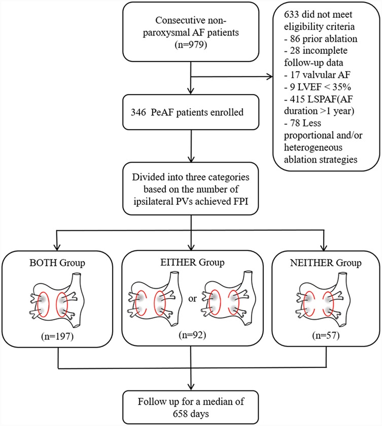
Figure 1. Study flowchart. Abbreviations: AF, atrial fibrillation; PeAF, persistent atrial fibrillation; LSPAF, long-standing persistent atrial fibrillation; LVEF, left ventricular injection fraction; PV, pulmonary vein; PVI, pulmonary vein isolation; FPI, first-pass isolation; BOTH group, patients with FPI in bilateral PVs group; EITHER group, patients with FPI in either ipsilateral PVs group; NEITHER group, patients without FPI in bilateral PVs group.
All patients provided written informed consent for AF ablation. In line with the Declaration of Helsinki's ethical principles, this study received approval from the hospital's Ethics Committee, with informed consent waived for the observational study due to anonymized data.
2.2 Radiofrequency ablation and first-pass pulmonary vein isolation
A transesophageal echocardiogram (TEE) was performed within 48 h before the procedure to confirm the absence of a thrombus in the left atrium (LA) and left atrial appendage. Antiarrhythmic drugs (AADs), except Amiodarone, were discontinued for five half-lives before the procedure. RFA was performed under deep conscious sedation and local anesthesia. After femoral venous access, intravenous heparin was administered to maintain an intraprocedural activated coagulation time (ACT) of 250–350 s. A steerable deca-polar catheter was introduced into the coronary sinus vein for intracardiac recording. Intracardiac electrograms were recorded using the multi-channel electrophysiological system (EP-Workmate, Abbott, USA). Three-dimensional electroanatomic mapping (EAM) systems (CARTO 3 version 6, Biosense Webster, USA) provided the operators with anatomic reconstructions. Multipolar mapping catheters (Pentaray Nav, Biosense-Webster, USA) were used for geometry reconstruction and mapping after respiratory gating settings. An open irrigated-tip contact force (CF)-sensing catheter (Thermocool SmartTouch, Biosense-Webster, USA) was employed to deliver RFA. Ablation lesions were delivered at quantitative indices [for LA anterior/roof segments: ablation index (AI) of 500–550; for LA posterior/inferior segments: AI of 350–400] in conjunction with an interlesion distance not more than 5 mm and a target CF of 5–20 g, following the CLOSE protocol (10).
All patients underwent PVI. Contiguous, encircling, ablation lesions were created around ipsilateral PV pairs to achieve isolation. Acute PVI was defined as a bidirectional conduction block between the PVs and LA following sequentially applying point-by-point ablation at the PV antrum. An entrance block was identified by the absence of PV potentials recorded by the multipolar mapping catheter. Electric cardioversion was applied to restore the sinus rhythm if the AF could not be terminated spontaneously. Intra-PV pacing (output of 20 mA at a pulse width of 2 ms) without capture of the LA was performed to identify the exit block after the restoration of sinus rhythm. In cases where isolation was not achieved, conduction gaps were tagged on the EAM system, and additional RF applications with the same AI targets were delivered at the gap sites. After a waiting period of 20 min since the last ablation, all PVs were assessed again and adenosine testing was performed to reveal dormant electrical conduction. FPI was defined as the completion of first-pass circumferential ablation lesion sets and isolation of ipsilateral PV without the need for additional touch-up ablations at the intervenous carina or gaps in the initial circumferential ablation circle. Given that the previous study has mentioned that FPI achievements in at least one of the two ipsilateral PVs (at least ipsilateral FPI, IFPI, inclusive of FPI in bilateral PVs and either ipsilateral PVs) to be significantly associated with ablation outcomes in the PAF population (9), its integration into our research was deemed necessary. PVI alone, PVI Plus LA roof line (LARL), and PVI Plus posterior wall isolation (PWI) were included as primary ablation strategies. The study was conducted by four experienced practitioners at our center with extensive experience performing AF ablation procedures, performing more than 200 procedures individually per year.
2.3 Post-ablation management and follow-up
Oral anticoagulants (OAC) and AAD were prescribed within the 90-day blanking period after ablation. Continuous OAC was recommended following the current guideline if the patient was at high risk of thromboembolism. AAD was discontinued after the blanking period. Outpatient clinic visits were scheduled at 1, 3, and 6 months after the procedure, followed by biannual visits. Each visit included physical examinations, 12-lead ECG, and 24-h Holter monitoring. Patients reporting symptoms of palpitations underwent a 24-h Holter recording and were evaluated for the possibility of arrhythmia recurrence. The primary study endpoint was the recurrence of atrial tachyarrhythmias (ATAs), defined as documented AF and organized atrial tachycardias (AT, including atrial flutter and tachycardia) lasting ≥30 s beyond the 90-day blanking period.
2.4 Statistical analysis
Variables conforming to a normal distribution were reported as mean ± standard deviation (SD), while non-normally distributed variables were reported as median and interquartile range (IQR). Dichotomous and categorical variables were presented in percentages. The Kruskal-Wallis test or ANOVA was used for continuous variable comparisons, and the chi-square test or Fisher's exact test for categorical variable comparisons. Freedom from ATAs/AF/AT recurrence was analyzed using the Kaplan–Meier method. Univariate and multivariate Cox proportional hazard models were used to evaluate predictors of ATAs/AF/AT recurrences. Subgroup analysis of the primary endpoint indicating hazard ratios and P-values for interaction was performed based on Cox regression analysis. Subgroup selection was determined by clinical experience and potential risk factors suggested by Cox regression. Restricted cubic spline was performed on continuous variables to find the appropriate thresholds. Propensity score matching was employed to address potential confounding. Propensity scores were calculated using logistic regression, incorporating variables such as age, gender, BMI, duration of AF persistence and left atrium diameter (LAD). Matched cohorts were created using nearest neighbor matching with a caliper of 0.2 standard deviations of the logit of the propensity score. IBM SPSS Statistics (version 26; SPSS Inc, USA) was employed for statistical analyses, and figures were created with GraphPad Prism (version 9.1; GraphPad Software, USA).
3 Results
3.1 Baseline characteristics
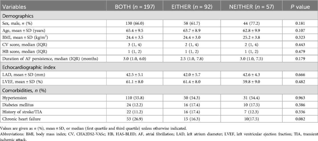
A total of 346 patients were available for analysis, which included 197 patients in the BOTH group, 92 in the EITHER group, and 57 in the NEITHER group. A detailed presentation of the demographic and baseline characteristics is available in Table 1. Patients with PeAF had a median AF episode duration of 3.0 (1.0, 6.0) months with 162 (46.8%) patients experiencing duration for less than 3 months. No significant differences were observed among the three groups for the all variables.
3.2 The first-pass pulmonary vein isolation and touch-up sites
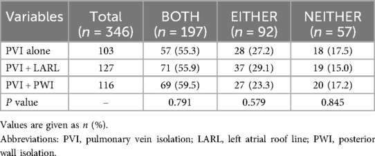
The PVI was successfully performed in all patients. Residual conduction gaps present upon completion of the encircling lesion sets were ablated, resulting in bilateral PVI. The FPI was effectively achieved in 251 left PVs (72.5%) and 235 right PVs (67.9%) in patients with PeAF. The spatial distribution of the sites for touch-up RF application in the case of non-FPI is illustrated in Supplementary Figure S1. Additional RF lesions to eliminate conduction gaps were mostly located at the carina between ipsilateral PVs. No significant difference in the proportion of different ablation strategies across the three groups (P > 0.05; Table 2).
3.3 Follow-up and redo ablation findings
The median duration of follow-up after the ablation procedure was 658 (402, 970) days. As depicted in Figures 2A,B, the Kaplan–Meier survival analysis indicated that the NEITHER group exhibited less freedom from ATAs and AF recurrence compared to the BOTH group (ATA: 57.9% vs. 75.1%, log-rank P < 0.001; AF: 63.2% vs. 76.6%, log-rank P = 0.007) or the EITHER group (ATAs: 57.9% vs. 70.7%, log-rank P = 0.036; AF: 63.2% vs. 76.1%, log-rank P = 0.041). However, there was no statistically significant difference in AT-free survival among the groups (BOTH vs. EITHER vs. NEITHER, 93.9% vs. 91.3% vs. 93.0%; all log-rank P > 0.05; Figure 2C). After matching, the baseline characteristics between PVI alone and PVI Plus LARL, as well as PVI alone and PVI Plus PWI cohorts, were well balanced. It demonstrated a similar pattern in freedom from ATAs recurrence between PVI alone and PVI Plus LARL (69.5% vs. 72.6%, log-rank P = 0.575; Supplementary Figure S2A). Similarly, there was no significant difference between PVI alone and PVI Plus PWI (67.9% vs. 69.1%, log-rank P = 0.892; Supplementary Figure S2B).

Figure 2. Kaplan–Meier analysis. Comparison of freedom from ATAs (A), AF (B) or AT (C) recurrences among PeAF patients with FPI achieved in different numbers of ipsilateral PVs. Abbreviations: PeAF, persistent atrial fibrillation; ATAs, atrial tachyarrhythmias; AF, atrial fibrillation; AT, atrial tachycardia.
Univariate Cox regression analysis revealed that LAD (HR, 1.06; 95% CI, 1.02–1.10; P = 0.004), achieving FPI in the left PVs (HR, 0.59; 95% CI, 0.39–0.89; P = 0.011), achieving FPI in the right PVs (HR, 0.58; 95% CI, 0.39–0.87; P = 0.008), BOTH (HR, 0.62; 95% CI, 0.42–0.92; P = 0.016), and IFPI (HR, 0.47; 95% CI, 0.29–0.74; P = 0.001) were significantly associated with ATAs recurrence in PeAF patients undergoing their initial RFA. In multivariate Cox regression analysis, LAD (HR, 1.05; 95% CI, 1.01–1.10; P = 0.016) and IFPI (HR, 0.46; 95% CI, 0.29–0.74; P = 0.001) were independent predictors of ATAs recurrence after adjustment for several factors including gender, age, BMI, duration of AF persistence and ablation strategies (Table 3).
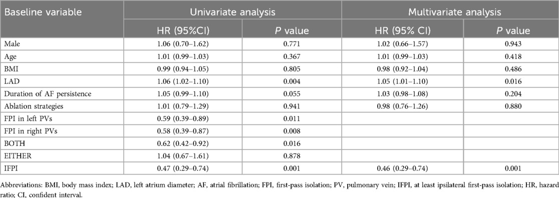
Table 3. Univariate and multivariate Cox regression analyses for atrial tachyarrhythmias recurrence.
Overall, 22 (22.0%) of 100 patients with arrhythmia relapse underwent repeat ablation. The time taken from arrhythmia recurrence to redo procedure was a median of 453 (204, 698) days. Notably, 21 (47.7%) of 44 ipsilateral PVs showed electrical reconnection. The most common site of reconnection was the intervenous carina (56.8%), followed by the posteroinferior aspect (13.6%) and the roof (9.1%) of the circle. The PV reconnection rate in the second procedure was significantly lower in PVs with successful FPI in the first procedure than in others (35.7% vs. 68.8%, p = 0.035; Figure 3).

Figure 3. Pulmonary vein reconnection rates in the redo procedure per ipsilateral PVs. Abbreviations: FPI, first-pass isolation; PVR, pulmonary vein reconnection.
3.4 Subgroup analysis of primary endpoint
The risk of ATAs recurrence during the followup was evaluated across a range of subgroups, including age, gender, BMI, duration of AF persistence, LAD and different ablation strategies, as depicted in Figure 4. The findings revealed a consistency across all prespecified subgroups, reinforcing the role of IFPI in reducing ATAs recurrence over the non-FPI group.
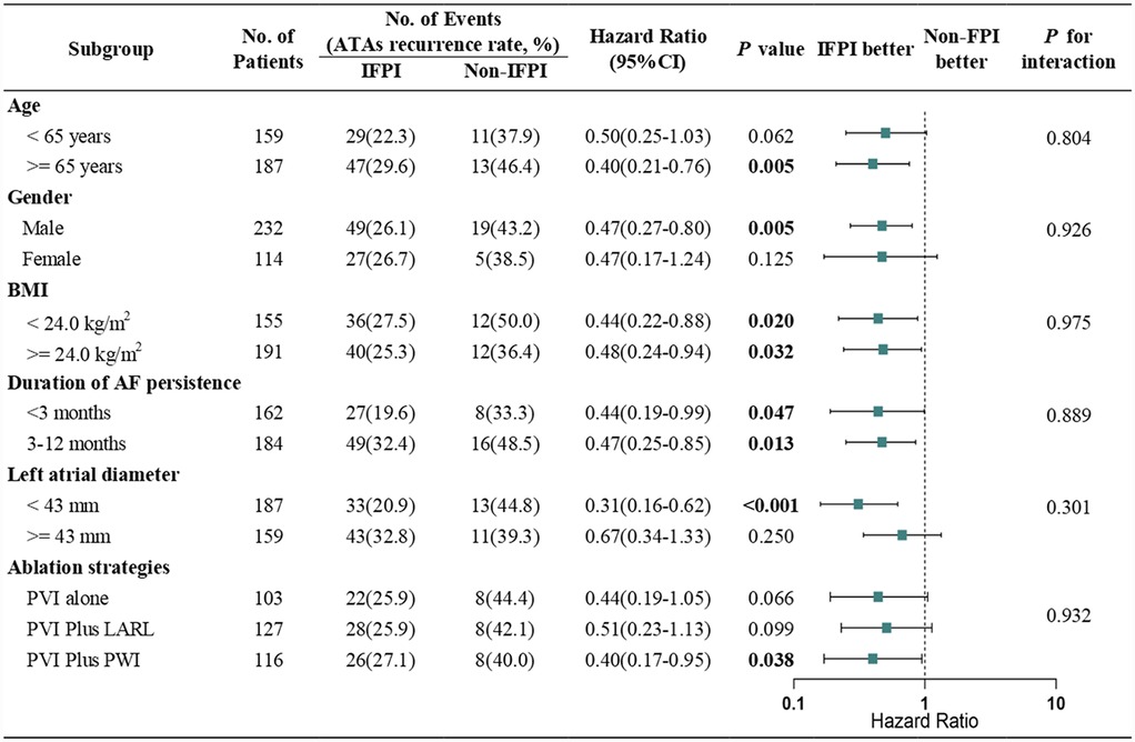
Figure 4. Subgroup analysis of primary endpoint. Hazard ratios and P for interaction are based on Cox regression analyses. Abbreviations: BMI, body mass index; AF, atrial fibrillation; PVI, pulmonary vein isolation; LARL, left atrial roof line; PWI, posterior wall isolation.
4 Discussion
4.1 Main findings
This study provides significant implications for the clinical management of PeAF through the lens of PVI and the achievement of FPI. Our findings suggested several insights: (1) after AI-guided ipsilateral encircling, the site most frequently subjected to touch-up ablation appeared to be the intervenous carina; (2) achieving IFPI was an independent predictor of freedom from ATAs recurrence in PeAF patients undergoing their initial RFA; (3) the prognostic benefit from FPI was primarily applicable to the freedom from recurrence of AF, rather than AT; (4) PV reconnection rate substantially decreased in the second procedure in ipsilateral PVs where FPI was achieved during the initial ablation.
4.2 Pulmonary vein isolation as a key success factor
Our study adds to the ongoing discourse surrounding the optimal approach for RFA in PeAF by underscoring the critical role of FPI during PVI. Our findings corroborate the prevailing view that PVI continues to be a cornerstone of successful RFA for PeAF (1). Achieving FPI proved to be a significant factor in enhancing ablation outcomes, as evidenced by the improved freedom from ATAs in our patient cohort successfully undergoing FPI. Contrary to expectations that adjunctive ablation strategies targeting non-PV triggers and substrates could yield better outcomes, our data reflect a lack of compelling evidence supporting these interventions. Despite extensive research over the past two decades, as demonstrated in several renowned randomized controlled trials, there has been negligible impact on reducing recurrent AF rates with additional ablation beyond PVI (3–7). This paradox can be largely attributed to two primary factors: the ambiguity in identifying effective targets for ablation outside the PVs and the technical challenges associated with achieving durable, transmural lesions encircling the PVs. The advent of advanced technologies, such as irrigated RF catheters with contact force-sensing, AI algorithms, and the implementation of the CLOSE protocol, has markedly enhanced the durability and efficacy of PVI. These innovations have facilitated a significant increase in arrhythmia-free survival rates, noted to have risen from approximately 40%–70% at 1-year follow-up in PeAF populations, employing PVI alone (6, 11–14). This substantial improvement highlights the crucial importance of achieving a high-quality lesion set at initial encirclement, as denoted by FPI, thus questioning the necessity of more complex adjunctive strategies. Our study supports the notion that durable PVI is paramount, positing that uncomplicated PVI may suffice in achieving favorable outcomes without the need for complex adjunctive ablation strategies which have not demonstrated clear added benefit in PeAF settings, particularly early-stage PeAF with a normal left atrial substrate.
4.3 Impact of first-pass isolation on ablation outcome
FPI has been shown to be associated with a diminished likelihood of acute and chronic pulmonary vein reconnection and favorable clinical results (8, 9, 15, 16). The pathophysiological basis can be attributed to the higher likelihood of creating transmural and contiguous lesions during the initial RF encirclement, thereby reducing the risk of PV reconnection and subsequent AF recurrence. Interestingly, our data reveal that patients from the NEITHER group, who did not achieve FPI in bilateral PVs, exhibited a markedly lower rate of freedom from ATAs. This suggests that the complexity of the left atrial and PV electroanatomy in these patients might impede effective lesion formation, as hypothesized in previous studies (9, 15). The presence of residual conduction gaps and non-transmural lesions, particularly in areas of increased PV wall thickness due to tissue edema, seems to play a crucial role in compromised ablation outcomes (17). Although additional RF applications may sometimes block these epicardial gaps, the transient nature of electrical blocks caused by edema often results in PV reconnection and subsequent AF recurrence (18, 19).
Moreover, the correlation between the absence of FPI and increased AF recurrence, contrasted with a lack of association with AT recurrence, presents a nuanced understanding of AF pathophysiology. Focal triggers originating from PVs are known to be predominant in the early stages of PeAF, especially with a relatively healthier atrial substrate. Consequently, situations where FPI is not achieved allow for easier electrical reconnection between the PVs and the left atrium (8, 15, 16), thereby fostering AF recurrence. In contrast, the recurrence of AT appears to depend less on these initial focal triggers and more on the structural and electrophysiological substrate set by previous ablation lesions (20), indicating a different pathophysiological mechanism. Notably, the study did not find a significant difference in AT recurrence among the different groups, suggesting that FPI's influence may not extend to perturbations in AT etiology. This points to the complex interplay between lesion sets, substrate properties, and arrhythmogenic foci, necessitating further investigation into the mechanistic pathways that could differentiate outcomes for AT and AF post-ablation.
The absence of a statistically significant difference in ablation outcomes between the BOTH group and the EITHER group may stem from multiple interrelated mechanisms: (1) the dominance of unilateral PV triggers in persistent AF, particularly from the left PVs (21–23), may diminish the incremental benefit of bilateral FPI. Anatomically, the left PVs are closer to arrhythmogenic substrates such as the left atrial posterior wall. It can be hypothesized that left PV reconnection is a stronger predictor of AF recurrence than right PV reconnection, but this has not been well documented. In our cohort, left PV FPI success rates were higher than right PVs (72.5% vs. 67.9%), implying that isolating the left PVs alone may suffice to suppress dominant triggers in a subset of patients; (2) Progressive atrial substrate remodeling in PeAF shifts arrhythmia maintenance from PV-dependent triggers to self-sustaining mechanisms (2). Even with bilateral FPI, residual substrate abnormalities could perpetuate AF/AT, reducing the relative advantage of complete PV isolation. This hypothesis is supported by the lack of difference in AT-free survival across groups (Figure 2C), as AT recurrence is more dependent on substrate than PV triggers (20). However, the precise mechanisms underlying this consistency remain elusive and warrant further mechanistic investigation.
It observed that most of touch-up ablation sites in non-FPI PVs were localized to the intervenous carina (Supplementary Figure S1), a region anatomically predisposed to epicardial muscle bundles bridging the LA and PVs (24). The clustering of residual conduction gaps at this epicardial hotspot suggests that incomplete lesion transmurality in this region may underlie FPI failure. Epicardial muscle bundles, which are not fully targeted by endocardial ablation, could perpetuate electrical reconnection and AF recurrence (25, 26). This hypothesis is supported by the significantly higher PV reconnection rate in non-FPI patients (68.8% vs. 35.7%, P = 0.035; Figure 3), consistent with prior studies implicating epicardial connections in AF recurrence due to their resistance to endocardial ablation (27). Future prospective studies integrating high-density mapping are needed to directly quantify the role of these pathways in FPI failure.
4.4 Perpetuation of atrial fibrillation and its impact on first-pass isolation
The distinct challenges presented by the long-standing nature of AF, especially in cases advancing towards LSPAF, require a nuanced strategy beyond PVI alone (28). As PeAF progresses, the atrial substrate undergoes significant electrical and structural remodeling (2), which can diminish the effectiveness of FPI by extending the pathological mechanisms beyond simple PV triggers. This progression complicates intervention strategies, necessitating the potential incorporation of more aggressive ablation approaches targeting non-PV areas (29, 30). We performed additional statistical analyses for excluded population with LSPAF and found that comparisons amongst the groups (BOTH vs. EITHER vs. NEITHER) revealed no statistically significant difference in the freedom from ATAs recurrence (P > 0.05), as expected. This suggests that while FPI is critical, its effectiveness might be compromised in advanced PeAF cases. The findings imply that for advanced cases of PeAF, particularly those approaching LSPAF, optimization of adjunctive ablation strategies might be necessary, even though current evidence regarding these strategies presents a limited prognostic benefit (3–7). Thus, our work not only reinforces the value of achieving FPI but also advocates for targeted exploration into personalized ablation plans as PeAF evolves toward chronicity.
Prolonged duration of AF leads to progressive deterioration of the atrial substrate (31, 32). While our study did not directly assess LA voltage or low-voltage areas (LVA), we propose that LAD, a surrogate for structural remodeling, may reflect underlying substrate heterogeneity. LA enlargement is strongly associated with fibrotic remodeling and larger LVA (33). In our cohort, baseline LAD was comparable across groups (BOTH: 42.5 ± 5.1 mm, EITHER: 42.0 ± 5.7 mm, NEITHER: 42.6 ± 4.3 mm; P = 0.666; Table 1), suggesting that differences in LVA burden among groups were likely minimal. However, the relationship between FPI success and LA substrate remains incompletely understood. For instance, Pérez-Pinzón et al. reported that higher LA voltage (indicative of healthier substrate) paradoxically predicted lower FPI rates in the right pulmonary veins, while left PV FPI remained unaffected (34)—a finding that underscores the complex regional interplay between substrate properties and ablation efficacy. Although LVA is a well-established predictor of poor ablation outcomes (35, 36), it remains unclear whether adverse LA substrate directly attenuates the prognostic benefit of FPI or acts as a confounding factor. Larger studies integrating voltage mapping and advanced imaging are needed to dissect these mechanisms.
4.5 Study limitation
Several limitations require emphasis in our study. Firstly, this study was retrospective and non-randomized, rendering our conclusions as hypothesis-generating, and selection bias concerning the study population remains a potential concern. Secondly, this study did not explore the impact of FPI on ablation outcomes in patients undergoing additional extra-PV ablation strategies, such as mitral isthmus line, cavotricuspid isthmus line or CFAE ablation, which suggests caution when extrapolating these results to the populations with other ablation strategies. Thirdly, the absence of specific data on the incidence of epicardial connections and LA voltage/fibrosis limited our ability to assess substrate-specific predictors of FPI success; future studies incorporating delayed-enhancement MRI or high-density voltage mapping are warranted to clarify the role of epicardial pathways and substrate characteristics in FPI failure. Fourthly, the lack of continuous cardiac monitoring might have led to an underestimation of arrhythmia recurrence.
5 Conclusion
Achieving FPI for PVI is strongly associated with enhanced ablation outcomes in patients with PeAF. The presence of IFPI emerges as a critical predictor for the long-term freedom from ATAs in this patient population undergoing radiofrequency ablation. These findings again highlight the pivotal role of durable PVI in determining successful ablation results for PeAF.
Data availability statement
The raw data supporting the conclusions of this article will be made available by the authors, without undue reservation.
Ethics statement
The studies involving humans were approved by Ethics Committee of the First Affiliated Hospital of Ningbo University. The studies were conducted in accordance with the local legislation and institutional requirements. The ethics committee/institutional review board waived the requirement of written informed consent for participation from the participants or the participants' legal guardians/next of kin because the study involves the collection of anonymous data, and participants' confidentiality can be adequately protected without a signed consent form.
Author contributions
CL: Conceptualization, Data curation, Formal analysis, Investigation, Methodology, Writing – original draft, Writing – review & editing. BL: Data curation, Formal analysis, Investigation, Writing – original draft. XY: Data curation, Formal analysis, Methodology, Writing – original draft. XD: Conceptualization, Data curation, Funding acquisition, Methodology, Resources, Supervision, Validation, Writing – original draft, Writing – review & editing. HC: Conceptualization, Methodology, Project administration, Resources, Writing – original draft, Writing – review & editing. SZ: Data curation, Formal analysis, Writing – original draft. CS: Data curation, Methodology, Writing – original draft. MF: Data curation, Formal analysis, Writing – review & editing. YJ: Investigation, Visualization, Writing – review & editing. HJ: Investigation, Software, Writing – review & editing. GF: Conceptualization, Methodology, Writing – review & editing. LY: Data curation, Formal analysis, Writing – review & editing. BW: Investigation, Validation, Writing – review & editing. YY: Data curation, Investigation, Writing – review & editing. WZ: Methodology, Software, Writing – review & editing. FG: Data curation, Investigation, Writing – review & editing. YX: Investigation, Writing – review & editing. YS: Visualization, Writing – review & editing. JD: Validation, Writing – review & editing. LD: Conceptualization, Supervision, Validation, Writing – review & editing.
Funding
The author(s) declare that financial support was received for the research and/or publication of this article. This work was supported by Medical Health Science and Technology Projects of Zhejiang Province Health Commission (2022KY302 and 2024KY1519), the Key Technology R&D Program of Ningbo (2022Z149 and 2023Z188), and the Key Laboratory of Precision Medicine for Atherosclerotic Diseases of Zhejiang Province (2022E10026).
Conflict of interest
The authors declare that the research was conducted in the absence of any commercial or financial relationships that could be construed as a potential conflict of interest.
Generative AI statement
The author(s) declare that no Generative AI was used in the creation of this manuscript.
Publisher's note
All claims expressed in this article are solely those of the authors and do not necessarily represent those of their affiliated organizations, or those of the publisher, the editors and the reviewers. Any product that may be evaluated in this article, or claim that may be made by its manufacturer, is not guaranteed or endorsed by the publisher.
Supplementary material
The Supplementary Material for this article can be found online at: https://www.frontiersin.org/articles/10.3389/fcvm.2025.1588716/full#supplementary-material
Abbreviations
AF, atrial fibrillation; AT, atrial tachycardia; ATAs, atrial tachyarrhythmias; FPI, first-pass isolation; PVI, pulmonary vein isolation; PAF, paroxysmal atrial fibrillation; PeAF, persistent atrial fibrillation; LSPAF, long-standing persistent atrial fibrillation; RFA, radiofrequency ablation.
References
1. Tzeis S, Gerstenfeld EP, Kalman J, Saad EB, Shamloo AS, Andrade JG, et al. 2024 European Heart Rhythm Association/Heart Rhythm Society/Asia Pacific Heart Rhythm Society/Latin American Heart Rhythm Society expert consensus statement on catheter and surgical ablation of atrial fibrillation. Heart Rhythm. (2024) 21(9):e31–149. doi: 10.1016/j.hrthm.2024.03.017
2. Allessie M, Ausma J, Schotten U. Electrical, contractile and structural remodeling during atrial fibrillation. Cardiovasc Res. (2002) 54(2):230–46. doi: 10.1016/S00086363(02)00258-4
3. Fink T, Schlüter M, Heeger C-H, Lemes C, Maurer T, Reissmann B, et al. Stand-alone pulmonary vein isolation versus pulmonary vein isolation with additional substrate modification as index ablation procedures in patients with persistent and long-standing persistent atrial fibrillation: the randomized Alster-Lost-AF trial (ablation at St. Georg hospital for long-standing persistent atrial fibrillation). Circ Arrhythm Electrophysiol. (2017) 10(7):e005114. doi: 10.1161/CIRCEP.117.005114
4. Kistler PM, Chieng D, Sugumar H, Ling L-H, Segan L, Azzopardi S, et al. Effect of catheter ablation using pulmonary vein isolation with vs. without posterior left atrial wall isolation on atrial arrhythmia recurrence in patients with persistent atrial fibrillation: the CAPLA randomized clinical trial. JAMA. (2023) 329(2):127–35. doi: 10.1001/jama.2022.23722
5. Marrouche NF, Wazni O, McGann C, Greene T, Dean JM, Dagher L, et al. Effect of MRI-guided fibrosis ablation vs. conventional catheter ablation on atrial arrhythmia recurrence in patients with persistent atrial fibrillation: the DECAAF II randomized clinical trial. JAMA. (2022) 327(23):2296–305. doi: 10.1001/jama.2022.8831
6. Verma A, Jiang C-Y, Betts TR, Chen J, Deisenhofer I, Mantovan R, et al. Approaches to catheter ablation for persistent atrial fibrillation. N Engl J Med. (2015) 372(19):1812–22. doi: 10.1056/NEJMoa1408288
7. Yang G, Zheng L, Jiang C, Fan J, Liu X, Zhan X, et al. Circumferential pulmonary vein isolation plus low-voltage area modification in persistent atrial fibrillation: the STABLE-SR-II trial. JACC Clin Electrophysiol. (2022) 8(7):882–91. doi: 10.1016/j.jacep.2022.03.012
8. Ninomiya Y, Inoue K, Tanaka N, Okada M, Tanaka K, Onishi T, et al. Absence of first-pass isolation is associated with poor pulmonary vein isolation durability and atrial fibrillation ablation outcomes. J Arrhythm. (2021) 37(6):1468–76. doi: 10.1002/joa3.12629
9. Osorio J, Hunter TD, Rajendra A, Zei P, Silverstein J, Morales G. Predictors of clinical success after paroxysmal atrial fibrillation catheter ablation. J Cardiovasc Electrophysiol. (2021) 32(7):1814–21. doi: 10.1111/jce.15028
10. Taghji P, El Haddad M, Phlips T, Wolf M, Knecht S, Vandekerckhove Y, et al. Evaluation of a strategy aiming to enclose the pulmonary veins with contiguous and optimized radiofrequency lesions in paroxysmal atrial fibrillation: a pilot study. JACC Clin Electrophysiol. (2018) 4(1):99–108. doi: 10.1016/j.jacep.2017.06.023
11. Dixit S, Marchlinski FE, Lin D, Callans DJ, Bala R, Riley MP, et al. Randomized ablation strategies for the treatment of persistent atrial fibrillation: RASTA study. Circ Arrhythm Electrophysiol. (2012) 5(2):287–94. doi: 10.1161/CIRCEP.111.966226
12. Vogler J, Willems S, Sultan A, Schreiber D, Lüker J, Servatius H, et al. Pulmonary vein isolation versus defragmentation: the CHASE-AF clinical trial. J Am Coll Cardiol. (2015) 66(24):2743–52. doi: 10.1016/j.jacc.2015.09.088
13. Voskoboinik A, Moskovitch JT, Harel N, Sanders P, Kistler PM, Kalman JM. Revisiting pulmonary vein isolation alone for persistent atrial fibrillation: a systematic review and meta-analysis. Heart Rhythm. (2017) 14(5):661–7. doi: 10.1016/j.hrthm.2017.01.003
14. Yamaguchi J, Takahashi Y, Yamamoto T, Amemiya M, Sekigawa M, Shirai Y, et al. Clinical outcome of pulmonary vein isolation alone ablation strategy using VISITAG SURPOINT in nonparoxysmal atrial fibrillation. J Cardiovasc Electrophysiol. (2020) 31(10):2592–9. doi: 10.1111/jce.14673
15. Okamatsu H, Okumura K, Onishi F, Yoshimura A, Negishi K, Tanaka Y, et al. Predictors of pulmonary vein non-reconnection in the second procedure after ablation index-guided pulmonary vein isolation for atrial fibrillation and its impact on the outcome. J Cardiovasc Electrophysiol. (2023) 34(12):2452–60. doi: 10.1111/jce.16084
16. Sandorfi G, Rodriguez-Mañero M, Saenen J, Baluja A, Bories W, Huybrechts W, et al. Less pulmonary vein reconnection at redo procedures following radiofrequency point-by-point antral pulmonary vein isolation with the use of contemporary catheter ablation technologies. JACC Clin Electrophysiol. (2018) 4(12):1556–65. doi: 10.1016/j.jacep.2018.09.020
17. Gao X, Chang D, Bilchick KC, Hussain SK, Petru J, Skoda J, et al. Left atrial thickness and acute thermal injury in patients undergoing ablation for atrial fibrillation: laser versus radiofrequency energies. J Cardiovasc Electrophysiol. (2021) 32(5):1259–67. doi: 10.1111/jce.15011
18. Arujuna A, Karim R, Caulfield D, Knowles B, Rhode K, Schaeffter T, et al. Acute pulmonary vein isolation is achieved by a combination of reversible and irreversible atrial injury after catheter ablation: evidence from magnetic resonance imaging. Circ Arrhythm Electrophysiol. (2012) 5(4):691–700. doi: 10.1161/CIRCEP.111.966523
19. Knowles BR, Caulfield D, Cooklin M, Rinaldi CA, Gill J, Bostock J, et al. 3-D visualization of acute RF ablation lesions using MRI for the simultaneous determination of the patterns of necrosis and edema. IEEE Trans Biomed Eng. (2010) 57(6):1467–75. doi: 10.1109/TBME.2009.2038791
20. Mesas CE, Pappone C, Lang CCE, Gugliotta F, Tomita T, Vicedomini G, et al. Left atrial tachycardia after circumferential pulmonary vein ablation for atrial fibrillation: electroanatomic characterization and treatment. J Am Coll Cardiol. (2004) 44(5):1071–9. doi: 10.1016/j.jacc.2004.05.072
21. Chen SA, Hsieh MH, Tai CT, Tsai CF, Prakash VS, Yu WC, et al. Initiation of atrial fibrillation by ectopic beats originating from the pulmonary veins: electrophysiological characteristics, pharmacological responses, and effects of radiofrequency ablation. Circulation. (1999) 100(18):1879–86. doi: 10.1161/01.CIR.100.18.1879
22. Haïssaguerre M, Jaïs P, Shah DC, Takahashi A, Hocini M, Quiniou G, et al. Spontaneous initiation of atrial fibrillation by ectopic beats originating in the pulmonary veins. N Engl J Med. (1998) 339(10):659–66. doi: 10.1056/NEJM199809033391003
23. Kurotobi T, Iwakura K, Inoue K, Kimura R, Okamura A, Koyama Y, et al. Multiple arrhythmogenic foci associated with the development of perpetuation of atrial fibrillation. Circ Arrhythm Electrophysiol. (2010) 3(1):39–45. doi: 10.1161/CIRCEP.109.885095
24. Cabrera JA, Ho SY, Climent V, Fuertes B, Murillo M, Sánchez-Quintana D. Morphological evidence of muscular connections between contiguous pulmonary venous orifices: relevance of the interpulmonary isthmus for catheter ablation in atrial fibrillation. Heart Rhythm. (2009) 6(8):1192–8. doi: 10.1016/j.hrthm.2009.04.016
25. Piorkowski C, Kronborg M, Hourdain J, Piorkowski J, Kirstein B, Neudeck S, et al. Endo-/epicardial catheter ablation of atrial fibrillation: feasibility, outcome, and insights into arrhythmia mechanisms. Circ Arrhythm Electrophysiol. (2018) 11(2):e005748. doi: 10.1161/CIRCEP.117.005748
26. Maesen B, Zeemering S, Afonso C, Eckstein J, Burton RAB, van Hunnik A, et al. Rearrangement of atrial bundle architecture and consequent changes in anisotropy of conduction constitute the 3-dimensional substrate for atrial fibrillation. Circ Arrhythm Electrophysiol. (2013) 6(5):967–75. doi: 10.1161/CIRCEP.113.000050
27. Pérez-Castellano N, Villacastín J, Salinas J, Vega M, Moreno J, Doblado M, et al. Epicardial connections between the pulmonary veins and left atrium: relevance for atrial fibrillation ablation. J Cardiovasc Electrophysiol. (2011) 22(2):149–59. doi: 10.1111/j.1540-8167.2010.01873.x
28. Brooks AG, Stiles MK, Laborderie J, Lau DH, Kuklik P, Shipp NJ, et al. Outcomes of long-standing persistent atrial fibrillation ablation: a systematic review. Heart Rhythm. (2010) 7(6):835–46. doi: 10.1016/j.hrthm.2010.01.017
29. Hung Y, Lo L-W, Lin Y-J, Chang S-L, Hu Y-F, Chung F-P, et al. Characteristics and long-term catheter ablation outcome in long-standing persistent atrial fibrillation patients with non-pulmonary vein triggers. Int J Cardiol. (2017) 241:205–11. doi: 10.1016/j.ijcard.2017.04.050
30. Della Rocca DG, Tarantino N, Trivedi C, Mohanty S, Anannab A, Salwan AS, et al. Non-pulmonary vein triggers in nonparoxysmal atrial fibrillation: implications of pathophysiology for catheter ablation. J Cardiovasc Electrophysiol. (2020) 31(8):2154–67. doi: 10.1111/jce.14638
31. Wijffels MC, Kirchhof CJ, Dorland R, Allessie MA. Atrial fibrillation begets atrial fibrillation. A study in awake chronically instrumented goats. Circulation. (1995) 92(7):1954–68. doi: 10.1161/01.CIR.92.7.1954
32. Morillo CA, Klein GJ, Jones DL, Guiraudon CM. Chronic rapid atrial pacing. Structural, functional, and electrophysiological characteristics of a new model of sustained atrial fibrillation. Circulation. (1995) 91(5):1588–95. doi: 10.1161/01.CIR.91.5.1588
33. Huo Y, Gaspar T, Pohl M, Sitzy J, Richter U, Neudeck S, et al. Prevalence and predictors of low voltage zones in the left atrium in patients with atrial fibrillation. Europace. (2018) 20(6):956–62. doi: 10.1093/europace/eux082
34. Pérez-Pinzón J, Waks JW, Yungher D, Reynolds A, Maher T, Locke AH, et al. Predictors of first-pass isolation in patients with recurrent atrial fibrillation: a retrospective cohort study. Heart Rhythm O2. (2024) 5 (10):713–9. doi: 10.1016/j.hroo.2024.08.008
35. Rolf S, Kircher S, Arya A, Eitel C, Sommer P, Richter S, et al. Tailored atrial substrate modification based on low-voltage areas in catheter ablation of atrial fibrillation. Circ Arrhythm Electrophysiol. (2014) 7(5):825–33. doi: 10.1161/CIRCEP.113.001251
Keywords: persistent atrial fibrillation, pulmonary vein isolation, first-pass isolation, radiofrequency ablation, ablation outcome
Citation: Luo C, Leng B, Yu X, Du X, Chu H, Zhou S, Shen C, Feng M, Jiang Y, Jin H, Fu G, Yu L, Wang B, Yu Y, Zhuo W, Gao F, Xu Y, Sun Y, Dai J and Di Biase L (2025) Impact of the first-pass pulmonary vein isolation on ablation outcomes in persistent atrial fibrillation. Front. Cardiovasc. Med. 12:1588716. doi: 10.3389/fcvm.2025.1588716
Received: 6 March 2025; Accepted: 8 May 2025;
Published: 5 June 2025.
Edited by:
Pietro Francia, University Sapienza, ItalyReviewed by:
Naotaka Hashiguchi, Japanese Red Cross Narita Hospital, JapanAnkit Jain, Government of India, India
Copyright: © 2025 Luo, Leng, Yu, Du, Chu, Zhou, Shen, Feng, Jiang, Jin, Fu, Yu, Wang, Yu, Zhuo, Gao, Xu, Sun, Dai and Di Biase. This is an open-access article distributed under the terms of the Creative Commons Attribution License (CC BY). The use, distribution or reproduction in other forums is permitted, provided the original author(s) and the copyright owner(s) are credited and that the original publication in this journal is cited, in accordance with accepted academic practice. No use, distribution or reproduction is permitted which does not comply with these terms.
*Correspondence: Xianfeng Du, ZHJkdXhpYW5mZW5nQDEyNi5jb20=; Huimin Chu, ZXBuYmhlYXJ0QDE2My5jb20=
†These authors have contributed equally to this work and share first authorship
 Chenxu Luo
 Bing Leng1,4,†
 Xianfeng Du
 Huimin Chu
 Shenyuan Zhou
 Caijie Shen
 Guohua Fu
 Binhao Wang
 Yibo Yu
 Yijun Sun
 Luigi Di Biase