- 1Graduate School, Beijing University of Chinese Medicine, Beijing, China
- 2Beijing Hospital of Traditional Chinese Medicine, Capital Medical University, Beijing, China
- 3Aviation Health Medical Center, Air China Limited, Beijing, China
- 4Guang’anmen Hospital, China Academy of Chinese Medical Sciences, Beijing, China
- 5Department of Traditional Chinese Medicine, Lhasa People’s Hospital, Tibet Autonomous Region, China
Background: Coronary microvascular dysfunction (CMD) in post-percutaneous coronary intervention (PCI) target vessels is increasingly recognized as a critical determinant of adverse cardiovascular outcomes, yet its prevalence and prognostic implications remain poorly characterized. We conducted a systematic review and meta-analysis to determine the prevalence of CMD in post-PCI target vessels and its associated clinical outcomes.
Methods: We conducted a systematic review and meta-analysis of observational studies using quantitative coronary physiological assessments to evaluate CMD in post-PCI target vessels. Databases (PubMed, Embase, Web of Science) were searched from inception to January 2025. The pooled Prevalence, multivariable-adjusted hazard ratio (HR) and 95% confidence interval (CI) for clinical outcomes were calculated using random-effects models.
Results: A total of 21 observational studies involving 6,632 patients were included. The pooled prevalence of CMD in post-PCI target vessels was 41.66% (95% CI: 34.18%–49.34%). Subgroup analyses revealed numerical variations in CMD prevalence across assessment methods, sex, clinical diagnoses, and target vessels, though intergroup differences did not reach statistical significance (all P > 0.05). The pooled prevalence of CMD was numerically higher in females (46.22% vs. 36.73% in males), patients with acute coronary syndrome (42.37% vs. 36.04% in chronic coronary syndrome), and those assessed via non-wire-based methods (44.72% vs. 35.65% in wire-based methods). CMD prevalence was comparable across target vessels (left anterior descending artery: 37.34%, left circumflex artery: 38.50%, right coronary artery: 39.09%). Patients with post-PCI thrombolysis in myocardial infarction (TIMI) flow grade ≤2 exhibited higher CMD prevalence than those with TIMI flow grade 3, with a statistically significant difference (75.36% vs. 37.26%, P = 0.0012). CMD in post-PCI target vessels was independently associated with a 3.10-fold increased risk of major adverse cardiovascular events (95% CI: 2.06–4.67) and a 4.66-fold risk of cardiac death or heart failure readmission (95% CI: 3.13–6.93).
Conclusion: CMD in post-PCI target vessels is prevalent (approximately 40%) and independently associated with a elevated risk of adverse cardiovascular outcomes. Standardized diagnostic criteria and targeted interventions are urgently needed to improve outcomes in this population.
Systematic review registration: https://www.crd.york.ac.uk/PROSPERO/, PROSPERO CRD42025637496.
1 Introduction
Coronary microvascular dysfunction (CMD), defined as a clinical syndrome characterized by structural or functional abnormalities of the coronary microvasculature and microvascular obstruction leading to myocardial ischemia, has been increasingly recognized as a critical determinant of adverse cardiovascular outcomes, even following successful epicardial coronary revascularization (1–3). While percutaneous coronary intervention (PCI) effectively restores macrovascular blood flow in patients with coronary artery disease (CAD), emerging evidence suggests that persistent microvascular dysfunction in post-PCI target vessels may compromise myocardial perfusion, resulting in ischemia, cardiac dysfunction, and adverse ventricular remodeling (4, 5).
To date, the true prevalence of CMD in post-PCI target vessels and its long-term prognostic implications remain incompletely understood. Current investigations have reported that CMD in post-PCI target vessels is observed across nearly all diagnostic subtypes of CAD, including acute coronary syndrome (ACS) and chronic coronary syndrome (CCS), with substantial variability in prevalence ranging from 20% to 80% (6–10). This heterogeneity may arise from multiple factors. Some studies indicate that ACS patients exhibit a higher prevalence of post-PCI CMD compared to CCS patients, likely due to differences in target lesion composition and thrombotic burden (11, 12). Methodological disparities in assessments and study designs further contribute to this variability. Direct testing of coronary microvascular function is now considered necessary to confirm the diagnosis of CMD (13). Quantitative coronary physiological assessments have revealed that a substantial proportion of post-PCI CMD cases occur in vessels with angiographically normal flow, suggesting that studies relying on non-physiological assessments systematically underestimate the true prevalence of CMD in post-PCI target vessels (13–15). Conversely, some studies have overestimated CMD prevalence by failing to differentiate between pre-existing non-target vessel CMD and post-PCI target vessel CMD, thereby conflating distinct pathophysiological entities (16–18).
However, large-scale clinical investigations comprehensively covering CAD subtypes and utilizing quantitative coronary physiological assessments to evaluate post-PCI target vessel CMD are still lacking. To address this research gap, we conducted a systematic review and meta-analysis of observational studies employing quantitative coronary physiological assessments to evaluate CMD in post-PCI target vessels, aiming to determine the prevalence of CMD and its associated clinical outcomes. These findings provide novel insights into the true burden and clinical implications of CMD in post-PCI target vessels.
2 Methods
This systematic review was performed according to the Preferred Reporting Items for Systematic Review and Meta-Analysis (PRISMA) (19), and the protocol was registered at PROSPERO (No. CRD42025637496).
2.1 Search strategy
Studies that assessed CMD in post-PCI target vessels of patients after PCI were systematically searched in PubMed, Embase, and Web of Science from from inception through January 20, 2025. We used the following search terms: “percutaneous coronary intervention”, and “coronary microvascular dysfunction”. The detailed search strategy is provided in Supplementary Table S1.
2.2 Selection criteria
Inclusion criteria were as follows: (1) reported the prevalence of CMD based on quantitative coronary physiological assessments of post-PCI target vessels (15); (2) prespecified diagnostic criteria for CMD were clearly defined; (3) provided data exclusively comprising of patients undergoing PCI.
Exclusion criteria were as follows: (1) the study was a review, case report, conference abstract, protocol, or non-observational study; (2) duplicate publications; (3) data or full-text were not available; (4) non-English language publications; (5) the study used multiple measurements but did not report composite diagnostic outcomes.
It should be noted that, due to the lack of systematic pre-PCI CMD assessment in many studies, the included cases included both de novo post-PCI CMD and pre-existing CMD in target vessels.
2.3 Study selection and data extraction
Two investigators independently screened the titles and abstracts and removed irrelevant studies according to the inclusion and exclusion criteria. Two investigators extracted the following data: first author's name, study design, year of publication, geographic region, the characteristics of patients undergoing PCI, the method and cut-off value used to diagnose CMD, sample size, prevalence, multivariable-adjusted hazard ratio (HR) for clinical outcome, and relevant follow-up duration. The threshold of diagnostics tests used to define the presence of CMD were based on each individual study. Any inconsistencies were resolved by discussion with a third investigator.
2.4 Quality assessment
Two investigators independently assessed methodological quality using the “Joannagen Briggs Institute (JBI) Critical Appraisal Checklist for Studies Reporting Prevalence Data” (20). The checklist consists of nine questions (1): Was the sample frame appropriate to address the target population? (2) Were study participants recruited in an appropriate way? (3) Was the sample size adequate? (4) Were the study subjects and setting described in detail? (5) Was data analysis conducted with sufficient coverage of the identified sample? (6) Were valid methods used for the identification of the condition? (7) Was the condition measured in a standard, reliable way for all participants? (8) Was there appropriate statistical analysis? and (9) Was the response rate adequate, and if not, was the low response rate managed appropriately? Each item has four options: yes, no, unclear, or not applicable. An overall score was used to reflect the number of questions with an option of “yes”.
2.5 Statistical analysis
To mitigate the risk of duplicate patient inclusion across overlapping studies, we systematically retained the study with the largest sample size for each meta-analysis when multiple publications potentially shared identical or overlapping populations (21). In cases of equivalent sample sizes, studies using index of microcirculatory resistance (IMR) or coronary flow reserve (CFR) were prioritized. To stabilize variance in proportion data, prevalence estimates were transformed using the Freeman-Tukey double arcsine method prior to pooling. Between-study heterogeneity was quantified via τ2 for variance estimation and I2 statistic for inconsistency assessment, with I2 > 50% indicating substantial heterogeneity. A random-effects model was applied to account for anticipated variability across studies, given the expected clinical and methodological diversity among included studies. The multivariable-adjusted hazard ratio (HR) and 95% confidence interval (CI) were used to pool clinical outcomes. Subgroup analyses were conducted according to the following: (1) assessment methods: non-wire-based and wire-based; (2) sex: male and female; (3) diagnosis: ACS and CCS; (4) target vessels: left anterior descending artery (LAD), left circumflex artery (LCX), and right coronary artery (RCA); (5) post-PCI thrombolysis in myocardial infarction (TIMI) flow grade: TIMI flow grade 3 and TIMI flow grade ≤2. Meta-analyses were pooled using the R Software (version 4.4.2 R Project for Statistical Computing, Vienna, Austria) and the “meta” R package (22). P value <0.05 was considered statistically significant.
3 Results
3.1 Literature search
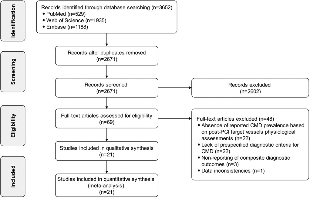
A total of 3,652 records were identified through systematic database searches, comprising 529 from PubMed, 1,935 from Web of Science, and 1,188 from Embase. After removing 981 duplicates, 2,671 records proceeded to title/abstract screening. Of these, 2,602 records were excluded due to irrelevance to CMD assessment in post-PCI target vessels. Subsequently, 69 full-text articles were assessed for eligibility. Ultimately, 21 studies (4, 5, 9, 10, 23–39) met all inclusion criteria and were incorporated into both qualitative synthesis and quantitative meta-analysis. The study selection process is detailed in Figure 1.
3.2 Characteristics of the included studies
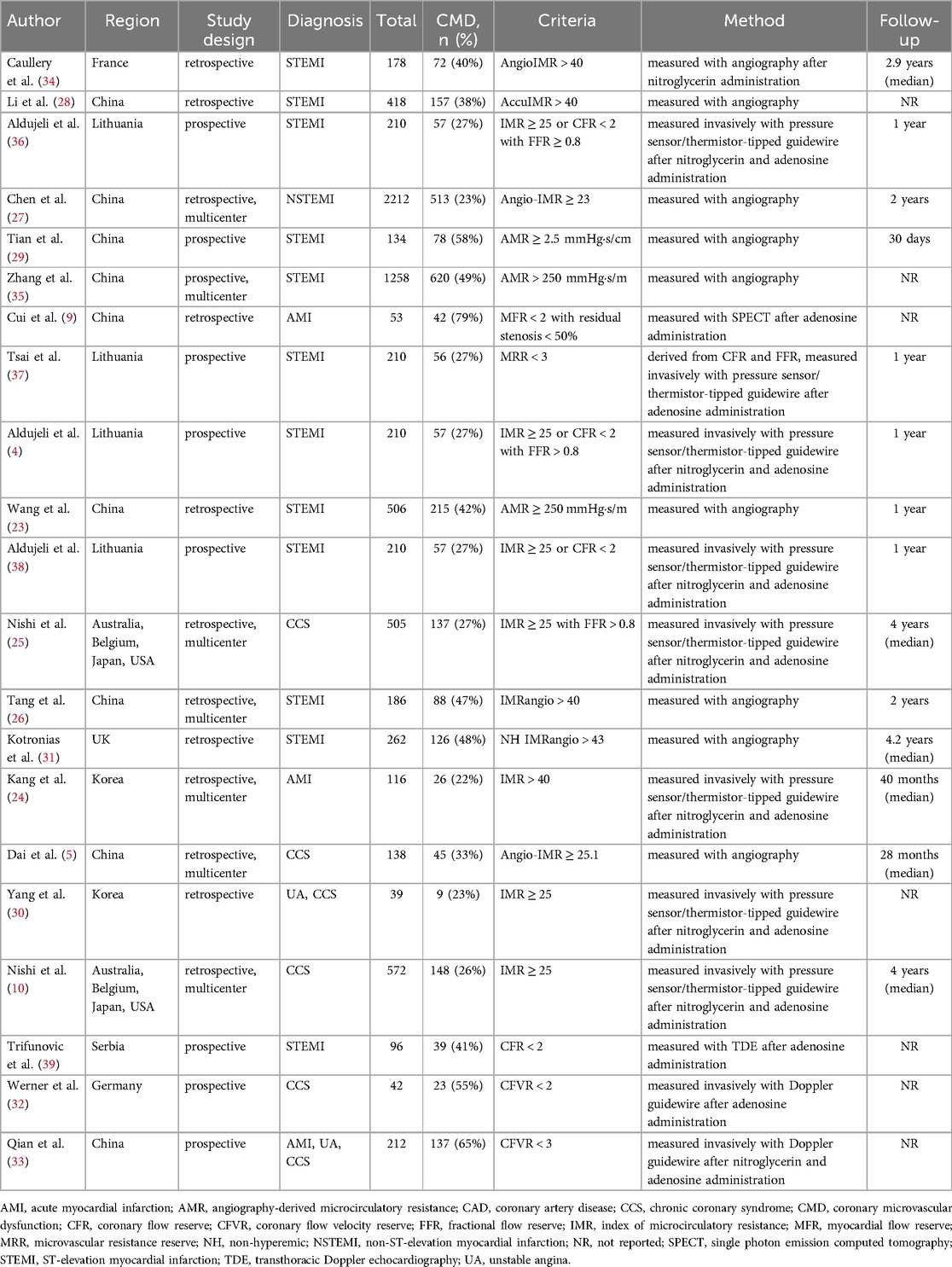
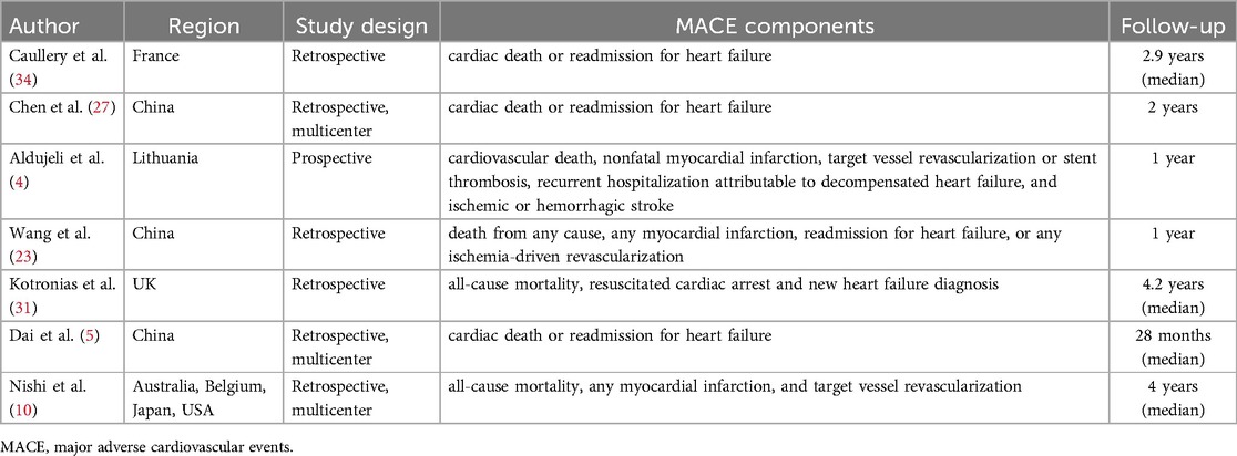
The 21 included studies were conducted across diverse geographic regions, including France (34), China (5, 9, 23, 26–29, 33, 35), Lithuania (4, 36–38), Australia (10, 25), Belgium (10, 25), Japan (10, 25), the United States (10, 25), the United Kingdom (31), Germany (32), Serbia (39), and Korea (24, 30). These studies comprehensively covered diagnostic subtypes of CAD. All studies utilized coronary physiological assessments to diagnose CMD in post-PCI target vessels, employing either wire-based or non-wire-based methods. 9 studies (4, 29, 32, 33, 35–39) were prospective, and 12 were retrospective (5, 9, 10, 23–28, 30, 31, 34). 7 studies were multicenter investigations (5, 10, 24–27, 35). 14 studies (4, 5, 10, 23–27, 29, 31, 34, 36–38) reported post-PCI follow-up, with durations ranging from 30 days to 4.2 years (median). Notably, 4 single-center studies from Lithuania (4, 36–38) and 2 multicenter studies from Australia, Belgium, Japan, and the United States (10, 25) utilized overlapping populations. The characteristics of the included studies are presented in Table 1.
3.3 Methodological quality of the included studies
19 studies (4, 5, 9, 10, 23–29, 31, 33–39) with scores above 7 points indicated that most studies were of a high quality (21). 2 studies (30, 32) scored 6 points. 9 studies (4, 5, 10, 24, 25, 34, 36–38) did not report the number of patients lost to follow-up, and 7 studies (9, 28, 30, 32, 33, 35, 39) were not applicable (absence of follow-up). 4 studies (9, 30, 32, 39) did not have sufficient sample size. 7 studies (24, 30, 32, 33, 35, 36, 38) did not report the demographic and clinical characteristics of CMD patients in detail. The detailed assessment results are shown in Supplementary Table S2.
3.4 Meta-analysis
3.4.1 Prevalence of CMD in post-PCI target vessels
To mitigate duplication bias, we included only studies with the largest sample sizes in the meta-analysis. Specifically, among 4 single-center (4, 36–38) studies and 2 multicenter studies (10, 25) that utilized overlapping populations, we prioritized those with the highest enrollment.
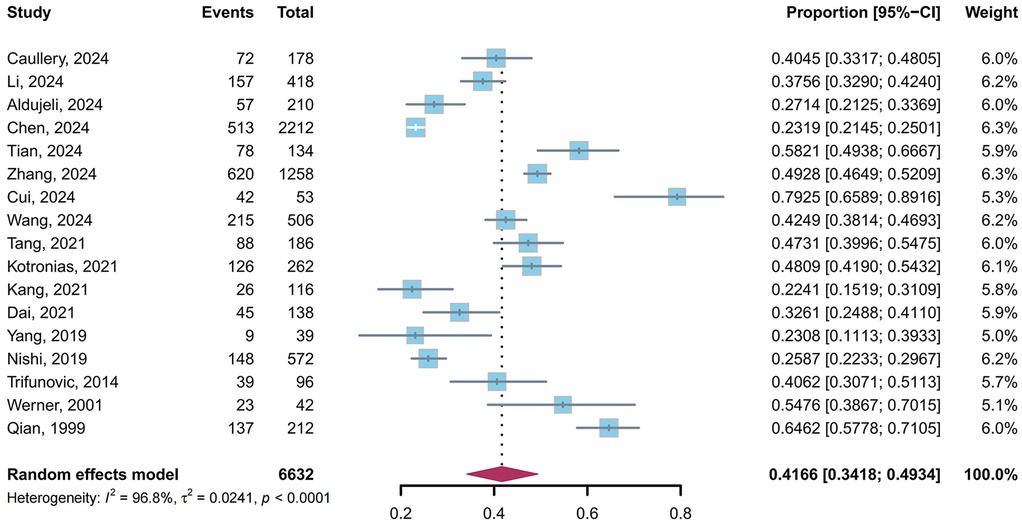
We performed a pooled analysis of 17 studies (5, 9, 10, 23, 24, 26–36, 39) comprising 6,632 patients. The results of the random-effects model (I2 = 96.8%) showed that 41.66% (95% CI: 34.18%–49.34%) of post-PCI patients exhibited CMD in target vessels (Figure 2).
3.4.2 Sensitivity analysis and publication bias
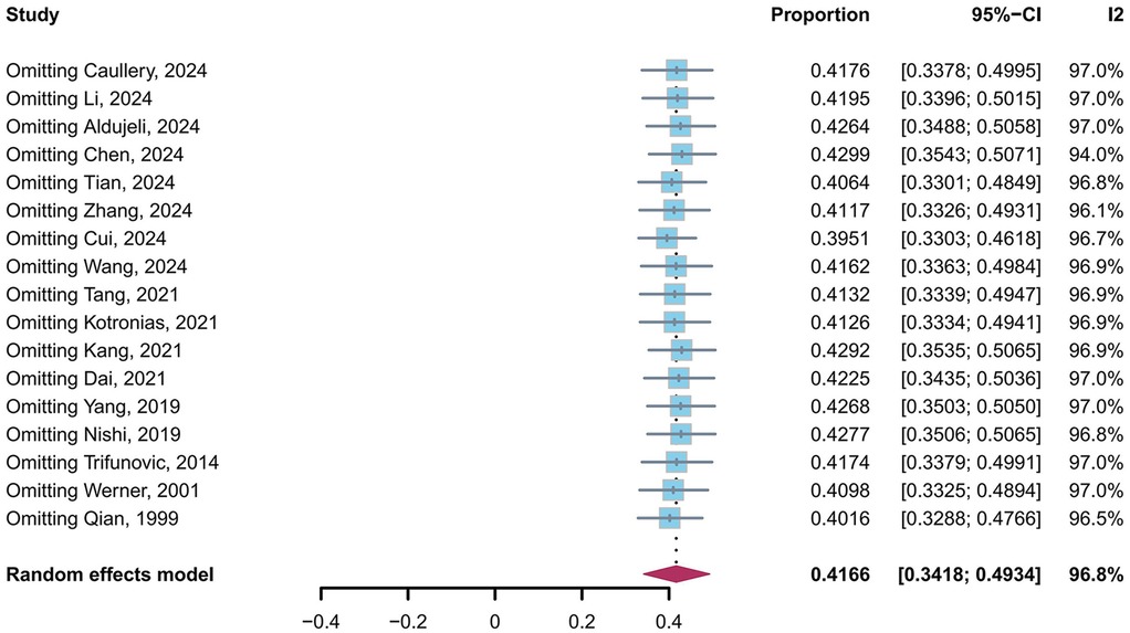
A leave-one-out sensitivity analysis confirmed the robustness of the pooled prevalence estimate (Figure 3). The prevalence of CMD in post-PCI target vessels remained stable, ranging from 39.51% (95% CI: 33.03%–46.18%) to 42.99% (95% CI: 35.43%–50.71%), with no single study altering the estimate beyond this range. Despite substantial heterogeneity (I2 = 96.8%, τ2 = 0.0241, P < 0.0001), the consistency across sensitivity analyses supports that approximately 40% of post-PCI patients exhibit CMD in target vessels. Egger's tests were performed for the prevalence of CMD in post-PCI target vessels, and the results revealed that no publication bias was observed (t = 1.76, p = 0.10).
3.4.3 Subgroup analysis
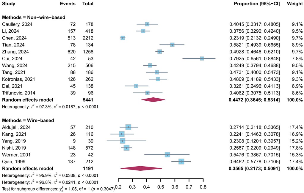
A total of 11 studies (5, 9, 23, 26–29, 31, 34, 35, 39) utilized non-wire-based methods, and 6 studies (10, 24, 30, 32, 33, 36) employed wire-based methods to evaluate the prevalence of CMD in post-PCI target vessels. Our meta-analysis showed that the pooled prevalence of CMD was 44.72% (95% CI: 36.45%–53.14%, I2 = 97.3%) in non-wire-based methods and 35.65% (95% CI: 21.73%–50.91%, I2 = 95.9%) in wire-based methods, with no statistically significant difference between subgroups (P = 0.30) (Figure 4).
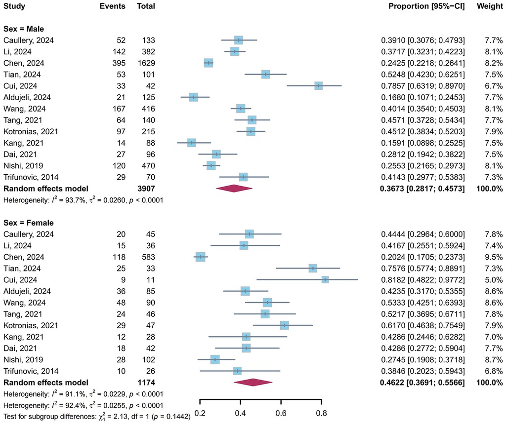
A total of 13 studies (4, 5, 9, 10, 23, 24, 26–29, 31, 34, 39) reported the prevalence of CMD in post-PCI target vessels stratified by sex. Our meta-analysis showed that the pooled prevalence of CMD was 36.73% (95% CI: 28.17%–45.73%, I2 = 93.7%) in males and 46.22% (95% CI: 36.91%–55.66%, I2 = 91.1%) in females, with no statistically significant difference between subgroups (P = 0.14) (Figure 5).
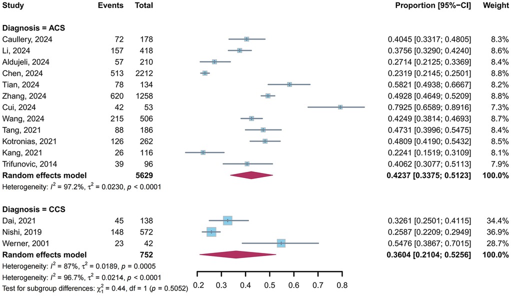
A total of 15 studies (5, 9, 10, 23, 24, 26–29, 31, 32, 34–36, 39) reported the prevalence of CMD in post-PCI target vessels stratified by diagnosis: 12 studies (9, 23, 24, 26–29, 31, 34–36, 39) for ACS and 3 studies (5, 10, 32) for CCS. Our meta-analysis showed that the pooled prevalence of CMD in post-PCI target vessels was 42.37% (95% CI: 33.75%–51.23%, I2 = 97.2%) in ACS and 36.04% (95% CI: 21.04%–52.56%, I2 = 87%) in CCS, with no statistically significant difference between subgroups (P = 0.51) (Figure 6).
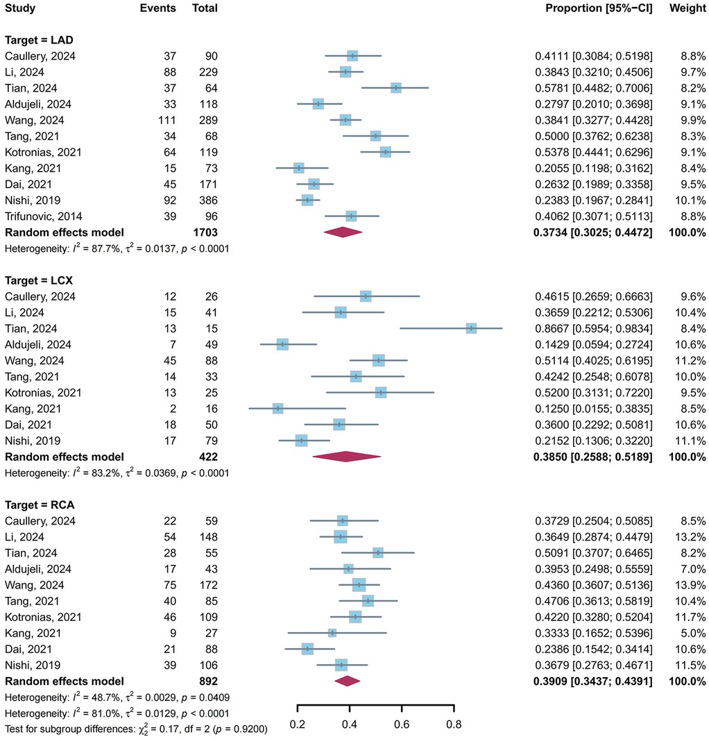
A total of 11 studies (4, 5, 10, 23, 24, 26, 28, 29, 31, 34, 39) reported the prevalence of CMD in post-PCI target vessels stratified by target vessels: 11 studies (4, 5, 10, 23, 24, 26, 28, 29, 31, 34, 39) for LAD, 10 studies (4, 5, 10, 23, 24, 26, 28, 29, 31, 34) for LCX and 10 studies (4, 5, 10, 23, 24, 26, 28, 29, 31, 34) for RCA. Our meta-analysis showed that the pooled prevalence of CMD in post-PCI target vessels was 37.34% (95% CI: 30.25%–44.72%, I2 = 87.7%) in LAD, 38.50% (95% CI: 25.88%–51.89%, I2 = 83.2%) in LCX, and 39.09% (95% CI: 34.37%–43.91%, I2 = 48.7%) in RCA, with no statistically significant difference between subgroups (P = 0.92) (Figure 7).
A total of 10 studies (4, 9, 23–27, 31, 34, 39) reported the prevalence of CMD in post-PCI target vessels stratified by post-PCI TIMI flow grade: 10 studies (4, 9, 23–27, 31, 34, 39) for TIMI flow grade 3 and 7 studies (4, 9, 23, 25, 31, 34, 39) for TIMI flow grade ≤2. Our meta-analysis demonstrated that the pooled prevalence of CMD was 37.26% (95% CI: 28.03%–46.98%, I2 = 95%) in TIMI flow grade 3 and 75.36% (95% CI: 54.38%–92.11%, I2 = 73%) in TIMI flow grade ≤2, with a statistically significant difference between subgroups (P = 0.0012) (Figure 8).
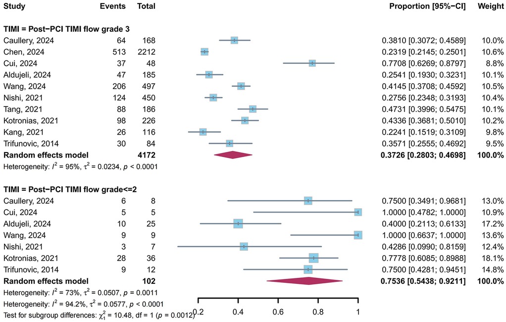
Figure 8. Prevalence of CMD in post-PCI target vessels: TIMI grade 3 vs. TIMI flow grade ≤2 subgroups.
3.4.4 The clinical outcomes in patients with CMD in post-PCI target vessels
A meta-analysis of multivariable-adjusted HRs was performed to evaluate the association between CMD in post-PCI target vessels and clinical outcomes (Figure 9). The pooled HR for major adverse cardiovascular events (MACE) was 3.10 (95% CI: 2.06–4.67, I2 = 67.9%) based on data from 7 studies (4, 5, 10, 23, 27, 31, 34). For the composite outcome of cardiac death or heart failure readmission, the pooled HR was 4.66 (95% CI: 3.13–6.93, I2 = 0%) derived from 3 studies (5, 27, 34). Table 2 summarized the specific components of MACE in pooled study.
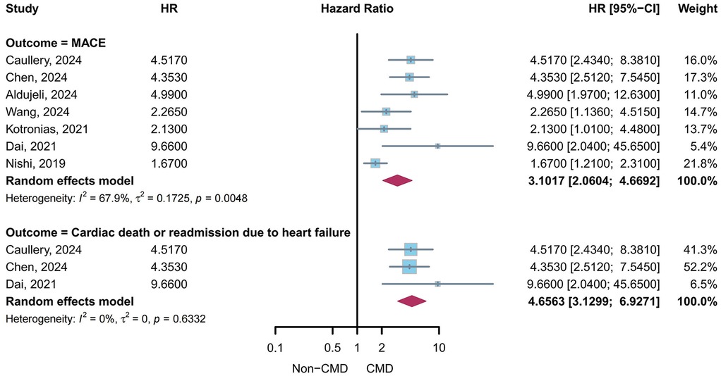
Figure 9. Pooled multivariable-adjusted HR for clinical outcomes in patients with CMD in post-PCI target vessels.
Outcomes unsuitable for meta-analysis due to insufficient data or methodological heterogeneity were described separately. Kang et al. (24) identified that high platelet-fibrin clot strength (P-FCS) (≥68 mm) significantly increased the risk of CMD [odds ratio (OR): 4.35, 95% CI: 1.74–10.89] post-PCI. Acute myocardial infarction (AMI) patients with both CMD and high P-FCS had a higher rate of MACE compared to non-CMD subjects with low P-FCS (OR: 5.58, 95% CI: 1.31–23.68). Chen et al. (27) demonstrated that the combination of diabetes mellitus (DM) and CMD in non-ST-elevation myocardial infarction (NSTEMI) patients was identified as the most powerful independent predictor of cardiac death or heart failure readmission at 2-year follow-up (adjusted HR: 7.894, 95% CI: 4.251–14.659, P < 0.001). Dai et al. (5) reported that in patients with CCS and CMD, the incidence of angina-related readmission was significantly higher during a median 28-month follow-up after the index procedure compared to CCS patients without CMD (46.7% vs. 19.4%, adjusted HR: 3.66, 95% CI: 1.68–7.97, P = 0.001). Aldujeli et al. (4) reported that in ST-elevation myocardial infarction (STEMI) patients with persistent CMD at 3 months post-PCI, those with CMD exhibited significantly poorer outcomes at 12-month follow-up compared to non-CMD counterparts. Specifically, CMD patients demonstrated markedly impaired recovery of left ventricular systolic function (−10.00% vs. +8.00%, P < 0.001), a higher prevalence of diastolic dysfunction (73.08% vs. 1.32%, P < 0.001), and elevated rates of adverse ventricular remodeling (11.32% vs. 7.28% and 22.64% vs. 1.99%, respectively, P < 0.001). Furthermore, IMR was independently associated with both left ventricular diastolic dysfunction and adverse remodeling (P < 0.001 for all associations).
4 Discussion
This systematic review and meta-analysis, encompassing 21 studies and 6,632 patients, consolidated the understanding that CMD in post-PCI target vessels is prevalent (41.66%, 95% CI: 34.18–49.34%) and independently associated with adverse clinical outcomes. Subgroup analyses revealed numerical variations in CMD prevalence across assessment methods, sex, clinical diagnoses, and target vessels, though intergroup differences did not reach statistical significance (all P > 0.05). Specifically, the pooled prevalence of CMD was numerically higher in females (46.22% vs. 36.73% in males), patients with ACS (42.37% vs. 36.04% in CCS), and those assessed via non-wire-based methods (44.72% vs. 35.65% in wire-based methods). CMD prevalence was comparable across target vessels (LAD: 37.34%, LCX: 38.50%, RCA: 39.09%), suggesting its nature irrespective of lesion location. Patients with suboptimal post-PCI TIMI flow (grade ≤2) exhibited a markedly higher prevalence of CMD (75.36%, 95% CI: 54.38%–92.11%) compared to those with TIMI grade 3 flow (37.26%, 95% CI: 28.03%–46.98%), with a statistically significant intergroup difference (P = 0.0012). Moreover, multivariable-adjusted meta-analysis demonstrated that CMD in post-PCI target vessels independently conferred a 3.10-fold increased risk of MACE (95% CI: 2.06–4.67) and a 4.66-fold risk of cardiac death or heart failure readmission (95% CI: 3.13–6.93), underscoring its prognostic significance.
Through technological advancements, CMD in target vessels after PCI has been substantiated as diagnosable via multiple coronary physiological assessments (15, 40). Among pressure wire-based methods, the index of microvascular resistance (IMR) and microvascular resistance reserve (MRR) specifically evaluate microvascular function independently of epicardial vessels. Conversely, assessments of CFR and coronary flow velocity reserve (CFVR) are confounded by epicardial blood flow and therefore must be conducted following PCI-mediated resolution of obstructive lesions in target vessels (41–43). Non-wire-based methods have garnered substantial attention owing to their procedural convenience and cost-effectiveness. Physiological coronary indices acquired through non-invasive techniques such as single photon emission computed tomography (SPECT) and transthoracic Doppler echocardiography (TDE) have demonstrated efficacy in delayed detection of post-PCI microvascular dysfunction in target vessels, yet remain inadequate for immediate intraprocedural identification of potential CMD (6). Importantly, despite potential influences from differing assumed boundary conditions (44), angiography-based microcirculation assessment techniques including angiography-derived microcirculatory resistance (AMR) have established diagnostic value for CMD. These techniques show strong correlation with IMR, the reference standard for specific coronary microvascular function evaluation (40), thereby enabling both immediate intraprocedural detection and subsequent management of CMD, along with epidemiological investigations leveraging historical coronary angiography data.
The marked heterogeneity in reported prevalence of post-PCI CMD (20%–80%) reflects not only differences in patient clinical characteristics but also methodological variability in diagnostic methods and study designs (6–10). Current coronary physiological assessments reveal that a substantial proportion of post-PCI CMD cases occur in vessels with angiographically normal flow, suggesting potential systematic underdiagnosis in studies relying on non-physiological assessments (13, 14). Furthermore, prior research often failed to distinguish between pre-existing non-target vessel CMD and post-PCI target vessel CMD, which likely conflatesdistinct pathophysiological entities (16–18). To address these limitations, investigations were exclusively included in our study if they utilized quantitative coronary physiological assessments to evaluate CMD in post-PCI target vessels, thereby minimizing diagnostic variability and confounding from non-target vessel pathology. This rigorous approach, when combined with subgroup analyses, provides novel insights into the true burden and clinical implications of CMD in post-PCI target vessels.
Subgroup analyses provided nuanced insights into the distribution and mechanisms of CMD across different clinical contexts. In the assessment method subgroup, although no statistical difference was observed between non-wire-based and wire-based methods in CMD prevalence (44.72% vs. 35.65%, P = 0.30), the numerical disparity may reflect differences in technical applicability and sensitivity (45). Non-wire-based methods, especially those relying on angiographic image analysis, offer procedural simplicity and are suitable for emergency PCI but may be confounded by vasospasm or collateral circulation (23, 45). In contrast, wire-based methods directly measure coronary physiological parameters with higher precision but require invasive equipment and complex operations (46). It is worth emphasizing that most non-wire-based methods utilize angiography-derived microcirculatory assessment techniques, which have been validated to exhibit diagnostic accuracy comparable to the traditional wire-based gold-standard IMR measurements (40). These non-wire-based methods provide advantages in operational simplicity and enable dynamic assessment during PCI, potentially enhancing real-time detection of subtle microvascular dysfunction. Conversely, wire-based methods, while precise, are technically complex and costly, with practical limitations in routine clinical use, which may contribute to underdiagnosis of CMD in real-world settings (11).
In the sex subgroup analysis, the prevalence of CMD in post-PCI target vessels was numerically higher in females than in males (46.22% vs. 36.73%, P = 0.14). Although this difference did not reach statistical significance, the trend aligns with prior studies reporting sex-specific disparities in CMD (47, 48). The mechanisms underlying these differences remain incompletely understood, though evidence suggests that hormonal fluctuations during peri- and post-menopausal transitions may contribute to coronary endothelial dysfunction and abnormal vasomotor regulation (49, 50). Notably, prior studies demonstrated that myocardial ischemia in males predominantly originated from obstructive epicardial coronary disease, whereas in females it more frequently stemmed from non-obstructive coronary disease (51, 52). This resulted in female patients undergoing coronary angiography requiring PCI less frequently than their male counterparts. Consequently, pre-existing CMD in target vessels might be detected less often in females after PCI, potentially obscuring sex-based differences and contributing to underestimation of the overall prevalence.
The prevalence of CMD was numerically higher in ACS patients compared to CCS patients (42.37% vs. 36.04%, P = 0.51), consistent with studies linking ACS to microvascular dysfunction (11). This discrepancy may arise from ACS-specific mechanisms, including thrombotic lesion instability, ischemia-reperfusion injury, and lipid-rich plaque composition (11, 53). Intracoronary imaging studies have demonstrated that lipid-rich plaque morphology is independently associated with post-PCI microvascular dysfunction (11, 12). Mechanistically, lipid-core embolization during stent deployment may trigger distal microvascular obstruction and activate pro-inflammatory pathways, exacerbating endothelial injury and perpetuating microvascular dysfunction (6, 49). It should be noted that diagnostic criteria for CMD remain controversial across clinical subtypes of CAD. Based on the association with adverse long-term outcomes in prior studies, higher diagnostic thresholds than those used under general circumstances tend to be selected for immediate post-PCI diagnosis of STEMI-related CMD (54–56). This situation was prevalent in our included studies, potentially reducing the pooled prevalence in the ACS subgroup to some extent and indirectly leading to blurring of differences in post-PCI target-vessel CMD prevalence between ACS and CCS subgroups.
Notably, CMD prevalence exhibited consistency across target vessels (LAD: 37.34%, LCX: 38.50%, RCA: 39.09%, P = 0.92). Although the LAD supplies a larger myocardial territory and is associated with a well-developed coronary microcirculation (57, 58), the uniform distribution of CMD suggests that microvascular dysfunction post-PCI might occur independently of lesion location or vessel-specific anatomical characteristics.
Patients with TIMI flow grade ≤2 had a significantly higher CMD prevalence than those with grade 3 (75.36% vs. 37.26%, P = 0.0012), highlighting the clinical relevance of TIMI flow grade. This observation aligns with the pathophysiological mechanisms whereby suboptimal TIMI flow may reflect impaired myocardial perfusion secondary to microvascular obstruction or reperfusion injury (14, 59). Importantly, such perfusion deficits are strongly associated with adverse ventricular remodeling and progression to heart failure, thereby providing a mechanistic explanation for their established correlation with poor clinical outcomes (60).
Our meta-analysis suggested that CMD in post-PCI target vessels is an independent predictor of adverse clinical outcomes. The pooled multivariable-adjusted hazard ratios revealed a 3.10-fold increased risk of MACE and a 4.66-fold risk of the composite outcome of cardiac death or heart failure readmission in patients with CMD. These findings suggest that microvascular dysfunction post-PCI is a critical determinant of long-term prognosis. The heightened risk may stem from persistent myocardial ischemia, adverse ventricular remodeling, and endothelial dysfunction, which collectively exacerbate cardiovascular vulnerability even after successful revascularization of epicardial arteries. During the peri-PCI period, severe post-PCI CMD may cause significant impairment of microcirculatory perfusion in target vessels through mechanisms including mechanical obstruction induced by distal embolization of the epicardial coronary artery, external compression due to tissue edema, localized in situ thrombosis, vascular spasm, leukocyte stagnation, activation of inflammatory cascades, and reperfusion injury (61, 62). This may promote peri-procedural myocardial injury and even type 4a myocardial infarction, leading to further increases in cardiovascular adverse events (56, 63). Notably, studies such as Chen et al. (27) highlighted the synergistic effect of CMD and DM, amplifying the risk of cardiac death or heart failure readmission by nearly 8-fold. This underscores the importance of integrating CMD assessment into risk stratification paradigms.
Our study has several limitations. First, significant heterogeneity (I2 > 90%) existed across studies due to differences in diagnostic methods and study design, which may limit the generalizability of pooled estimates. Non-uniformity in detection details and timing was likely a primary source of heterogeneity, aligned with the inherent variability among current diagnostic modalities. Discrepancies in CMD diagnostic standards across clinical subtypes of CAD also contributed partially to heterogeneity, though this requires further research for clarification. Additionally, while the use of vasodilation induced by papaverine and continuous thermodilution might enhance the stability of microvascular measurements, the absence of these methods in the included studies could have increased heterogeneity (64–66). Second, the exclusive inclusion of observational studies utilizing quantitative coronary physiological assessments with prespecified diagnostic criteria, while enhancing diagnostic consistency, might have introduced selection bias by excluding populations where such evaluations were unavailable or clinically unfeasible. Third, despite comprehensive coverage of diagnostic subtypes of CAD, studies focusing on non-ST-elevation acute coronary syndrome (NSTE-ACS) remained underrepresented, potentially limiting insights into related subtypes. Fourth, all included studies explicitly reporting elective PCI were limited to patients with CCS, which may not fully reflect the broader clinical significance of elective PCI in other populations. Additionally, emergency PCI studies predominantly included cases of primary PCI for STEMI, which is not yet sufficient to further differentiate between primary PCI and rescue PCI post-thrombolysis. Consequently, our analysis did not provide subgroup results stratified by PCI type, which may limit the applicability of findings to specific clinical scenarios. Fifth, although we excluded CMD in non-target vessels, the data proved insufficient to distinguish between de novo post-PCI CMD and pre-existing CMD in target vessels because many studies lacked systematic pre-PCI CMD assessment. Sixth, the absence of standardized follow-up protocols and inconsistent reporting of MACE components complicated the interpretation of results. To scientifically and reliably assess the impact of post-PCI target vessel CMD on clinical outcomes while minimizing confounding effects from other factors, our study exclusively included and pooled HRs with 95% CIs that underwent multivariate adjustment. Given that most incorporated studies adjusted solely for MACE, this resulted in isolated outcomes that failed to meet the predefined pooling criteria of this study. Furthermore, even with multivariable adjustments, unmeasured confounders may persist, potentially influencing the observed associations between CMD and clinical outcomes. Finally, regarding outcomes, we did not analyze the impact of epicardial vascular functional parameters such as fractional flow reserve (FFR) on clinical outcomes. Given that our study primarily focused on the independent effect of post-PCI target vessel CMD on clinical outcomes, and considering that the direct association between epicardial vascular functional parameters and CMD remains unclear, we excluded this analysis from the present investigation.
5 Conclusion
This systematic review and meta-analysis demonstrated that CMD in post-PCI target vessels is prevalent (approximately 40%) and independently associated with a elevated risk of adverse cardiovascular outcomes, especially MACE and the composite outcome of cardiac death or heart failure readmission. Future efforts should focus on standardizing diagnostic criteria and developing tailored interventions to improve outcomes in this population.
Data availability statement
The raw data supporting the conclusions of this article will be made available by the authors, without undue reservation.
Author contributions
YL: Software, Methodology, Writing – original draft, Investigation, Supervision, Data curation, Visualization, Resources, Validation, Formal analysis, Writing – review & editing, Project administration, Conceptualization. JD: Conceptualization, Writing – original draft, Writing – review & editing, Software, Investigation, Methodology, Data curation. HL: Formal analysis, Methodology, Conceptualization, Writing – original draft, Funding acquisition, Writing – review & editing, Resources. SL: Writing – original draft, Methodology, Software, Data curation, Investigation. HX: Methodology, Data curation, Writing – original draft, Software. XL: Funding acquisition, Resources, Formal analysis, Validation, Data curation, Conceptualization, Writing – original draft, Writing – review & editing, Investigation, Methodology.
Funding
The author(s) declare that financial support was received for the research and/or publication of this article. This work was supported by Grants from the National Natural Science Foundation of China (No. 82104767, No. 82174315); Beijing major and difficult diseases collaborative research project of traditional Chinese and Western medicine (No. 2023BJSZDYNJBXTGG-011); Beijing “The Fourteenth Five-Year” key specialty of Traditional Chinese Medicine Cardiovascular Department (demonstration class) (BJZKLC0011); Research and Translational Application of Clinical Specialized Diagnosis and Treatment Techniques in the Capital (No. Z221100007422127); Scientific Research Program of Hebei Provincial Administration of Traditional Chinese Medicine (No. B2025055); and Training Fund for Open Projects at Clinical Institutes and Departments of Capital Medical University (No. CCMU2024ZKYXY012); Young Talent Lifting Project of China Association of Chinese Medicine (No. CACM-2024-QNRC2-B20); Beijing University of Chinese Medicine XinShiDai 125 Program Talent Support Initiative; Capital Health Development Research Special Project (No. 2022-1-2231); Medical Aid Program for Tibet Grant from Natural Science Foundation of Tibet Autonomous Region (No. XZZR202402037[W]); Major Science and Technology Project of Lhasa (No. LSKJ202521).
Acknowledgements
The authors sincerely thank the support of Beijing University of Chinese Medicine and Beijing Hospital of Traditional Chinese Medicine.
Conflict of interest
JD was employed by company Air China Limited.
The remaining authors declare that the research was conducted in the absence of any commercial or financial relationships that could be construed as a potential conflict of interest.
Generative AI statement
The author(s) declare that no Generative AI was used in the creation of this manuscript.
Publisher's note
All claims expressed in this article are solely those of the authors and do not necessarily represent those of their affiliated organizations, or those of the publisher, the editors and the reviewers. Any product that may be evaluated in this article, or claim that may be made by its manufacturer, is not guaranteed or endorsed by the publisher.
Supplementary material
The Supplementary Material for this article can be found online at: https://www.frontiersin.org/articles/10.3389/fcvm.2025.1620204/full#supplementary-material
References
1. Canu M, Khouri C, Marliere S, Vautrin E, Piliero N, Ormezzano O, et al. Prognostic significance of severe coronary microvascular dysfunction post-PCI in patients with STEMI: a systematic review and meta-analysis. PLoS One. (2022) 17:e0268330. doi: 10.1371/journal.pone.0268330
2. Pries AR, Reglin B. Coronary microcirculatory pathophysiology: can we afford it to remain a black box? Eur Heart J. (2017) 38:478–88. doi: 10.1093/eurheartj/ehv760
3. Gdowski MA, Murthy VL, Doering M, Monroy-Gonzalez AG, Slart R, Brown DL. Association of isolated coronary microvascular dysfunction with mortality and major adverse cardiac events: a systematic review and meta-analysis of aggregate data. J Am Heart Assoc. (2020) 9:e014954. doi: 10.1161/JAHA.119.014954
4. Aldujeli A, Tsai T, Haq A, Tatarunas V, Knokneris A, Briedis K, et al. Impact of coronary microvascular dysfunction on functional left ventricular remodeling and diastolic dysfunction. J Am Heart Assoc. (2024) 13:e033596. doi: 10.1161/JAHA.123.033596
5. Dai N, Che W, Liu L, Zhang W, Yin G, Xu B, et al. Diagnostic value of angiography-derived IMR for coronary microcirculation and its prognostic implication after PCI. Front Cardiovasc Med. (2021) 8:735743. doi: 10.3389/fcvm.2021.735743
6. Chen W, Ni M, Huang H, Cong H, Fu X, Gao W, et al. Chinese expert consensus on the diagnosis and treatment of coronary microvascular diseases (2023 edition). MedComm. (2023) 4:e438. doi: 10.1002/mco2.438
7. Kunadian V, Chieffo A, Camici PG, Berry C, Escaned J, Maas AHEM, et al. An EAPCI expert consensus document on ischaemia with non-obstructive coronary arteries in collaboration with European society of cardiology working group on coronary pathophysiology & microcirculation endorsed by coronary vasomotor disorders international study group. Eur Heart J. (2020) 41:3504–20. doi: 10.1093/eurheartj/ehaa503
8. Vrints C, Andreotti F, Koskinas KC, Rossello X, Adamo M, Banning AJ, et al. 2024 ESC guidelines for the management of chronic coronary syndromes: developed by the task force for the management of chronic coronary syndromes of the European Society of Cardiology (ESC) endorsed by the European Association for Cardio-Thoracic Surgery (EACTS). Eur Heart J. (2024) 45(36):3415–537. doi: 10.1093/eurheartj/ehae177
9. Cui L, Wang Y, Chen W, Huang P, Tang Z, Wang J, et al. Coronary microvascular dysfunction and myocardial area at risk assessed by cadmium zinc telluride single photon emission computed tomography after primary percutaneous coronary intervention in acute myocardial infarction patients. Quant Imaging Med Surg. (2024) 14:3816–27. doi: 10.21037/qims-23-1260
10. Nishi T, Murai T, Ciccarelli G, Shah SV, Kobayashi Y, Derimay F, et al. Prognostic value of coronary microvascular function measured immediately after percutaneous coronary intervention in stable coronary artery disease: an international multicenter study. Circ Cardiovasc Interv. (2019) 12:e007889. doi: 10.1161/CIRCINTERVENTIONS.119.007889
11. Xie J, He Y, Ji H, Hu Q, Chen S, Gao B, et al. Impact of plaque characteristics on percutaneous coronary intervention-related microvascular dysfunction: insights from angiographic microvascular resistance and intravascular ultrasound. Quant Imaging Med Surg. (2023) 13:6037–47. doi: 10.21037/qims-23-414
12. Kozuki A, Shinke T, Otake H, Shite J, Matsumoto D, Kawamori H, et al. Feasibility of a novel radiofrequency signal analysis for in vivo plaque characterization in humans: comparison of plaque components between patients with and without acute coronary syndrome. Int J Cardiol. (2013) 167:1591–6. doi: 10.1016/j.ijcard.2012.04.102
13. Ong P, Camici PG, Beltrame JF, Crea F, Shimokawa H, Sechtem U, et al. Coronary vasomotion disorders international study group (COVADIS). international standardization of diagnostic criteria for microvascular angina. Int J Cardiol. (2018) 250:16–20. doi: 10.1016/j.ijcard.2017.08.068
14. Taqueti VR, Di Carli MF. Coronary microvascular disease pathogenic mechanisms and therapeutic options: JACC state-of-the-art review. J Am Coll Cardiol. (2018) 72:2625–41. doi: 10.1016/j.jacc.2018.09.042
15. Koo B-K, Lee JM, Hwang D, Park S, Shiono Y, Yonetsu T, et al. Practical application of coronary physiologic assessment. JACC Asia. (2023) 3:689–706. doi: 10.1016/j.jacasi.2023.07.003
16. Nogami K, Kanaji Y, Usui E, Hada M, Nagamine T, Ueno H, et al. Prognostic value of endogenous-type coronary microvascular dysfunction after elective percutaneous coronary intervention. Circ J Off J Jpn Circ Soc. (2025) 89(3):292–302. doi: 10.1253/circj.CJ-24-0482
17. Aldujeli A, Patel R, Grabauskyte I, Hamadeh A, Lieponyte A, Tatarunas V, et al. The impact of trimethylamine N-oxide and coronary microcirculatory dysfunction on outcomes following ST-elevation myocardial infarction. J Cardiovasc Dev Dis. (2023) 10:197. doi: 10.3390/jcdd10050197
18. Chao H, Jun-Qing G, Hong Z, Zhen Q, Hui Z, Wen A, et al. Prognostic value of coronary microvascular dysfunction assessed by coronary angiography-derived Index of microcirculatory resistance in patients with ST-segment elevation myocardial infarction. Clin Cardiol. (2024) 47:e24318. doi: 10.1002/clc.24318
19. Page MJ, McKenzie JE, Bossuyt PM, Boutron I, Hoffmann TC, Mulrow CD, et al. The PRISMA 2020 statement: an updated guideline for reporting systematic reviews. Br Med J. (2021) 372:n71. doi: 10.1136/bmj.n71
20. Munn Z, Moola S, Lisy K, Riitano D, Tufanaru C. Methodological guidance for systematic reviews of observational epidemiological studies reporting prevalence and cumulative incidence data. JBI Evid Implement. (2015) 13:147. doi: 10.1097/XEB.0000000000000054
21. Xu H, Lu T, Liu Y, Yang J, Ren S, Han B, et al. Prevalence and risk factors for long COVID among cancer patients: a systematic review and meta-analysis. Front Oncol. (2024) 14:1506366. doi: 10.3389/fonc.2024.1506366
22. Shim SR, Kim S-J. Intervention meta-analysis: application and practice using R software. Epidemiol Health. (2019) 41:e2019008. doi: 10.4178/epih.e2019008
23. Wang H, Wu Q, Yang L, Chen L, Liu W-Z, Guo J, et al. Application of AMR in evaluating microvascular dysfunction after ST-elevation myocardial infarction. Clin Cardiol. (2024) 47:e24196. doi: 10.1002/clc.24196
24. Kang MG, Koo B-K, Tantry US, Kim K, Ahn J-H, Park HW, et al. Association between thrombogenicity indices and coronary microvascular dysfunction in patients with acute myocardial infarction. JACC Basic Transl sci. (2021) 6:749–61. doi: 10.1016/j.jacbts.2021.08.007
25. Nishi T, Murai T, Waseda K, Hirohata A, Yong ASC, Ng MKC, et al. Association of microvascular dysfunction with clinical outcomes in patients with non-flow limiting fractional flow reserve after percutaneous coronary intervention. Int J Cardiol Heart Vasc. (2021) 35:100833. doi: 10.1016/j.ijcha.2021.100833
26. Tang J, Chu J, Hou H, Lai Y, Tu S, Chen F, et al. Clinical implication of QFR in patients with ST-segment elevation myocardial infarction after drug-eluting stent implantation. Int J Cardiovasc Imaging. (2021) 37:755–66. doi: 10.1007/s10554-020-02068-0
27. Chen D, Zhang Y, Yidilisi A, Hu D, Zheng Y, Fang J, et al. Combined risk estimates of diabetes and coronary angiography-derived index of microcirculatory resistance in patients with non-ST elevation myocardial infarction. Cardiovasc Diabetol. (2024) 23:300. doi: 10.1186/s12933-024-02400-1
28. Li J, Zhao W, Tian Z, Hu Y, Xiang J, Cui M. Correlation between coronary microvascular dysfunction and cardiorespiratory fitness in patients with ST-segment elevation myocardial infarction. Sci Rep. (2024) 14:26564. doi: 10.1038/s41598-024-74948-7
29. Tian R, Wang Z, Zhang S, Wang X, Zhang Y, Yuan J, et al. Growth differentiation factor-15 as a biomarker of coronary microvascular dysfunction in ST-segment elevation myocardial infarction. Heliyon. (2024) 10:e35476. doi: 10.1016/j.heliyon.2024.e35476
30. Yang HM, Yoon MH, Lim HS, Seo KW, Choi BJ, Choi SY, et al. Lipid-core plaque assessed by near-infrared spectroscopy and procedure related microvascular injury. Korean Circ J. (2019) 49:1010–8. doi: 10.4070/kcj.2019.0072
31. Kotronias RA, Terentes-Printzios D, Shanmuganathan M, Marin F, Scarsini R, Bradley-Watson J, et al. Long-term clinical outcomes in patients with an acute ST-segment-elevation myocardial infarction stratified by angiography-derived index of microcirculatory resistance. Front Cardiovasc Med. (2021) 8:717114. doi: 10.3389/fcvm.2021.717114
32. Werner GS, Ferrari M, Richartz BM, Gastmann O, Figulla HR. Microvascular dysfunction in chronic total coronary occlusions. Circulation. (2001) 104:1129–34. doi: 10.1161/hc3401.095098
33. Qian J, Ge J, Baumgart D, Sack S, Haude M, Erbel R. Prevalence of microvascular disease in patients with significant coronary artery disease. Herz. (1999) 24:548–57. doi: 10.1007/BF03044227
34. Caullery B, Riou L, Marliere S, Vautrin E, Piliero N, Ormerzzano O, et al. Prognostic impact of coronary microvascular dysfunction in patients with myocardial infarction evaluated by new angiography-derived index of microvascular resistance. Int J Cardiol Heart Vasc. (2025) 56:101575. doi: 10.1016/j.ijcha.2024.101575
35. Zhang S, Lin Z, Yu B, Liu J, Jin J, Li G, et al. Smoking paradox in coronary function and structure of acute ST-segment elevation myocardial infarction patients treated with primary percutaneous coronary intervention. BMC Cardiovasc Disord. (2024) 24:427. doi: 10.1186/s12872-024-04093-6
36. Aldujeli A, Tsai T-Y, Haq A, Tatarunas V, Garg S, Hughes D, et al. The association between trimethylamine N-oxide levels and coronary microvascular dysfunction and prognosis in patients with ST-elevation myocardial infarction. Atherosclerosis. (2024) 398:118597. doi: 10.1016/j.atherosclerosis.2024.118597
37. Tsai T-Y, Aldujeli A, Haq A, Knokneris A, Briedis K, Hughes D, et al. The impact of microvascular resistance reserve on the outcome of patients with STEMI. JACC Cardiovasc Interv. (2024) 17:1214–27. doi: 10.1016/j.jcin.2024.03.024
38. Aldujeli A, Haq A, Tsai T-Y, Grabauskyte I, Tatarunas V, Briedis K, et al. The impact of primary percutaneous coronary intervention strategies during ST-elevation myocardial infarction on the prevalence of coronary microvascular dysfunction. Sci Rep. (2023) 13:20094. doi: 10.1038/s41598-023-47343-x
39. Trifunovic D, Stankovic S, Marinkovic J, Beleslin B, Banovic M, Djukanovic N, et al. Time-dependent changes of plasma adiponectin concentration in relation to coronary microcirculatory function in patients with acute myocardial infarction treated by primary percutaneous coronary intervention. J Cardiol. (2015) 65:208–15. doi: 10.1016/j.jjcc.2014.05.011
40. Wang D, Li X, Feng W, Zhou H, Peng W, Wang X. Diagnostic and prognostic value of angiography-derived index of microvascular resistance: a systematic review and meta-analysis. Front Cardiovasc Med. (2024) 11:1360648. doi: 10.3389/fcvm.2024.1360648
41. De Bruyne B, Pijls NHJ, Gallinoro E, Candreva A, Fournier S, Keulards DCJ, et al. Microvascular resistance reserve for assessment of coronary microvascular function: JACC technology corner. J Am Coll Cardiol. (2021) 78:1541–9. doi: 10.1016/j.jacc.2021.08.017
42. Mahendiran T, Bertolone D, Viscusi MM, Gallinoro E, Keulards DCJ, Collet C, et al. The influence of epicardial resistance on microvascular resistance reserve. J Am Coll Cardiol. (2024) 84:512–21. doi: 10.1016/j.jacc.2024.05.004
43. Denby KJ, Zmaili M, Datta S, Das T, Ellis S, Ziada K, et al. Developments and controversies in invasive diagnosis of coronary microvascular dysfunction in angina with nonobstructive coronary arteries. Mayo Clin Proc. (2024) 99:1469–81. doi: 10.1016/j.mayocp.2024.04.022
44. Lodi Rizzini M, Candreva A, Chiastra C, Gallinoro E, Calò K, D’Ascenzo F, et al. Modelling coronary flows: impact of differently measured inflow boundary conditions on vessel-specific computational hemodynamic profiles. Comput Methods Programs Biomed. (2022) 221:106882. doi: 10.1016/j.cmpb.2022.106882
45. Fan Y, Fezzi S, Sun P, Ding N, Li X, Hu X, et al. In vivo validation of a novel computational approach to assess microcirculatory resistance based on a single angiographic view. J Pers Med. (2022) 12:1798. doi: 10.3390/jpm12111798
46. Fearon WF, Kobayashi Y. Invasive assessment of the coronary microvasculature: the index of microcirculatory resistance. Circ Cardiovasc Interv. (2017) 10:e005361. doi: 10.1161/CIRCINTERVENTIONS.117.005361
47. Mileva N, Nagumo S, Mizukami T, Sonck J, Berry C, Gallinoro E, et al. Prevalence of coronary microvascular disease and coronary vasospasm in patients with nonobstructive coronary artery disease: systematic review and meta-analysis. J Am Heart Assoc. (2022) 11:e023207. doi: 10.1161/JAHA.121.023207
48. Shimokawa H, Suda A, Takahashi J, Berry C, Camici PG, Crea F, et al. Clinical characteristics and prognosis of patients with microvascular angina: an international and prospective cohort study by the coronary vasomotor disorders international study (COVADIS) group. Eur Heart J. (2021) 42:4592–600. doi: 10.1093/eurheartj/ehab282
49. Li H, Gao Y, Lin Y. Progress in molecular mechanisms of coronary microvascular dysfunction. Microcirculation. (2023) 30:e12827. doi: 10.1111/micc.12827
50. Taylor DJ, Aubiniere-Robb L, Gosling R, Newman T, Hose DR, Halliday I, et al. Sex differences in coronary microvascular resistance measured by a computational fluid dynamics model. Front Cardiovasc Med. (2023) 10:1159160. doi: 10.3389/fcvm.2023.1159160
51. Millett ERC, Peters SAE, Woodward M. Sex differences in risk factors for myocardial infarction: cohort study of UK biobank participants. BMJ (Clin Res Ed). (2018) 363:k4247. doi: 10.1136/bmj.k4247
52. Canton L, Fedele D, Bergamaschi L, Foà A, Di Iuorio O, Tattilo FP, et al. Sex- and age-related differences in outcomes of patients with acute myocardial infarction: MINOCA vs. MIOCA. Eur Heart J Acute Cardiovasc Care. (2023) 12:604–14. doi: 10.1093/ehjacc/zuad059
53. Lerman A, Holmes DR, Herrmann J, Gersh BJ. Microcirculatory dysfunction in ST-elevation myocardial infarction: cause, consequence, or both? Eur Heart J. (2007) 28:788–97. doi: 10.1093/eurheartj/ehl501
54. Maznyczka AM, Oldroyd KG, Greenwood JP, McCartney PJ, Cotton J, Lindsay M, et al. Comparative significance of invasive measures of microvascular injury in acute myocardial infarction. Circ Cardiovasc Interv. (2020) 13:e008505. doi: 10.1161/CIRCINTERVENTIONS.119.008505
55. Konijnenberg LSF, Damman P, Duncker DJ, Kloner RA, Nijveldt R, van Geuns R-JM, et al. Pathophysiology and diagnosis of coronary microvascular dysfunction in ST-elevation myocardial infarction. Cardiovasc Res. (2020) 116:787–805. doi: 10.1093/cvr/cvz301
56. Fearon WF, Dash R. Index of microcirculatory resistance and infarct size. JACC Cardiovasc Imaging. (2019) 12:849–51. doi: 10.1016/j.jcmg.2018.04.004
57. Xing W-L, Wu Y-J, Liu H-X, Liu Q-R, Qi Z, Li A-Y, et al. Effects of danhong injection (丹红注射液) on peri-procedural myocardial injury and microcirculatory resistance in patients with unstable angina undergoing elective percutaneous coronary intervention:a pilot randomized study. Chin J Integr Med. (2021) 27:846–53. doi: 10.1007/s11655-021-2872-1
58. Donato P, Coelho P, Santos C, Bernardes A, Caseiro-Alves F. Correspondence between left ventricular 17 myocardial segments and coronary anatomy obtained by multi-detector computed tomography: an ex vivo contribution. Surg Radiol Anat: SRA. (2012) 34:805–10. doi: 10.1007/s00276-012-0976-1
59. Khan JN, Razvi N, Nazir SA, Singh A, Masca NG, Gershlick AH, et al. Prevalence and extent of infarct and microvascular obstruction following different reperfusion therapies in ST-elevation myocardial infarction. J Cardiovasc Magn Reson. (2014) 16:38. doi: 10.1186/1532-429X-16-38
60. Morishima I, Sone T, Okumura K, Tsuboi H, Kondo J, Mukawa H, et al. Angiographic no-reflow phenomenon as a predictor of adverse long-term outcome in patients treated with percutaneous transluminal coronary angioplasty for first acute myocardial infarction. J Am Coll Cardiol. (2000) 36:1202–9. doi: 10.1016/S0735-1097(00)00865-2
61. Reffelmann T, Kloner RA. The no-reflow phenomenon: a basic mechanism of myocardial ischemia and reperfusion. Basic Res Cardiol. (2006) 101:359–72. doi: 10.1007/s00395-006-0615-2
62. Jaffe R, Dick A, Strauss BH. Prevention and treatment of microvascular obstruction-related myocardial injury and coronary No-reflow following percutaneous coronary intervention: a systematic approach. JACC Cardiovasc Interventions. (2010) 3:695–704. doi: 10.1016/j.jcin.2010.05.004
63. Armillotta M, Bergamaschi L, Paolisso P, Belmonte M, Angeli F, Sansonetti A, et al. Prognostic relevance of type 4a myocardial infarction and periprocedural myocardial injury in patients with non-ST-segment-elevation myocardial infarction. Circulation. (2025) 151:760–72. doi: 10.1161/CIRCULATIONAHA.124.070729
64. Belmonte M, Gallinoro E, Pijls NHJ, Bertolone DT, Keulards DCJ, Viscusi MM, et al. Measuring absolute coronary flow and microvascular resistance by thermodilution: JACC review topic of the week. J Am Coll Cardiol. (2024) 83:699–709. doi: 10.1016/j.jacc.2023.12.014
65. Kodeboina M, Nagumo S, Munhoz D, Sonck J, Mileva N, Gallinoro E, et al. Simplified assessment of the index of microvascular resistance. J Intervent Cardiol. (2021) 2021:9971874. doi: 10.1155/2021/9971874
Keywords: coronary microvascular dysfunction, percutaneous coronary intervention, prevalence, outcomes, meta-analysis
Citation: Li Y, Duan J, Liu H, Lin S, Xu H and Li X (2025) Coronary microvascular dysfunction in post-PCI target vessels: a systematic review and meta-analysis of prevalence and associated outcomes. Front. Cardiovasc. Med. 12:1620204. doi: 10.3389/fcvm.2025.1620204
Received: 20 May 2025; Accepted: 18 July 2025;
Published: 4 August 2025.
Edited by:
Emanuele Gallinoro, Cardiovascular Center, OLV Aalst, BelgiumReviewed by:
Matteo Armillotta, University of Bologna, ItalyMarta Belmonte, Cardiovascular Center, OLV Aalst, Belgium
Copyright: © 2025 Li, Duan, Liu, Lin, Xu and Li. This is an open-access article distributed under the terms of the Creative Commons Attribution License (CC BY). The use, distribution or reproduction in other forums is permitted, provided the original author(s) and the copyright owner(s) are credited and that the original publication in this journal is cited, in accordance with accepted academic practice. No use, distribution or reproduction is permitted which does not comply with these terms.
*Correspondence: Hongxu Liu, bGh4X0AyNjMubmV0; Xiang Li, bGl4aWFuZzExODk3QGJqemhvbmd5aS5jb20=
†These authors have contributed equally to this work
 Yunze Li
 Jinlan Duan3,†
 Hongkun Xu
 Xiang Li