- 1Department of Dermatology, Osaka City University Graduate School of Medicine, Osaka, Japan
- 2Kurume University School of Medicine, Kurume, Japan
- 3Kurume University Institute of Cutaneous Cell Biology, Kurume, Japan
- 4Department of Life Sciences, Graduate School of Arts and Sciences, University of Tokyo, Tokyo, Japan
- 5Department of Dermatology and Immunodermatology, Medical University of Warsaw, Warsaw, Poland
- 6Ueo Dermatology Clinic, Saiki, Japan
- 7Department of Dermatology, Faculty of Medicine, Oita University, Yufu, Japan
- 8Department of Dermatology, Kimitsu Chuo Hospital, Kimitsu, Japan
- 9Department of Dermatology, Osaka University Graduate School of Medicine, Suita, Japan
- 10University Hospital Centre Zagreb, Department of Dermatology and Venereology, School of Medicine, University of Zagreb, Zagreb, Croatia
- 11Pediatric Dermatology, University of Colorado School of Medicine, Children’s Hospital Colorado, Denver, CO, United States
- 12Department of Dermatology, St. Marriana Medical University, Kawasaki, Japan
- 13Department of Dermatology, Kurume University School of Medicine, Fukuoka, Japan
Several sporadic cases, in which direct and indirect immunofluorescence studies simultaneously detected IgG and IgA autoantibodies to keratinocyte cell surfaces, have been reported mainly under the name of IgG/IgA pemphigus. However, there have been no systematic studies for this condition. In this study, we collected 30 cases of this condition from our cohort of more than 5,000 autoimmune bullous disease cases, which were consulted for our diagnostic methods from other institutes, and summarized their clinical and immunological findings. Clinically, there was no male–female prevalence, mean age of disease onset was 55.6 years, and mean duration before this condition was suspected was 18 months. The patients showed clinically bullous and pustular skin lesions preferentially on the trunk and extremities, and histopathologically intraepidermal pustules and blisters with infiltration of neutrophils and eosinophils. Immunologically, ELISAs frequently detected IgG and IgA autoantibodies to both desmogleins and desmocollins. From the characteristic clinical, histopathological, and immunological features, which are considerably different from those in classical IgG types of pemphigus, we propose this disease as a new disease entity with preferential name of intercellular IgG/IgA dermatosis (IGAD). This was the largest study of IGAD to date.
Introduction
Autoimmune bullous disease (AIBD) is divided into pemphigus group with autoantibodies to keratinocyte cell surfaces (CSs) and pemphigoid group with autoantibodies to epidermal basement membrane zone (BMZ) (1, 2). Two representative classical IgG types of pemphigus are pemphigus vulgaris (PV) and pemphigus foliaceus (PF), which react with desmoglein 3 (Dsg3) and Dsg1, respectively, although there are many other forms of pemphigus (3, 4). Among them, cases with anti-CS antibodies exclusively of IgA class had been called as IgA pemphigus (5–8), for which we proposed intercellular IgA dermatosis (IAD) as a preferable name (5, 7, 8).
IgG/IgA pemphigus is the name given to an atypical form of pemphigus characterized by in vivo bound and/or circulating anti-keratinocyte CS antibodies of both IgG and IgA classes (1). The results in approximately 20 reports indicated that IgG/IgA pemphigus is an atypical form of pemphigus with heterogeneous clinical and histopathological features (9–30). However, because this condition is extremely rare, there is no systematic study and disease entity of this condition has not been established. At Kurume University, we have examined more than 5,000 cases of various AIBDs, which were consulted at other institutes for our diagnostic studies (1, 31). Therefore, in this study, we attempted to determine the characteristic clinical, histopathological, and immunological features of all patients with both IgG and IgA anti-keratinocyte CS antibodies as the first step to establish this disease entity.
In this retrospective study, we selected 30 cases with IgG and IgA anti-CS antibodies from our AIBD cohort, and characterized them clinically, histopathologically, and immunologically. Considerably distinct features found in these cases indicated this condition as a new disease entity, and we propose the term “intercellular IgG/IgA dermatosis (IGAD)” to this disorder, following the designation of IAD.
Materials and Methods
This study was performed following Declaration of Helsinki and guidelines of local ethics committees of Kurume University School of Medicine. Informed consents were provided by all patients and normal individuals.
Cases and Sera
In this study, we used our AIBD cohort of 5,402 cases. Information and sera for these cases were sent to us from other institutes in either Japan or other countries between 1996 and 2015. Information of clinical and histopathological findings and direct immunofluorescence (IF) was obtained from consultation letters.
Various Immunological Methods
IF Studies
Direct IF for IgG, IgA, IgM, and C3 using skin biopsies was performed mainly at other institutes. Indirect IF studies of normal human skin and monkey esophagus for both IgG and IgA antibodies were performed by standard method. In cases with autoimmune reactivity with epidermal BMZ, indirect IF of 1 M NaCl-split normal human skin for IgG and IgA antibodies were also performed (32).
Immunoblotting Studies
Immunoblotting of normal human epidermal extract was performed as described previously (33, 34). In cases with reactivity with BMZ, we also performed IB analyses using BP180 NC16a domain recombinant protein (RP) (35), BP180 C-terminal domain RP (36), concentrated culture supernatant of HaCaT cells (37), normal human dermal extract (38), and purified human laminin-332 (39) for both IgG and/or IgA antibodies.
ELISA Studies
Commercially available IgG ELISAs of Dsg1 and Dsg3 (cutoff: <index 12) (MESACUP, MBL, Nagoya, Japan) (40) were conducted according to the manufacturer’s instruction. Using the same ELISA kits, IgA antibodies to Dsg1 and Dsg3 (cutoff: <OD 0.15) were also examined (41). In addition, ELISAs of mammalian RPs of human desmocollin 1 (Dsc1)-Dsc3 were performed for both IgG antibodies (42) and IgA antibodies (43). Cutoff OD values were 0.2 for Dsc1, 0.07 for Dsc2, and 0.12 for Dsc3 for IgG antibodies, and 0.123 for Dsc1, 0.048 for Dsc2, and 0.074 for Dsc3 for IgA antibodies. Furthermore, in cases with reactivity with BMZ, we performed IgG ELISAs of BP180 NC16a domain RP (cutoff: <index 15) (44), BP230 RPs (cutoff values: <index 9) (45), and type VII collagen RP (MBL) (46). OD at 490 nm was measured by ELISA reader.
COS-7 Cell cDNA Transfection Method
COS-7 cell cDNA transfection method using cDNAs of human Dsc1–Dsc3 was performed as described previously (47).
Statistical Analyses
We statistically analyzed correlations between the results in Dsg and Dsc ELISAs and clinical parameters. Differences among qualitative results were compared using the chi-square test. Differences among quantitative parameters between groups were assessed using the Mann–Whitney U test. p Values less than 0.05 were considered significant.
Results
Diagnoses
In this study, we suspected the diagnosis of IGAD for 30 cases, which showed simultaneous IgG and IgA immunoreactivity with keratinocyte CS antigens in direct IF and various serological studies (whole clinical and immunological data are shown in Tables S1 and S2 in Supplementary Material, respectively). In addition, four cases showed IgG and/or IgA anti-BMZ immunoreactivity by IF and/or molecular studies.
Clinical diagnosis in the consultation letters for the 30 cases included IGAD (12 cases), IAD (9 cases), and other AIBDs (7 cases), while no clinical diagnosis was given in 2 cases (Table S3A in Supplementary Material).
Regarding the diagnoses after the characterization, 26 IGAD cases had sole IGAD, while bullous pemphigoid, linear IgA bullous dermatosis, and linear IgA/IgG bullous dermatosis were concomitant in 1, 1, and 2 cases, respectively (Table S3B in Supplementary Material). In addition, one case each was suggested to be named as IgG/IgA PF and IgG/IgA PV.
Because of the large cohort size and long surveillance time, most of the clinical, histopathological, and immunological results were not obtained from all cases. Therefore, we summarize only available results for each parameter in the following sections.
Detailed Reports for Six New IGAD Cases
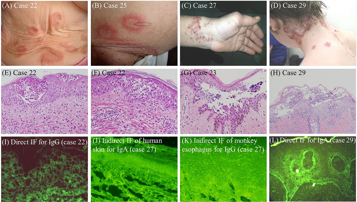
Clinical and histopathological findings and disease courses for six patients are described in Supplementary data (cases 17, 22, 23, 25, 27, and 29 in Tables S1 and S2 in Supplementary Material), as well as corresponding figures (Figures S1–S6 in Supplementary Material). Clinical, histopathological, and IF features in representative IGAD cases are also depicted in Figure 1.

Figure 1. Clinical (A–D), histopathological (E–H), and IF (I–L) features in representative IGAD cases examined in this study. (A) Vesicles were seen on the peripheries of annular erythemas on the abdomen (case 22). (B) Annular erythemas with superficial pustules and desquamation were seen (case 25). (C) Erythematous skin lesions with pustules and crusts on the left wrist were seen (case 27). (D) Pustular skin lesions on the peripheries of annular erythemas were seen (case 29). (E,F) Eosinophilic pustules in the upper epidermis (E) and eosinophilic spongiosis in the middle epidermis (F) were seen (case 22). (G) Acantholytic blisters and pustules in the middle epidermis were seen (case 23). (H) Subcorneal pustules with predominant neutrophils and a few eosinophils were seen. (I) The result of direct IF for IgG (case 22). (J) The result of indirect IF of normal human skin for IgA antibodies (case 27). (K) The results of indirect IF of monkey esophagus for IgG antibodies (case 27). (L) The result of direct IF for C3 (case 29).
Clinical Findings in 30 IGAD Cases
Backgrounds of the Cases
The 30 IGAD cases included 18 Japanese, 6 European, 3 US, 2 Indian, and 1 Australian. There were 15 males and 13 females, with 2 cases without information of gender. Mean ages of 30 cases were 55.6 years (53.4 years for males and 57.8 years for females). The durations between the disease onset and the consultation to us for 21 cases were from 2 weeks to 10 years (mean 18 months).
Before the cases visited the practitioners, who consulted us, one each case was treated with systemic steroid, antihistamine, combination of systemic steroid and antihistamine, and minocycline. Administration of vancomycin was not reported.
Underlying diseases included six malignant tumors, i.e., two cases of lung cancer and one case each of uterus carcinoma, breast carcinoma, gallbladder carcinoma, and malignant lymphoma. In addition, one case each had diabetes mellitus, IgA nephropathy, myasthenia gravis, Sweet’s syndrome, Sjogren’s syndrome, and PV.
Clinical Manifestations
Involved sites available from consultation letters for the 30 IGAD cases were the whole body (2 cases), the trunk (19 cases), the extremities (17 cases), the intertriginous areas (4 cases), and the head/face (6 cases) (Figure 1; Table S1 in Supplementary Material). Mucosal lesions were found in oral mucosae (11 cases) and conjunctiva (2 cases), with 1 case each with genital, nasal, and esophageal mucosae.
Regarding clinical manifestations, 19 cases showed blister formation with vesicles (5 cases), tense bullae (2 cases) and flaccid bullae (3 cases), 10 cases showed pustule formation, 10 cases showed erosions, and 17 cases showed erythemas with annular erythemas (9 cases). Other clinical manifestations were crust formation (3 cases), pigmentation (2 cases), and vegetating lesions (1 case). Itch was complained by 6 patients.
Histopathological Features
Among 23 IGAD cases with histopathological information for skin biopsies, 10 cases showed intraepidermal blister formation at the upper epidermis or middle epidermis, while 12 cases showed intraepidermal pustules at the upper epidermis, middle or lower epidermis (Figure 1; Table S1 in Supplementary Material). Spongiosis and acantholysis were observed in three and four cases, respectively. Intraepidermal infiltrations of neutrophils, eosinophils, and lymphocytes were found in 11, 8, and 4 cases, respectively. Presence and absence of acantholysis were described in one and three cases, respectively. Two cases showed subepidermal blisters.
Treatments, Responsiveness, and Complications
Sole therapy of oral steroids, dapsone (DDS), minocycline, and combinations of these drugs and other treatments were performed as described in 13 cases (Tables S1 and S4 in Supplementary Material). The responsiveness to these therapies was variable (Table S4 in Supplementary Material).
Immunological Findings
Direct IF
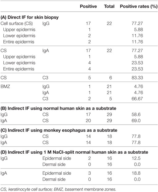
The results of direct IF were described in the consulting letters for 22 patients. IgG, IgA, and C3 depositions to keratinocyte CSs were positive in 17, 17, and 5 cases, respectively (Table 1A). IgG deposition was seen at upper epidermis in one case, lower epidermis in two cases, and entire epidermis in two cases. IgA deposition was seen at upper epidermis in one cases, lower epidermis in four cases, and entire epidermis in four cases. IgG, IgA, and C3 depositions to epidermal BMZ were observed in one, one, and two cases, respectively.
Indirect IF
In indirect IF of normal human skin performed for 29 IGAD cases, IgG and IgA anti-keratinocyte CS antibodies were positive in 17 and 20 cases, respectively (Figure S3 in Supplementary Material) (Table 1B–D). In addition, two and one cases showed IgG and IgA anti-BMZ antibodies, respectively. Indirect IF of monkey esophagus performed for 18 cases detected anti-epithelial CS antibodies of both IgG and IgA classes in 14 cases. In addition, two cases each showed IgG and IgA anti-BMZ antibodies. In indirect IF of 1 M NaCl-split human skin performed for 16 cases, IgG and IgA reactivity with epidermal side was found in two and three cases, respectively.
IB Studies of Normal Human Epidermal Extract and Other Substrates for BMZ Autoantigens
In immunoblotting of normal human epidermal extract performed for all 30 IGAD cases, few cases showed positive reactivity with various epidermal autoantigens for both IgG and IgA classes (Table S5 in Supplementary Material). For IgG antibodies, three cases each reacted with Dsg1 and Dsg3 and one case each reacted with both a and b forms of Dsc. One case each reacted with desmoplakin I, BP230, and envoplakin. For IgA antibodies, one case reacted with Dsg3 and two cases each reacted with both a and b forms of Dsc.
The four cases with positive reactivity with BMZ in IF studies were further examined by immunoblotting for the reactivity with various BMZ autoantigens. Two cases showed positive IgG, but not IgA, reactivity with BP180 NC16a domain RP, while none reacted with BP180 C-terminal domain RP for either IgG or IgA antibodies. One case was positive for IgG, but not IgA, for the 120-kDa LAD-1 in concentrated culture supernatant of HaCaT cells. No positive reactivity was found in immunoblotting of both purified human laminin-332 and normal human dermal extracts for either IgG or IgA antibodies.
ELISAs
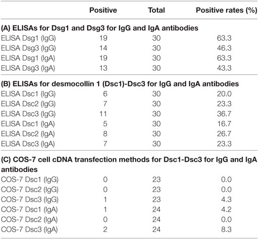
In commercially available IgG ELISAs for Dsg1 and Dsg3 for all 30 cases, 19 (63.6%) and 14 (46.7%) cases showed IgG reactivity with Dsg1 and Dsg3, respectively, and 19 (63.3%) and 13 (43.3%) cases showed IgA reactivity with Dsg1 and Dsg3, respectively (Table 2A). In ELISAs of mammalian RPs of Dsc1–Dsc3, 6 (20.0%), 7 (23.3%), and 11 (36.7%) cases showed IgG reactivity with Dsc1, Dsc2, and Dsc3, respectively, while 5 (16.7%), 8 (26.7%), and 7 (23.3%) cases showed IgA reactivity with Dsc1, Dsc2, and Dsc3, respectively (Table 2B).
Interestingly, there were very high association of detection of IgG and IgA antibodies to the same Dsgs, i.e., 18 (94.7%) of 19 cases with IgG antibodies to Dsg1 had IgA antibodies to Dsg1, 18 (94.7%) of 19 cases with IgA anti-Dsg1 antibodies had IgG anti-Dsg1 antibodies, 11 (78.6%) of 14 cases with IgG anti-Dsg3 antibodies had IgA anti-Dsg3 antibodies, and 11 (84.6%) of 13 cases with IgA anti-Dsg3 antibodies had IgA anti-Dsg3 antibodies. By contrast, rates of coexistence of IgG and IgA antibodies to Dsc1–Dsc3 were not very high, i.e., only 50.0% (3/6), 57.1% (4/7), and 54.5% (6/11) of cases with IgG antibodies to Dsc1, Dsc2, and Dsc3 had IgA antibodies to the same Dscs (Table S2 in Supplementary Material).
One of the four cases with positive reactivity with BMZ in IF studies was positive in IgG ELISA of BP180 NC16a RP (data not shown).
COS-7 Cell cDNA Transfection Methods
In COS-7 cell cDNA transfection method performed for 23 and 24 cases for IgG and IgA antibodies, respectively, one case showed IgG reactivity with Dsc3, and one and two cases showed IgA reactivity with Dsc1 and Dsc3, respectively (Table 2C).
Statistical Analyses Between the Dsg and Dsc ELISAs and Clinical Parameters
The case with positive reactivity with Dsg3 for both IgG (p = 0.0103) and IgA antibodies had oral mucosal lesions significantly more frequently. Female cases had both IgA anti-Dsg1 antibodies (p = 0.0351) and IgG anti-Dsc1 antibodies (p = 0.0409) more frequently (chi-square for independence test). No statistically significant correlations were found for any other clinical parameters, including ages, disease durations, sites and manifestations of skin lesions, and responses to various treatments, as well as histopathological features.
Discussion
In this study, although the diagnosis of IGAD was suspected by atypical clinico-histopathological features, we made possible diagnosis of IGAD for patients showing simultaneously IgG and IgA reactivity in various immunological examinations, including IF, immunoblotting, ELISA, and cDNA transfection studies. Particularly, ELISAs were sensitive and detected IgG and IgA antibodies to Dsgs and Dscs in considerable numbers of patients. Finally, using the tentative diagnostic criteria, we diagnosed 30 patients as IGAD.
Clinically, skin lesions developed mainly on the trunk and extremities, and 11 cases showed oral mucosal lesions. Most patients showed blisters, erosions, and/or pustules with erythematous lesions. In most cases, histopathology showed intraepidermal blister and/or pustules at various layers in the epidermis. The major infiltrating cells were neutrophils, followed by eosinophils and lymphocytes. Acantholysis was not clearly seen in some of the patients. Spongiosis were seen occasionally. These clinico-histopathological findings were generally compatible with those in previously reported cases (9–30). In 11 cases, various therapies, mainly oral steroids and DDS, were performed with different efficacy.
Immunologically, direct IF and indirect IF of normal human skin and monkey esophagus were the most sensitive methods with positive rates from 60 to 80%. Regarding antigen detection methods, IgG and IgA ELISAs for Dsgs and Dscs were very sensitive. The IgG and IgA ELISAs detected antibodies to Dsg1 and Dsg3 in 63 and 45% of the patients, respectively, and antibodies to Dsc1–Dsc3 in 20–40% of the patients. Because anti-Dsc antibodies are rarely detected in classical IgG type pemphigus diseases (42), the frequent detection of anti-Dsc antibodies for both IgG and IgA antibodies was considered the significant feature in IGAD. The presence of autoantibodies to other than Dsgs may account for the absence of acantholysis in some patients.
By contrast, sensitivity of immunoblotting of normal epidermal extract and cDNA transfection was very low.
Statistical analyses revealed that anti-Dsg3 antibodies of both IgG and IgA classes were more frequently detected in the cases with oral mucosal lesions. Because Dsg3 is autoantigens found in mucosal-type PV, the results further suggested the significant involvement of IgG anti-Dsg3 antibodies in oral involvement in IGAD. This result may also suggest the pathogenic role of IgA anti-Dsg3 antibodies in development of oral lesions. However, because of the high rates of simultaneous detection of IgG and IgA anti-Dsg3 antibodies in the same sera, the oral lesions might be produced by IgG, but not IgA, anti-Dsg3 antibodies.
Thus, IGAD patients tended to show clinico-histopathological features of both blisters and pustules. IgG-type pemphigus usually shows blisters, while IAD shows pustules. Therefore, IGAD seems to show mixed features of IgG-type pemphigus and IAD. Immunologically, IgG and IgA antibodies tended to react with both Dsgs and Dscs, which also indicates mixed immunological features of IgG-type pemphigus and IAD. These clinical, histopathological, and immunological features are different from either IgG-type pemphigus or IAD. Therefore, we concluded that IGAD is a distinct clinical entity with unique clinical and immunological features.
In this study, we performed no disease model experiments for the pathogenic role of IgG and IgA antibodies in IGAD. However, several IGAD cases showed clear acantholytic histology, suggesting that IgG antibodies to Dsg1 and/or Dsg3 induced the cell detachment similar to IgG-type pemphigus. In addition, because most IGAD cases showed extensive pustular lesions, IgA antibodies to either Dsgs or Dscs may produce the pustular lesions similar to IAD.
We should also consider the molecular mechanisms of simultaneous production of autoantibodies of IgG and IgA classes to various keratinocyte CS antigens, particularly class switch recombination (CSR) for antibody class switching (48, 49). CSR occurs through a genomic rearrangement within constant region locus of immunoglobulin heavy chain, where gene segments of all immunoglobulin classes are tandemly located downstream of the VDJ variable region locus (Figure S7 in Supplementary Material). The upstream classes are looped out through the CSR and the downstream class is docked into the VDJ region (Figure S7 in Supplementary Material). Thus, the class switching is irreversible and a class must be switched from left to right. Although a class has been considered to be switched from IgM/IgD-producing B-cells to IgG-, IgA-, and IgE-producing B-cells (50), a recent study of comprehensive antibody repertoire sequencing followed by lineage tracing revealed more variable class switch pathways, including pathway from IgG1 to IgA1 (Figure S7 in Supplementary Material) (51).
The extremely high rates of simultaneous detection of IgG and IgA anti-Dsg3 antibodies in the same sera found in this study may indicate that IgG-producing B-cells converted to IgA-producing B-cells. Although we could not determine the ancestral class, predominance of IgG1 class in human serum may suggest that the autoantibody switch from IgG1 to IgA1. By contrast, concurrence of IgG and IgA antibodies to the same Dscs was not very high, suggesting that different immunological IgG and IgA autoantibodies may develop by different molecular mechanisms between Dsgs and Dscs. In future studies, these CSR-related molecular mechanisms in IGAD should be examined more extensively using subclass-specific secondary antibodies or methods to detect the epitopes in more detail.
In conclusion, the present study was the first systematic study for IGAD, and suggested that IGAD is a distinct disease entity with characteristic clinical, histopathological, and immunological features. Combination methods of direct IF, indirect IF of normal human skin/monkey esophagus and ELISAs of Dsgs and Dscs are useful to make the diagnosis of IGAD.
Author Contributions
TH, DT, and NI designed the study. KW, DU, SF, KI, YK, IJ, BM, AB, and TK gave clinical and histopathological information of the patients. KT and NI performed the experiments. TH, DT, KT, and NI analyzed the data. TH and KH prepared the figures. TK performed the statistical analyses. TH, KT, KH, DT, and NI wrote the paper.
Conflict of Interest Statement
The authors declare that the research was conducted in the absence of any commercial or financial relationships that could be construed as a potential conflict of interest.
The reviewer MD declared a past co-authorship with one of the authors KW to the handling Editor.
Acknowledgments
We gratefully appreciate Ms. Tomoko Tashima and Ms. Shinobu Ide for secretarial work and Ms. Michiru Kubo and Ms. Kyoko Hiromatsu for conducting experiments. We thank all patients for their participation, all members at both Department of Dermatology, Kurume University School of Medicine, and Kurume University Institute of Cutaneous Cell Biology for long lasting efforts for this study, and all researchers and practitioners in other institutes for generously sending us sera and information of the patients.
Funding
This study was supported by Grants-in-Aid for Scientific Research (No. 20390308, 20591331, 21659271, 23591634, 23791298, 23791299, 23791300, 23791301, 24659534, 24591672, 24591640, 24791185), and Supported Program for the Strategic Research Foundation at Private Universities from the Ministry of Education, Culture, Sports, Science and Technology; and by “Research on Measures for Intractable Diseases” Project: matching fund subsidy to Takashi Hashimoto and Masayuki Amagai from the Ministry of Health, Labor and Welfare. The study was also supported by grants from Takeda Science Foundation.
Supplementary Material
The Supplementary Material for this article can be found online at https://www.frontiersin.org/articles/10.3389/fimmu.2018.00994/full#supplementary-material.
References
1. Hashimoto T, Tsuruta D, Koga H, Fukuda S, Ohyama B, Komai A, et al. Summary of results of serological tests and diagnoses for 4774 cases of various autoimmune bullous diseases consulted to Kurume University. Br J Dermatol (2016) 175:953–65. doi:10.1111/bjd.14692
2. Hashimoto T, Ishii N, Ohata C, Furumura M. Pathogenesis of epidermolysis bullosa acquisita, an autoimmune subepidermal bullous disease. J Pathol (2012) 228:1–7. doi:10.1002/path.4062
3. Hashimoto T. Treatment strategies for pemphigus vulgaris in Japan. Expert Opin Pharmacother (2008) 9:1519–30. doi:10.1517/14656566.9.9.1519
4. Tsuruta D, Ishii N, Hashimoto T. Diagnosis and treatment of pemphigus. Immunotherapy (2012) 4:735–45. doi:10.2217/imt.12.67
5. Nishikawa T, Shimizu H, Hashimoto T. Role of IgA intercellular antibodies: report of clinically and immunopathologically atypical cases. Proc XVII World Congr Dermatol (1987):383–4.
6. Hashimoto T. Immunopathology of IgA pemphigus. Clin Dermatol (2001) 19:683–9. doi:10.1016/S0738-081X(00)00193-0
7. Hashimoto T, Nishikawa T. Nomenclature for diseases with IgA antikeratinocyte cell surface autoantibodies. Br J Dermatol (2015) 173:868–9. doi:10.1111/bjd.13813
8. Hashimoto T, Teye K, Ishii N. Clinical and immunological studies of 49 cases of various types of intercellular IgA dermatosis and 13 cases of classical subcorneal pustular dermatosis examined at Kurume University. Br J Dermatol (2017) 176:168–75. doi:10.1111/bjd.14780
9. Chorzelski TP, Hashimoto T, Nishikawa T, Ebihara T, Dmochowski M, Ismail M, et al. Unusual acantholytic bullous dermatosis associated with neoplasia and IgG and IgA antibodies against bovine desmocollins I and II. J Am Acad Dermatol (1994) 31:351–5. doi:10.1016/S0190-9622(94)70171-7
10. Ohno H, Miyagawa S, Hashimoto T, Nakagawa A, Watanabe K, Nishikawa T, et al. Atypical pemphigus with concomitant IgG and IgA anti-intercellular autoantibodies associated with monoclonal IgA gammopathy. Dermatology (1994) 189(Suppl 1):115–6. doi:10.1159/000246948
11. Oiso N, Yamashita C, Yoshioka K, Amagai M, Komai A, Nagata Y, et al. IgG/IgA pemphigus with IgG and IgA antidesmoglein 1 antibodies detected by enzyme-linked immunosorbent assay. Br J Dermatol (2002) 147:1012–7. doi:10.1046/j.1365-2133.2002.04984.x
12. Kozlowska A, Hashimoto T, Jarzabek-Chorzelska M, Amagai A, Nagata Y, Strasz Z, et al. Pemphigus herpetiformis with IgA and IgG antibodies to desmoglein 1 and IgG antibodies to desmocollin 3. J Am Acad Dermatol (2003) 48:117–22. doi:10.1067/mjd.2003.23
13. Bruckner AL, Fitzpatrick JE, Hashimoto T, Weston WL, Morelli JG. Atypical IgA/IgG pemphigus involving the skin, oral mucosa, and colon in a child: a novel variant of IgA pemphigus? Pediatr Dermatol (2005) 22:321–7. doi:10.1111/j.1525-1470.2005.22408.x
14. Morizane S, Yamamoto T, Hisamatsu Y, Tsuji K, Oono T, Hashimoto T, et al. Pemphigus vegetans with IgG and IgA antidesmoglein 3 antibodies. Br J Dermatol (2005) 153:1236–7. doi:10.1111/j.1365-2133.2005.06956.x
15. Heng A, Nwaneshiudu A, Hashimoto T, Amagai M, Stanley JR. Intraepidermal neutrophilic IgA/IgG antidesmocollin 1 pemphigus. Br J Dermatol (2006) 154:1018–20. doi:10.1111/j.1365-2133.2006.07226.x
16. Kowalewski C, Hashimoto T, Amagai M, Jablonska S, Mackiewicz W, Wozniak K. IgA/IgG pemphigus: a new atypical subset of pemphigus? Acta Derm Venereol (2006) 86:357–8. doi:10.2340/00015555-0060
17. Maruyama H, Kawachi Y, Fujisawa Y, Itoh S, Furuta J, Ishii Y, et al. IgA/IgG pemphigus positive for anti-desmoglein 1 autoantibody. Eur J Dermatol (2007) 17:94–5. doi:10.1684/ejd.2007.0176
18. Nakajima K, Hashimoto T, Nakajima H, Yokogawa M, Ikeda M, Kodama H. IgG/IgA pemphigus with dyskeratotic acantholysis and intraepidermal neutrophilic microabscesses. J Dermatol (2007) 34:757–60. doi:10.1111/j.1346-8138.2007.00378.x
19. Mentink LF, de Jong MC, Kloosterhuis GJ, Zuiderveen J, Jonkman MF, Pas HH. Coexistence of IgA antibodies to desmogleins 1 and 3 in pemphigus vulgaris, pemphigus foliaceus and paraneoplastic pemphigus. Br J Dermatol (2007) 156:635–41. doi:10.1111/j.1365-2133.2006.07717.x
20. Tajima M, Mitsuhashi Y, Irisawa R, Amagai M, Hashimoto T, Tsuboi R. IgA pemphigus reacting exclusively to desmoglein 3. Eur J Dermatol (2010) 20:626–9. doi:10.1684/ejd.2010.1021
21. Santiago-et-Sanchez-Mateos D, Juarez Martin A, Gonzalez De Arriba A, Delgado Jimenez Y, Fraga J, Hashimoto T, et al. IgG/IgA pemphigus with IgA and IgG antidesmoglein 1 antibodies detected by enzyme-linked immunosorbent assay: presentation of two cases. J Eur Acad Dermatol Venereol (2011) 25:110–2. doi:10.1111/j.1468-3083.2010.03686.x
22. Hosoda S, Suzuki M, Komine M, Murata S, Hashimoto T, Ohtsuki M. A case of IgG/IgA pemphigus presenting malar rash-like erythema. Acta Derm Venereol (2012) 92:164–6. doi:10.2340/00015555-1258
23. Ueda A, Ishii N, Temporin K, Yamazaki R, Murakami F, Fukuda S, et al. IgA pemphigus with paraneoplastic pemphigus-like clinical features showing IgA antibodies to desmoglein 1/3 and desmocollin 3, and IgG and IgA antibodies to the basement membrane zone. Clin Exp Dermatol (2013) 38:370–3. doi:10.1111/ced.12050
24. Uchiyama R, Ishii N, Arakura F, Kiniwa Y, Nakazawa K, Uhara H, et al. IgA/IgG pemphigus with infiltration of neutrophils and eosinophils in an ulcerative colitis patient. Acta Derm Venereol (2014) 94:737–8. doi:10.2340/00015555-1836
25. Furuya A, Takahashi E, Ishii N, Hashimoto T, Satoh T. IgG/IgA pemphigus recognizing desmogleins 1 and 3 in a patient with Sjogren’s syndrome. Eur J Dermatol (2014) 24:512–3. doi:10.1684/ejd.2014.2391
26. Watkins C, West C, Kosari P, Ali S, Sangüeza O, Huang W. IgG/IgA pemphigus: report of a rare variant of atypical pemphigus and a review of the literature. J Dermatol Clin Res (2014) 2:1011.
27. Lane N, Parekh P. IgG/IgA pemphigus. Am J Dermatopathol (2014) 36:1002–4. doi:10.1097/DAD.0000000000000058
28. Cetkovska P, Komorousova M, Lomicova I. Management of a pemphigus with IgA and IgG antibodies and coexistent lung cancer. Dermatol Ther (2014) 27:236–9. doi:10.1111/dth.12126
29. Kanwar AJ, Vinay K, Saikia UN, Koga H, Teye K, Tsuruta D, et al. IgG/IgA pemphigus reactive with desmoglein 1 with additional undetermined reactivity with epidermal basement membrane zone. Indian J Dermatol Venereol Leprol (2014) 80:46–50. doi:10.4103/0378-6323.125499
30. Inoue-Nishimoto T, Hanafusa T, Hirohata A, Mabuchi-Kiyohara E, Mizoguchi N, Matsumoto K, et al. IgG/IgA pemphigus representing pemphigus vegetans caused by low titres of IgG and IgA antibodies to desmoglein 3 and IgA antibodies to desmocollin 3. J Eur Acad Dermatol Venereol (2016) 30:1229–31. doi:10.1111/jdv.13158
31. Ohzono A, Sogame R, Li X, Teye K, Tsuchisaka A, Numata S, et al. Clinical and immunological findings in 104 cases of paraneoplastic pemphigus. Br J Dermatol (2015) 173:1447–52. doi:10.1111/bjd.14162
32. Woodley DT. Immunofluorescence on salt-split skin for the diagnosis of epidermolysis bullosa acquisita. Arch Dermatol (1990) 126:229–31. doi:10.1001/archderm.126.2.229
33. Sugi T, Hashimoto T, Hibi T, Nishikawa T. Production of human monoclonal anti-basement membrane zone (BMZ) antibodies from a patient with bullous pemphigoid (BP) by Epstein-Barr virus transformation. Analyses of the heterogeneity of anti-BMZ antibodies in BP sera using them. J Clin Invest (1989) 84:1050–5. doi:10.1172/JCI114266
34. Hashimoto T, Ogawa MM, Konohana A, Nishikawa T. Detection of pemphigus vulgaris and pemphigus foliaceus antigens by immunoblot analysis using different antigen sources. J Invest Dermatol (1990) 94:327–31. doi:10.1111/1523-1747.ep12874456
35. Matsumura K, Amagai M, Nishikawa T, Hashimoto T. The majority of bullous pemphigoid and herpes gestationis serum samples react with the NC16a domain of the 180-kDa bullous pemphigoid antigen. Arch Dermatol Res (1996) 288:507–9. doi:10.1007/BF02505245
36. Nie Z, Hashimoto T. IgA antibodies of cicatricial pemphigoid sera specifically react with C-terminus of BP180. J Invest Dermatol (1999) 112:254–5. doi:10.1046/j.1523-1747.1999.00501.x
37. Ishii N, Ohyama B, Yamaguchi Z, Hashimoto T. IgA autoantibodies against the NC16a domain of BP180 but not 120-kDa LAD-1 detected in a patient with linear IgA disease. Br J Dermatol (2008) 158:1151–3. doi:10.1111/j.1365-2133.2008.08492.x
38. Ishii N, Yoshida M, Hisamatsu Y, Ishida-Yamamoto A, Nakane H, Iizuka H, et al. Epidermolysis bullosa acquisita sera react with distinct epitopes on the NC1 and NC2 domains of type VII collagen: study using immunoblotting of domain-specific recombinant proteins and postembedding immunoelectron microscopy. Br J Dermatol (2004) 150:843–51. doi:10.1111/j.1365-2133.2004.05933.x
39. Hisamatsu Y, Nishiyama T, Amano S, Matsui C, Ghohestani R, Hashimoto T. Usefulness of immunoblotting using purified laminin 5 in the diagnosis of anti-laminin 5 cicatricial pemphigoid. J Dermatol Sci (2003) 33:113–9. doi:10.1016/S0923-1811(03)00158-0
40. Ishii K, Amagai M, Hall RP, Hashimoto T, Takayanagi A, Gamou S, et al. Characterization of autoantibodies in pemphigus using antigen-specific enzyme-linked immunosorbent assays with baculovirus-expressed recombinant desmogleins. J Immunol (1997) 159:2010–7.
41. Hashimoto T, Komai A, Futei Y, Nishikawa T, Amagai M. Detection of IgA autoantibodies to desmogleins by an enzyme-linked immunosorbent assay: the presence of new minor subtypes of IgA pemphigus. Arch Dermatol (2001) 137:735–8. doi:10-1001/pubs.Arch Dermatol
42. Ishii N, Teye K, Fukuda S, Uehara R, Hachiya T, Koga H, et al. Anti-desmocollin autoantibodies in nonclassical pemphigus. Br J Dermatol (2015) 173:59–68. doi:10.1111/bjd.13711
43. Teye K, Numata S, Ohzono A, Ohyama B, Tsuchisaka A, Koga H, et al. Establishment of IgA ELISAs of mammalian recombinant proteins of human desmocollins 1-3. J Dermatol Sci (2016) 83:75–7. doi:10.1016/j.jdermsci.2016.04.001
44. Kobayashi M, Amagai M, Kuroda-Kinoshita K, Hashimoto T, Shirakata Y, Hashimoto K, et al. BP180 ELISA using bacterial recombinant NC16a protein as a diagnostic and monitoring tool for bullous pemphigoid. J Dermatol Sci (2002) 30:224–32. doi:10.1016/S0923-1811(02)00109-3
45. Yoshida M, Hamada T, Amagai M, Hashimoto K, Uehara R, Yamaguchi K, et al. Enzyme-linked immunosorbent assay using bacterial recombinant proteins of human BP230 as a diagnostic tool for bullous pemphigoid. J Dermatol Sci (2006) 41:21–30. doi:10.1016/j.jdermsci.2005.11.002
46. Saleh MA, Ishii K, Kim YJ, Murakami A, Ishii N, Hashimoto T, et al. Development of NC1 and NC2 domains of type VII collagen ELISA for the diagnosis and analysis of the time course of epidermolysis bullosa acquisita patients. J Dermatol Sci (2011) 62:169–75. doi:10.1016/j.jdermsci.2011.03.003
47. Hashimoto T, Kiyokawa C, Mori O, Miyasato M, Chidgey MA, Garrod DR, et al. Human desmocollin 1 (Dsc1) is an autoantigen for the subcorneal pustular dermatosis type of IgA pemphigus. J Invest Dermatol (1997) 109:127–31. doi:10.1111/1523-1747.ep12319025
48. Muramatsu M, Kinoshita K, Fagarasan S, Yamada S, Shinkai Y, Honjo T. Class switch recombination and hypermutation require activation-induced cytidine deaminase (AID), a potential RNA editing enzyme. Cell (2000) 102:553–63. doi:10.1016/S0092-8674(00)00078-7
49. Stavnezer J, Guikema JE, Schrader CE. Mechanism and regulation of class switch recombination. Annu Rev Immunol (2008) 26:261–92. doi:10.1146/annurev.immunol.26.021607.090248
50. Tangye SG, Hodgkin PD. Divide and conquer: the importance of cell division in regulating B-cell responses. Immunology (2004) 112:509–20. doi:10.1111/j.1365-2567.2004.01950.x
Keywords: autoimmune bullous diseases, desmocollin, desmoglein, ELISA, intercellular IgG/IgA dermatosis
Citation: Hashimoto T, Teye K, Hashimoto K, Wozniak K, Ueo D, Fujiwara S, Inafuku K, Kotobuki Y, Jukic IL, Marinović B, Bruckner A, Tsuruta D, Kawakami T and Ishii N (2018) Clinical and Immunological Study of 30 Cases With Both IgG and IgA Anti-Keratinocyte Cell Surface Autoantibodies Toward the Definition of Intercellular IgG/IgA Dermatosis. Front. Immunol. 9:994. doi: 10.3389/fimmu.2018.00994
Received: 13 December 2017; Accepted: 20 April 2018;
Published: 07 May 2018
Edited by:
Falk Nimmerjahn, Friedrich-Alexander-Universität Erlangen-Nürnberg, GermanyReviewed by:
Frank Antonicelli, Université de Reims Champagne Ardenne, FranceMarian Dmochowski, Poznan University of Medical Sciences, Poland
Copyright: © 2018 Hashimoto, Teye, Hashimoto, Wozniak, Ueo, Fujiwara, Inafuku, Kotobuki, Jukic, Marinović, Bruckner, Tsuruta, Kawakami and Ishii. This is an open-access article distributed under the terms of the Creative Commons Attribution License (CC BY). The use, distribution or reproduction in other forums is permitted, provided the original author(s) and the copyright owner are credited and that the original publication in this journal is cited, in accordance with accepted academic practice. No use, distribution or reproduction is permitted which does not comply with these terms.
*Correspondence: Takashi Hashimoto, aGFzaHl0QGdtYWlsLmNvbQ==
 Takashi Hashimoto
 Kwesi Teye3
 Koji Hashimoto
 Daisuke Ueo
 Kazuhiro Inafuku
 Ines Lakos Jukic
 Branka Marinović
 Anna Bruckner
 Daisuke Tsuruta
