- 1Division of Medical Oncology, Ente Ospedaliero (E.O.), Ospedali Galliera, Genoa, Italy
- 2Dipartimento di Medicina Sperimentale (DIMES), Università degli Studi di Genova, Genova, Italy
- 3Division of Pathology, Ente Ospedaliero (E.O.) Ospedali Galliera, Genoa, Italy
- 4Centre for Cancer Prevention, Wolfson Institute of Preventive Medicine, Barts and the London School of Medicine and Dentistry, Queen Mary University of London, London, United Kingdom
- 5IRCCS Ospedale Policlinico San Martino, Genova, Italy
Despite recent advances in ovarian cancer (OC) treatment, including the introduction of bevacizumab and PARP-inhibitors, OC remains a lethal disease. Other therapeutic options are being explored, such as immunotherapy (IT), which has been proved effective in many solid tumors. Findings about tumor-infiltrating cytotoxic and regulatory T cells, together with the expression of PD-1 on immune cells and of PD-L1 on tumor cells, gave the rationale for an attempt to the use of IT also in OC. We treated two patients with avelumab, an anti-PD-L1 monoclonal antibody, after the first line of chemotherapy: Patient A underwent 19 cycles of maintenance therapy with avelumab with a disease-free interval of 12 months, whereas patient B showed a slight progression of disease after only eight cycles. A higher PD-L1 expression in tumor cells of patient A was detected. She also underwent a genomic assessment that described the presence of a high Tumor Mutational Burden (TMB) and a status of Loss of Heterozygosity (LoH). This different response to the same treatment puts in evidence that some genomic and immune features might be investigated.
Introduction
Ovarian cancer (OC) is the third most common but the first most lethal gynecologic malignancy all over the world, as it represents the fifth cause of death by cancer in women, with 21,750 new cases and 13,940 deaths estimated in the USA in 2020 (1).
The treatment of OC has always consisted in the combination of surgery and platinum-based chemotherapy (2). The therapeutic innovations in the past decade consisted in the introduction of bevacizumab (3) and, only recently, in the advent of PARP inhibitors (PARPis) (4).
However, despite the availability of these new therapeutical options, the prognosis for women affected by advanced OC is still poor. Therefore, other strategies, such as targeting specific molecules on cancer cells or harnessing the host’s immune system, need to be explored more carefully.
Immunotherapy (IT) based on the stimulation of the endogenous immune response against tumor cells is the last frontier in cancer treatments and it is now widely used in many solid tumors, with results so satisfactory that the natural history of some types of cancers, such as non-small cell lung carcinoma (NSCLC) and melanoma, has dramatically changed. The most common type of IT consists in the use of monoclonal antibodies, which can be directed against immunosuppressive receptors (ICI, immune checkpoint inhibitors), expressed not only by activated T cells but also by Natural Killer cells, such as PD-1 (5) (6), NKG2A (7), and CTLA4 (8) or against their ligands, such as PD-L1 (9), expressed by tumor and immune cells.
Based on this evidence, IT has now been included in the standard of care for several malignancies. The possibility of considering IT as a viable option also for OC is based on the finding that the presence of tumor-infiltrating lymphocytes is correlated with better survival, whereas the presence of regulatory T cells is a negative prognostic factor (10–12). In addition, the existence of several escape mechanisms exploited by OC cells to prevent T and NK cell–mediated attack strongly suggests a critical role for these adaptive and innate cells in OC immunosurveillance (13–17).
In addition, patients with the BRCA 1/2 mutation showed high expression of PD-1 on immune cells and of PD-L1 on tumor cells (18). Taken together, all this evidence has motivated the exploration of a possible application of IT also to OC. Several clinical trials exploring various immune-based strategies have been conducted for this purpose; single agent therapies including ICI, vaccines, monoclonal antibodies, adoptive cell therapy have shown modest effects (19, 20), but their combination in a synergistic treatment, directed toward either tumor cells or the immune microenvironment could lead to better clinical response, so further explorations are needed (21, 22).
Many phases II and III trials investigating IT in OC are still ongoing, mainly addressing recurrent disease and exploring various possible combination of IT with not only the standard of care, for example, ICI with chemotherapy and/or anti-angiogenic agents and/or PARPis, but also the association of different immune approaches (23).
Here, we report our experience with the use of IT in OC, reporting the cases of two patients who showed an almost opposite response to treatment, despite an almost overlapping therapeutic path. We will furthermore describe some histological, genetic, and molecular characteristics that diversify these patients in an attempt to identify potential predictive and prognostic factors of the response to IT.
Case description
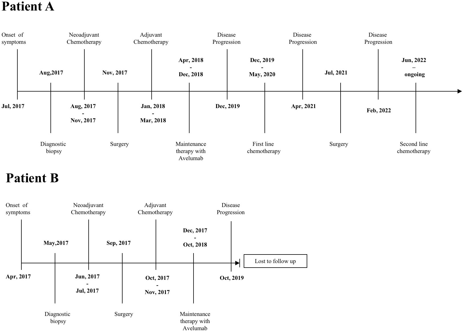
Patient A was a 71-year-old woman with a history of ocular glaucoma, whereas Patient B was a 75-year-old woman in good health; neither patient had oncological familiarity or gene mutations already known at the time of diagnosis. Details of the patient’s clinical course are outlined in Figure 1.
On 19 July 2017, Patient A presented with dyspnea and abdominal pain. A total body CT scan showed ascites, bilateral pleural effusion, and peritoneal nodules with inhomogeneous ovaries suggestive of cancer. Biopsy of peritoneum confirmed histological diagnosis of advanced high-grade serous OC (HGSOC).
Patient B was admitted to the Emergency Room for abdominal pain and constipation on 11 April 2017. A CT scan revealed the presence of suspicious ovaries, omental cakes, peritoneal nodules, and multiple pathological abdominal lymph nodes. Again, a peritoneal biopsy confirmed the diagnosis of malignancy of probable ovarian origin. For both the patients, the definitive diagnosis of HGSOC was subsequently confirmed by the histology performed following debulking surgery.
Both the patients were enrolled in JAVELIN Ovarian 100 trial, a phase III study comparing avelumab (anti–PD-L1 monoclonal antibody) in combination with chemotherapy followed by avelumab maintenance (arm A), or chemotherapy alone followed by avelumab (arm B), versus chemotherapy alone (arm C) in patients with previously untreated OC (24). Both the patients were randomized in the arm B of the trial.
Patient A underwent three cycles of neoadjuvant chemotherapy (carboplatin AUC 5 intravenously, every 3 weeks, with paclitaxel 80 mg/m2 weekly) from 24 August 2017 to 2 November 2017 with a partial response on both ovarian/peritoneal disease and pleural effusion. She was candidate for debulking surgery performed on 23 November 2017 and completed chemotherapy with three more adjuvant cycles from 18 January 2018 to 19 March 2018. The CT scan performed after the completion of chemotherapy showed a complete response. Thus, she started maintenance therapy with avelumab 10 mg/kg every 2 weeks for 19 cycles from 5 April 2018 to 11 December 2018. The treatment was then interrupted because of early termination of the trial due to ineffectiveness of therapy, as demonstrated by interim analysis. At that time, Patient A had still no evidence of disease; the first progression was recorded on December 2019 when a CT scan showed peritoneal lesions, with a disease-free interval of 12 months. She underwent second-line chemotherapy with carboplatin AUC 5 and Caelyx 30 mg/mq day 1 every 28 for six cycles from 9 December 2019 to 5 May 2020 and a CT scan performed after the treatment showed a complete response again. Disease free survival time was of 11 months, then a PET scan performed in April 2021 revealed the appearance of disease on the right adrenal gland which was surgically removed in July 2021. Patient was disease free until February 2022 when a PET scan showed a single peritoneal lesion. On 3 June 2022, Patient A started chemotherapy again with carboplatin AUC 5 day 1 plus gemcitabine 800 mg/m2 days 1 and 8 every 21, and she is undergoing treatment at the moment.
Patient B underwent three cycles of neoadjuvant chemotherapy (Carboplatin AUC 5 and paclitaxel 175 mg/m2 every 3 weeks) from 14 June 2017 to 26 July 2017, with partial response on peritoneum and mediastinal nodes and disease progression on ovary; surgery was performed on 4 September 2017, followed by three more cycles of adjuvant chemotherapy from 18 October 2017 to 29 November 2017 obtaining a complete response. Maintenance with avelumab was started on 29 December 2017. On April 2017, after eight cycles of therapy, CA 125 serum levels started to increase and a CT scan and a PET scan both confirmed the appearance of small peritoneal lesions. In consideration of a slight progression of disease against a subjective clinical benefit of the patient, it was decided to continue the therapy with avelumab, in accordance with the trial’s medical monitor and the patient, until 17 October 2018, for a total of 20 cycles, when the treatment was definitely stopped. In this case, the interruption of therapy was due to an evident disease progression in peritoneum, mediastinal, and abdominal lymph nodes. Shortly after, this patient was lost to follow up due to moving to Romania, her native country.
Faced with two such different responses despite a substantially overlapping and common therapeutic path, we tried to retrospectively analyze some histological and biological characteristics of the patients that could explain such a difference in treatment efficacy.
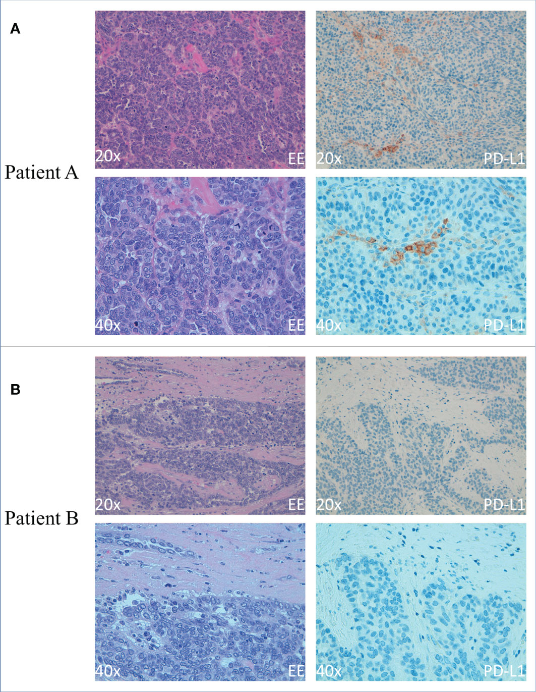
Regarding the contribution of PD-1/PD-L1 blockade IT in the overall survival and progression-free survival of patients, it is of note that the most widely used biomarker with some prediction capabilities for the outcome of the treatment is PD-L1 expression in tumor biopsies. For this reason, we performed immunohistochemistry (IHC) analysis of this marker in tumor biopsies derived from Patients A and B before and after IT. Tumor proportion score (TPS) has been used to evaluate PD-L1 expression. In particular, PD-L1 expression was calculated as the percentage of tumor cells with membrane staining of any intensity for each core; the final score was calculated as the average of all available cores. Cases were considered positive for PD-L1 when ≥ 1% of the tumor cells expressed PD-L1. We observed a higher expression of PD-L1–positive tumor cells in Patient A. In particular, immunohistochemical staining was performed on 2-µm thick FFPE serial with an automated IHC staining system (Ventana BenchMark ULTRA, Ventana Medical Systems, Italy). Sequential IHC was performed on Ventana BenchMark ULTRA, using a ultraView Universal DAB detection Kit. Afterward, slides were incubated with VENTANA PD-L1 (SP263) CE IVD US EXPORT antibody. (Figure 2).

Figure 2 Immunohistochemical analysis of PD-L1 expression on OC cells from Patients A and B (A) Smaller (20×) (upper panels) and larger (40×) (lower panels) magnification of primary OC showing hematoxylin-eosin staining (left panels) and PD-L1+ tumoral cells (right panels) of Patient A (B) Smaller (20×) (upper panels) and larger (40×) (lower panels) magnification of primary OC showing hematoxylin-eosin staining (left panels) and PD-L1+ tumoral cells (right panels) of Patient B Scale bars in A and B are 100 μm.
Evaluation of the peritumoral inflammatory infiltrate indicated an increase in infiltrating lymphocytes in Patient A compared with Patient B, mainly T cells (CD3+ cells) and a minor but detectable proportion of innate lymphocytes (NKp46+ CD3− cells) (data not shown).
As it was not yet in use in clinical practice, neither patient had been tested for the BRCA 1/2 gene mutation. However, Patient A agreed to undergo FoundationOne® CDx (Table 1), a validated Comprehensive Genomic Profile able to detect four classes of genomic alterations targeting the entire coding sequence of 324 cancer-related genes plus select introns from 36 genes frequently rearranged in cancer. The application of this test to patients with solid tumors may be particularly useful in identifying those that show some genetic alterations that could potentially make them susceptible to IT, such as the TMB, the Microsatellite Instability (MSI) or the LoH (25, 26).
The test showed the presence of an increased TMB, which is now a recognized factor in the identification of tumors, which are most responsive to both anti–PD-1 and anti–PD-L1 agents (27). On the contrary, the tumor was also characterized by Microsatellite Stability (MSS), which is a well-known negative predictive factor of response to ICIs, differently from MSI. This evidence has been validated above all in colorectal cancer for which the determination of the state of MSS is carried out routinely in common clinical practice (28).
Discussion
Regarding these two patients treated with the anti–PD-L1 monoclonal antibody avelumab, after the first line of chemotherapy, Patient A showed progression after 12 months and 19 cycles, whereas Patient B showed a slight progression of disease after only eight cycles. Interestingly, our data showed that Patient A tumor, which showed a better response to IT, was characterized by a high expression of PD-L1. In contrast, Patient B, who showed a worse outcome and a weak advantage from IT, was consistently negative for PD-L1. While PD-L1 was proven to be remarkable as a target for IT in melanoma (29) and lung cancer (30), its importance in OC is yet to be proven. These data confirm the importance of analysis of PD-L1 expression for the selection of therapeutic approach.
Nevertheless, PD-L1, as a biomarker for clinical diagnostic, shows some limitations, including differences among PD-L1 assays and scoring methods, as each method of PD-L1 detection has been developed by a different pharmaceutical company and the protocols and thresholds for positivity are associated with the methodology used in each trial (31–33). Other concerns to be considered are the dynamic and heterogeneous PD-L1 expression within tumors, which might differ between the biopsy and the rest of the tumor tissue, the time gap between the biopsy and therapeutic decisions (34–38).
In addition, in the case of patients treated with IT as a second or further line of treatment, there is a time gap between the diagnosis and the clinical decisions during which intermediate treatments such as conventional chemotherapy may alter PD-L1 expression in tumors. In fact, as in the case of the two patients discussed here, if the disease is too bulky to undergo upfront surgery, a first biopsy is obtained for diagnostic purposes but the definitive histological examination on the surgical tissue takes place after the administration of neo-adjuvant chemotherapy. Furthermore, it is possible, in case of disease recurrence, that new histological tissue is obtained from a metastatic lesion after further chemotherapy or other maintenance therapy such as bevacizumab or PARPis. The dynamic regulation of PD-L1 expression could explain clinical cases showing that patients diagnosed as tumor PD-L1 negative show objective responses to an anti–PD-L1 antibody as a second-line treatment (39).
Another concern is related to the heterogeneous nature of the tumor, which may affect PD-L1 quantification depending on the origin of the biopsy (primary tumor or metastasis), the degree of intratumoral heterogeneity and the sampling methodology (biopsy or tumor resection) (40). In conclusion, these data show that inconsistent response to IT in OC might be related to significant differences in PD-L1 expression in the tumor tissue that may humper its predictive potential. However, in OC, neither PD-L1 expression is always considered before the start of the therapy nor a consistent standardization for PD-L1 testing has been defined to obtain clear and comparable results.
Furthermore, considering that the immune infiltrate within tumors has proved to be very powerful in the prognostic stratification of patients, much attention should also be paid to its predictive value (41).
With a view to a possible future introduction of IT for the treatment of OC, considering the high cost of these therapies and the risk of immune-related adverse events during therapy, it will be necessary to identify the best combination of biomarkers that would facilitate the identification of potential responders and non-responders before the start of IT and a standardized evaluation of PD-L1 expression should become part of the routinely evaluated biomarkers in OC to better identify possible responders.
Data availability statement
The original contributions presented in the study are included in the article/supplementary material. Further inquiries can be directed to the corresponding authors.
Ethics statement
The studies involving human participants were reviewed and approved by ethics committee of the Liguria Region, Genova, Italy (n. 326/2018 and n127/2022-DB id12223 and B9991010 trial - JAVELIN Ovarian 100(EudraCT: 2015-003239-36). The B9991010 trial was supported by Pfizer, as part of an alliance between Pfizer and Merck (CrossRef Funder ID: 10.13039/100009945). The patients/participants provided their written informed consent to participate in this study. Written informed consent was obtained from the individual(s) for the publication of any potentially identifiable images or data included in this article.
Author contributions
NP, MG, and SP interpreted data, and wrote the article; MR performed immunohistochemical analyses; NP, MR, and AD provided samples and managed patient’s profile; TW managed patient’s profile; IB and MF revised the article; EM financed, designed, interpreted data, and wrote the article. All authors contributed to the article and approved the submitted version.
Funding
The research leading to these results has received funding from AIRC under IG 2021 – ID. 26037 project – P.I. EM. Additional grants from ROCHE 2017 (P.I. SP); Compagnia di San Paolo (2019.866) (G.L. E.M). MG was supported by a FIRC-AIRC fellowship for Italy.
Acknowledgments
The JAVELIN Ovarian 100 trial was sponsored by Pfizer, as part of an alliance between Pfizer and Merck (CrossRef Funder ID: 10.13039/100009945). Pfizer and Merck (CrossRef Funder ID: 10.13039/100009945) reviewed this manuscript for medical accuracy only before journal submission.
Conflict of interest
The authors declare that the research was conducted in the absence of any commercial or financial relationships that could be construed as a potential conflict of interest.
Publisher’s note
All claims expressed in this article are solely those of the authors and do not necessarily represent those of their affiliated organizations, or those of the publisher, the editors and the reviewers. Any product that may be evaluated in this article, or claim that may be made by its manufacturer, is not guaranteed or endorsed by the publisher.
Author disclaimer
The authors are fully responsible for the content of this manuscript, and the views and opinions described in the publication reflect solely those of the authors.
References
1. Siegel RL, Miller KD, Jemal A. Cancer statistics. CA Cancer J Clin (2020) 70:7–30. doi: 10.3322/caac.21590
2. Ledermann JA, Raja FA, Fotopoulou C, Gonzalez-Martin A, Colombo N, Sessa C. ESMO guidelines working group. newly diagnosed and relapsed epithelial ovarian carcinoma: ESMO clinical practice guidelines for diagnosis, treatment and follow-up. Ann Oncol (2013) 24(Suppl 6):vi24–32. doi: 10.1093/annonc/mdt333
3. Oza AM, Cook AD, Pfisterer J, Embleton A, Ledermann JA, Pujade-Lauraine E, et al. ICON7 trial investigators. standard chemotherapy with or without bevacizumab for women with newly diagnosed ovarian cancer (ICON7): overall survival results of a phase 3 randomised trial. Lancet Oncol (2015) 16:928–36. doi: 10.1016/S1470-2045(15)00086-8
4. Mirza MR, Coleman RL, González-Martín A, Moore KN, Colombo N, Ray-Coquard I, et al. The forefront of ovarian cancer therapy: update on PARP inhibitors. Ann Oncol (2020) 31(9):1148–59. doi: 10.1016/j.annonc.2020.06.004
5. Pesce S, Greppi M, Tabellini G, Rampinelli F, Parolini S, Olive D, et al. Identification of a subset of human natural killer cells expressing high levels of programmed death 1: A phenotypic and functional characterization. J Allergy ClinImmunol (2017) 139(1):335–346.e3. doi: 10.1016/j.jaci.2016.04.025
6. Topalian SL, Hodi FS, Brahmer JR, Gettinger SN, Smith DC, McDermott DF, et al. Safety, activity, and immune correlates of anti-PD-1 antibody in cancer. N Engl J Med (2012) 366:2443e54. doi: 10.1056/NEJMoa1200690
7. Braud VM, Allan DS, O’Callaghan CA, Soderstrom K, D’Andrea A, Ogg GS, et al. HLA-e binds to natural killer cell receptors CD94/NKG2A, b and c. Nature (1998) 391:795–9. doi: 10.1038/35869
8. Phan GQ, Yang JC, Sherry RM, Hwu P, Topalian SL, Schwartzentruber DJ, et al. Cancer regression and autoimmunity induced by cytotoxic T lymphocyte-associated antigen 4 blockade in patients with metastatic melanoma. Proc Natl AcadSci USA (2003) 100:8372e7. doi: 10.1073/pnas.1533209100
9. Brahmer JR, Tykodi SS, Chow LQ, Hwu WJ, Topalian SL, Hwu P, et al. Safety and activity of anti-PD-L1 antibody in patients with advanced cancer. N Engl J Med (2012) 366:2455e65. doi: 10.1056/NEJMoa1200694
10. Zhang L, Conejo-Garcia JR, Katsaros D, Gimotty PA, Massobrio M, Regnani G, et al. Intratumoral T cells, recurrence, and survival in epithelial ovarian cancer. N Engl J Med (2003) 348:203–13. doi: 10.1056/NEJMoa020177
11. Curiel TJ, Coukos G, Zou L, Alvarez X, Cheng P, Mottram P, et al. Specific recruitment of regulatory T cells in ovarian carcinoma fosters immune privilege and predicts reduced survival. Nat Med (2004) 10:942–9. doi: 10.1038/nm1093
12. Sterre T, Paijens ST, Vledder A, de Bruyn M, Nijman HW. Tumor-infiltrating lymphocytes in the immunotherapy era. Cell Mol Immunol (2021) 18:842–59. doi: 10.1038/s41423-020-00565-9
13. Pesce S, Tabellini G, Cantoni C, Patrizi O, Coltrini D, Rampinelli F, et al. B7-H6-mediated downregulation of NKp30 in NK cells contributes to ovarian carcinoma immune escape. Oncoimmunology (2015) 4(4):e1001224. doi: 10.1080/2162402X.2014.1001224
14. Carlsten M, Norell H, Bryceson YT, Poschke I, Schedvins K, Ljunggren HG, et al. Primary human tumor cells expressing CD155 impair tumor targeting by down-regulating DNAM-1 on NK cells. J Immunol (2009) 183(8):4921–30. doi: 10.4049/jimmunol.0901226
15. Wang J-J, Kwan-Yee Siu M, Yu-Xin Jiang Y-X, Wai Chan D, Nga-Yin Cheung A, Yuen-Sheung Ngan H, et al. Infiltration of T cells promotes the metastasis of ovarian cancer cells via the modulation of metastasis-related genes and PD-L1 expression. Cancer Immunol Immunother (2020) 69:2275–89. doi: 10.1007/s00262-020-02621-9
16. Greppi M, Tabellini G, Patrizi O, Candiani S, Decensi A, Parolini S, et al. Strengthening the AntiTumor NK cell function for the treatment of ovarian cancer. Int J Mol Sci (2019) 20:890. doi: 10.3390/ijms20040890
17. Patrizi O, Rampinelli F, Coltrini D, Pesce S, Carlomagno S, Sivori S, et al. Natural killer cell impairment in ovarian clear cell carcinoma. J Leukocyte Biol 2020 (2020) 108(4):1425–34. doi: 10.1002/JLB.5MA0720-295R
18. Wieser V, Gaugg I, Fleischer M, Shivalingaiah G, Wenzel S, Sprung S, et al. BRCA1/2 and TP53 mutation status associates with PD-1 and PD-L1 expression in ovarian cancer. Oncotarget (2018) 9:17501–11. doi: 10.18632/oncotarget.24770
19. Krishnan V, Berek JS, Dorigo O. Immunotherapy in ovarian cancer. CurrProbl Cancer (2017) 41(1):48–63. doi: 10.1016/j.currproblcancer.2016.11.003
20. Ghisoni E, Imbimbo M, Zimmermann S, Valabrega G. Ovarian cancer immunotherapy: Turning up the heat. Int J Mol Sci (2019) 20(12):2927. doi: 10.3390/ijms20122927
21. Marth C, Wieser V, Tsibulak I, Zeimet AG. Immunotherapy in ovarian cancer: fake news or the real deal? Int J Gynecol Cancer (2019) 29(1):201–11. doi: 10.1136/ijgc-2018-000011
22. Hartnett EG, Knight J, Radolec M, Buckanovich RJ, Edwards RP, Vlad AM. Immunotherapy advances for epithelial ovarian cancer. Cancers (Basel). (2020) 12(12):3733. doi: 10.3390/cancers12123733
23. Polastro L, Closset C, Kerger J. Immunotherapy in gynecological cancers: where are we? Curr Opin Oncol (2020) 32(5):459–70. doi: 10.1097/CCO.0000000000000661
24. Ledermann JA, Colombo N, Oza AM, Fujiwara K, Birrer MJ, Randall LM. Et al, avelumab in combination with and/or following chemotherapy vs chemotherapy alone in patients with previously untreated epithelial ovarian cancer: Results from the phase 3 javelin ovarian 100 trial. Gynecol. Oncol (2020) 159:13–4. doi: 10.1016/j.ygyno.2020.06.025
25. . Available at: https://www.fda.gov/medical-devices/recently-approved-devices/foundationone-cdx-p170019.
26. Chalmers ZR, Connelly CF, Fabrizio D, Gay L, Ali SM, Ennis R, et al. Analysis of 100,000 human cancer genomes reveals the landscape of tumor mutational burden. Genome Med (2017) 9(1):34. doi: 10.1186/s13073-017-0424-2
27. Goodman AM, Kato S, Bazhenova L, Patel SP, Frampton GM, Miller V, et al. Tumor mutational burden as an independent predictor of response to immunotherapy in diverse cancers. Mol Cancer Ther (2017) 16(11):2598–608. doi: 10.1158/1535-7163.MCT-17-0386
28. Lumish MA, Cercek A. Immunotherapy for the treatment of colorectal cancer. J SurgOncol (2021) 123(3):760–74. doi: 10.1002/jso.26357
29. Mahoney KM, Freeman GJ, McDermott DF. The next immune-checkpoint inhibitors: PD-1/PD-L1 blockade in melanoma. Clin Ther (2015) 37(4):764–82. doi: 10.1016/j.clinthera.2015.02.018
30. Dantoing E, Piton N, Salaün M, Thiberville L, Guisier F. Anti-PD1/PD-L1 immunotherapy for non-small cell lung cancer with actionable oncogenic driver mutations. Int J Mol Sci (2021) 22(12):6288. doi: 10.3390/ijms22126288
31. Munari E, Mariotti FR, Quatrini L, Bertoglio P, Tumino N, Vacca P, et al. PD-1/PD-L1 in cancer : Pathophysiological, diagnostic and therapeutic aspects. Int J Mol Sci (2021) 22(10):5123. doi: 10.3390/ijms22105123
32. Mariotti FR, Petrini S, Ingegnere T, Tumino N, Besi F, Scordamaglia F, et al. PD-1 in human NK cells: evidence of cytoplasmic mRNA and protein expression. Oncoimmunology (2018) 8(3):1557030. doi: 10.1080/2162402X.2018.1557030
33. Munari E, Rossi G, Zamboni G, Lunardi G, Marconi M, Sommaggio M, et al. PD-L1 assays 22C3 and SP263 are not interchangeable in non-small cell lung cancer when considering clinically relevant cutoffs: An interclone evaluation by differently trained pathologists. Am J Surg Pathol (2018) 42(10):1384–9. doi: 10.1097/PAS.0000000000001105
34. Kaderbhaï C, Tharin Z, Ghiringhelli F. The role of molecular profiling to predict the response to immune checkpoint inhibitors in lung cancer. Cancers (2019) 11:201. doi: 10.3390/cancers11020201
35. Hofman P, Heeke S, Alix-Panabières C, Pantel K. Liquid biopsy in the era of immuno-oncology: Is it ready for prime-time use for cancer patients? Ann Oncol (2019) 30:1448–59. doi: 10.1093/annonc/mdz196
36. Evans M, O’Sullivan B, Smith M, Taniere P. Predictive markers for anti-PD-1/PD-L1 therapy in non-small cell lung cancer-where are we? Transl Lung Cancer Res (2018) 7:682–90. doi: 10.21037/tlcr.2018.06.09
37. Munari E, Zamboni G, Lunardi G, Marchionni L, Marconi M, Sommaggio M, et al. PD-L1 expression heterogeneity in non-small cell lung cancer: Defining criteria for harmonization between biopsy specimens and whole sections. J Thorac Oncol (2018) 13(8):1113–20. doi: 10.1016/j.jtho.2018.04.017
38. Munari E, Zamboni G, Sighele G, Marconi M, Sommaggio M, Lunardi G, et al. Expression of programmed cell death ligand 1 in non-small cell lung cancer: Comparison between cytologic smears, core biopsies, and whole sections using the SP263 assay. Cancer Cytopathol. (2019) 127(1):52–61. doi: 10.1002/cncy.22083
39. Bocanegra A, Fernandez G, Zuazo M, Arasanz H, Garcia-Granda MJ, Hernandez C, et al. PD-L1 expression in systemic immune cell populations as a potential predictive biomarker of responses to PD-L1/PD-1 blockade therapy in lung cancer. Int J Mol Sci (2019) 20:1631. doi: 10.3390/ijms20071631
40. Kim H, Chung JH. PD-L1 testing in non-small cell lung cancer: Past, present, and future. J Pathol Transl Med (2019) 53:199–206. doi: 10.4132/jptm.2019.04.24
Keywords: ovarian cancer, immunotherapy, avelumab, PD-1, PD-L1
Citation: Provinciali N, Greppi M, Pesce S, Rutigliani M, Briata IM, Buttiron Webber T, Fava M, DeCensi A and Marcenaro E (2022) Case report: Variable response to immunotherapy in ovarian cancer: Our experience within the current state of the art. Front. Immunol. 13:1094017. doi: 10.3389/fimmu.2022.1094017
Received: 09 November 2022; Accepted: 05 December 2022;
Published: 19 December 2022.
Edited by:
Nicola Tumino, Bambino Gesù Children’s Hospital (IRCCS), ItalyReviewed by:
Silvano Sozzani, Department of Molecular Medicine, Sapienza University of Rome, ItalyEnrico Munari, University of Brescia, Italy
Copyright © 2022 Provinciali, Greppi, Pesce, Rutigliani, Briata, Buttiron Webber, Fava, DeCensi and Marcenaro. This is an open-access article distributed under the terms of the Creative Commons Attribution License (CC BY). The use, distribution or reproduction in other forums is permitted, provided the original author(s) and the copyright owner(s) are credited and that the original publication in this journal is cited, in accordance with accepted academic practice. No use, distribution or reproduction is permitted which does not comply with these terms.
*Correspondence: Emanuela Marcenaro, ZW1hbnVlbGEubWFyY2VuYXJvQHVuaWdlLml0; Silvia Pesce, c2lsdmlhLnBlc2NlQHVuaWdlLml0
†These authors share first authorship
 Nicoletta Provinciali
 Marco Greppi
 Silvia Pesce
 Mariangela Rutigliani
 Irene Maria Briata
 Tania Buttiron Webber
 Marianna Fava1
 Andrea DeCensi
 Emanuela Marcenaro
