 Débora Zini Baldasso1
 João Antônio Guizzo2
 Cláudia Cerutti Dazzi1,2
 Gabriela Carolina Paraboni Frandoloso2
 César Feronato3
 Stephan von Berg4
 Roberto Maurício Carvalho Guedes5
 Heather Lynne Wilson6
 Luiz Carlos Kreutz1,2
 Rafael Frandoloso1,2*
- 1Laboratory of Microbiology and Advanced Immunology, Faculty of Veterinary Medicine, University of Passo Fundo, Passo Fundo, Brazil
- 2Section of Immunology, AFK Imunotech, Passo Fundo, Brazil
- 3Swine Technical Department, Merck Sharp & Dohme (MSD), São Paulo, Brazil
- 4Global Marketing Swine, Merck Sharp & Dohme (MSD), Animal Health, Munich, Germany
- 5Department of Veterinary Clinic and Surgery, Veterinary School, Universidade Federal de Minas Gerais, Belo Horizonte, Brazil
- 6Vaccine and Infectious Disease Organization (VIDO), University of Saskatchewan, Saskatoon, SK, Canada
Lawsonia intracellularis is the etiologic agent of porcine proliferative enteropathy (PPE), an inflammatory bowel disease with a major economic impact on the pig industry. The serological diagnosis of PPE can be performed using Blocking or Indirect ELISA, Immunoperoxidase Monolayer Assay (IPMA) and Indirect Fluorescence Antibody Test (IFAT). Here, we designed a most sophisticated immunological method for the detection of porcine anti-L. intracellularis IgGs, named Flow Cytometry Antibody Test - FCAT. This assay uses whole, live-attenuated L. intracellularis bacteria derived from a commercial vaccine. For the assay, we set up the optimal antigen concentration (106 bacterium/assay), primary antibody dilution (1:100), time of incubation (20 min), antigen stability (15 days), precision (coefficient of variation - CV < 10%), reproducibility (CV ≤ 13%) and Receiver Operating Characteristic (ROC). When using a cut-off of >15.15% for FCAT, we determined that it showed a sensitivity of 98.8% and specificity of 100%. The rate of agreement with IPMA was 84.09% with a kappa index of 0.66. FCAT was used to screen 1,000 sera from non-vaccinated pigs housed in 22 different farms and we found that 730 pigs (73%) from 16 farms (72.7%) had L. intracellularis IgG. This high prevalence confirms that L. intracellularis is endemic on Brazilian pig farms. Finally, we determined that FCAT is an easy to perform diagnostic assay and we would highly recommend it for: i) seroepidemiological studies; ii) evaluation of infection dynamics; and iii) characterization of the humoral response profile induced by vaccines.
1 Introduction
Lawsonia intracellularis is an obligate intracellular Gram-negative bacterium that causes porcine proliferative enteropathy (PPE), an enteric disease with a major economic impact on the pig industry (1, 2). Pigs infected with L. intracellularis can develop two clinical forms of the disease depending on their previous immunological condition and the dose of bacteria ingested (3–5). The acute form, known as proliferative hemorrhagic enteropathy (6), affects mainly adult (> 4 months) animals (replacement gilts; growers) and, although clinically less frequent, it can cause high rates of mortality (up to 50%) (7, 8). The chronic form of PPE, in contrast, known as porcine proliferative enteropathy affects growing pigs (6 – 16 weeks of age) which has a low mortality rate, close to 1%, and might cause (but not always) a grey-green, semi-solid to liquid diarrhea (9).
Although the clinical presentations of the disease are very important, the silent (subclinical) course of the infection is most commonly observed on farms and, in this case, the presence of L. intracellularis can be demonstrated only by laboratory diagnosis assays as qPCR (if the bacteria are present) and serology (if the bacteria remain present for several months after infection/exposure) (10). The distribution of L. intracellularis in the global pig production system has been demonstrated and the prevalence of infected herds in Germany (91.7%), Denmark (95.8%), Spain (83.3%), France (79.2%), Netherlands (91.7%), United Kingdom (100%), Brazil (37%), China (77%), Canada (>50%) and Australia (100%) (10–14) illustrates the epidemiological importance of this microorganism. Now that pig farming is progressively reducing the use of antimicrobials as preventive measure, more prevalence of pathogenic bacteria in herds will be present.
The diagnosis of L. intracellularis can be confirmed by different assays, which can be used according to the type of biological sample collected. During post-mortem investigations, histological analysis complemented with Warthin-Starry silver staining (15), immunohistochemistry (16), and in situ hybridization (17) can be used to directly detect the bacteria in tissues. During in vivo monitoring, qPCR is the best technique to detect and determine the load of L. intracellularis in feces (4). ELISA (18), immunofluorescence antibody test (IFAT) (19), and immunoperoxidase monolayer assay (IPMA) (20) can be used to assess antibodies against the microorganism to establish whether the animals were exposed naturally (infection) or artificially [experimental challenges (controlled infection) and vaccination (Porcilis® Ileitis and Enterisol® Ileitis vaccines)].
Serological diagnosis is an excellent tool for assessing the kinetics of infection, the profile and duration of passive immunity, and the potency and duration of the vaccine-induced antibody response. Although these data can be easily obtained for other pathogens such as porcine circovirus type 2 and Influenza A virus, for which several commercial diagnostic kits with high sensitivity and specificity are available, this is not the case for L. intracellularis. For this microorganism, only two commercial ELISA kits (Blocking ELISA - Svanovir® L. intracellularis/Ileitis-Ab, Sweden, and Indirect ELISA for research use only - Biostone™ Animal Health, USA) are available and whose sensitivity (Blocking ELISA) close to 72% is considered low. Furthermore, although it is possible to use IFAT and IPMA techniques for detecting anti-L. intracellularis IgG, in practice, few laboratories around the world offer these assays as a service, mainly due to the difficulty of cultivating L. intracellularis. Therefore, the use of serology to understand the infection dynamic or general sanitary condition of the farm for L. intracellularis is limited.
In order to overcome the limitations described above, in this study we used an innovative strategy to develop a new serological diagnostic test to detect antibodies against L. intracellularis. This test, named Flow Cytometry Antibody Test (FCAT), uses a live-attenuated strain of L. intracellularis as detecting antigen readily available from a commercial vaccine. FCAT has high sensitivity and specificity, is easy to perform, and can be promptly incorporated into the diagnostic routine of any specialized laboratory around the world.
2 Materials and methods
2.1 Statement of institutional animal care
All sera used in this study came from: i) other studies previously approved by the Ethics Committee for the Use of Animals in Research at the Universidade de Passo Fundo (CEUA no. 10/2020, 19/2020 & 20/2020); ii) routine of diagnosis of the AFK Imunotech Laboratory; iii) other studies previously approved by the Ethics Committee for the Use of Animals in Research at the Universidade Federal de Minas Gerais (CEUA n° 133/2018, 36/2016).
2.2 Antigen
The in vitro cultivation of L. intracellularis is extremely difficult, which limits the production of this bacterium to few laboratories in the world. Since our objective was to develop a serological assay that could be easily performed in any laboratory, we used the vaccine strain of L. intracellularis from Enterisol® Ileitis (Boehringer Ingelheim), a live attenuated vaccine, as a source of antigen. This licensed vaccine is distributed worldwide, and all new batches of live antigen are qualified by the manufacturer, an ideal situation to guarantee antigen quality and ensure repeatability between antigens batches and laboratories that will perform this diagnosis.
2.3 Sera samples
A total of 246 serum samples obtained from pigs with different serological condition were used to determine the preliminary cut-off values of this flow cytometry antibody test (Table 1, Phase I). The second set of specimens included 1,200 serum samples collected between 2018 and 2021 from 32 farms located in Southern region of Brazil (Paraná, Santa Catarina and Rio Grande do Sul) (Table 1, Phase II); this panel of sera was used to analyze the ability of the established cut-off to identify conventional pigs with or without anti-L. intracellularis IgGs.
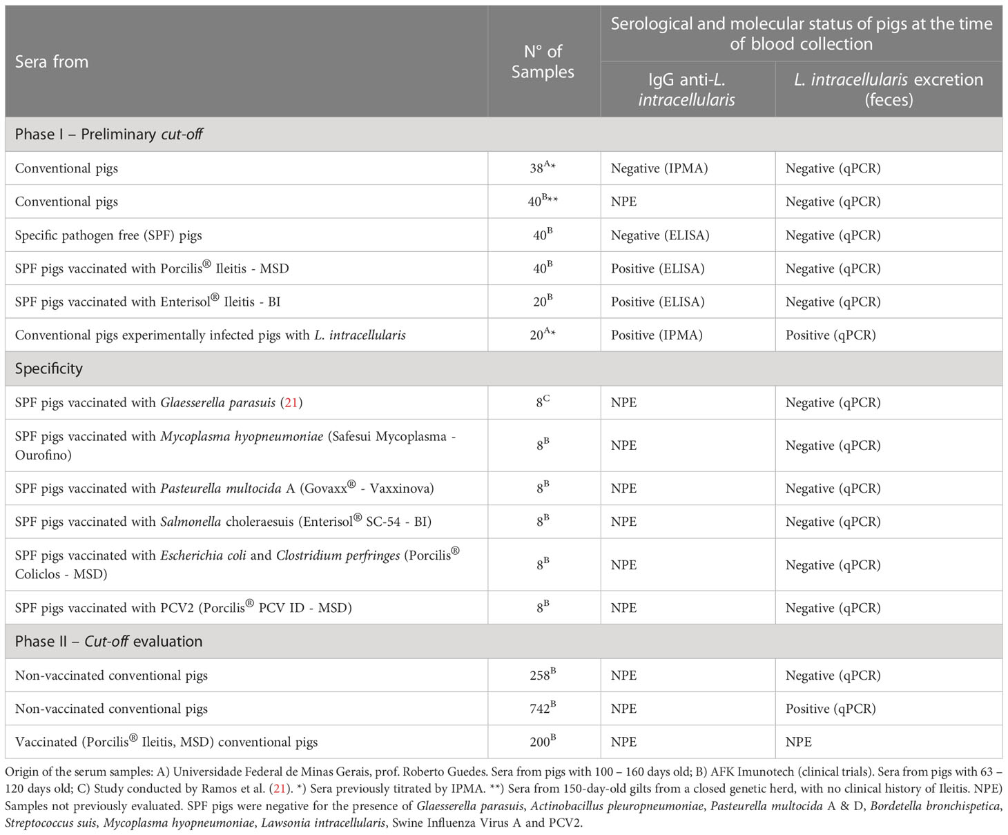
Table 1 Description of the sera used in this study.
2.4 Antigen stability
The stability of the reconstituted antigen was evaluated daily for a period of 30 days. Three parameters were analyzed by flow cytometry: i) absolute count of L. intracellularis; ii) bacterial morphology; and iii) antigenicity. For the first two analyses, a daily aliquot of the antigen was diluted 1:1,000 in filtered PBS pH 7.4 and acquired in the cytometer. The number of bacteria per microliter of acquired samples was calculated automatically by the cytometer which was equipped with flow sensor used for volumetric measurement. Therefore, to calculate the total number of bacteria per mL of the reconstituted vaccine, we used the following mathematical formula: number of events per μL × inverse of the dilution factor (1,000) × per 1,000 (to convert μL to mL). The morphometric analysis was conducted analyzing the parameters of Forward Scatter (FSC) vs Side Scatter (SSC) of the daily aliquot of the antigen. For the antigenicity analysis, 3 sera from pigs immunized with the Porcilis® Ileitis vaccine were used; briefly, 106 bacteria were incubated into the wells of a 96 well conical-bottom polystyrene plates (cat. # K30-6096V, Olen, China) together with 100 µL of porcine serum diluted 1:100 in filtered (0,22 µm, cat. # GPWP04700, Millipore, Ireland) PBS pH 7.4 containing 1% bovine serum albumin (cat. # A2153, Sigma-Aldrich, USA) (FACS buffer) for 1 hour at 37°C. After three washing steps with 200 μL of FACS buffer (plate centrifuged at 1,200 × g for 5 min; centrifuge 5810R Eppendorf, Germany), 100 μL of FACS buffer containing 1 μg of Goat anti-Porcine IgG(H+L)-PE (cat. # 6050-09, SouthernBiotech, USA) was added and incubated for 1 hour at 37°C. Then, the washing steps were repeated, and the bacterial pellet resuspended in 350 µL of PBS for analysis. All parameters were analyzed by Flow Cytometry using a FACSVerse Cytometer (Becton Dickinson, USA) equipped with a 405 nm violet laser, 488 nm blue laser, 640 nm red laser and flow sensor. A total of 100,000 events were acquired in P1 region and analyzed. Each sample was analyzed in triplicate.
2.5 Determination of the ideal concentration of L. intracellularis for the assay
To determine the optimal amount of antigen that would be used in the assay, three different concentrations (105, 106 and 107) of L. intracellularis were evaluated. Each concentration was incubated with a panel of sera (n=5) with different titers of anti-L. intracellularis IgG as defined previously by the IPMA (Table 1): i) highly positive sera (titer between 1:960 and 1:1,920); ii) moderately positive sera (titer between 1:120 and 1:480); and iii) weakly positive sera (titer of 1:30). The immunostaining was performed as described above (subsection 2.3) and the ideal bacteria concentration was defined as the smallest number of bacteria that: i) allowed the statistical differentiation of three sera categories and ii) ensured enough bacteria to be analyzed after the washing steps.
2.6 Primary antibody working dilution
To define the optimal dilution of the primary antibody (porcine sera) different dilutions of a panel of highly positive (n=5, titer > 960), moderately positive (n=5, titer between 1:120 and 1:480, weakly positive (n=5, titer of 1:30) and negative (n=5) sera were analyzed. The dilutions evaluated were 1:50, 1:100 and 1:200 in a final volume of 100 µL. The immunostaining was performed as described above (subsection 2.3).
2.7 Evaluation of the incubation period of primary and secondary antibodies
To investigate the shortest incubation period necessary to guarantee the optimal interaction between the molecules involved in the assay, three incubation periods were evaluated: 60 min, 30 min and 20 min. These periods were evaluated for primary (10 different positive samples) and secondary (Goat Anti-Porcine IgG (H+L)-PE) antibodies. All incubations were performed at 37°C. The immunostaining was performed as described above (subsection 2.3).
2.8 Enzyme-linked immunosorbent assay - ELISA
A total of 100 samples (Table 1) were analyzed for the presence of anti-L. intracellularis antibodies using a commercial blocking ELISA Kit (SVANOVIR®, Boehringer Ingelheim Svanova, Sweden). The protocol was performed following the manufacturer’s recommendations.
2.9 Immunoperoxidase monolayer assay
The immunoperoxidase monolayer assay (IPMA) was used to detect porcine anti-L. intracellularis IgG. This assay was carried out by the laboratory of Prof. Roberto Guedes (Universidade Federal de Minas Gerais), according to the methodology previously described (20).
2.10 Flow cytometry antibody test
A total of 106 L. intracellularis suspended in 200 µL was added per well of a 96 well conical bottom polystyrene plates. The plate was centrifuged (3,200 × g, 5 min) and the bacterial pellet was resuspended in 100 µL of porcine serum diluted 1:100 in FACS buffer and incubated at 37°C during 20 min. After three washing steps with 200 μL of FACS buffer (step of centrifugation at 1,200 × g for 5 min between washes), 100 μL of FACS buffer containing 1 μg of Goat Anti-Porcine IgG(H+L)-PE was added and incubated for 20 min at 37°C. Then, the bacteria were washed 3 times, resuspended in 350 µL of FACS buffer and transferred into a round-bottom tube (Falcon®, cat. # 352052). A total of 100,000 events were acquired in a FACSVerse cytometer. The bacterial population was initially identified through a dot plot crossing the FSC and SSC parameters; where the study region, designated P1, was established. Subsequently, a second dot plot was created, in this case, crossing the FL-2 (Phycoerythrin, PE) and FL-4 (Allophycocyanin, APC) channels and having P1 as the study region. The events considered positive were those enclosed within the quadrant representative of PE (low right). The results are expressed as the percentage of positive bacteria in relation to the total bacteria contained in the P1 region. The step-by-step execution of this diagnosis as well as the analysis strategy are illustrated in Figure 1.
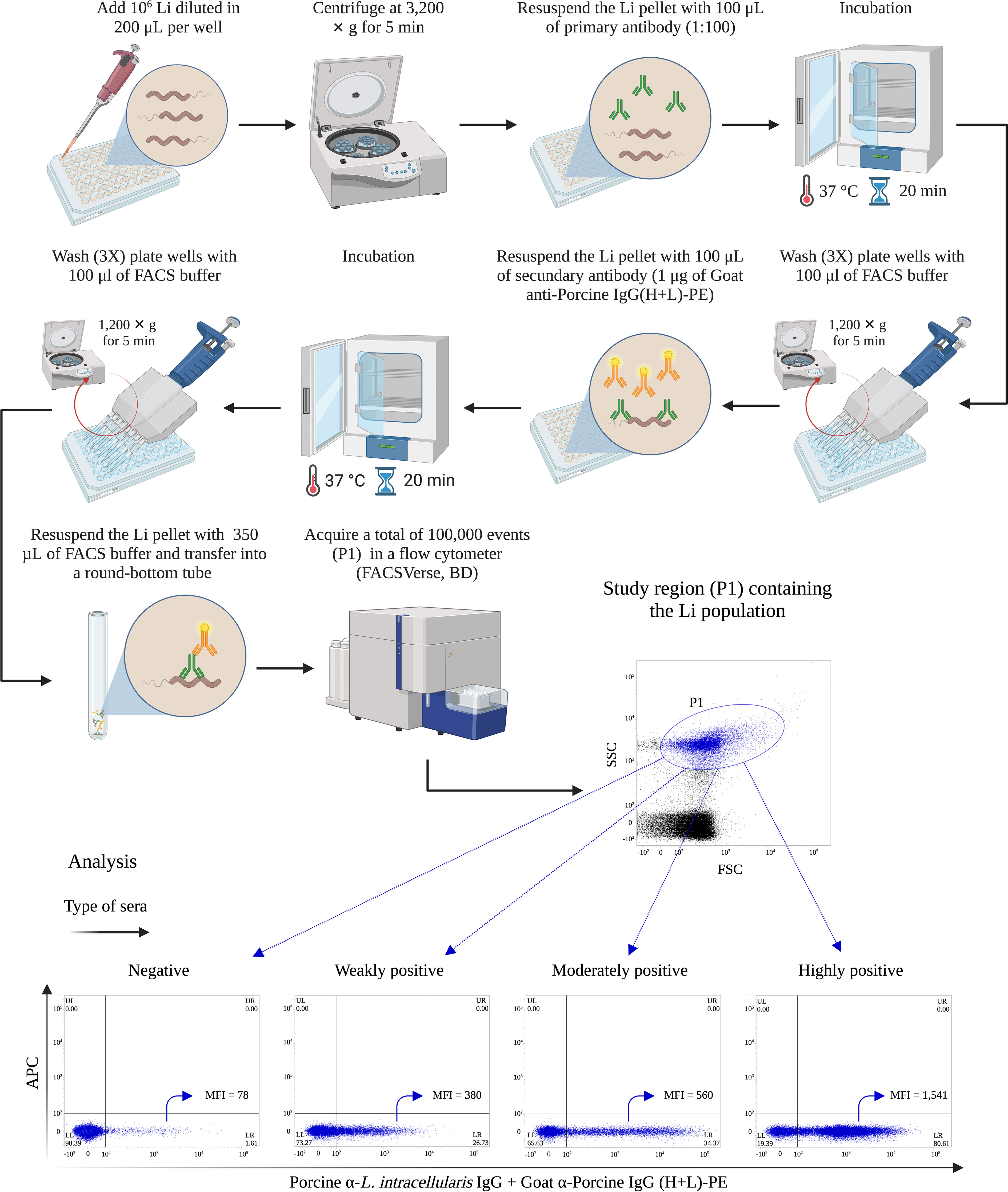
Figure 1 Quick protocol of flow cytometry antibody test for Lawsonia intracellularis. The brief protocol of immunostaining and cytometric analysis for the detection of porcine anti-L. intracellularis (Li) IgG. MFI, Mean Fluorescence Intensity. This figure was created with BioRender.com.
2.11 Receiver operating characteristic
The determination of the cut-off was performed using a panel of 198 porcine sera (Table 1). Briefly, sera from 78 animals known to be negative for anti-Lawsonia intracellularis IgG and molecularly (qPCR) negative for L. intracellularis were obtained. We also obtained sera from 40 animals from a genetic nucleus herd with no history of Ileitis; these animals were molecularly negative for L. intracellularis; collectively sera from a total of 118 negative animals. We used sera from 80 animals serologically positive for L. intracellularis; of these total, 60 SPF pigs were vaccinated (qPCR negative), and 20 were experimentally infected (and consequently qPCR-positive). Therefore, the cut-off calculation, performed using the Receiver Operating Characteristic (ROC) Curve, included 118 negative and 80 positive animals. All sera were tested by this new assay and the results were used to generate a Receiver Operating Characteristic (ROC) Curve. The selected cut-off was based on the sensitivity and specificity values. The ROC Curve analysis was performed using Graph Pad Prism Software Version 9.2.0.
2.12 Specificity
The specificity analysis was conducted to evaluate the ability of this assay to identify IgG specifically to L. intracellularis, which is the analyte of interest. The analysis was performed as recommended by Selliah, Eck (22). Briefly, a panel of 48 sera (Table 1, Specificity) from SPF pigs immunized with Glaesserella parasuis (bacterin prepared with SV7 (21), n=8), Mycoplasma hyopneumoniae (Safesui Mycoplasma, Ourofino, n=8), Pasteurella multocida A (Bacterin formulated with a Brazilian clinical strain of P. multocida A, pfhA+, Vaxxinova, n=8), Salmonella enterica, serovar Choleraesuis (Enterisol® SC-54 vaccine, Boehringer Ingelheim, n=8), Clostridium perfringes type C and Escherichia coli (Porcilis® Coliclos, MSD, n=8) and Porcine circovirus type 2 (Porcilis® PCV ID, MSD, n=8) were assessed using the protocol described in subsection 2.8.
2.13 Precision and reproducibility
Precision is one of the most critical parameters in flow cytometry assay (22). In order to know the intra-assay precision (precision), samples from 3 positive pigs (vaccinated with Porcilis® Ileitis) and negative SPF pigs were tested twice under the same conditions by a single analyst. With the values obtained from each sample, the mean and coefficient of variation were calculated. For reproducibility analysis (inter-assay precision) the same samples were analyzed by 2 different analysts on 2 different days. Again, with the values obtained from each sample, the mean and the coefficient of variation were calculated. The accuracy acceptance criterion was established by accepting a coefficient of variation between the analyses of the same analyst or between the analysts of a maximum of 25%.
2.14 Agreement analysis between FCAT and IPMA techniques
To analyze the level of agreement between the FCAT and IPMA, we used a panel of sera from two experimental groups: G1) pigs (n = 8) immunized with the Porcilis® Ileitis vaccine at 21 days of life and G2) pigs (n = 8) inoculated with PBS. Serum samples were collected prior to vaccination (D0) and at the following post-vaccination times: D7, D14, D21, D28 and D55 contributing to 88 sera for comparative purposes. Agreement analysis (Kappa) was performed as described by Landis and Koch (23).
2.15 Statistical analysis
All data were analyzed using GraphPad Prism™ (GraphPad Software, San Diego, California, USA). One or two-way ANOVA with Tukey’s multiple comparisons test were used to assess significance between the different variables analyzed in this study. The specific test used in each analysis, as well as the significance, is indicated in the figure legends.
3 Results
3.1 Antigen stability
L. intracellularis obtained from the attenuated vaccine Enterisol® Ileitis was used as a diagnostic antigen. The antigen used in our study is destined to immunizing pigs and should be immediately used upon reconstitution. In the test designed here, the amount of antigen used in each assay is low and a single reconstituted bottle contains enough antigen to be used along a few weeks. Thus, the stability and quality of the reconstituted and refrigerated (4 – 8°C) antigen was analyzed daily along a 30 days period. During this time, changes in the morphology of the bacterial population (analyzed in the P1 region) were observed after 15 days of antigen storage (Figure 2B); a subpopulation of larger and more complex L. intracellularis was clearly detected, which may represent loss of bacteria cell wall integrity. Although we observed morphological changes in the bacterial population, the number of bacteria detected at each time point was similar with no significant reduction at any time (Figure 2A). The antigenicity analysis revealed that antigen stability follows the same trend as bacteria reconstituted for more than two weeks were significantly more antigenic compared to previous time points (Figure 3), which indicated that storage time alters the antigenic characteristics of the reconstituted antigen; thus, for all assays the reconstituted antigen was used for 15 days only.
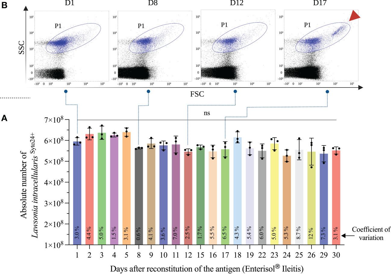
Figure 2 Antigen stability analysis. An aliquot of Enterisol® Ileitis vaccine reconstituted and stored at 4-8°C was analyzed daily by flow cytometry. The aliquot diluted 1:1,000 was analyzed in triplicate and the mean of L. intracellularis absolute count plus standard deviation is shown in “(A)”. The coefficient of variation of each quantification is represented at the foot of the bars. The morphological characteristic of the bacterial population at 4 different times is illustrated in “(B)”. The subpopulation of L. intracellularis with greatest size and complexity is indicated with the arrowhead highlighted in red. Statistical comparison was performed using one-way ANOVA with Tukey’s multiple comparisons test and no significant (ns) differences were found between the moments compared.
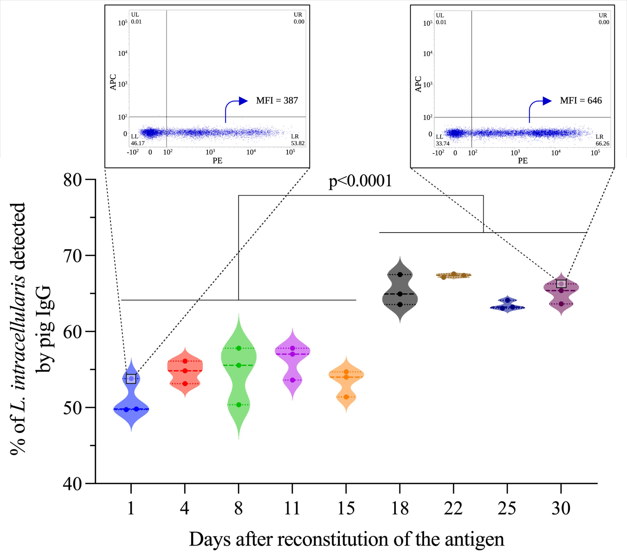
Figure 3 Antigenicity analysis of the reconstituted antigen. This analysis was performed by flow cytometry using sera (n=3) from pigs immunized with the Porcilis® Ileitis vaccine. The results express the percentage of L. intracellularis bacteria (total population containing 106 bacteria) containing antibodies (IgGs) surface. Statistical comparison was performed using one-way ANOVA with Tukey’s multiple comparisons, and statistical differences are represented in the figure. Dot plots showing antigenic differences between time D1 and D30 are highlighted in zoom format within the figure.
3.2 Determination of the ideal concentration of L. intracellularis for the assay
The amount of antigen used in a diagnostic test must be evaluated with two objectives: i) functionally (to ensure maximum resolution of the test) and ii) economically (antigen saving). Taking these premises into account, we analyzed 3 concentrations of antigens (105, 106, and 107) immunostained with 3 categories of sera, weakly, moderately, and highly positive as determined by IPMA. As illustrated in Figure 4, regardless of the bacterial concentration it is possible to differentiate the 3 serum categories, except for the concentration of 107, in which weakly and moderately positive sera could not be statistically differentiated. There is a reduction in the percentage of detection of L. intracellularis between concentrations of 106 and 107, when using weakly and moderately positive sera (Figure 4). Additionally, we noticed that the number of bacteria acquired from immunostaining using 105 bacteria was lower (long acquisition period) compared to 106 and 107, indicating that part of the bacteria was lost during the washing steps; therefore, the concentration of 106 was defined as the ideal concentration of antigen.
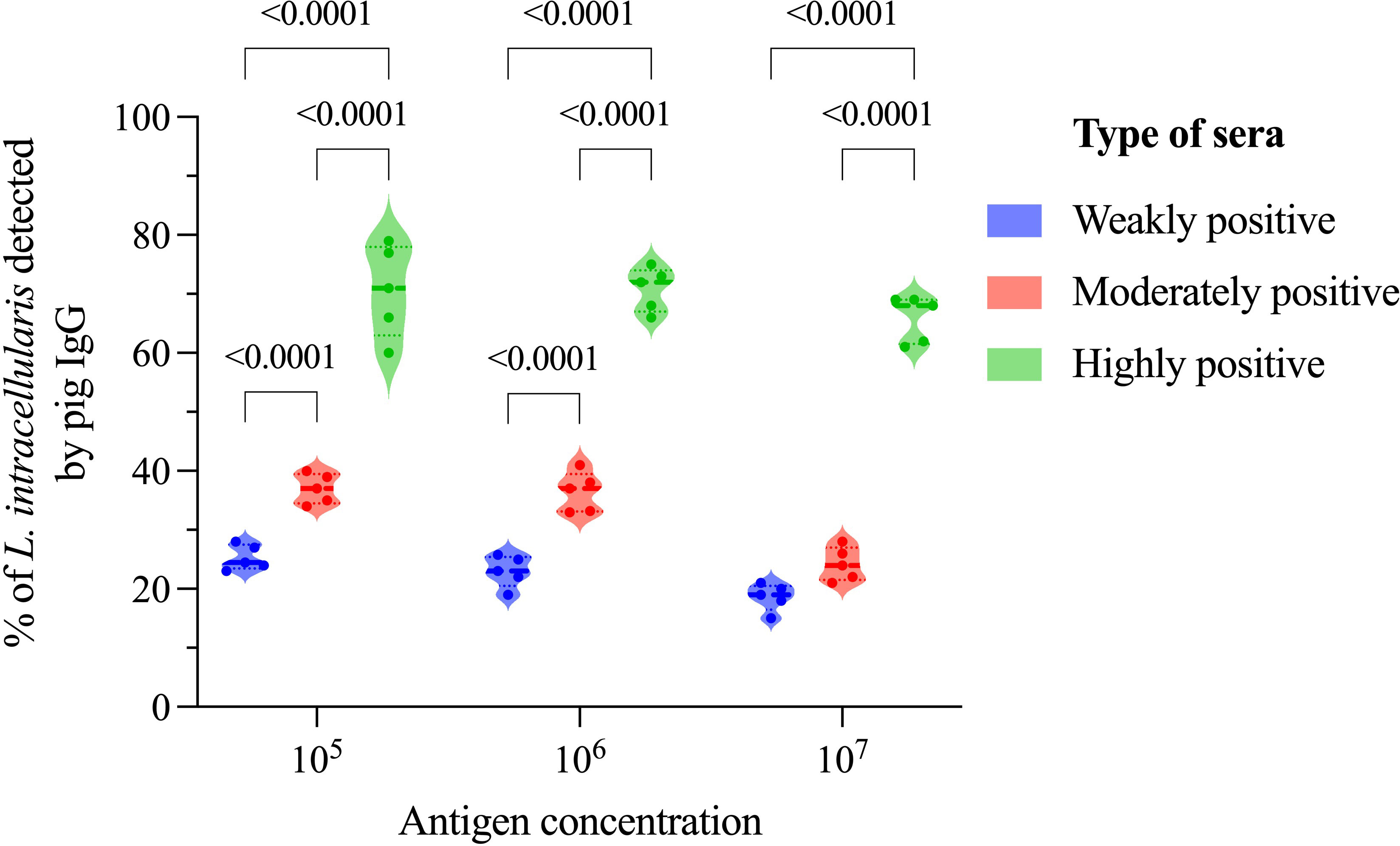
Figure 4 Impact of antigen concentration on anti-L. intracellularis IgG detection. In this analysis 3 different concentrations of antigens (105, 106 and 107) and 3 different categories of sera (weakly, moderately, and highly positive for L. intracellularis) were analyzed by flow cytometry after immunostaining. Statistical comparison was performed using two-way ANOVA with Tukey’s multiple comparisons. Statistical differences are indicated in the figure.
3.3 Assessment of primary antibody dilution
The dilution rate of the serum (primary antibody) is essential in any diagnostic test and should be set to mitigate false positive results, usually associated with polyreactive IgGs. Three serum dilutions were analyzed; 1:50, 1:100 and 1:200. We observed that there was no reduction in the percentage of L. intracellularis associated to porcine IgGs between the 1:50 and 1:100 dilutions, regardless of the serum category. On the other hand, the percentage of L. intracellularis with associated IgGs was significantly lower (p < 0.0001) when sera diluted 1:200 were used, and the number of sera considered positive (cut-off set at 15.15% as described in the section 3.5) at lower dilutions (5/5) decreased when the weakly positive sera category was tested (2/5) (Figure 5). Therefore, the dilution of the primary serum was set at 1:100.

Figure 5 Primary antibody dilution and its impact on serology interpretation. In this analysis 3 different dilution (1:50, 1:100 and 1:200) of primary antibody (pig sera) classified in 3 different categories (weakly, moderately, and highly positive for L. intracellularis) were analyzed by flow cytometry after immunostaining. Statistical comparison was performed using two-way ANOVA with Tukey’s multiple comparisons. Statistical differences are indicated in the figure.
3.4 Selection of the best antibody incubation time
Evaluating antibody incubation time (primary and secondary) is essential in any diagnostic test under development; in general, it is always recommended to select the shortest time, as long as there are no changes in the final result of the diagnosis. Three incubation times (20, 30 and 60 min) for both the primary (pig IgG anti-L. intracellularis) and secondary (Goat anti-Porcine IgG-PE) antibody were tested. As illustrated in Figure 6, the mean percentage of L. intracellularis with associated IgGs at the 3 different incubation times was 47.5%, 47.1% and 48.0%; additionally, the same dispersion profile of the results was observed, and no significant differences were found between the analyzed times. In view of these results, an incubation time of 20 min was selected for the primary and secondary antibodies.
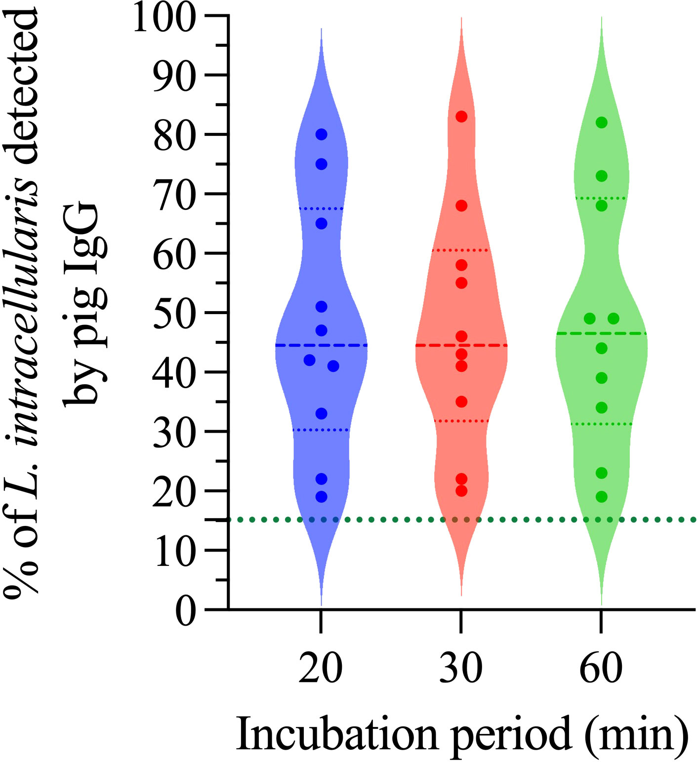
Figure 6 Outcome of incubation time on the detection of anti-L. intracellularis IgG. A total of 10 pig sera positive for IgG anti-L. intracellularis were included in this analysis. Pig sera were incubated for 20, 30 and 60 minutes with 106 L. intracellularis at 37°C. Subsequently, the samples were incubated with the secondary antibody for the same times. Statistical comparison was performed using one-way ANOVA with Tukey’s multiple comparisons. No statistical differences (ns) were observed between the different times analyzed.
3.5 Specificity and sensitivity of the flow cytometry antibody test
Once the definition of all parameters and conditions of the Flow Cytometry Antibody Test (FCAT) was concluded, a wide panel of swine sera was used (Table 1, Phase I) aiming to find the cut-off value capable of truly differentiating a positive from a negative serum sample. For this purpose, a ROC curve analysis was conducted using 118 serum samples negative and 80 serum samples positive to L. intracellularis antibodies (Figure 7A). The ROC curve analysis of the FCAT data generated paired estimates of relative sensitivity and relative specificity at different cut-off values. A cut-off of 15.15% (percentage of L. intracellularis with associated IgG) was recommended; and at this cut-off value, the relative sensitivity and specificity estimates were 98.8% [95% confidence interval (CI) = 93.2% to 99.9%] and 100% (95% CI = 96.9% to 100%), respectively. The ROC curve had an Area Under the Curve (AUC) value of 0.9963 (95% CI = 0.99 to 1.0), which indicated a high level of accuracy for this FCAT (Figure 7B). Thereafter, using the established cut-off value, a second panel consisting of 48 sera (Table 1, Specificity) was tested on the FCAT. As illustrated in Figure 7C, none of the sera reached the cut-off of 15.15%; therefore, all sera were considered negative for L. intracellullaris. This result demonstrates that there is no cross-reactivity between L. intracellularis and G. parasuis, M. hyopneumoniae, P. multocida A, Salmonella enterica, serovar Choleraesuis, E. coli, C. perfringes type C and Porcine circovirus type 2. Finally, a third panel of sera (n=1,200, Table 1, Phase II) from: i) non-vaccinated conventional pigs [L. intracellularis (qPCR) negative feces]; ii) non-vaccinated conventional pigs [feces positive for L. intracellularis (qPCR)]; and iii) Vaccinated (Porcilis® Ileitis, MSD) conventional pigs, was evaluated in the FCAT. As shown in Figure 7D, all animals vaccinated with the Porcilis® Ileitis vaccine developed antibodies against L. intracellularis and were therefore considered positive. Similarly, 85.3% of the animals that were shedding L. intracellularis in the feces had anti-L. intracellularis IgG at a level higher than the FCAT cut-off; and 68.7% of the animals that were not shedding L. intracellularis at the time of blood collection had previous contact with L. intracellularis.
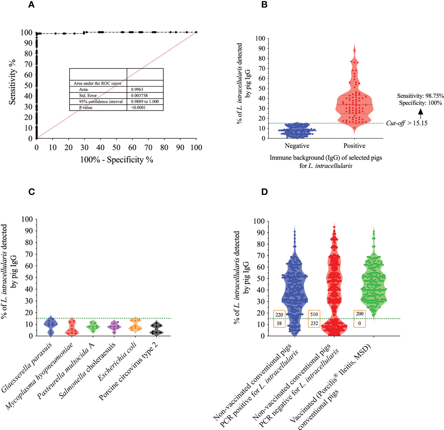
Figure 7 Basic immunological characteristics of the FCAT method. (A) Receiver Operating Characteristic (ROC) analysis. (B) Representation of the percentage values (dispersion) of L. intracellularis recognition of the samples used to define the cut-off of the FCAT test. The sensitivity and specificity of the test using the cut-off of > 15.15% are described in the figure. (C) FCAT specificity analysis using sera from pigs immunized with other pathogens. (D) Analysis of clinical samples collected from pigs with different immunological backgrounds against L. intracellularis.
3.6 Precision and reproducibility of the FCAT
The precision (intra-assay) and reproducibility (inter-assay) analysis were performed by two different analysts (A and B). As described in the Table 2, the coefficient of variation (CV) of the samples analyzed in triplicate by each of the analysts was always less than 10%, indicating that analysts performed the immunostaining and acquisition of samples with precision. Furthermore, when the same samples were repeated by the same analyst on the following day, a CV of less than 10% was again observed. It is important to highlight that the results of immunostaining performed by the two analysts varied little, with the smallest variation being 0.9% and the maximum 13%; these data indicate that FCAT has high accuracy.
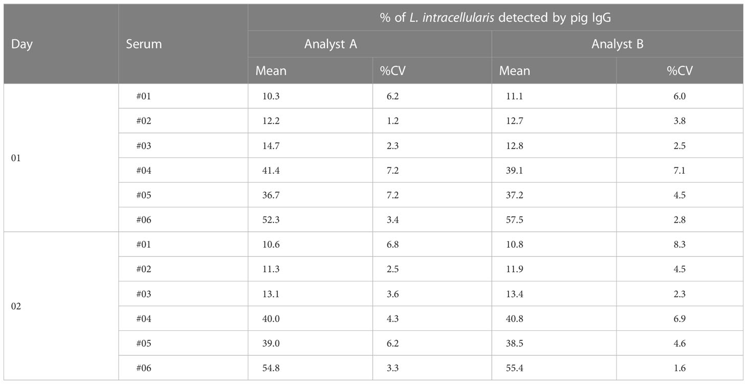
Table 2 Precision and reproducibility analysis of pig serum by different analyst at two different days.
3.7 Comparison between FCAT and IPMA techniques
To analyze the level of agreement between FCAT and IPMA, sera collected from two groups of pigs (vaccinated and unvaccinated) at different post-vaccination periods were analyzed by these two methodologies in two different laboratories (FCTA conducted at the Frandoloso’s lab and IPMA conducted at Guedes’s lab). To avoid any bias, the analysis was blind, and the history of the sera was not revealed to the analysts. As described in Table 3, out of the total number of samples tested (n=88), 74 had the same result in both methodologies, which represents a direct agreement of 84.09%. Using all the data (samples with the same and different results according to the technique used) the Kappa index was generated resulting in a general agreement of 0.66 ± 0.08 (95% CI = 0.50 to 0.82) (Table 4), which represent a substantial agreement taken into consideration the Kappa scale (0 to 1).
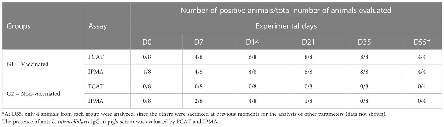
Table 3 Serological evolution of pigs immunized or non-immunized pigs with Lawsonia intracellularis.
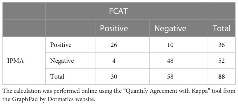
Table 4 Matrix used to calculate the Kappa index.
Considering that the pigs used in this experiment were Specific Pathogen Free and kept in a controlled facility (BSL-2) throughout the study, our expectation was that all animals, regardless of the diagnostic technique, would be negative for L. intracellularis. As illustrated in Table 3, we observed that a considerable number of pigs from group G2 (non-vaccinated) were positive for anti-L. intracellularis IgG as determined by the IPMA day (D) 7 (n=2, 25%), D14 (n=4, 50%) and D21 (n=1, 12.5%) post inoculation with PBS (Table 3). We understand that these IPMA results are false positives, and this interpretation is supported by two observations: i) samples collected from the same pigs at days D35 and D55 were negative by IPMA (Table 3); ii) all sera samples from this group were negative for anti-L. intracellularis IgG by FCAT.
The sensitivity to specifically detect the analytical, in our case the anti-L. intracellularis IgG during the genesis of humoral response induced by vaccination is one of the most important features. We noticed that both techniques are very similar in terms of sensitivity; however, FCAT standing out slightly over IPMA on D14, at that moment the FCAT classified more animals as positive than the IPMA.
4 Discussion
Lawsonia intracellularis is a complex intracellular bacterium ubiquitously found in the pig production system worldwide (11–13, 24). This microorganism is the causative agent of PPE which is considered one of most important enteric diseases of pigs in the growing and finishing phases. The presence of uncontrolled L. intracellularis infection in pigs has significant economic impacts due to its negative effect on daily gain, feed conversion rate and mortality (4, 25–27).
Currently, the control of PPE can be performed satisfactorily by using licensed vaccines, and in this line, three antigen delivery platforms are available to immunize pigs. Boehringer Ingelheim commercializes the Enterisol® Ileitis vaccine, which is based on an attenuated strain of L. intracellularis and whose administration is carried out through the drinking water or by directly administration into the oral cavity of piglets. In parallel, MSD Animal Health has two inactivated vaccines, which can be administered intramuscularly (Porcilis® Ileitis & Porcilis® Lawsonia) and intradermally (Porcilis® Lawsonia ID). The effectiveness of these vaccines has already been scientifically demonstrated (28–31) and their use in pig production is a valuable tool for the progressive reduction of antimicrobial usage as a measure to prevent this disease (32). Additionally, it is important to recognize that vaccines applied by the oral and intradermal routes are still scarce in pigs (and in mammals in general). Due to their demonstrated efficacy, they represent two platforms that should be priority for the veterinary pharmaceuticals industry. These vaccines are safe from the point of view of application (without needles, and therefore mitigate iatrogenic transmission of pathogens) and painless, which contemplates one of the premises of animal welfare.
During infection, L. intracellularis stimulates B lymphocytes to produce mucosal (IgA) (33) and systemic antibodies (IgG) (34). The immunization using inactivated-based vaccines consistently induces systemic IgG (28–30), a feature that is also observed in animals immunized with the attenuated vaccine (34). Therefore, antibodies (IgG) are a valuable marker to demonstrate the circulation of the agent in the farm, or even to monitor the antibody response after the application of vaccines.
Since the negative impact of L. intracellularis infection for pig production is unquestionable, in this study we present a new method of serological diagnosis for L. intracellularis, named Flow Cytometry Antibody Test. Flow cytometry has been used to quantify the percentage of eukaryotic cells which had taken up or were associated with fluorescent L. intracellularis (35). As described in Table 5, there are different diagnostic tests available on the market for the detection of anti-L. intracellularis IgG, which differ regarding sensitivity, specificity, execution time and complexity. Among these tests, the FCAT stands out in all parameters; the method is highly sensitive (98.8%), specific (100%) and takes approximately only 1:15 hours to run, which represents a time saving compared to Blocking ELISA, Indirect ELISA, IPMA and IFAT of 47%, 8%, 33% and 12%, respectively. Although the specificity of IPMA and FCAT are the same (100%), the sensitivity of the FCAT method (98.8%) is much higher than IPMA (89%), IFAT (91%) and Blocking ELISA (72%) (Table 5). In practical terms it represents a greater ability to detect truly positive samples even if they contain low levels of antibodies to the bacterium.
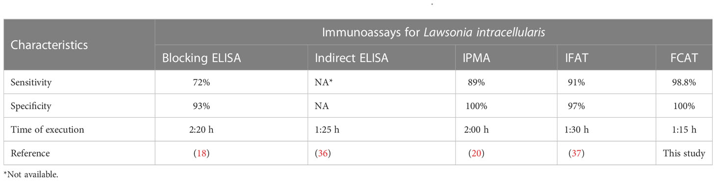
Table 5 Types and main characteristics of available serological assays for L. intracellularis.
The reason why FCAT is superior to other tests is related to the basic characteristics of the assay, such as: i) live L. intracellularis is used in the test (the antigen source for this test is available globally – Enterisol® Ileitis, BI); ii) the antigen is not chemically (acetone and methanol) treated during the test [all antigens (protein lipids and saccharides) remain native]; iii) the entire surface of the antigen is accessible to antibodies (increases the chance of antibodies specifically binding to the surface of L. intracellularis); and iv) the fluorescence reading is performed automatically by Flow Cytometry Equipment, and therefore, mitigates human errors (e.g. subjective counting) (Figure 8A). In contrast to FCAT, in the case of IPMA and IFAT, the cells are fixed with acetone and methanol, which promotes disruption of the cytoplasmic membrane of infected eukaryotic cells and might reduce the antigenic quality of the antigens (38) (Figure 8B). Additionally, in the case of IPMA, as the reading is performed manually with an inverted microscope, and as endogenous cell-derived peroxidases might be present on the assay, some false positive results can be expected (39). In this line, in our study, when analyzing sera from SPF piglets that were truly negative for L. intracellularis, we observed false positive results at four different times by the IPMA technique (Table 3); and, therefore, considering its biological limitation, the use of IPMA in the certification of negative farms for L. intracellularis should be carried out with caution.
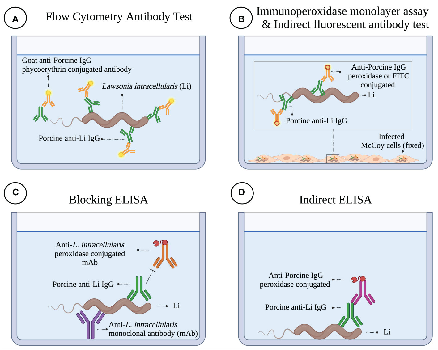
Figure 8 Illustration of different serological diagnostic tests for Lawsonia intracellularis. (A) Flow Cytometry Antibody Test. In this assay, porcine antibodies interact with L. intracellularis floating in the well of the plate. The binding of specific antibodies to L. intracellularis is demonstrated with a phycoerythrin-conjugated antibody specific for porcine IgG. The test is read on the flow cytometer. (B) Immunoperoxidase Monolayer Assay (IPMA) & Indirect Fluorescent Antibody Test (IFAT). In these assays, cells (i.e., McCoy cells) infected with L. intracellularis are chemically fixed with acetone-methanol, which promotes a structural change in the cell (cytoplasmic membrane damage, as indicated in figure). This damage is essential for porcine antibodies to interact with L. intracellularis. The presence of antibodies bound to L. intracellularis is demonstrated by a peroxidase conjugate antibody plus chromogen (3-amino-9-ethyl-carbazole) solution (IPMA). In the case of IFAT, a fluorescein isothiocyanate conjugated antibody is used to reveal the presence of porcine IgG bound to the pathogen. Plates are read under an inverted microscope. (C) Blocking ELISA. In this ELISA, a monoclonal antibody bound to the ELISA plate is used to capture L. intracellularis. Porcine antibodies compete with the peroxidase-conjugated monoclonal antibody for a specific epitope present on a surface antigen of L. intracellularis. The presence of porcine antibodies to L. intracellularis is determined by the potential to reduce or inhibit binding of the monoclonal antibody and, therefore, the enzymatic reaction associated with the competing conjugated antibody. (D) ELISA Indirect. In this ELISA, L. intracellularis is immobilized directly on the ELISA plate; and for this reason, a considerable surface area of the antigen is not accessible to antibodies (physical masking of antigens). Porcine antibodies that recognize L. intracellularis are detected with a peroxidase-conjugated anti-porcine IgG antibody. This figure was created with BioRender.com.
In addition to the two mentioned techniques, the detection of porcine anti-L. intracellularis antibodies can be performed by ELISA. As illustrated in Figure 8C, the blocking ELISA is based on i) monoclonal antibody-coated wells for capturing of cell-cultured antigen and ii) utilizes peroxidase-conjugated monoclonal antibodies as competitive antibodies. In this case, one of the disadvantages of this method in relation to FCAT is that the blocking of the conjugated monoclonal antibody is restricted to a single epitope, and therefore, for an animal to be diagnosed as positive, it must produce antibodies against this epitope during infection and/or vaccination. As it is a single epitope, any antigenic variation in this epitope region could generate false negative results. On the other hand, in FCAT, all antibodies generated against the surface antigens of L. intracellularis have accessibility to the diagnostic antigen, and therefore, the sensitivity of the technique is greater. The Indirect ELISA (Figure 8D) presents as its main limitation the masking of epitopes, due to the adherence of the bacteria on the plate. An alternative to overcome this limitation is the preparation of biotinylated protein and its immobilization in the correct orientation on streptavidin plates, as described by our group (40). For the development of the latter ELISA, it is first necessary to characterize an immunogenic, specific, and conserved protein; and efforts in this line need to be carried out, although promising results are already available (41).
As already mentioned, the main objective of this study was to develop a method of serological diagnosis for L. intracellularis that could be widely disseminated among diagnostic laboratories. Our results demonstrated that the objective was achieved and that FCAT is the most sophisticated immunological technique for the detection of anti-Lawsonia intracellularis IgGs. It is important to mention that this technique can be used to detect other classes of antibodies, such as systemic monomeric IgA, IgG1 and IgG2, as well as those associated with the ileum mucosa (data not shown). For this purpose, it is necessary to use specific antibodies (mouse anti-Pig IgG1, clone K139 3C8; mouse anti-Pig IgG2, clone K68 Ig2; mouse anti-Pig IgA, clone K61 1B4, Bio-Rad). The flexibility of FCAT is ideal for studying the immunological profile of licensed vaccines as well as vaccines under development against L. intracellularis. Furthermore, as described in Table 2, the FCAT is highly accurate, and comparison of precision (intra-assay) and reproducibility (inter-assay) parameters of the other techniques is not available, which makes it difficult to predict the results obtained by the other methods when performed in different laboratories.
A limitation of FCTA, like ELISA, IPMA and IFAT is that it is not possible to differentiate, based on the presence of systemic IgG, vaccinated animals from those naturally infected with L. intracellularis. Additionally, one disadvantage of FCAT relative to other diagnostic techniques is the need for the diagnostic laboratory to have a flow cytometer instrument, which is more expensive than an ELISA reader (for the ELISA technique) or a microscope (for the IPMA and IFAT techniques). However, what may be considered expensive today may become affordable in the medium term (such as the situation with polymerase chain reaction machines for example).
We recognize that we have taken an important step towards improving L. intracellularis diagnosis, but future studies need to be carried out to develop, on an industrial scale, the other reagents for this diagnosis, such as positive and negative control sera, and conjugated secondary antibodies, which will increase the repeatability of the results and avoid any variations related to the quality and specificity of the commercial reagents (controls and secondary antibodies).
L. intracellularis, in addition to produce Ileitis in pigs, also causes an intestinal disease in horses called equine proliferative enteropathy (EPE), which cases have been increasing in the last few years, especially in post-weaning foals and occasionally in adult horses (42). Although the presumptive diagnosis of EPE can be established based on age of the affected animal, clinical signs, and imaging (ultrasonographic evaluation), the confirmation of infection needs to be conducted in vitro using molecular or serological techniques. In this scenario, although in this study we did not evaluate FCAT to analyze equine serum, we can speculate, based on existing data on the use of IPMA for the detection of anti-L. intracellularis (20, 43), that FCAT has a high potential to work for horses. In this case, future studies need to be carried out to demonstrate the application of this technique in this animal species.
Finally, using the FCAT we analyzed the prevalence of circulating antibodies in a total of 1,000 pigs not vaccinated against L. intracellularis from 22 different farms located in 8 Brazilian states. As illustrated in Figure 7D, a total of 730 pigs were found to be positive for anti-Lawsonia intracellularis IgG; these sera were from animals of 16 different farms (72.7%) and indicates a high prevalence of L. intracellularis in Brazilian pig farms. The national prevalence found here is higher than those previously reported in the state of Minas Gerais (37%) (14), which is the fourth main pig producer region of Brazil. Therefore, our results suggest that Brazil is an endemic country for L. intracellularis, and that producers should implement biological strategies (vaccines) to mitigate the economic losses caused by this pathogen. Future seroepidemiological studies using the FCAT are needed to better estimate the impact of L. intracellularis on Brazilian pig herds.
Data availability statement
The original contributions presented in the study are included in the article/supplementary material. Further inquiries can be directed to the corresponding author.
Ethics statement
The animal study was reviewed and approved by Ethics Committee for the Use of Animals in Research at the University of Passo Fundo (CEUA no. 10/2020, 19/2020 & 20/2020). Ethics Committee for the Use of Animals in Research at the Federal University of Minas Gerais (CEUA n° 133/2018. 36/2016).
Author contributions
Conceived and designed the experiments: RF. Performed the experiments: DZB, JAG, and GCPF. Contributed reagents/materials/analysis tools: RF, LCK, CF, and RMCG. Wrote the paper: DZB, RF, and LCK. Critical review and editing of the manuscript: RMCG, HLW, and SV. All authors discussed the results and commented on the manuscript. All authors contributed to the article and approved the submitted version.
Funding
This study was funded by MSD Animal Health. The funder was not involved in the study design, collection, analysis, interpretation of data, the writing of this article, or the decision to submit it for publication.
Acknowledgments
DB was supported by CAPES – Brazilian Federal Agency for Support and Evaluation of Graduate Education within the Ministry of Education of Brazil. LK and RC have a research fellowship from CNPq.
Conflict of interest
Authors CF and SV were employed by company Merck Sharp & Dohme (MSD) Animal Health.
The remaining authors declare that the research was conducted in the absence of any commercial or financial relationships that could be construed as a potential conflict of interest.
Publisher’s note
All claims expressed in this article are solely those of the authors and do not necessarily represent those of their affiliated organizations, or those of the publisher, the editors and the reviewers. Any product that may be evaluated in this article, or claim that may be made by its manufacturer, is not guaranteed or endorsed by the publisher.
References
1. Collins M A. Advances in ileitis control, diagnosis, epidemiology and the economic impacts of disease in commercial pig herds. Agriculture. (2013) 3:536–55. doi: 10.3390/agriculture3030536
2. Holyoake PK, Mullan BP, Cutler RS. Simulation of the economic impact of proliferative enteritis on pig production in Australia. Aust Vet J (1996) 73(3):89–92. doi: 10.1111/j.1751-0813.1996.tb09983.x
3. Collins AM, Love RJ. Re-challenge of pigs following recovery from proliferative enteropathy. Vet Microbiol (2007) 120(3-4):381–6. doi: 10.1016/j.vetmic.2006.11.004
4. Collins AM, Barchia IM. The critical threshold of Lawsonia intracelullaris in pig faeces that causes reduced average daily weight gains in experimentally challenged pigs. Vet Microbiol (2014) 168(2-4):455–8. doi: 10.1016/j.vetmic.2013.12.003
5. Guedes RM, Winkelman NL, Gebhart CJ. Relationship between the severity of porcine proliferative enteropathy and the infectious dose of Lawsonia intracelullaris. Vet Rec (2003) 153(14):432–3. doi: 10.1136/vr.153.14.432
6. Lawson GH, Rowland AC, Roberts L, Fraser G, McCartney E. Proliferative haemorrhagic enteropathy. Res Vet Sci (1979) 27(1):46–51. doi: 10.1016/S0034-5288(18)32858-3
7. Rowland AC, Rowntree PG. A haemorrhagic bowel syndrome associated with intestinal adenomatosis in the pig. Vet Rec (1972) 91(10):235–41. doi: 10.1136/vr.91.10.235
8. Love DN, Love RJ. Pathology of proliferative haemorrhagic enteropathy in pigs. Vet Pathol (1979) 16(1):41–8. doi: 10.1177/030098587901600104
9. Lawson GH, Gebhart CJ. Proliferative enteropathy. J Comp Pathol (2000) 122(2-3):77–100. doi: 10.1053/jcpa.1999.0347
10. Arnold M, Crienen A, Swam H, von Berg S, Jolie R, Nathues H. Prevalence of Lawsonia intracellularis in pig herds in different European countries. Porcine Health Manage (2019) 5:31. doi: 10.1186/s40813-019-0137-6
11. Wu Z, Ling Y, Tian D, Pan Q, Heegaard PM, He C. Seroprevalence of Lawsonia intracellularis antibodies in intensive pig farms in China. BMC Vet Res (2014) 10:100. doi: 10.1186/1746-6148-10-100
12. Paradis MA, Gottschalk M, Rajic A, Ravel A, Wilson JB, Aramini J, et al. Seroprevalence of Lawsonia intracellularis in different swine populations in 3 provinces in Canada. Can Vet J (2007) 48(1):57–62. doi: 10.4141/cjas68-008
13. Holyoake PK, Emery D, Gonsalves J, Dona hoo M, Collins A. Prevalence of antibodies to Lawsonia intracellularis in pig herds in Australia. Aust Vet J (2010) 88(5):186–8. doi: 10.1111/j.1751-0813.2010.00558.x
14. Resende TP, Pereira CE, Gabardo Mde P, Haddad JP, Lobato ZI, Guedes RM. Serological profile, seroprevalence and risk factors related to Lawsonia intracellularis infection in swine herds from minas gerais state, Brazil. BMC Vet Res (2015) 11:306. doi: 10.1186/s12917-015-0618-z
15. Huerta B, Arenas A, Carrasco L, Maldonado A, Tarradas C, Carbonero A, et al. Comparison of diagnostic techniques for porcine proliferative enteropathy (Lawsonia intracellularis infection). J Comp Pathol (2003) 129(2-3):179–85. doi: 10.1016/S0021-9975(03)00030-6
16. Szczotka A, Stadejek T, Zmudzki J, Nowak A, Osinski Z, Pejsak Z. Immunohistochemical detection of Lawsonia intracellularis in tissue sections from pigs. Pol J Vet Sci (2011) 14(4):531–8. doi: 10.2478/v10181-011-0079-7
17. Jensen TK, Boesen HT, Vigre H, Boye M. Detection of Lawsonia intracellularis in formalin-fixed porcine intestinal tissue samples: Comparison of immunofluorescence and in-situ hybridization, and evaluation of the effects of controlled autolysis. J Comp Pathol (2010) 142(1):1–8. doi: 10.1016/j.jcpa.2009.04.001
18. Jacobson M, Wallgren P, Nordengrahn A, Merza M, Emanuelson U. Evaluation of a blocking ELISA for the detection of antibodies against Lawsonia intracellularis in pig sera. Acta Vet Scand (2011) 53:23. doi: 10.1186/1751-0147-53-23
19. Guedes RM, Gebhart CJ. Preparation and characterization of polyclonal and monoclonal antibodies against Lawsonia intracellularis. J Vet Diagn Invest (2003) 15(5):438–46. doi: 10.1177/104063870301500506
20. Guedes RM, Gebhart CJ, Deen J, Winkelman NL. Validation of an immunoperoxidase monolayer assay as a serologic test for porcine proliferative enteropathy. J Vet Diagn Invest (2002) 14(6):528–30. doi: 10.1177/104063870201400618
21. Ramos Prigol S, Klein R, Chaudhuri S, Paraboni Frandoloso G, Guizzo JA, Gutierrez Martin CB, et al. TbpB(Y167A)-based vaccine can protect pigs against glasser's disease triggered by Glaesserella parasuis SV7 expressing TbpB cluster I. Pathogens (2022) 11(7):766. doi: 10.3390/pathogens11070766
22. Selliah N, Eck S, Green C, Oldaker T, Stewart J, Vitaliti A, et al. Flow cytometry method validation protocols. Curr Protoc Cytom (2019) 87(1):e53. doi: 10.1002/cpcy.53
23. Landis JR, Koch GG. The measurement of observer agreement for categorical data. Biometrics. (1977) 33(1):159–74. doi: 10.2307/2529310
24. Dazzi CC, Baldasso D, Frandoloso GP, Kreutz LC RF. Brazilian Growing and finishing pig farms are endemic for Lawsonia intracellularis IPVS2022. (Rio de Janeiro, Brazil: The International Pig Veterinary Society - IPVS) (2022).
25. Jensen HM. Health management with reduced antibiotic use - experiences of a Danish pig vet. Anim Biotechnol (2006) 17(2):189–94. doi: 10.1080/10495390600957142
26. McOrist S, Smith SH, Green LE. Estimate of direct financial losses due to porcine proliferative enteropathy. Vet Rec (1997) 140(22):579–81. doi: 10.1136/vr.140.22.579
27. Jacobson M, Fellstrom C, Jensen-Waern M. Porcine proliferative enteropathy: An important disease with questions remaining to be solved. Vet J (2010) 184(3):264–8. doi: 10.1016/j.tvjl.2009.05.010
28. Roerink F, Morgan CL, Knetter SM, Passat MH, Archibald AL, Ait-Ali T, et al. A novel inactivated vaccine against Lawsonia intracellularis induces rapid induction of humoral immunity, reduction of bacterial shedding and provides robust gut barrier function. Vaccine. (2018) 36(11):1500–8. doi: 10.1016/j.vaccine.2017.12.049
29. Jacobs AAC, Harks F, Hazenberg L, Hoeijmakers MJH, Nell T, Pel S, et al. Efficacy of a novel inactivated Lawsonia intracellularis vaccine in pigs against experimental infection and under field conditions. Vaccine. (2019) 37(15):2149–57. doi: 10.1016/j.vaccine.2019.02.067
30. Jacobs AAC, Harks F, Pauwels R, Cao Q, Holtslag H, Pel S, et al. Efficacy of a novel intradermal Lawsonia intracellularis vaccine in pigs against experimental infection and under field conditions. Porcine Health Manage (2020) 6:25. doi: 10.1186/s40813-020-00164-0
31. Leite FLL, Singer RS, Ward T, Gebhart CJ, Isaacson RE. Vaccination against Lawsonia intracellularis decreases shedding of Salmonella enterica serovar Typhimurium in Co-infected pigs and alters the gut microbiome. Sci Rep (2018) 8(1):2857. doi: 10.1038/s41598-018-21255-7
32. Karuppannan AK, Opriessnig T. Lawsonia intracellularis: Revisiting the disease ecology and control of this fastidious pathogen in pigs. Front Vet Sci (2018) 5:181. doi: 10.3389/fvets.2018.00181
33. Guedes RM, Gebhart CJ. Evidence of cell-mediated immune response and specific local mucosal immunoglobulin (Ig) a production against Lawsonia intracellularis in experimentally infected swine. Can J Vet Res (2010) 74(2):97–101.
34. Guedes RM, Gebhart CJ. Onset and duration of fecal shedding, cell-mediated and humoral immune responses in pigs after challenge with a pathogenic isolate or attenuated vaccine strain of Lawsonia intracellularis. Vet Microbiol (2003) 91(2-3):135–45. doi: 10.1016/S0378-1135(02)00301-2
35. Obradovic M, Pasternak JA, Ng SH, Wilson HL. Use of flow cytometry and PCR analysis to detect 5-carboxyfluoroscein-stained obligate intracellular bacteria Lawsonia intracellularis invasion of McCoy cells. J Microbiol Methods (2016) 126:60–6. doi: 10.1016/j.mimet.2016.04.015
36. Biostone. In: AsurDx™ Lawsonia intracellularis antibody test. (Southlake, Texas, USA, BioStone Animal Health) (2022) Available at: https://biostoneah.com/how-to-order/.
37. Knittel JP, Jordan DM, Schwartz KJ, Janke BH, Roof MB, McOrist S, et al. Evaluation of antemortem polymerase chain reaction and serologic methods for detection of Lawsonia intracellularis-exposed pigs. Am J Vet Res (1998) 59(6):722–6.
38. Chao Y, Zhang T. Optimization of fixation methods for observation of bacterial cell morphology and surface ultrastructures by atomic force microscopy. Appl Microbiol Biotechnol (2011) 92(2):381–92. doi: 10.1007/s00253-011-3551-5
39. Guedes RM, Gebhart CJ, Winkelman NL, Mackie-Nuss RA. A comparative study of an indirect fluorescent antibody test and an immunoperoxidase monolayer assay for the diagnosis of porcine proliferative enteropathy. J Vet Diagn Invest (2002) 14(5):420–3. doi: 10.1177/104063870201400512
40. Frandoloso R, Schryvers AB. Structure-based antigen design: Targeting transferrin receptors to prevent respiratory and systemic disease of swine. In: International pig veterinary society congress – IPVS2020. Rio de Janeiro: IPVS2020 (2020).
41. Watarai M, Yamato Y, Horiuchi N, Kim S, Omata Y, Shirahata T, et al. Enzyme-linked immunosorbent assay to detect Lawsonia intracellularis in rabbits with proliferative enteropathy. J Vet Med Sci (2004) 66(6):735–7. doi: 10.1292/jvms.66.735
42. Pusterla N, Gebhart C. Lawsonia intracellularis infection and proliferative enteropathy in foals. Vet Microbiol (2013) 167(1-2):34–41. doi: 10.1016/j.vetmic.2013.06.017
Keywords: Lawsonia intracellularis, Ileitis, pig, diagnostic test, flow cytometry assay, serological antibody detection
Citation: Baldasso DZ, Guizzo JA, Dazzi CC, Paraboni Frandoloso GC, Feronato C, von Berg S, Carvalho Guedes RM, Wilson HL, Kreutz LC and Frandoloso R (2023) Development and validation of a flow cytometry antibody test for Lawsonia intracellularis. Front. Immunol. 14:1145072. doi: 10.3389/fimmu.2023.1145072
Received: 15 January 2023; Accepted: 06 March 2023;
Published: 21 March 2023.
Edited by:
Felix Ngosa Toka, Ross University School of Veterinary Medicine, Saint Kitts and NevisReviewed by:
Eduardo Ruben Cobo, University of Calgary, CanadaUwe Müller, Leipzig University, Germany
François JMA Meurens, INRA Ecole nationale vétérinaire, agroalimentaire et de l’alimentation de Nantes-Atlantique (Oniris), France
Copyright © 2023 Baldasso, Guizzo, Dazzi, Paraboni Frandoloso, Feronato, von Berg, Carvalho Guedes, Wilson, Kreutz and Frandoloso. This is an open-access article distributed under the terms of the Creative Commons Attribution License (CC BY). The use, distribution or reproduction in other forums is permitted, provided the original author(s) and the copyright owner(s) are credited and that the original publication in this journal is cited, in accordance with accepted academic practice. No use, distribution or reproduction is permitted which does not comply with these terms.
*Correspondence: Rafael Frandoloso, cmZyYW5AdXBmLmJy