 Zehong Wei
 Huaiyu Li1
- 1Graduate School, Jiangxi University of Chinese Medicine, Nanchang, Jiangxi, China
- 2Clinical Laboratory, Affiliated Hospital of Jiangxi University of Chinese Medicine, Nanchang, Jiangxi, China
Background: Rheumatoid arthritis (RA) is a chronic, systemic autoimmune disease that affects multiple joints and has adverse effects on various organs throughout the body, often leading to a poor prognosis. Recent studies have shown significant progress in the research of non-coding RNAs (ncRNAs) in RA. Therefore, this study aims to comprehensively assess the current status and research trends of ncRNAs in RA through a bibliometric analysis.
Methods: This study retrieved articles relevant to ncRNAs and RA from the Science Citation Index Expanded Database of the Web of Science Core Collection between January 1st, 2003, and July 31st, 2023. The relevant articles were screened based on the inclusion criteria. VOSviewer and CiteSpace are utilized for bibliometric and visual analysis.
Results: A total of 1697 publications were included in this study, and there was a noticeable increase in annual publications from January 1st, 2003, to July 31st, 2023. China, the United States, and the United Kingdom were the most productive countries in this field, contributing to 43.81%, 13.09%, and 3.87% of the publications. Anhui Medical University and Lu Qianjin were identified as the most influential institution and author. Frontiers In Immunology stood out as the most prolific journal, while Arthritis & Rheumatology was the most co-cited journal. Additionally, the research related to “circular RNA”, “oxidative stress”, “proliferation”, and “migration” have emerged as new hotspots in the field.
Conclusion: In this study, we have summarized the publication characteristics related to ncRNA and RA and identified the most productive countries, institutions, authors, journals, hot topics, and trends.
1 Introduction
Rheumatoid arthritis (RA) is a chronic autoimmune disease characterized by symmetric polyarthritis. RA exhibits synovial inflammation which is the fundamental pathological alteration in the early stage of disease, then synovium proliferates and pannus forms that damages joints and impairs joint function, ultimately results in disability (1). RA is a major global public health challenge, according to the global epidemiological study on RA in 2017, the age-standardized global prevalence and annual incidence rates of RA were 0.246% and 0.015%, which show an increasing trend (2, 3). The etiology of RA has not yet been fully clarified, but it generally involves a complex interplay among genetic susceptibility, environmental exposures, microbial infections, and immune dysregulation (4, 5). American college of rheumatology guideline for the treatment of rheumatoid arthritis indicates that the treatment of RA includes nonsteroidal anti-inflammatory drugs (NSAIDs), glucocorticoids (GC), conventional synthetic disease-modifying antirheumatic drugs (csDMARDs), biologic DMARDs (bDMARDs) and targeted synthetic DMARDs (tsDMARDs), as well as joint replacement surgery (6, 7).However, RA cannot be cured entirely, and it is essential to elucidate the pathogenesis of RA to update therapeutic strategies and develop novel medicine.
Non-coding RNAs (ncRNAs) which are not translated to protein have a crucial role in regulating gene expression, mainly include microRNA (miRNA), long non-coding RNA (lncRNA), and circular RNA (circRNA) (8). In recent years, an increasing number of genetic studies related to the pathogenesis of RA (including gene-arrays and new generation sequencing studies) revealed gene expression signatures and networks in synovial tissue and peripheral blood mononuclear cells (PBMCs) (9–11). Furthermore, research on post-transcriptional mechanisms of gene regulation has identified ncRNAs associated with disease activity, inflammation, pannus formation and bone destruction (12). These ncRNAs play a significant role in the diagnosis and treatment of RA as biomarkers, mediators of pathogenesis and potential therapeutic targets (12, 13).
Bibliometrics is an important method of quantitatively and qualitatively analyzing publications within a specific field via mathematical and statistical approaches (14). It aims to uncover the research hotspots and trends by acquiring, processing, and analyzing the data of countries, institutions, author, journals, and keywords (15). Despite the intrinsic mechanisms of ncRNAs in the pathogenesis of RA have been continuously unveiled over the past two decades, there have no bibliometric analysis has been published focusing on the field. In this study, we aimed to use bibliometric method and mapping knowledge domain through softwares (such as VOSviewer (16) and CiteSpace (17)), to visually analyze the publications related to ncRNAs in RA, and identify the global research status, trends, and hotspots in the field.
2 Materials and methods
2.1 Data collection
In this study, we retrieved and downloaded the data from the Science Citation Index Expanded (SCI-Expanded) Database of the Web of Science Core Collection (WoSCC) on August 20, 2023. The procedure for data retrieval and collection is illustrated in Figure 1. The search strategy was as follows: TS=((“rheumatoid arthritis”) AND (“non-coding RNA” OR “noncoding RNA” OR miR-* OR miRNA$ OR microRNA$ OR circ_* OR circRNA$ OR Circular RNA$ OR lnc-* OR lncRNA$ OR LincRNA$ OR “long noncoding RNA” OR “long non-coding RNA” OR “long intergenic non-coding RNA” OR “long ncRNA”)), and the timespan of the index date was set from January 1st, 2003, to July 31st, 2023. Afterwards, the document types were limited to “articles” and “reviews”, and the language type of documents was restricted to English. The full record and cited references of all retrieved documents were saved as “plain text” files which were used for further analysis. Data including countries, institutions, authors and co-cited authors, journals and co-cited journals, as well as Hirsch’s hybrid index (H-index) were extracted from the included publications.
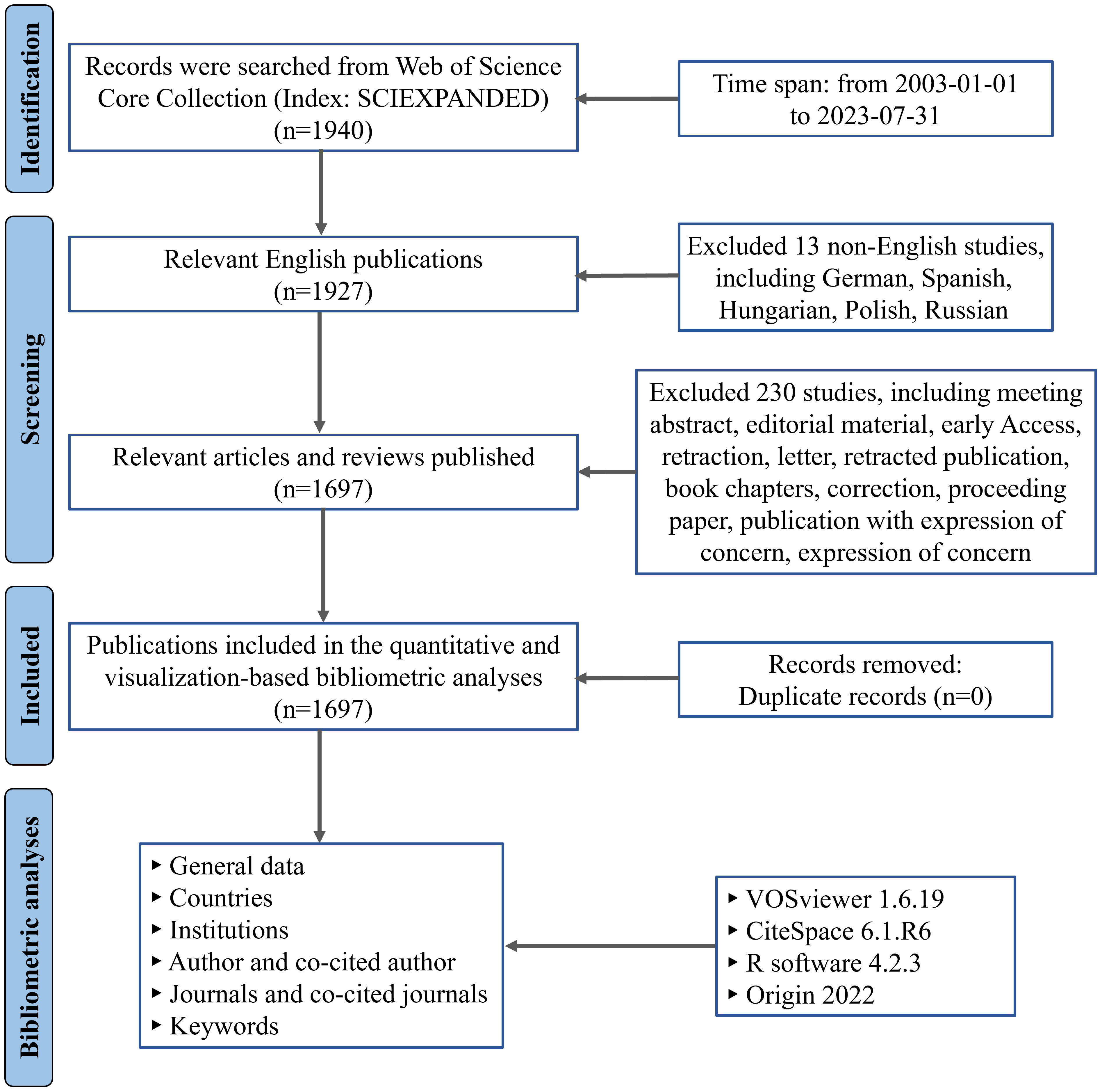
Figure 1 Flowchart for the research’s search procedure.
2.2 Visualized analysis
The Web of Science (WOS) database is widely recognized as the primary source of citation information in bibliometric research, providing general statistical data including publication year, citation count, H-index, etc (18, 19). VOSviewer (version 1.6.19), CiteSpace (version 6.1. R6), R (version 4.2.3), and Origin 2022 were used to analyze data and generate visualization map.
R was used to generate geographical distribution map based on the total publications of different countries between January 1st, 2003, and July 31st, 2023. Origin was used to generate the trend chart of global publications and citations for each year, and the international collaborations’ chord diagram of countries. VOSviewer was used to generate visualization map and analyze the co-occurrence of productive countries, institutions, authors and co-authors, journals and co-cited journals. CiteSpace was used to construct the superposition of journal double maps and the keyword network visualization map from the time zone perspective to display the evolution of research hotspots over time.
In the visualization map, node size is correlated with the type of data being analyzed, typically determined by factors such as the counts of publications, the frequency of occurrence, or the number of citations. Nodes representing correlated data have connecting lines and share the same color. The lines generally represent the cooperation between countries, the association between keywords, or other relevant connections, and the thickness of line indicates the strength of cooperation, which was assessed by total link strength (TLS).
2.3 Research ethics
This study does not require ethical approval because it does not involve human or animal experiments, and the data was obtained from publicly accessible databases.
3 Results
3.1 Global trend in publications and citations
After excluding 243 publications that did not meet the inclusion criteria, a total of 1697 publications between January 1st, 2003, and July 31st, 2023, were obtained from WoSCC database, including 1272 articles and 412 reviews. Figure 2A shows the global trend in publications and total citations for ncRNA-related RA research from January 1st, 2003, to July 31st, 2023. The annual publication count has shown a growing trend over the past two decades, with particularly notable growth in the last five years, during which more than half of all publications were released. All publications have been cited 58522 times as of the search date, with 34.49 average citations per document. Figure 2B illustrates a polynomial fitting curve (R2 = 0.99005) depicting the annual growth trend of publications. As of date (July 31, 2023), there have been over 73 articles published in 2023, with the publication count steadily increasing. Overall, these findings indicate that research on RA-related ncRNA remains highly innovative. Figure 2C shows the annual publication trends related to the main types of ncRNAs (miRNA, lncRNA, and circRNA) in RA. It can be observed that the focus of ncRNA research in the RA field is gradually shifting from miRNA to lncRNA and circRNA. Although there has been an overall decline in the number of ncRNA publications in the field of RA in recent years due to the impact of the COVID-19 pandemic, it is expected that future research on ncRNAs in RA will primarily focus on lncRNA and circRNA and continue to increase.
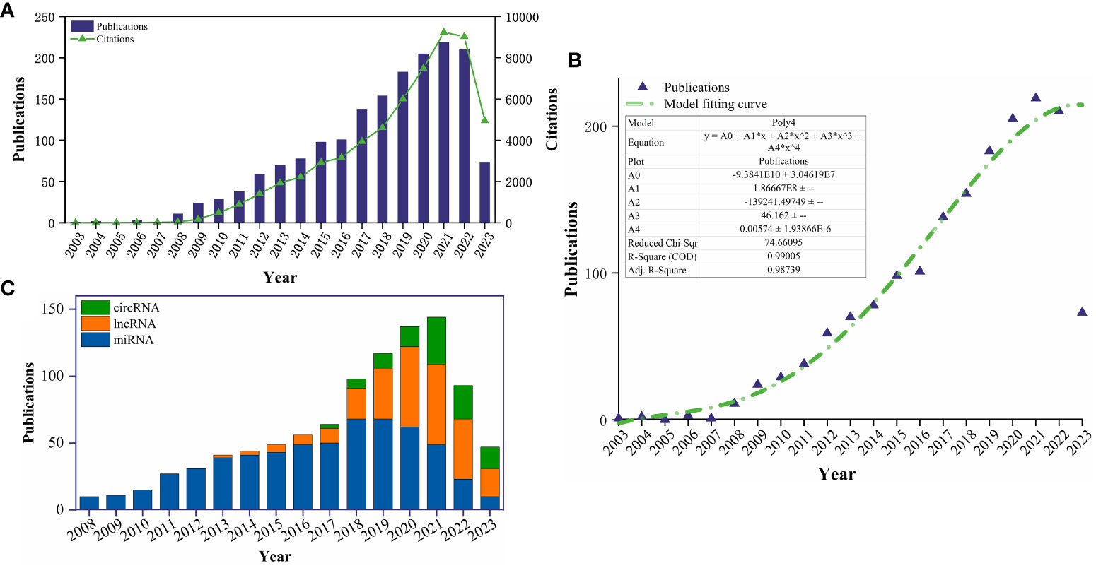
Figure 2 (A) The global annual number of publications and citation on ncRNA-related RA research from 2003-01-01 to 2023-07-31; (B) Model fitting curves of global trends in publications; (C) The annual publication trends related to the main ncRNAs (miRNA, lncRNA, and circRNA) in RA.
3.2 Analysis of countries of the publications
A total of 61 different countries have published publications relating to this topic. According to the geographical distribution map based on the total publications of different countries (Figure 3A), documents related to ncRNA-related RA research were primarily published in China and the United States. Table 1 lists the top ten most productive countries regarding ncRNA and RA research. China had most publications (917, 43.81%), followed by the United States (203, 13.09%), and the United Kingdom (81, 3.87%). Figure 3B shows the trend in annual publication counts of the top 10 most productive countries throughout the period from January 1st, 2003, to July 31st, 2023, and China has the highest annual published growth rate. Moreover, China has most total citations (21811) and the highest H-index (69), indicating China’s positive influence and importance in the field of ncRNA research for RA. Figures 3C, D depict the international cooperation among different countries, the United States had the strongest international cooperation network (TLS=203) and cooperated most closely with China (TLS=113).
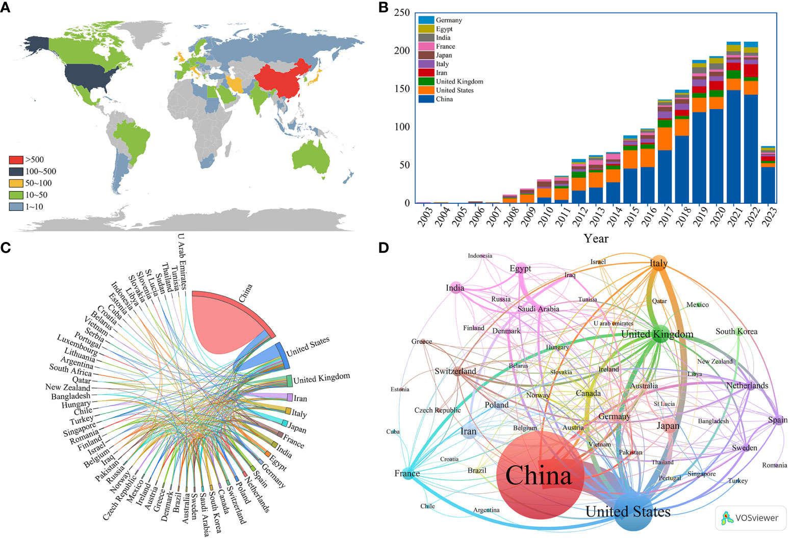
Figure 3 (A) Geographical distribution map based on the total publications of different countries; (B) The trend in annual publication counts of the top 10 most productive countries throughout the period 2003-01-01~2023-07-31; (C) The international collaborations’ visualization map of countries; (D) The co-authorship network visualization map of countries generated by VOSviewer.
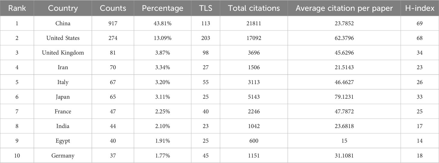
Table 1 The top 10 most productive countries regarding ncRNA and RA research from 2003-01-01 to 2023-07-31.
3.3 Analysis of institutions of the publications
A total of 1827 institutions have contributed to the publications on ncRNA-related RA research. Table 2 shows the top 10 most productive institutions, while Figure 4 illustrates the co-authorship network visualization map of institutions. China has conducted extensive research on ncRNA in RA. The top ten most productive institutions are from China, contributing 345 articles, accounting for 20.57% of the total publications. Most articles were produced by Anhui Medical University, Central South University, Shanghai Jiao Tong University, and China Medical University. Central South University had the highest H-index (23), followed by Anhui Medical University (22) and Shanghai Jiao Tong University (22). Additionally, Shanghai Jiao Tong University had the highest number of total citations, while the Chinese Academy of Sciences had the highest average number of citations per paper. Institutions with at least five publications were presented in the co-authorship network visualization map (Figure 4), which contains 161 nodes and 1478 linkages. China Medical University (TLS = 91), Shanghai Jiao Tong University (TLS = 83), and Anhui Medical University (TLS = 65) are the top three institutions with the highest TLS.
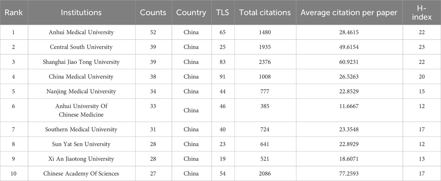
Table 2 The top 10 most productive institutions regarding ncRNA and RA research from 2003-01-01 to 2023-07-31.
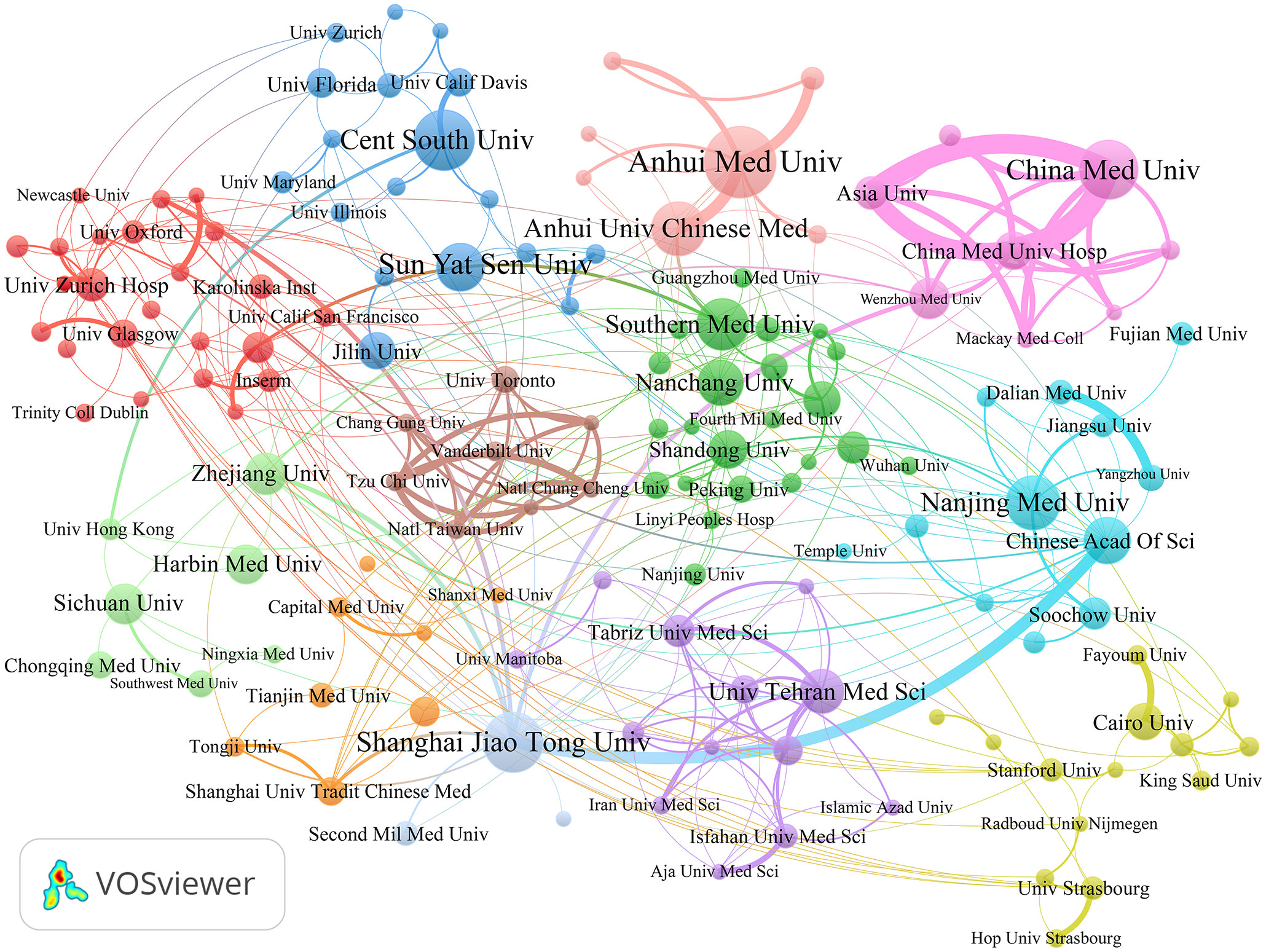
Figure 4 The co-authorship network visualization map of institutions generated by VOSviewer.
3.4 Analysis of authors and co-cited authors of the publications
This study investigates 8589 authors and 40806 co-cited authors (Figure 5). Table 3A shows the top ten most productive authors regarding ncRNA and RA research. Lu, Qianjin (from Central South University) had most publications (21), followed by Gay, Steffen (from University Zurich Hospital) with 19 documents, and Liu, Jian (from Anhui University of Chinese Medicine) with 16 documents. In addition, Lu, Qianjin also had the highest H-index (19), while Gay, Steffen had the most total citations and the highest average number of citations per paper. Their research has a significant impact on the field of ncRNA in RA. Authors who are simultaneously referenced by two (or more) authors in one or even more publications are known as co-cited authors. Table 3B shows the top ten co-cited authors. Stanczyk, J (441 citation), Pauley, Km (386 citation), and Nakasa, T (314 citation) are the top three co-cited authors. They had made an indispensable contribution to the knowledge base of ncRNA-related RA research.
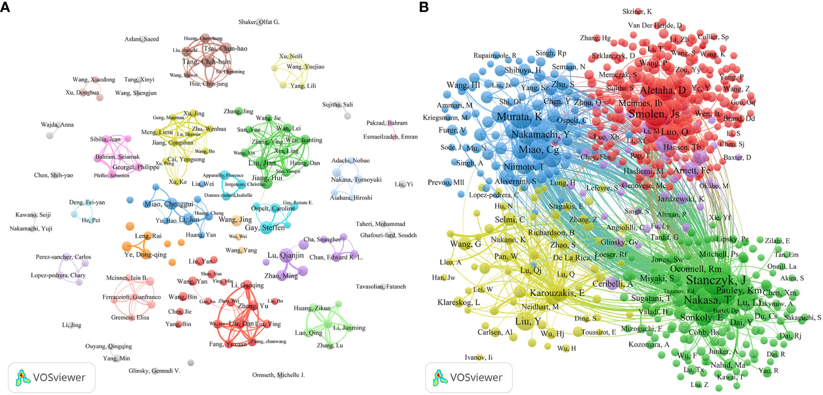
Figure 5 The co-authorship network visualization map of authors (A) and co-cited authors (B) generated by VOSviewer.
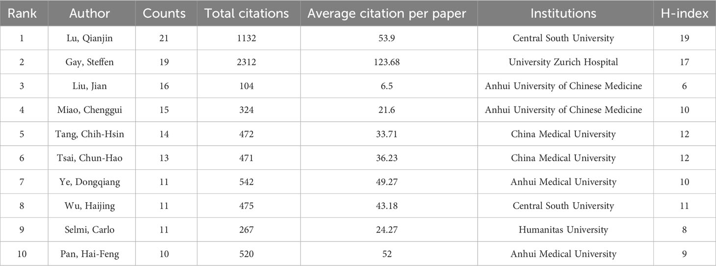
Table 3a The top 10 most productive authors regarding ncRNA and RA research from 2003-01-01 to 2023-07-31.
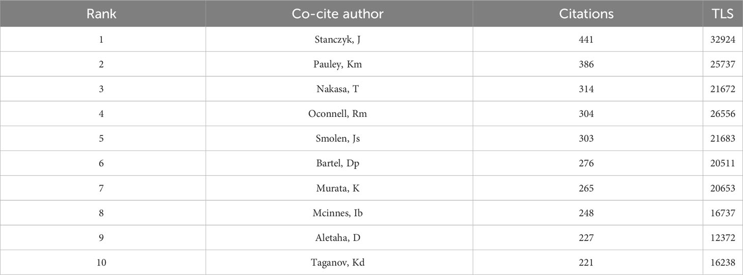
Table 3b The top 10 co-cited authors regarding ncRNA and RA research from 2003-01-01 to 2023-07-31.
3.5 Analysis of journals and co-cited journals of the publications
The top ten most productive journals contributed 327 articles, accounting for 19.27% of the total publications. Frontiers In Immunology (n=73), International Journal Of Molecular Sciences (n=41), Plos One (n=32) are the top three academic journals that publish articles on ncRNA-related RA research out of 504 academic journals. Frontiers In Immunology also had the highest H-index (26), followed by Arthritis Research & Therapy (24) and Plos One (21). Additionally, Arthritis Research & Therapy had the most total citations and the highest average number of citations per paper.
The journal impact factor (IF) is also one of the indicators used to assess the influence of academic journals, including measuring the journal’s reputation, evaluating research quality, and assessing the impact of research outcomes (20). Journal Of Autoimmunity had the highest impact factor (IF 2023 = 12.8), followed by Frontiers In Immunology (IF 2023 = 7.3) and International Journal Of Molecular Sciences (IF 2023 = 5.6). Figure 2B shows the co-occurrence network visualization map of co-cited journals. The top three co-cited journals are Arthritis & Rheumatology (4696 citations), Journal Of Immunology (2457 citations), Arthritis Research & Therapy (2447 citations).
The superposition of journal double maps (Figure 6C) shows the distribution of journals’ topics. Various research fields covered by all the journals are displayed on the labels of the map. The citing journals are displayed on the left side of the map, while the cited journals are shown on the right side. The connecting lines represent that research in certain topics of the cited journals is often cited by research in certain topics of the citing journals, and the width of the lines is closely related to the frequency of citations. There were two main connecting lines on Figure 6C, indicating that research published in the Molecular/Biological/Immunology journals and the Medicine/Medical/Clinical journals generally referenced studies published in the Molecular/Biology/Genetics journals.
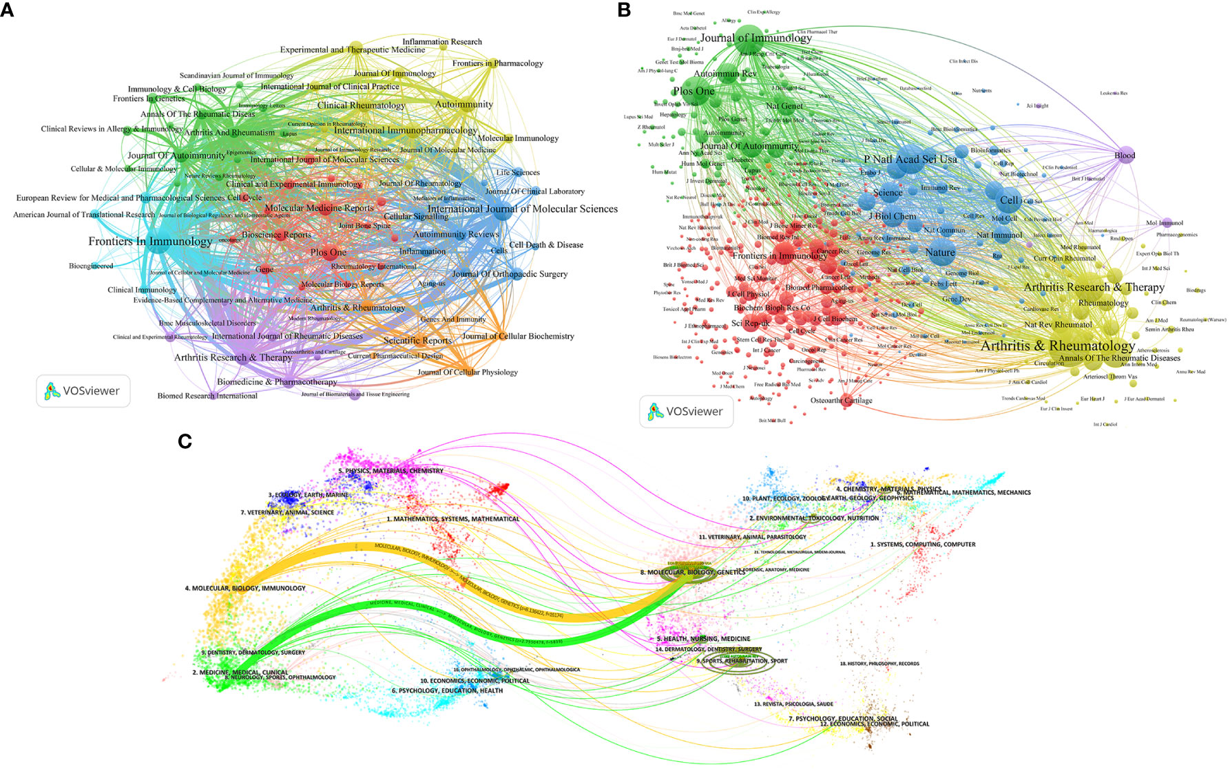
Figure 6 The co-occurrence network visualization map of journals (A) and co-cited journals (B) generated by VOSviewer; (C) The superposition of journal double maps.
3.6 Analysis of keywords of the publications
Keyword co-occurrence analysis is a common method for investigating popular research directions and fields. Figure 7A shows the co-occurrence network visualization map of keywords in cluster view generated by CiteSpace. Through combining the synonyms and analogous keywords and changing g-index from k = 25 to k = 12, a clearer view of the keyword distribution was obtained.
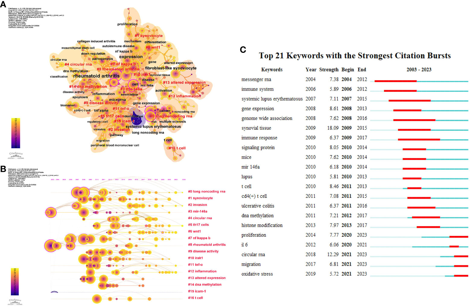
Figure 7 The co-occurrence network visualization map of keywords in Cluster view (A) and timeline view (B) generated by CiteSpace; (C) The top 21 keywords with the strongest citation bursts.
As a result, a total of 319 keywords and 598 links extracted from all 1690 publications were visualized and analyzed. The nodes represent keywords, whose sizes are proportional to the frequency of keywords’ occurrence, and the time span of keywords ranges from January 1st, 2003, to July 31st, 2023. Each node consists of multiple concentric circles, whose color represents the publication time of the article containing the corresponding keywords, and the width of the circles represents the number of articles published within specific time frame. The link between nodes indicates that two keywords appear in the literature simultaneously, and the thickness of the link indicates the frequency of their occurrence, which is known as the degree of association. The top ten keywords (Table 4) are rheumatoid arthritis (1276 times), expression (545 times), fibroblast-like synoviocyte (322 times), microRNA (303 times), cell (226 times), systemic lupus erythematosus (197 times), inflammation (186 times), t cell (157 times), activation (151 times), nf kappa B (144 times). Evidently, research related to non-coding RNA in rheumatoid arthritis primarily involves the altered expression of disease-related genes in fibroblast-like synoviocytes and the activation of immune cells, specifically T cells. These events commonly lead to a series of inflammatory responses which closely associated with the NF-κB pathway.
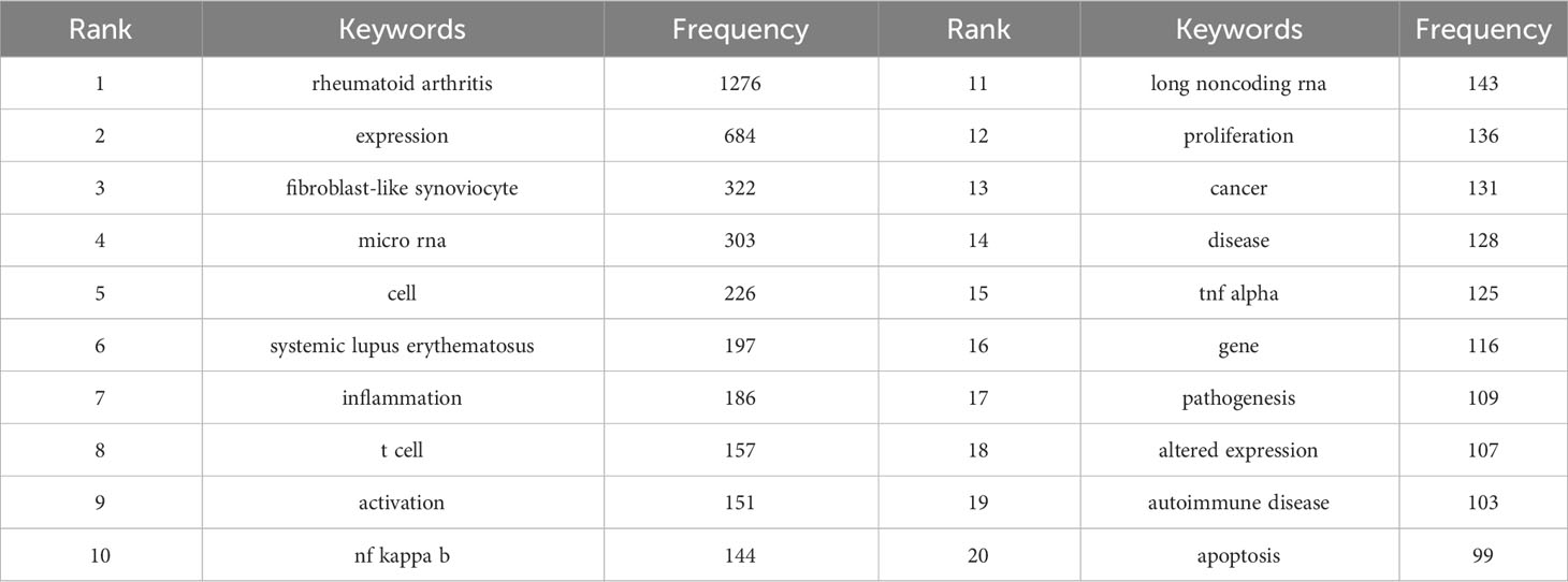
Table 4 The top 20 frequency keywords regarding ncRNA and RA research from 2003-01-01 to 2023-07-31.
Figure 7B presented the visualization map of the keyword evolution in timeline view. The research on topics such as long noncoding RNA, synoviocyte, invasion, miR-146a, and circular RNA has received considerable attention. The keywords with the strongest citation burst are another crucial sign of research frontiers, hotspots, and emerging trends over time. The keywords such as “circular RNA” (2018-2023), “proliferation” (2014-2023), “migration” (2017-2023), and “oxidative stress” (2019-2023) burst continued in 2023, indicating that they are currently hot topics.
In the keywords analysis by CiteSpace, some ncRNAs (especially miRNAs and lncRNAs) had been investigated by many publications, revealing their crucial roles in the onset and diagnosis of RA. Table 5 lists some of the mechanisms of these ncRNAs in the pathogenesis of RA. Clearly, RA-FLSs, with their aberrant proliferation, migration, invasion functions and the ability to secrete a substantial amount of inflammatory factors, play a significant role in the pathogenesis of RA. Additionally, it is worth noting that the keyword analysis did not comprises circRNAs mentioned in several studies. The primary reason is that research on circRNAs in RA is still in nascent stage, requiring additional time to accumulate more relevant research findings. Therefore, we can anticipate more studies in the future elucidating the specific roles of circRNAs in RA.
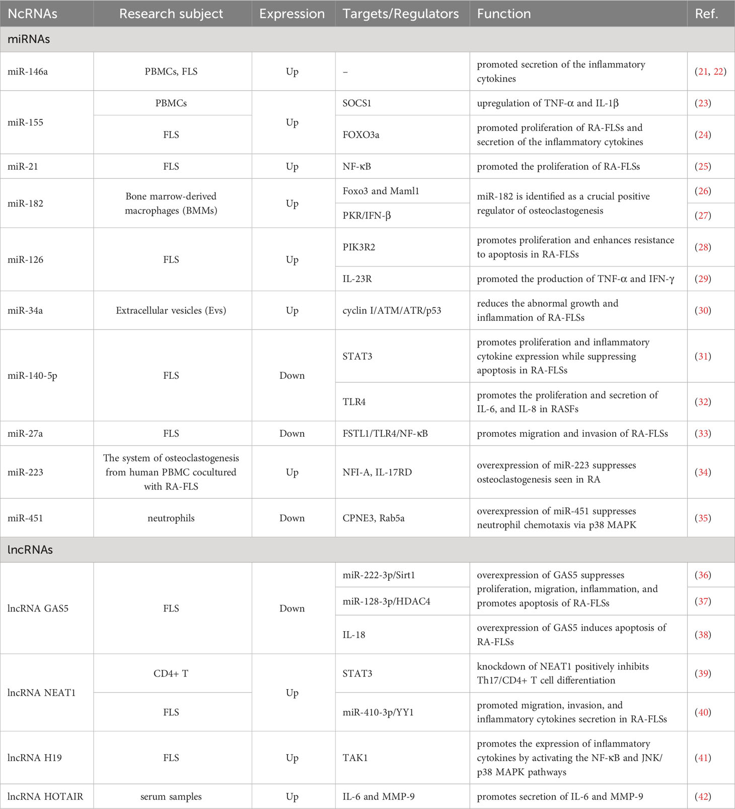
Table 5 The important ncRNAs identified through citespace’s keyword analysis that are associated with the pathogenesis of RA.
4 Discussion
4.1 General information of ncRNA-related RA research
This study performed a systematic literature search of publications on ncRNA-related RA research from January 1, 2003, to July 31, 2023, in the SCI-Expanded database of WOSCC. After excluding publications that did not meet the inclusion criteria, this bibliometric study comprised 1697 English papers published in 504 journals from 1872 institutions in 61 countries. Figures 2A, B show that the number of publications was generally on the rise from 2003 to 2023, which suggests that the research has promising prospects and potential. Although there are no wide variations in the number of publications in the past three years, a new burst may appear in “circular RNA”, “oxidative stress”, “proliferation” and “inflammation” in just a few years.
China has the highest number of publications, citations, and H-Index among all 61 countries, with all the top ten institutions located in China, indicating that China has significant influence in this field. However, the United States, with approximately one-third of the publication count of China, has achieved a similar H-Index, the highest level of collaboration, and the highest average number of citations per paper compared to China, suggesting that the United States continues to dominate in ncRNA-related RA research. This may be attributed to China’s academic evaluation system placing excessive emphasis on the quantity of SCI papers while overlooking quality and innovation, which has resulted in repetitive research lacking innovation. Additionally, there is relatively less international collaboration among Chinese institutions, which also limits China’s influence in the international academic community. Therefore, Chinese institutions and scholars need to focus more on conducting high-quality and innovative research on issues of significant scientific and societal value and expand international cooperation to increase their impact. In addition, with China’s economic development and increasing financial support for medical research, it is foreseeable that China’s academic productivity will continue to rise, and its influence on global academic research will become increasingly significant.
Based on the results of the top ten most productive journals, Frontiers In Immunology and International Journal Of Molecular Sciences can provide a wealth of high-quality literature resources for scholars who seek the latest frontier knowledge on ncRNA-related RA research. The top ten most influential co-cited journals include Arthritis & Rheumatology, Annals Of The Rheumatic Diseases, Nature, Cell, and Proceedings Of The National Academy Of Sciences Of The United States Of America, which cover a wide range of disciplines such as rheumatology, immunology, cell biology, and biochemistry. This indicates that ncRNA is widely recognized and studied in both basic and clinical research, which holds great research prospects significant prospects for RA diagnosis and treatment.
As for the authors, Lu, Qian-Jin is the most productive author and has the highest H-index, who provided an overview of the relationship between the dysregulation of miRNAs and the pathogenesis of autoimmune diseases, as well as the potential applications of miRNAs as biomarkers and therapeutic targets in autoimmune diseases (43–46), which provides insights into miRNA-related RA research.
4.2 Hotspots and emerging frontiers in ncRNA-related RA research
The visualization analysis of keywords (Figure 6C) in miRNA-related RA research by CiteSpace reveals that the latest hot research keywords primarily include “circular RNA”, “oxidative stress”, “proliferation”, and “migration”, and research in these areas has emerged as new hotspots in RA.
Specifically, researchers delved into the biological significance of ncRNAs in RA. In this field, circRNAs have emerged as a new focal point in gene expression regulation owing to its unique circular structure and enhanced stability. In RA, ncRNAs play a crucial role in regulating key cellular processes, encompassing immune response, inflammation, oxidative stress, and tissue damage (Table 5) (47). This emphasizes the potential of ncRNAs as more effective targets for the clinical treatment of RA. The proliferation, migration, and oxidative stress of pathological cells have garnered significant attention as critical steps in disease progression. Additionally, ncRNAs demonstrate specific expression patterns and high stability in disease conditions, making them precisely detectable in certain bodily fluids (such as blood, urine, etc.) (48). Therefore, ncRNA holds significant clinical value in the specific diagnosis of RA.
Overall, these studies lay the foundation for a deeper understanding of the pathogenesis of RA, the development of new treatment strategies, and the application of precision medicine. The continuous and in-depth research in these areas is expected to provide a more comprehensive understanding and effective intervention methods for future rheumatology research and clinical practice.
4.2.1 The role and diagnostic value of circRNA in RA
RA is a severe chronic autoimmune disease that leads to joint inflammation and damage, as well as systemic impairment, which significantly impact patients’ health and quality of life. The early diagnosis and treatment of RA play a crucial role in delaying its progression, helping to prevent irreversible joint damage and disability (49).
The current diagnosis of RA is primarily based on the classification criteria of the American College of Rheumatology (ACR)/European League Against Rheumatism (EULAR) and certain serological markers, including rheumatoid factor (RF) and Anti-cyclic citrullinated peptide antibody (ACPA) (50). However, the limited sensitivity and specificity of these diagnostic markers often result in misdiagnosis and poor prognosis for RA patients (51). CircRNAs are ncRNAs that have been investigated extensively in RA following miRNAs and LncRNAs. Compared to traditional linear RNAs, circRNAs form a covalently closed loop structure which lacks 3’ PolyA tails and 5’ caps, making them effectively resistant to degradation by the ribonuclease R (RNase R) (52). Furthermore, circRNAs exhibit characteristics such as tissue specificity, conservation, and ubiquitous expression (53–56). Due to these characteristics, circRNAs hold greater potential as a diagnostic biomarker for diseases (48, 57–59). Several studies (Table 6) had explored the circRNA expression profiles in peripheral blood mononuclear cells of RA patients by microarray assay and receiver operating characteristic (ROC) analysis, and identified circRNAs such as hsa_circ_0005008, hsa_circ_0140271, and hsa_circ_101328 exhibit specificity and sensitivity in RA, which indicates that circRNAs hold significant promise as a diagnostic biomarker in RA with broad application prospects (9, 67, 68).

Table 6 AUC for circRNAs in PBMCs in discriminating RA patients from healthy controls.
Furthermore, circRNAs offer new perspectives and insights in research related to the pathogenesis and treatment of RA (69). Current studies are primarily focused on the competitive endogenous RNA (ceRNA) mechanism of circRNAs. CircRNAs can function by sponging miRNAs to reduce the number of miRNAs available to target mRNA, thus contributing to mRNA stability or protein expression, which in turn influences the onset and development of RA (70). CircRNAs act as ceRNAs in various cell types associated with RA, including macrophages, helper T cells, chondrocytes, fibroblast-like synoviocyte (FLS), and others, and fibroblast-like synoviocytes are the primary focus of research in this context (Table 7). For example, CircRNA_09505 can promote AKT1 expression by acting as a miR-6089 sponge through the IκBα/NF-κB signaling pathway in macrophages, resulting in increased the production of TNF-α, IL-6, and IL-12, which cause joint inflammation and joint damage (77); CircNUP214 functioned as a miR-125a-3p sponge in RA, which promotes the expression of IL-17A and IL-23R in PBMCs and increases the proportion of Th17 cells, resulting in immune dysregulation and inflammatory responses (92); CircFADS2 promotes the expression of type II collagen, MMP-13, COX-2, and IL-6 via miR-498/mTOR axis, which leads to extracellular matrix (ECM) degradation, inflammation, and cell apoptosis in chondrocytes (72); Circ-PTTG1IP can promote TLR4 expression by acting as a miR-671-5p sponge, promoting the proliferation, migration, invasion and inflammatory response of FLSs in RA (89).
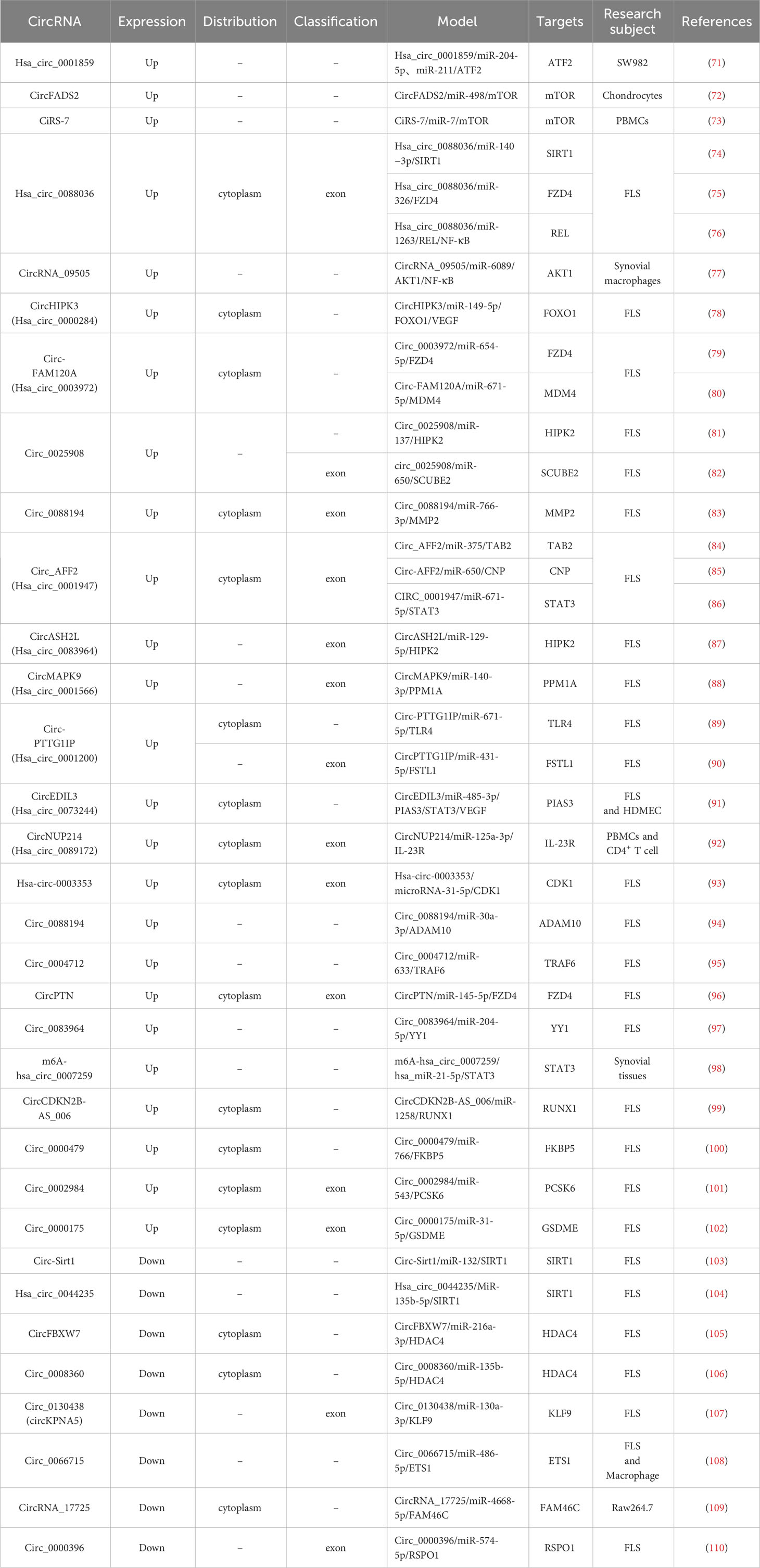
Table 7 CircRNAs related to the Pathogenesis of RA.
Based on the extensive research on circRNAs, researchers are further exploring the clinical applications of circRNAs as therapeutic targets. Compared to mRNA, miRNA, or lncRNA, circRNAs exhibit higher stability in the cytoplasm as therapeutic targets (111, 112). Additionally, circRNAs have lower immunogenicity of unmodified transcripts which will help to avoid activation of the innate immune responses that can decrease gene editing capabilities and disrupt protein production (113, 114). These two advantages can fully realize the potential of circRNAs in gene editing therapies and long-term protein expression regulation (115). Recent research has found that exosomes from circEDIL3-overexpressing synovial mesenchymal stem cells (SMSCs) can reduce VEGF expression by acting as a miR-485-3p sponge through the PIAS3/STAT3 signaling pathway in RA-FLS, and suppress inflammation-induced angiogenesis, which promotes pannus formation, ultimately ameliorated RA (91).
Consequently, circRNAs have started to exhibit the therapeutic potential in vitro. However, the application of circRNAs in clinical settings still requires further research, including how to construct more efficient and specific carriers for the delivery of circRNAs to specific cells or tissues. Additionally, it is important to clarify circRNAs’ pharmacokinetics, biodistribution in the body, and adverse reactions (116).
4.2.2 Oxidative stress is a pivotal factor in rheumatoid arthritis progression, modulated by ncRNAs
Oxidative stress is characterized by the disruption of the intracellular redox balance, resulting in an excessive production of reactive oxygen species (ROS), leading to protein and DNA degradation, as well as lipid peroxidation (117). It plays a crucial role in accelerating the progression of RA and exacerbating clinical symptoms, which includes triggering immune cell activation, promoting synovial inflammation, inducing cell apoptosis and necrosis, increasing joint pain, and initiating joint damage (118–121). Research has revealed that the inflammatory synovial cavity in RA patients is a severely hypoxic environment, which serves as the primary site for oxidative stress and is the fundamental condition for the accumulation of ROS and mitochondrial damage (122). FLS play a pivotal role in the pathogenesis of RA (123). Under hypoxic conditions, FLS exhibit an increased frequency of mitochondrial DNA (mtDNA) mutations, leading to mitochondrial dysfunction, which results in the downregulation of mitochondrial respiration and a decrease in ATP production, along with an upregulation of glycolysis and the accumulation of ROS (124).
Oxidative stress directly activates redox-sensitive transcription factors such as NF-κB, Hypoxia-inducible factor-1α (HIF-1α), Activator Protein-1 (AP-1), and Nuclear factor erythroid2-related factor 2 (Nrf2), which exacerbate the inflammatory responses, oxidative stress, proliferation, invasion, and migration of FLS, induce the recruitment and activation of macrophages and neutrophils that contributing to oxidative stress and inflammatory responses, as well as directly or indirectly contribute to cartilage and bone damage (125–130).
ROS can induce the tyrosine phosphorylation of IκBα by activating Syk kinase, which leads to the dissociation, phosphorylation, and nuclear translocation of NF-κB p65 subunit, thereby stimulating the NF-κB pathway (131, 132). In RA, NF-κB is involved in the induction of various pro-inflammatory cytokines and chemokines, including TNF-α, IL-1β, IL-6, GM-CSF, and others, within monocytes, macrophages, and synovial cells (133). Consequently, these cytokines recruit additional immune cells from the immune system and activate the NF-κB factor, resulting in the initiation of an inflammatory loop (134–136). Furthermore, NF-κB also participates in the regulation of Cyclin D1, Matrix Metalloproteinases (MMPs), and Vascular Endothelial Growth Factor (VEGF), which contribute to the proliferation, migration, and invasion of FLS, as well as cartilage tissue damage (137–139).
HIF-1α is highly regulated by oxygen levels, and in the hypoxic environment of the synovium, HIF-1α accumulates and translocates to the nucleus, where it forms a heterodimer with HIF-1β, known as HIF-1 (140, 141). HIF-1 binds to the HRE promoter thus mediating VEGF expression, thereby leading to angiogenesis (142). Moreover, the upregulation of HIF-1α and VEGF leads to an increase in the expression of stromal cell–derived factor 1α (SDF1α)/CXCL12 mRNA in RA-FLS, which facilitates the recruitment of leukocytes to RA synovial tissue and triggers the release of matrix metalloproteinase 3 (MMP-3) by chondrocytes (143). Furthermore, through the overexpression of HIF-1α in FLS, the upregulation of MMP-1, MMP-3, and IL-8 expression was induced, indicating that HIF-1 can recruit neutrophils, exacerbate inflammation, and induce bone and cartilage damage (144). Studies had found that HIF-1α also plays a significant role in bone cells. Under hypoxic conditions, HIF-1α can upregulate receptor activator of nuclear factor‐κB ligand (RANKL) expression through the JAK2/STAT3 pathway, thereby inducing osteocyte‐mediated osteoclastogenesis (145). HIF-1α can also reduce osteoclast iron death by inhibiting osteoclast iron-starvation response and RANKL-induced ferritinophagy (146). Additionally, HIF-1α can increase the expression and release of angiopoietin-like 4 (ANGPTL4) in various cell types within the rheumatoid synovium, thereby promoting osteoclast-mediated bone resorption (147, 148).
AP-1 is typically a homodimer/heterodimer composed of c-Jun and c-Fos proteins. ROS can activate AP-1 in RA-FLS, leading to the upregulation of IL-1β, MMP-1, MMP-3, and MMP-9 expression (149, 150). Additionally, AP-1 promotes cPLA2 expression by binding to the cPLA2 promoter region, and subsequently increasing the production of eicosanoids like prostaglandin E2 (PGE2), which contributes to inflammation and exacerbates pain (151).
Nrf2 is a crucial factor in the RA antioxidant system, regulating the expression of multiple antioxidant genes (152–154). It is also an important target in current research on ncRNA regulation of oxidative stress in RA. In the cytoplasm, Kelch-like ECH-associated protein-1 (keap1) binds to Nrf2 and promotes its degradation, while ROS can directly activate Nrf2, leading to the dissociation of Nrf2 from Keap1 and its translocation into the cell nucleus, where it interacts with antioxidant response elements (ARE), such as heme oxygenase-1 (HO-1) and superoxide dismutase (SOD), to exert its antioxidant effects (155–157). Currently, multiple studies have revealed the involvement of ncRNAs in regulating Nrf2 expression in RA. For instance, overexpression of miR-30a-3p in RA-FLS of rat can downregulate the expression of Keap1 and cullin 3 (cul3), promoting the expression of Nrf2 and its downstream targets, HO-1 and NAD(P)H: Quinone Oxidoreductase 1 (NQO1), thus exerting antioxidant effects (158). Additionally, overexpression of LINC00638 in RA-FLS activates the Nrf2/HO-1 pathway, leading to the upregulation of SOD2 expression and a reduction in levels of ROS and reactive nitrogen species (RNS) (159).
NcRNAs have the capability to regulate the expression of multiple target genes. Currently, extensive and comprehensive studies have been conducted on ncRNAs in the context of synovial inflammation, vascular angiogenesis, and the destruction of bone and cartilage joints in RA. However, much remains to be discovered about oxidative stress in RA. Oxidative stress can expedite the progression of RA by intensifying the inflammatory loop and causing irreversible joint damage. Hence, the exploration of ncRNAs’ role in regulating oxidative stress in RA shows promise in offering new potential therapeutic approaches to manage the advancement of the disease.
4.2.3 NcRNAs influence the development of RA by regulating the proliferation, migration, and invasion of FLS
FLSs are the predominant cells in synovial tissue, and in RA, they exhibit a high proliferative capacity and an aggressive pathogenic phenotype, invading joint cartilage and bone while secreting MMPs, resulting in joint damage (123, 160, 161). Consequently, it is important to target RA-FLS to suppress their proliferation, migration, and invasive capabilities to counteract joint damage.
Currently, several studies have revealed that ncRNAs play a role in regulating proteins associated with proliferation, migration, and invasion in RA-FLS. MiRNAs can bind to complementary base sequences on target mRNA molecules, resulting in decreased stability, degradation, or inhibition of the translation process of the target mRNA, ultimately reducing the expression level of the target protein (162). For example, miR-4423-3p inhibits the proliferation, migration and invasion of RA-FLS by downregulating the expression level of MMP13 (163); MiR-449a downregulates the expression of High Mobility Group Box 1 (HMGB1) to inhibit the proliferation, migration, and invasion of RA-FLS (164); MicroRNA-133 downregulates mesenchymal-epithelial transition factor (MET), Epidermal Growth Factor Receptor (EGFR), and Fascin Actin-Bundling Protein 1 (FSCN1) to suppress the proliferation, migration, and invasion of RA-FLS (165).
LncRNAs and circRNAs primarily act as sponges for miRNAs, indirectly regulating the expression of target gene mRNAs, thereby promoting the progression of RA (166, 167). Circ_0088194 regulates proliferation, migration, and invasion in RA-FLS through the miR-30a-3p/ADAM10 axis (94). Long non-coding RNA NR-133666 promotes proliferation and migration in RA-FLS through the miR-133c/MAPK1 axis (168). In addition, LncRNAs can also form complexes with proteins, collaboratively participating in the regulation of specific gene expression (166). The elevated expression of LncRNA HAFML in RA-FLS promotes the formation of the HAFML-HuR complex, facilitating the binding of HuR (Human antigen R) to APPL2 mRNA and enhancing its translation, which promotes the migration and invasion of RA-FLS (169). The decreased expression of LncRNA LERFS in RA-FLS leads to a reduction in the complex formation between LERFS and heterogeneous nuclear ribonucleoprotein Q (hnRNP Q), which reduces the binding of the complex to RhoA, Rac1, and Cdc42 mRNA, subsequently promoting the translation process and leading to the proliferation, migration, and invasion of RA-FLS (170).
Evidently, non-coding RNAs (ncRNAs) can effectively modulate the proliferation, migration, and invasion capacities of RA-FLSs through various pathways and targets, thus contributing to the reduction of irreversible damage to joint cartilage and bone (167). Moreover, drugs specifically tailored to target the biological functions of RA-FLS can be locally administered, enabling higher drug concentrations and prolonged therapeutic durations, which enhances treatment efficacy while reducing systemic drug distribution, thereby minimizing adverse effects on other tissues and organs (116, 171). This comprehensive therapeutic strategy represents a promising avenue for RA management.
4.3 Summary and prospects of ncRNA-Related Research in RA
The second part of the discussion primarily emphasizes the role of ncRNAs (particularly circRNAs) in oxidative stress in RA and its impact on the biological functions of RA-FLSs, including proliferation, migration, and invasion. Here, I will summarize the ncRNAs (miRNA and lncRNA) in the pathogenesis of RA that haven’t been extensively detailed in this text.
4.3.1 miRNAs in the pathogenesis of RA
MiRNAs are small, single-stranded transcripts of about 18-24 nucleotides in length. They do not directly encode proteins, instead, they regulate gene expression at both the transcriptional and translational levels (172, 173).
Several studies have indicated abnormal expression of miRNAs in blood or tissue samples from RA patients, suggesting their potential role in the pathogenesis of RA. As listed in Table 5, miR-146a and miR-155 had been consistently identified through multiple studies as being overexpressed in PBMCs (21, 23, 174–176). However, isolating PBMCs and fresh plasma is not a common practice in routine clinical procedures, and there is a possibility of significant alteration in miRNA biomarker level caused by minor variations in cell counts or hemolysis during the isolation of cell-free plasma or blood-derived immune cells (such as PBMCs) (177, 178). Based on these two points, further research has revealed a strong correlation between whole blood samples and PBMC expression levels of miR-155 and miR-146 in healthy controls and RA patients (179). This discovery offers a new perspective on the application of miRNAs as clinical biomarkers in RA, facilitating the use of miRNA-based diagnostics in routine clinical practice by using easily accessible samples, such as whole blood. Moreover, recent evidence has demonstrated a promising platform for detecting certain cancers through profiling miRNAs derived from whole blood (180, 181). This significantly broadens the scope for the application of miRNAs as clinical biomarkers.
Additionally, miRNAs are also aberrantly expressed in RA-FLSs and regulate the inflammatory responses, proliferation, migration, and invasion of RA-FLSs. For example, miR-21 promotes the proliferation of RA-FLS by mediating NF-κB nuclear translocation (25); overexpression of miR-27a inhibits migration and invasion of RA-FLS and simultaneously decreases the expression of MMP2, MMP9, and MMP13 via the FSTL1/TLR4/NF-κB axis (33); miR-34a can be transported to RA-FLS via extracellular vesicles (EVs), which reduced inflammation, proliferation, and resistance to apoptosis of RA-FLS by inhibiting the cyclin I/ATM/ATR/p53 signaling pathway (30). MiRNAs can also regulate immune cells and the differentiation of bone cells to modulate the progression of RA. For instance, miR-146a enhances a pro-inflammatory phenotype in Tregs by upregulating the expression of STAT1 (182); miR-182 had been identified as a critical positive regulator of osteoclastogenesis, which is associated with the Foxo3, Maml1 and PKR/IFN-β signaling pathways (26, 27).
4.3.2 LncRNAs in the pathogenesis of RA
LncRNAs are transcripts with a length larger than 200 nucleotides that regulate gene expression through various mechanisms, including epigenetic regulation, transcriptional control, and post-transcriptional regulation. The regulatory mechanisms of lncRNAs mainly encompass (183, 184): (1) LncRNAs can act as sponges for miRNAs, indirectly regulating the expression of target gene mRNAs; (2) LncRNAs can interact with proteins, influencing the binding of proteins to chromatin DNA, altering chromatin structure, and affecting the activity of transcription factors, thus regulating gene transcription; (3) LncRNAs recruit chromatin-modifying enzymes (such as methyltransferases and deacetylases) to specific gene regions, altering histone modification patterns, and modulating chromatin status and gene expression; (4) LncRNAs can form complexes with transcription factors or other RNAs, serving as part of ribonucleoprotein complexes, recruiting other proteins or RNAs, thereby influencing the formation and function of transcription complexes; (5) LncRNAs can regulate mRNA processing, splicing, stability, and translation in the post-transcriptional phase, affecting the final products of gene expression. Currently, the dysregulation of lncRNAs is also observed in the PBMCs, FLS, and immune cells of RA patients, suggesting that lncRNAs might influence the progression of RA by impacting the function of these cells.
Table 5 shows the important LncRNAs associated with the pathogenesis of RA. LncRNA GAS5 can be upregulated by Tanshinone IIA, thereby enhancing the expression of caspase-3/caspase-9 and suppressing the PI3K/AKT signaling pathway, resulting in the apoptosis of RA-FLS (185). This demonstrates the functional capacity of LncRNAs as potential therapeutic targets and their effectiveness as biomarkers to evaluate treatment responses in RA patients. Shui X (39) exploring the dysregulated lncRNAs in PBMCs and differentiated Th17 cells of RA patients and observed an upregulation of LncRNA NEAT1. Additionally, knockdown of lncRNA NEAT1 inhibits Th17/CD4+ T cell differentiation through reducing the STAT3 protein level (39). LncRNA H19 expression was found to be higher in PBMCs of RA patients compared to healthy volunteers (186). Overexpression of H19 in PMA-induced THP-1 cells (a common model used to study macrophage activity modulation) upregulates KDM6A and various M1 macrophage-associated factors (particularly CCL8, CXCL9, CXCL10, and CXCL11) which promote M1 macrophage activity and macrophage migration (186). LncRNA H19 can also aggravate TNF-α-induced inflammatory injury via TAK1 pathway in MH7A cells (41). LncRNA Hotair exhibited notably high expression levels in the PBMCs and serum exosomes of RA patients, promoting the migration of activated macrophages (187). Meanwhile, markedly lower levels of LncRNA Hotair were detected in differentiated osteoclasts and RA-FLS, and upregulating LncRNA Hotair can decrease the activation of MMP-2 and MMP-13 in both differentiated osteoclasts and RA-FLS (187).
4.3.3 Summary and prospects
In general, whether it’s miRNAs, lncRNAs, or circRNAs, their roles in the pathogenesis of RA are quite similar and can be summarized in the following three points: (1) ncRNAs regulates the inflammatory response of immune cells in the synovium (188); (2) the expression of ncRNAs influences the biological behavior of RA-FLSs, including affecting inflammation, proliferation, migration, and invasion functions; (3) ncRNAs participate in bone metabolism in RA, promoting the secretion of MMPs by RA-FLSs to disrupt the bone matrix and regulating the activation of osteoclasts. During the process in which non-coding RNA exerts its influence, miRNA, being relatively short, exhibits high specificity when binding to target genes. However, this characteristic also limits its diversity in molecular interactions. On the other hand, lncRNA, usually longer and forming stable secondary or tertiary structures, remains relatively stable in cells, resisting degradation by RNase R. Its more complex structure and diverse functional domains allow it to interact with multiple molecules. CircRNAs, share similar functions with lncRNAs, have a circular structure that provides an advantage in resisting degradation compared to linear RNA. While circRNAs and lncRNAs have become hot topics in research due to their structural and functional advantages, it is undeniable that the regulatory roles played by circRNA, lncRNA, and miRNA at the cellular and molecular levels should not be overlooked. Further clarification of the regulatory network constituted by circRNA, lncRNA, and miRNA will be a major research direction in the future (10).
In addition to understanding the role of ncRNAs in the pathogenesis of RA, exploring their application in the diagnosis and treatment of RA is equally crucial. The future application of ncRNAs in RA might develop in several aspects: (1) ncRNA as a disease biomarker for RA diagnosis; (2) ncRNA as a predictor of complications in RA patients, such as cardiovascular complications (189); (3) ncRNA as a biomarker for assessing the treatment response of RA patients (190); (4) the use of engineered exosomes containing ncRNA for RA treatment (13). Exploration in these areas is essential for advancing our comprehension of RA pathogenesis and enhancing diagnostic and treatment approaches.
5 Limitations
This study provides a bibliometric and visual analysis of the current research status and trends related to ncRNAs in RA. However, there are certain limitations to this research that need to be acknowledged. Firstly, the study exclusively gathered English articles and reviews from the WoSCC-SCI-EXPANDED database, with a literature collection cut-off date of July 31, 2023, potentially excluding some relevant publications. Secondly, software algorithms like Citespace and VOSviewer have limitations in that they cannot analyze the full text of publications, leading to the omission of certain details and some margin of error in the results. Thirdly, the total number of citations usually rises over time. Thus, early publications have a higher likelihood of being referenced compared to newly released ones, which enhancing the significance of information gathered from early publications in Tables 2 and 8. Fourthly, this study did not conduct a quality assessment of the included publications, treating all of them as having equal validity. It is hoped that future research can address these limitations by collecting literature data from additional databases and employing quality assessment methods to provide a more comprehensive and accurate understanding of non-coding RNA research in RA.
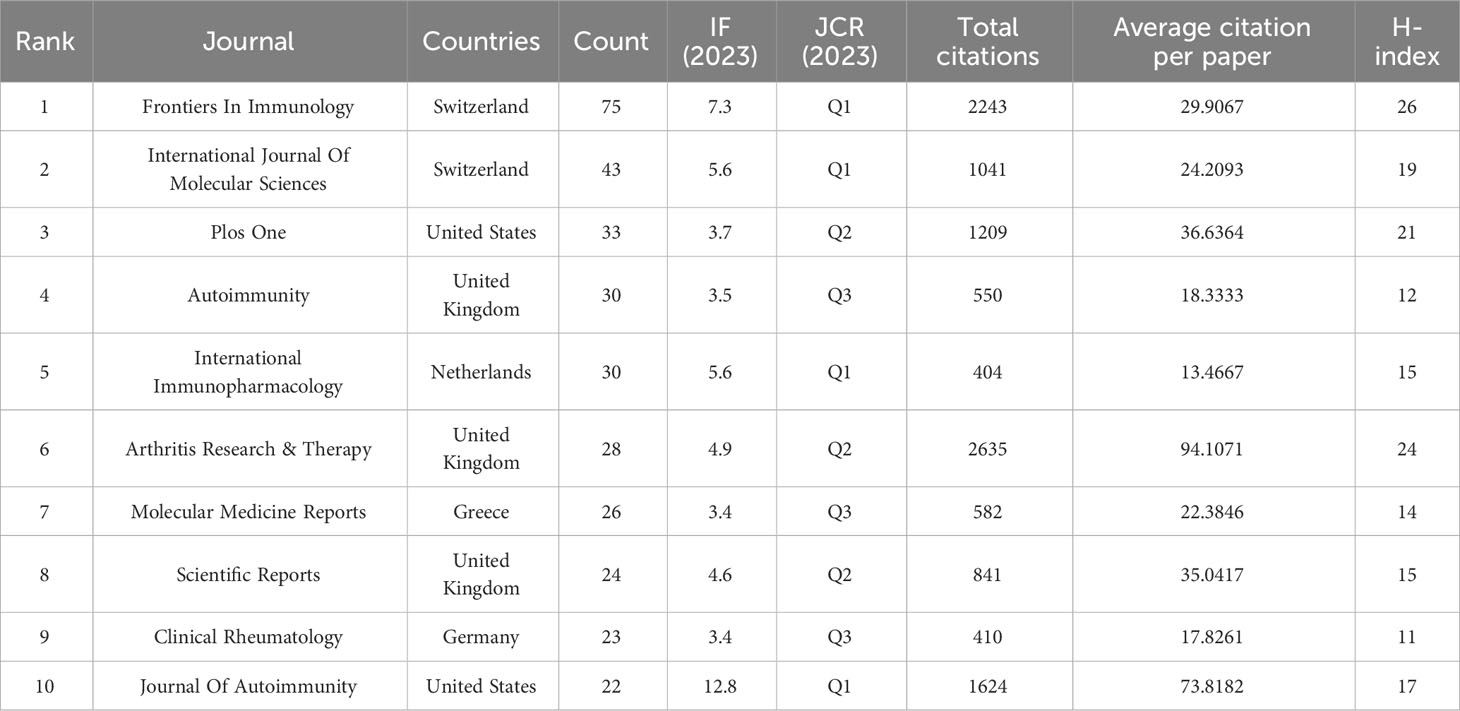
Table 8a The top 10 most productive journals regarding ncRNA and RA research from 2003-01-01 to 2023-07-31.
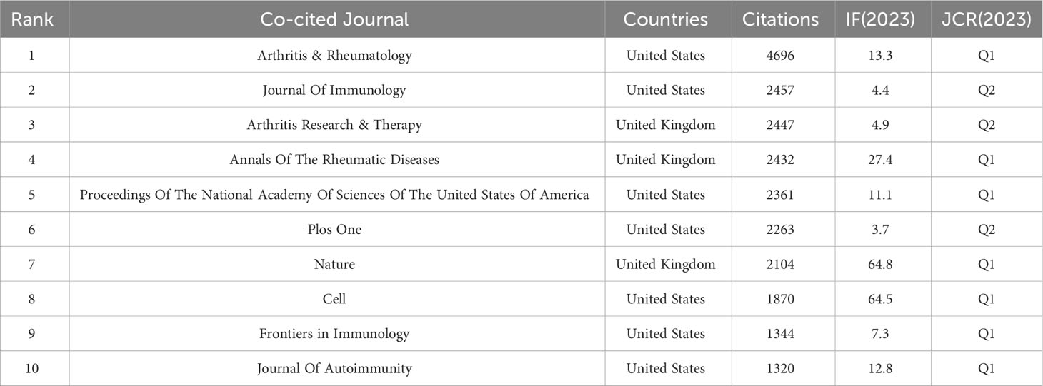
Table 8b The top 10 co-cited journals regarding ncRNA and RA research from 2003-01-01 to 2023-07-31.
6 Conclusion
Over the past two decades, as the role of non-coding RNAs in the pathogenesis of RA has been increasingly unveiled, their potential value in the diagnosis and treatment of RA has garnered growing attention. This study conducted a bibliometric analysis of literature related to ncRNAs in RA using various statistical analysis tools. It has revealed the countries, institutions, authors, and journals with the most significant impact in this field. Additionally, it has identified the hot trends in ncRNA research concerning the pathogenesis, diagnosis, and treatment of RA. Currently, circRNAs and the biological functions of RA-FLS have emerged as new research focal points. In the future, targeted drugs designed based on circRNA regulatory mechanisms may become potential therapeutic strategies for controlling the progression of RA by targeting RA-FLS. This trend underscores the importance of non-coding RNAs in the field of RA research and provides a promising direction for future therapeutic investigations.
Data availability statement
Publicly available datasets were analyzed in this study. This data can be found here: https://www.webofscience.com/wos/woscc/summary/868a42b7-1929-4c31-8a06-3b183a5293a1-9ea62624/relevance/1.
Author contributions
ZW: Data curation, Investigation, Software, Visualization, Writing – original draft, Writing – review & editing. HL: Methodology, Writing – review & editing. SL: Data curation, Investigation, Writing – review & editing. JY: Writing – review & editing, Funding acquisition, Project administration, Resources, Supervision.
Funding
The author(s) declare financial support was received for the research, authorship, and/or publication of this article. This work was supported by the National Natural Science Foundation of China (81460769), Jiangxi Provincial Department of Education Scientific and Technological Research Project (GJJ2200935) and 2023 Jiangxi University of Chinese Medicine Graduate Innovation Special Fund Project (JZYC23S12).
Conflict of interest
The authors declare that the research was conducted in the absence of any commercial or financial relationships that could be construed as a potential conflict of interest.
Publisher’s note
All claims expressed in this article are solely those of the authors and do not necessarily represent those of their affiliated organizations, or those of the publisher, the editors and the reviewers. Any product that may be evaluated in this article, or claim that may be made by its manufacturer, is not guaranteed or endorsed by the publisher.
References
1. Smolen JS, Aletaha D, Barton A, Burmester GR, Emery P, Firestein GS, et al. Rheumatoid arthritis. Nat Rev Dis Primers (2018) 4:18001. doi: 10.1038/nrdp.2018.1
2. Safiri S, Kolahi AA, Hoy D, Smith E, Bettampadi D, Mansournia MA, et al. Global, regional and national burden of rheumatoid arthritis 1990-2017: a systematic analysis of the Global Burden of Disease study 2017. Ann Rheum Dis (2019) 78(11):1463–71. doi: 10.1136/annrheumdis-2019-215920
3. Otón T, Carmona L. The epidemiology of established rheumatoid arthritis. Best Pract Res Clin Rheumatol (2019) 33(5):101477. doi: 10.1016/j.berh.2019.101477
4. Smolen JS, Aletaha D, Mcinnes IB. Rheumatoid arthritis. Lancet (2016) 388(10055):2023–38. doi: 10.1016/S0140-6736(16)30173-8
5. Mcinnes IB, Schett G. The pathogenesis of rheumatoid arthritis. N Engl J Med (2011) 365(23):2205–19. doi: 10.1056/NEJMra1004965
6. Fraenkel L, Bathon JM, England BR, St Clair EW, Arayssi T, Carandang K, et al. 2021American college of rheumatology guideline for the treatment of rheumatoid arthritis. Arthritis Rheumatol (2021) 73(7):1108–23. doi: 10.1002/art.41752
7. Goodman SM, Springer B, Guyatt G, Abdel MP, Dasa V, George M, et al. 2017 American college of rheumatology/American association of hip and knee surgeons guideline for the perioperative management of antirheumatic medication in patients with rheumatic diseases undergoing elective total hip or total knee arthroplasty. Arthritis Rheumatol (2017) 69(8):1538–51. doi: 10.1002/art.40149
8. Panni S, Lovering RC, Porras P, Orchard S. Non-coding RNA regulatory networks. Biochim Biophys Acta Gene Regul Mech (2020) 1863(6):194417. doi: 10.1016/j.bbagrm.2019.194417
9. Lu H, Yang Y, Kuang D, Liu P, Yang J. Expression profile of circRNA in peripheral blood mononuclear cells of patients with rheumatoid arthritis. BMC Med Genomics (2022) 15(1):77. doi: 10.1186/s12920-022-01225-9
10. Xu D, Jiang Y, Yang L, Hou X, Wang J, Gu W, et al. Long noncoding RNAs expression profile and functional networks in rheumatoid arthritis. Oncotarget (2017) 8(56):95280–92. doi: 10.18632/oncotarget.20036
11. Taheri M, Eghtedarian R, Dinger ME, Ghafouri-Fard S. Dysregulation of non-coding RNAs in Rheumatoid arthritis. BioMed Pharmacother (2020) 130:110617. doi: 10.1016/j.biopha.2020.110617
12. Ali SA, Peffers MJ, Ormseth MJ, Jurisica I, Kapoor M. The non-coding RNA interactome in joint health and disease. Nat Rev Rheumatol (2021) 17(11):692–705. doi: 10.1038/s41584-021-00687-y
13. Li C, Ni YQ, Xu H, Xiang QY, Zhao Y, Zhan JK, et al. Roles and mechanisms of exosomal non-coding RNAs in human health and diseases. Signal Transduct Target Ther (2021) 6(1):383. doi: 10.1038/s41392-021-00779-x
14. Xu J, Yu J, Jiao W, Chen G, Liu L, Zhang M, et al. Scientific knowledge of rheumatoid arthritis: A bibliometric analysis from 2011 to 2020. J Pain Res (2022) 15:2761–72. doi: 10.2147/JPR.S362717
15. Pei Z, Chen S, Ding L, Liu J, Cui X, Li F, et al. Current perspectives and trend of nanomedicine in cancer: A review and bibliometric analysis. J Control Release (2022) 352:211–41. doi: 10.1016/j.jconrel.2022.10.023
16. Van Eck NJ, Waltman L. Software survey: VOSviewer, a computer program for bibliometric mapping. Scientometrics (2010) 84(2):523–38. doi: 10.1007/s11192-009-0146-3
17. Chen C. Searching for intellectual turning points: progressive knowledge domain visualization. Proc Natl Acad Sci U.S.A. (2004) 101 Suppl 1(Suppl 1):5303–10. doi: 10.1073/pnas.0307513100
18. Liu J, Gao J, Niu Q, Wu F, Wu Z, Zhang L. Bibliometric and visualization analysis of mesenchymal stem cells and rheumatoid arthritis (from 2012 to 2021). Front Immunol (2022) 13:1001598. doi: 10.3389/fimmu.2022.1001598
19. Xu Y, Zhang Z, He J, Chen Z. Immune effects of macrophages in rheumatoid arthritis: A bibliometric analysis from 2000 to 2021. Front Immunol (2022) 13:903771. doi: 10.3389/fimmu.2022.903771
20. Garfield E. The history and meaning of the journal impact factor. Jama (2006) 295(1):90–3. doi: 10.1001/jama.295.1.90
21. Abou-Zeid A, Saad M, Soliman E. MicroRNA 146a expression in rheumatoid arthritis: association with tumor necrosis factor-alpha and disease activity. Genet Test Mol Biomarkers (2011) 15(11):807–12. doi: 10.1089/gtmb.2011.0026
22. Nakasa T, Miyaki S, Okubo A, Hashimoto M, Nishida K, Ochi M, et al. Expression of microRNA-146 in rheumatoid arthritis synovial tissue. Arthritis Rheum (2008) 58(5):1284–92. doi: 10.1002/art.23429
23. Li X, Tian F, Wang F. Rheumatoid arthritis-associated microRNA-155 targets SOCS1 and upregulates TNF-α and IL-1β in PBMCs. Int J Mol Sci (2013) 14(12):23910–21. doi: 10.3390/ijms141223910
24. Wang Y, Feng T, Duan S, Shi Y, Li S, Zhang X, et al. miR-155 promotes fibroblast-like synoviocyte proliferation and inflammatory cytokine secretion in rheumatoid arthritis by targeting FOXO3a. Exp Ther Med (2020) 19(2):1288–96. doi: 10.3892/etm.2019.8330
25. Chen Y, Xian PF, Yang L, Wang SX. MicroRNA-21 promotes proliferation of fibroblast-like synoviocytes through mediation of NF-κB nuclear translocation in a rat model of collagen-induced rheumatoid arthritis. BioMed Res Int (2016) 2016:9279078. doi: 10.1155/2016/9279078
26. Miller CH, Smith SM, Elguindy M, Zhang T, Xiang JZ, Hu X, et al. RBP-J-regulated miR-182 promotes TNF-α-induced osteoclastogenesis. J Immunol (2016) 196(12):4977–86. doi: 10.4049/jimmunol.1502044
27. Inoue K, Deng Z, Chen Y, Giannopoulou E, Xu R, Gong S, et al. Bone protection by inhibition of microRNA-182. Nat Commun (2018) 9(1):4108. doi: 10.1038/s41467-018-06446-0
28. Gao J, Zhou XL, Kong RN, Ji LM, He LL, Zhao DB. microRNA-126 targeting PIK3R2 promotes rheumatoid arthritis synovial fibro-blasts proliferation and resistance to apoptosis by regulating PI3K/AKT pathway. Exp Mol Pathol (2016) 100(1):192–8. doi: 10.1016/j.yexmp.2015.12.015
29. Gao J, Kong R, Zhou X, Ji L, Zhang J, Zhao D. MiRNA-126 expression inhibits IL-23R mediated TNF-α or IFN-γ production in fibroblast-like synoviocytes in a mice model of collagen-induced rheumatoid arthritis. Apoptosis (2018) 23(11-12):607–15. doi: 10.1007/s10495-018-1474-7
30. Wu H, Zhou X, Wang X, Cheng W, Hu X, Wang Y, et al. miR-34a in extracellular vesicles from bone marrow mesenchymal stem cells reduces rheumatoid arthritis inflammation via the cyclin I/ATM/ATR/p53 axis. J Cell Mol Med (2021) 25(4):1896–910. doi: 10.1111/jcmm.15857
31. Zhu J, Wang J, Huang J, Du W, He Y, Pan H, et al. MicroRNA-140-5p regulates the proliferation, apoptosis and inflammation of RA FLSs by repressing STAT3. Exp Ther Med (2021) 21(2):171. doi: 10.3892/etm.2020.9602
32. Li H, Guan SB, Lu Y, Wang F. MiR-140-5p inhibits synovial fibroblasts proliferation and inflammatory cytokines secretion through targeting TLR4. BioMed Pharmacother (2017) 96:208–14. doi: 10.1016/j.biopha.2017.09.079
33. Shi DL, Shi GR, Xie J, Du XZ, Yang H. MicroRNA-27a inhibits cell migration and invasion of fibroblast-like synoviocytes by targeting follistatin-like protein 1 in rheumatoid arthritis. Mol Cells (2016) 39(8):611–8. doi: 10.14348/molcells.2016.0103
34. Shibuya H, Nakasa T, Adachi N, Nagata Y, Ishikawa M, Deie M, et al. Overexpression of microRNA-223 in rheumatoid arthritis synovium controls osteoclast differentiation. Mod Rheumatol (2013) 23(4):674–85. doi: 10.3109/s10165-012-0710-1
35. Murata K, Yoshitomi H, Furu M, Ishikawa M, Shibuya H, Ito H, et al. MicroRNA-451 down-regulates neutrophil chemotaxis via p38 MAPK. Arthritis Rheumatol (2014) 66(3):549–59. doi: 10.1002/art.38269
36. Yang Z, Lin SD, Zhan F, Liu Y, Zhan YW. LncRNA GAS5 alleviates rheumatoid arthritis through regulating miR-222-3p/Sirt1 signalling axis. Autoimmunity (2021) 54(1):13–22. doi: 10.1080/08916934.2020.1846183
37. Peng T, Ji D, Jiang Y. Long non-coding RNA GAS5 suppresses rheumatoid arthritis progression via miR-128-3p/HDAC4 axis. Mol Cell Biochem (2021) 476(6):2491–501. doi: 10.1007/s11010-021-04098-1
38. Ma C, Wang W, Li P. LncRNA GAS5 overexpression downregulates IL-18 and induces the apoptosis of fibroblast-like synoviocytes. Clin Rheumatol (2019) 38(11):3275–80. doi: 10.1007/s10067-019-04691-2
39. Shui X, Chen S, Lin J, Kong J, Zhou C, Wu J. Knockdown of lncRNA NEAT1 inhibits Th17/CD4(+) T cell differentiation through reducing the STAT3 protein level. J Cell Physiol (2019) 234(12):22477–84. doi: 10.1002/jcp.28811
40. Wang Y, Hou L, Yuan X, Xu N, Zhao S, Yang L, et al. LncRNA NEAT1 Targets Fibroblast-Like Synoviocytes in Rheumatoid Arthritis via the miR-410-3p/YY1 Axis. Front Immunol (2020) 11:1975. doi: 10.3389/fimmu.2020.01975
41. Yang J, Li Y, Wang L, Zhang Z, Li Z, Jia Q. LncRNA H19 aggravates TNF-α-induced inflammatory injury via TAK1 pathway in MH7A cells. Biofactors (2020) 46(5):813–20. doi: 10.1002/biof.1659
42. Shaker OG, Mahmoud RH, Abdelaleem OO, Ahmed TI, Fouad NA, Hussein HA, et al. Expression profile of long noncoding RNAs, lnc-cox2, and HOTAIR in rheumatoid arthritis patients. J Interferon Cytokine Res (2019) 39(3):174–80. doi: 10.1089/jir.2018.0117
43. Long H, Wang X, Chen Y, Wang L, Zhao M, Lu Q. Dysregulation of microRNAs in autoimmune diseases: Pathogenesis, biomarkers and potential therapeutic targets. Cancer Lett (2018) 428:90–103. doi: 10.1016/j.canlet.2018.04.016
44. Zhang L, Wu H, Zhao M, Chang C, Lu Q. Clinical significance of miRNAs in autoimmunity. J Autoimmun (2020) 109:102438. doi: 10.1016/j.jaut.2020.102438
45. Zhang L, Wu H, Zhao M, Lu Q. Identifying the differentially expressed microRNAs in autoimmunity: A systemic review and meta-analysis. Autoimmunity (2020) 53(3):122–36. doi: 10.1080/08916934.2019.1710135
46. Zeng L, Cui J, Wu H, Lu Q. The emerging role of circulating microRNAs as biomarkers in autoimmune diseases. Autoimmunity (2014) 47(7):419–29. doi: 10.3109/08916934.2014.929667
47. Wang J, Yan S, Yang J, Lu H, Xu D, Wang Z. Non-coding RNAs in rheumatoid arthritis: from bench to bedside. Front Immunol (2019) 10:3129. doi: 10.3389/fimmu.2019.03129
48. Jiang Y, Zhong S, He S, Weng J, Liu L, Ye Y, et al. Biomarkers (mRNAs and non-coding RNAs) for the diagnosis and prognosis of rheumatoid arthritis. Front Immunol (2023) 14:1087925. doi: 10.3389/fimmu.2023.1087925
49. Aletaha D, Smolen JS. Diagnosis and management of rheumatoid arthritis: A review. Jama (2018) 320(13):1360–72. doi: 10.1001/jama.2018.13103
50. Aletaha D, Neogi T, Silman AJ, Funovits J, Felson DT, Bingham CO 3rd, et al. 2010 rheumatoid arthritis classification criteria: an American College of Rheumatology/European League Against Rheumatism collaborative initiative. Ann Rheum Dis (2010) 69(9):1580–8. doi: 10.1136/ard.2010.138461
51. Boeters DM, Gaujoux-Viala C, Constantin A, Van Der Helm-Van Mil AHM. The 2010 ACR/EULAR criteria are not sufficiently accurate in the early identification of autoantibody-negative rheumatoid arthritis: Results from the Leiden-EAC and ESPOIR cohorts. Semin Arthritis Rheum (2017) 47(2):170–4. doi: 10.1016/j.semarthrit.2017.04.009
52. Chen LL, Yang L. Regulation of circRNA biogenesis. RNA Biol (2015) 12(4):381–8. doi: 10.1080/15476286.2015.1020271
53. Patop IL, Wüst S, Kadener S. Past, present, and future of circRNAs. EMBO J (2019) 38(16):e100836. doi: 10.15252/embj.2018100836
54. Kristensen LS, Andersen MS, Stagsted LVW, Ebbesen KK, Hansen TB, Kjems J. The biogenesis, biology and characterization of circular RNAs. Nat Rev Genet (2019) 20(11):675–91. doi: 10.1038/s41576-019-0158-7
55. Li X, Yang L, Chen LL. The biogenesis, functions, and challenges of circular RNAs. Mol Cell (2018) 71(3):428–42. doi: 10.1016/j.molcel.2018.06.034
56. Salzman J, Chen RE, Olsen MN, Wang PL, Brown PO. Cell-type specific features of circular RNA expression. PloS Genet (2013) 9(9):e1003777. doi: 10.1371/journal.pgen.1003777
57. Zhou WY, Cai ZR, Liu J, Wang DS, Ju HQ, Xu RH. Circular RNA: metabolism, functions and interactions with proteins. Mol Cancer (2020) 19(1):172. doi: 10.1186/s12943-020-01286-3
58. Xu Y, Chen F. Current status of functional studies on circular RNAs in rheumatoid arthritis and their potential role as diagnostic biomarkers. J Inflammation Res (2021) 14:1185–93. doi: 10.2147/JIR.S302846
59. Pan YH, Wu WP, Xiong XD. Circular RNAs: promising biomarkers for age-related diseases. Aging Dis (2020) 11(6):1585–93. doi: 10.14336/AD.2020.0309
60. Ouyang Q, Wu J, Jiang Z, Zhao J, Wang R, Lou A, et al. Microarray expression profile of circular RNAs in peripheral blood mononuclear cells from rheumatoid arthritis patients. Cell Physiol Biochem (2017) 42(2):651–9. doi: 10.1159/000477883
61. Yang X, Li J, Wu Y, Ni B, Zhang B. Aberrant dysregulated circular RNAs in the peripheral blood mononuclear cells of patients with rheumatoid arthritis revealed by RNA sequencing: novel diagnostic markers for RA. Scand J Clin Lab Invest (2019) 79(8):551–9. doi: 10.1080/00365513.2019.1674004
62. Luo Q, Zhang L, Li X, Fu B, Deng Z, Qing C, et al. Identification of circular RNAs hsa_circ_0044235 in peripheral blood as novel biomarkers for rheumatoid arthritis. Clin Exp Immunol (2018) 194(1):118–24. doi: 10.1111/cei.13181
63. Luo Q, Liu J, Fu B, Zhang L, Guo Y, Huang Z, et al. Circular RNAs hsa_circ_0002715 and hsa_circ_0035197 in peripheral blood are novel potential biomarkers for new-onset rheumatoid arthritis. Dis Markers (2019) 2019:2073139. doi: 10.1155/2019/2073139
64. Luo Q, Zeng L, Zeng L, Rao J, Zhang L, Guo Y, et al. Expression and clinical significance of circular RNAs hsa_circ_0000175 and hsa_circ_0008410 in peripheral blood mononuclear cells from patients with rheumatoid arthritis. Int J Mol Med (2020) 45(4):1203–12. doi: 10.3892/ijmm.2020.4498
65. Wen J, Liu J, Wang X, Wang J. Expression and clinical significance of circular RNAs related to immunity and inflammation in patients with rheumatoid arthritis. Int Immunopharmacol (2021) 92:107366. doi: 10.1016/j.intimp.2021.107366
66. Jiang Z, Zhong Z, Miao Q, Zhang Y, Ni B, Zhang M, et al. circPTPN22 as a novel biomarker and ceRNA in peripheral blood mononuclear cells of rheumatoid arthritis. Mol Med Rep (2021) 24(2):617. doi: 10.3892/mmr.2021.12256
67. Ouyang Q, Liu C, Lu X, Liang R, Zhao J, Yang M. Identification of circular RNAs circ_0005008 and circ_0005198 in plasma as novel biomarkers for new-onset rheumatoid arthritis. Front Pharmacol (2021) 12:722017. doi: 10.3389/fphar.2021.722017
68. Chen Y, Xu X, Li X, Zhong J, Wu B, Shang J, et al. Identification of circular RNAs hsa_circ_0140271 in peripheral blood mononuclear cells as a novel diagnostic biomarker for female rheumatoid arthritis. J Orthop Surg Res (2021) 16(1):647. doi: 10.1186/s13018-021-02794-8
69. Xue L, Wang B, Zhu J, He Q, Huang F, Wang W, et al. Profiling of differentially expressed circRNAs and functional prediction in peripheral blood mononuclear cells from patients with rheumatoid arthritis. Ann Med (2023) 55(1):175–89. doi: 10.1080/07853890.2022.2156596
70. Hansen TB, Jensen TI, Clausen BH, Bramsen JB, Finsen B, Damgaard CK, et al. Natural RNA circles function as efficient microRNA sponges. Nature (2013) 495(7441):384–8. doi: 10.1038/nature11993
71. Li B, Li N, Zhang L, Li K, Xie Y, Xue M, et al. Hsa_circ_0001859 regulates ATF2 expression by functioning as an miR-204/211 sponge in human rheumatoid arthritis. J Immunol Res (2018) 2018:9412387. doi: 10.1155/2018/9412387
72. Li G, Tan W, Fang Y, Wu X, Zhou W, Zhang C, et al. circFADS2 protects LPS-treated chondrocytes from apoptosis acting as an interceptor of miR-498/mTOR cross-talking. Aging (Albany NY) (2019) 11(10):3348–61. doi: 10.18632/aging.101986
73. Tang X, Wang J, Xia X, Tian J, Rui K, Xu H, et al. Elevated expression of ciRS-7 in peripheral blood mononuclear cells from rheumatoid arthritis patients. Diagn Pathol (2019) 14(1):11. doi: 10.1186/s13000-019-0783-7
74. Zhong S, Ouyang Q, Zhu D, Huang Q, Zhao J, Fan M, et al. Hsa_circ_0088036 promotes the proliferation and migration of fibroblast-like synoviocytes by sponging miR-140-3p and upregulating SIRT 1 expression in rheumatoid arthritis. Mol Immunol (2020) 125:131–9. doi: 10.1016/j.molimm.2020.07.004
75. Geng X, Zhao C, Zhang Z, Liu Y, Zhang X, Ding P. Circ_0088036 facilitates the proliferation and inflammation and inhibits the apoptosis of fibroblast-like synoviocytes through targeting miR-326/FZD4 axis in rheumatoid arthritis. Autoimmunity (2022) 55(3):157–67. doi: 10.1080/08916934.2022.2027920
76. Wang X, Liu D, Cui G, Shen H. Circ_0088036 mediated progression and inflammation in fibroblast-like synoviocytes of rheumatoid arthritis by miR-1263/REL-activated NF-κB pathway. Transpl Immunol (2022) 73:101604. doi: 10.1016/j.trim.2022.101604
77. Yang J, Cheng M, Gu B, Wang J, Yan S, Xu D. CircRNA_09505 aggravates inflammation and joint damage in collagen-induced arthritis mice via miR-6089/AKT1/NF-κB axis. Cell Death Dis (2020) 11(10):833. doi: 10.1038/s41419-020-03038-z
78. Zhang J, Ma Y, Zhang Y, Niu S, Chu M, Zhang Z. Angiogenesis is inhibited by arsenic trioxide through downregulation of the CircHIPK3/miR-149-5p/FOXO1/VEGF functional module in rheumatoid arthritis. Front Pharmacol (2021) 12:751667. doi: 10.3389/fphar.2021.751667
79. Li J, Liu D, Wu K, Chen Q, Lei J. Circ_0003972 Promotes the Proliferation and Inflammation of Fibroblast-like Synovial Cells in Rheumatoid Arthritis through Regulation of the miR-654-5p/FZD4 Axis. Immunol Invest (2022) 51(5):1437–51. doi: 10.1080/08820139.2021.1958837
80. Ma J, Meng Q, Zhan J, Wang H, Fan W, Wang Y, et al. Paeoniflorin suppresses rheumatoid arthritis development via modulating the circ-FAM120A/miR-671-5p/MDM4 axis. Inflammation (2021) 44(6):2309–22. doi: 10.1007/s10753-021-01504-0
81. Wang X, Zhang Z, Liang H, Chen R, Huang Y. Circ_0025908 regulates cell vitality and proliferation via miR-137/HIPK2 axis of rheumatic arthritis. J Orthop Surg Res (2021) 16(1):472. doi: 10.1186/s13018-021-02615-y
82. Wang R, Li H, Han Y, Li L. Knockdown of circ_0025908 inhibits proliferation, migration, invasion, and inflammation while stimulates apoptosis in fibroblast-like synoviocytes by regulating miR-650-dependent SCUBE2. Autoimmunity (2022) 55(7):473–84. doi: 10.1080/08916934.2022.2102164
83. Cai Y, Liang R, Xiao S, Huang Q, Zhu D, Shi GP, et al. Circ_0088194 Promotes the Invasion and Migration of Rheumatoid Arthritis Fibroblast-Like Synoviocytes via the miR-766-3p/MMP2 Axis. Front Immunol (2021) 12:628654. doi: 10.3389/fimmu.2021.628654
84. Zhi L, Liang J, Huang W, Ma J, Qing Z, Wang X. Circ_AFF2 facilitates proliferation and inflammatory response of fibroblast-like synoviocytes in rheumatoid arthritis via the miR-375/TAB2 axis. Exp Mol Pathol (2021) 119:104617. doi: 10.1016/j.yexmp.2021.104617
85. Qu W, Jiang L, Hou G. Circ-AFF2/miR-650/CNP axis promotes proliferation, inflammatory response, migration, and invasion of rheumatoid arthritis synovial fibroblasts. J Orthop Surg Res (2021) 16(1):165. doi: 10.1186/s13018-021-02306-8
86. Yang Y, Lin S, Yang Z, Huang Y, Zhan F. Circ_0001947 promotes cell proliferation, invasion, migration and inflammation and inhibits apoptosis in human rheumatoid arthritis fibroblast-like synoviocytes through miR-671-5p/STAT3 axis. J Orthop Surg Res (2022) 17(1):54. doi: 10.1186/s13018-022-02939-3
87. Li X, Qu M, Zhang J, Chen K, Ma X. CircASH2L facilitates tumor-like biologic behaviours and inflammation of fibroblast-like synoviocytes via miR-129-5p/HIPK2 axis in rheumatoid arthritis. J Orthop Surg Res (2021) 16(1):302. doi: 10.1186/s13018-021-02432-3
88. Luo Z, Chen S, Chen X. CircMAPK9 promotes the progression of fibroblast-like synoviocytes in rheumatoid arthritis via the miR-140-3p/PPM1A axis. J Orthop Surg Res (2021) 16(1):395. doi: 10.1186/s13018-021-02550-y
89. Chen L, Huang H, Chen L, Xu L, Chen J, Lu Q. circ-PTTG1IP/miR-671-5p/TLR4 axis regulates proliferation, migration, invasion and inflammatory response of fibroblast-like synoviocytes in rheumatoid arthritis. Gen Physiol Biophys (2021) 40(3):207–19. doi: 10.4149/gpb_2021014
90. Yang C, Liu Q, Jiang Z. CircPTTG1IP knockdown suppresses rheumatoid arthritis progression by targeting miR-431-5p/FSTL1 axis. Transpl Immunol (2022) 75:101685. doi: 10.1016/j.trim.2022.101685
91. Zhang J, Zhang Y, Ma Y, Luo L, Chu M, Zhang Z. Therapeutic Potential of Exosomal circRNA Derived from Synovial Mesenchymal Cells via Targeting circEDIL3/miR-485-3p/PIAS3/STAT3/VEGF Functional Module in Rheumatoid Arthritis. Int J Nanomedicine (2021) 16:7977–94. doi: 10.2147/IJN.S333465
92. Peng H, Xing J, Wang X, Ding X, Tang X, Zou J, et al. Circular RNA circNUP214 modulates the T helper 17 cell response in patients with rheumatoid arthritis. Front Immunol (2022) 13:885896. doi: 10.3389/fimmu.2022.885896
93. Wen JT, Liu J, Wan L, Xin L, Guo JC, Sun YQ, et al. Triptolide inhibits cell growth and inflammatory response of fibroblast-like synoviocytes by modulating hsa-circ-0003353/microRNA-31-5p/CDK1 axis in rheumatoid arthritis. Int Immunopharmacol (2022) 106:108616. doi: 10.1016/j.intimp.2022.108616
94. Feng L, Jing W, Jin S, Wang B. Circ_0088194 regulates proliferation, migration, apoptosis, and inflammation by miR-30a-3p/ADAM10 axis in rheumatoid arthritis fibroblastic synovial cells. Inflammation (2023) 46(1):161–74. doi: 10.1007/s10753-022-01719-9
95. Zhang S, Shen Z, Chao G, Du X, Zhang W, Jin D, et al. Circ_0004712 silencing suppresses the aggressive changes of rheumatoid arthritis fibroblast-like synoviocytes by targeting miR-633/TRAF6 axis. Biochem Genet (2023) 61(2):521–37. doi: 10.1007/s10528-022-10265-w
96. Wang X, Zhou D, Zhou W, Liu J, Xue Q, Huang Y, et al. Clematichinenoside AR inhibits the pathology of rheumatoid arthritis by blocking the circPTN/miR-145-5p/FZD4 signal axis. Int Immunopharmacol (2022) 113(Pt A):109376. doi: 10.1016/j.intimp.2022.109376
97. Xiang L, Yang W, Wang F, Liu G. Circ_0083964 knockdown impedes rheumatoid arthritis progression via the miR-204-5p-dependent regulation of YY1. J Orthop Surg Res (2022) 17(1):558. doi: 10.1186/s13018-022-03353-5
98. Luo H, Wei J, Wu S, Zheng Q, Zhang N, Chen P. Exploring CircRNA N6-methyladenosine in human rheumatoid arthritis: Hyper-methylated hsa_circ_0007259 as a potential biomarker and its involvement in the hsa_circ_0007259/hsa_miR-21-5p/STAT3 axis. Int Immunopharmacol (2023) 124(Pt A):110938. doi: 10.1016/j.intimp.2023.110938
99. Xu Y, Zai Z, Lu Z, Zhang T, Wang L, Qian X, et al. Circular RNA circCDKN2B-AS_006 promotes the tumor-like growth and metastasis of rheumatoid arthritis synovial fibroblasts by targeting the miR-1258/RUNX1 axis. Int J Mol Sci (2023) 24(6):5880. doi: 10.3390/ijms24065880
100. Zhao P, Ma G, Ma L. Circ_0000479 promotes proliferation, invasion, migration and inflammation and inhibits apoptosis of rheumatoid arthritis fibroblast-like synoviocytes via miR-766/FKBP5 axis. J Orthop Surg Res (2023) 18(1):220. doi: 10.1186/s13018-023-03700-0
101. Lu JZ, Yang J, Zhou ST, Xie KL. Circ_0002984 promotes proliferation, migration and inflammatory cytokine secretion and inhibits apoptosis of rheumatoid arthritis fibroblast-like synoviocytes by inducing PCSK6 through miR-543. J Orthop Surg Res (2023) 18(1):335. doi: 10.1186/s13018-023-03823-4
102. Xin P, Tan Z, Wang Z, Chen Y, Zhuang Y. Circular RNA hsa_circ_0000175 Serves as a Potential Biomarker for Rheumatoid Arthritis via miR-31-5p/GSDME Axis. Biochem Genet (2023). doi: 10.1007/s10528-023-10576-6
103. Zhang S, Zhao J, Ma W. Circ-Sirt1 inhibits proliferation, induces apoptosis, and ameliorates inflammation in human rheumatoid arthritis fibroblast-like synoviocytes. Autoimmunity (2021) 54(8):514–25. doi: 10.1080/08916934.2021.1969550
104. Chen S, Luo Z, Chen X. Hsa_circ_0044235 regulates the pyroptosis of rheumatoid arthritis via MiR-135b-5p-SIRT1 axis. Cell Cycle (2021) 20(12):1107–21. doi: 10.1080/15384101.2021.1916272
105. Chang L, Kan L. Mesenchymal Stem Cell-Originated Exosomal Circular RNA circFBXW7 Attenuates Cell Proliferation, Migration and Inflammation of Fibroblast-Like Synoviocytes by Targeting miR-216a-3p/HDAC4 in Rheumatoid Arthritis. J Inflammation Res (2021) 14:6157–71. doi: 10.2147/JIR.S336099
106. Hao J, Chen Y, Yu Y. Circular RNA circ_0008360 Inhibits the Proliferation, Migration, and Inflammation and Promotes Apoptosis of Fibroblast-Like Synoviocytes by Regulating miR-135b-5p/HDAC4 Axis in Rheumatoid Arthritis. Inflammation (2022) 45(1):196–211. doi: 10.1007/s10753-021-01538-4
107. Li L, Zhan M, Li M. Circular RNA circ_0130438 suppresses TNF-α-induced proliferation, migration, invasion and inflammation in human fibroblast-like MH7A synoviocytes by regulating miR-130a-3p/KLF9 axis. Transpl Immunol (2022) 72:101588. doi: 10.1016/j.trim.2022.101588
108. Wan L, Liu J, Huang C, Zhu Z, Li F, Sun G, et al. Role of m6A modification and novel circ_0066715/miR-486-5p/ETS1 axis in rheumatoid arthritis macrophage polarization progression. Aging (Albany NY) (2022) 14(24):10009–26. doi: 10.18632/aging.204439
109. Yang C, Ni B, Li C, Sun W, Wang Z, Wang H, et al. circRNA_17725 promotes macrophage polarization towards M2 by targeting FAM46C to alleviate arthritis. Mediators Inflammation (2023) 2023:6818524. doi: 10.1155/2023/6818524
110. Yu H, Yang J, Chen K, Kang W, Zhu F. Circ_0000396 suppresses the proliferation and inflammation of rheumatoid arthritis synovial fibroblasts by targeting miR-574-5p/RSPO1 axis. J Orthop Surg Res (2023) 18(1):718. doi: 10.1186/s13018-023-04117-5
111. Wesselhoeft RA, Kowalski PS, Anderson DG. Engineering circular RNA for potent and stable translation in eukaryotic cells. Nat Commun (2018) 9(1):2629. doi: 10.1038/s41467-018-05096-6
112. Enuka Y, Lauriola M, Feldman ME, Sas-Chen A, Ulitsky I, Yarden Y. Circular RNAs are long-lived and display only minimal early alterations in response to a growth factor. Nucleic Acids Res (2016) 44(3):1370–83. doi: 10.1093/nar/gkv1367
113. Wesselhoeft RA, Kowalski PS, Parker-Hale FC, Huang Y, Bisaria N, Anderson DG. RNA circularization diminishes immunogenicity and can extend translation duration in vivo. Mol Cell (2019) 74(3):508–520.e4. doi: 10.1016/j.molcel.2019.02.015
114. Chen YG, Chen R, Ahmad S, Verma R, Kasturi SP, Amaya L, et al. N6-methyladenosine modification controls circular RNA immunity. Mol Cell (2019) 76(1):96–109.e9. doi: 10.1016/j.molcel.2019.07.016
115. Chen R, Wang SK, Belk JA, Amaya L, Li Z, Cardenas A, et al. Engineering circular RNA for enhanced protein production. Nat Biotechnol (2023) 41(2):262–72. doi: 10.1038/s41587-022-01393-0
116. Kumar D, Sahoo SS, Chauss D, Kazemian M, Afzali B. Non-coding RNAs in immunoregulation and autoimmunity: Technological advances and critical limitations. J Autoimmun (2023) 134:102982. doi: 10.1016/j.jaut.2022.102982
117. Hitchon CA, El-Gabalawy HS. Oxidation in rheumatoid arthritis. Arthritis Res Ther (2004) 6(6):265–78. doi: 10.1186/ar1447
118. Cui L, Weiyao J, Chenghong S, Limei L, Xinghua Z, Bo Y, et al. Rheumatoid arthritis and mitochondrial homeostasis: The crossroads of metabolism and immunity. Front Med (Lausanne) (2022) 9:1017650. doi: 10.3389/fmed.2022.1017650
119. Ng CT, Biniecka M, Kennedy A, Mccormick J, Fitzgerald O, Bresnihan B, et al. Synovial tissue hypoxia and inflammation in vivo. Ann Rheum Dis (2010) 69(7):1389–95. doi: 10.1136/ard.2009.119776
120. Kan S, Duan M, Liu Y, Wang C, Xie J. Role of mitochondria in physiology of chondrocytes and diseases of osteoarthritis and rheumatoid arthritis. Cartilage (2021) 13(2_suppl):1102s–21s. doi: 10.1177/19476035211063858
121. Bolduc JA, Collins JA, Loeser RF. Reactive oxygen species, aging and articular cartilage homeostasis. Free Radic Biol Med (2019) 132:73–82. doi: 10.1016/j.freeradbiomed.2018.08.038
122. Biniecka M, Canavan M, Mcgarry T, Gao W, Mccormick J, Cregan S, et al. Dysregulated bioenergetics: a key regulator of joint inflammation. Ann Rheum Dis (2016) 75(12):2192–200. doi: 10.1136/annrheumdis-2015-208476
123. Bartok B, Firestein GS. Fibroblast-like synoviocytes: key effector cells in rheumatoid arthritis. Immunol Rev (2010) 233(1):233–55. doi: 10.1111/j.0105-2896.2009.00859.x
124. Biniecka M, Fox E, Gao W, Ng CT, Veale DJ, Fearon U, et al. Hypoxia induces mitochondrial mutagenesis and dysfunction in inflammatory arthritis. Arthritis Rheum (2011) 63(8):2172–82. doi: 10.1002/art.30395
125. Konisti S, Kiriakidis S, Paleolog EM. Hypoxia–a key regulator of angiogenesis and inflammation in rheumatoid arthritis. Nat Rev Rheumatol (2012) 8(3):153–62. doi: 10.1038/nrrheum.2011.205
126. Fearon U, Canavan M, Biniecka M, Veale DJ. Hypoxia, mitochondrial dysfunction and synovial invasiveness in rheumatoid arthritis. Nat Rev Rheumatol (2016) 12(7):385–97. doi: 10.1038/nrrheum.2016.69
127. Akhavani MA, Madden L, Buysschaert I, Sivakumar B, Kang N, Paleolog EM. Hypoxia upregulates angiogenesis and synovial cell migration in rheumatoid arthritis. Arthritis Res Ther (2009) 11(3):R64. doi: 10.1186/ar2689
128. Kemble S, Croft AP. Critical role of synovial tissue-resident macrophage and fibroblast subsets in the persistence of joint inflammation. Front Immunol (2021) 12:715894. doi: 10.3389/fimmu.2021.715894
129. Floudas A, Neto N, Orr C, Canavan M, Gallagher P, Hurson C, et al. Loss of balance between protective and pro-inflammatory synovial tissue T-cell polyfunctionality predates clinical onset of rheumatoid arthritis. Ann Rheum Dis (2022) 81(2):193–205. doi: 10.1136/annrheumdis-2021-220458
130. Komatsu N, Takayanagi H. Mechanisms of joint destruction in rheumatoid arthritis - immune cell-fibroblast-bone interactions. Nat Rev Rheumatol (2022) 18(7):415–29. doi: 10.1038/s41584-022-00793-5
131. Takada Y, Mukhopadhyay A, Kundu GC, Mahabeleshwar GH, Singh S, Aggarwal BB. Hydrogen peroxide activates NF-kappa B through tyrosine phosphorylation of I kappa B alpha and serine phosphorylation of p65: evidence for the involvement of I kappa B alpha kinase and Syk protein-tyrosine kinase. J Biol Chem (2003) 278(26):24233–41. doi: 10.1074/jbc.M212389200
132. Oliver KM, Garvey JF, Ng CT, Veale DJ, Fearon U, Cummins EP, et al. Hypoxia activates NF-kappaB-dependent gene expression through the canonical signaling pathway. Antioxid Redox Signal (2009) 11(9):2057–64. doi: 10.1089/ars.2008.2400
133. Li J, Tang RS, Shi Z, Li JQ. Nuclear factor-κB in rheumatoid arthritis. Int J Rheum Dis (2020) 23(12):1627–35. doi: 10.1111/1756-185X.13958
134. Valcárcel-Ares MN, Riveiro-Naveira RR, Vaamonde-García C, Loureiro J, Hermida-Carballo L, Blanco FJ, et al. Mitochondrial dysfunction promotes and aggravates the inflammatory response in normal human synoviocytes. Rheumatol (Oxford) (2014) 53(7):1332–43. doi: 10.1093/rheumatology/keu016
135. Chen DP, Wong CK, Leung PC, Fung KP, Lau CB, Lau CP, et al. Anti-inflammatory activities of Chinese herbal medicine sinomenine and Liang Miao San on tumor necrosis factor-α-activated human fibroblast-like synoviocytes in rheumatoid arthritis. J Ethnopharmacol (2011) 137(1):457–68. doi: 10.1016/j.jep.2011.05.048
136. Parsonage G, Filer A, Bik M, Hardie D, Lax S, Howlett K, et al. Prolonged, granulocyte-macrophage colony-stimulating factor-dependent, neutrophil survival following rheumatoid synovial fibroblast activation by IL-17 and TNFalpha. Arthritis Res Ther (2008) 10(2):R47. doi: 10.1186/ar2406
137. Tong Y, Li X, Deng Q, Shi J, Feng Y, Bai L. Advances of the small molecule drugs regulating fibroblast-like synovial proliferation for rheumatoid arthritis. Front Pharmacol (2023) 14:1230293. doi: 10.3389/fphar.2023.1230293
138. Neumann E, Lefèvre S, Zimmermann B, Gay S, Müller-Ladner U. Rheumatoid arthritis progression mediated by activated synovial fibroblasts. Trends Mol Med (2010) 16(10):458–68. doi: 10.1016/j.molmed.2010.07.004
139. Wang CH, Yao H, Chen LN, Jia JF, Wang L, Dai JY, et al. CD147 induces angiogenesis through a vascular endothelial growth factor and hypoxia-inducible transcription factor 1α-mediated pathway in rheumatoid arthritis. Arthritis Rheum (2012) 64(6):1818–27. doi: 10.1002/art.34341
140. Hua S, Dias TH. Hypoxia-inducible factor (HIF) as a target for novel therapies in rheumatoid arthritis. Front Pharmacol (2016) 7:184. doi: 10.3389/fphar.2016.00184
141. Imtiyaz HZ, Simon MC. Hypoxia-inducible factors as essential regulators of inflammation. Curr Top Microbiol Immunol (2010) 345:105–20. doi: 10.1007/82_2010_74
142. Park SY, Lee SW, Kim HY, Lee WS, Hong KW, Kim CD. HMGB1 induces angiogenesis in rheumatoid arthritis via HIF-1α activation. Eur J Immunol (2015) 45(4):1216–27. doi: 10.1002/eji.201444908
143. Amin MA, Mansfield PJ, Pakozdi A, Campbell PL, Ahmed S, Martinez RJ, et al. Interleukin-18 induces angiogenic factors in rheumatoid arthritis synovial tissue fibroblasts via distinct signaling pathways. Arthritis Rheum (2007) 56(6):1787–97. doi: 10.1002/art.22705
144. Ahn JK, Koh EM, Cha HS, Lee YS, Kim J, Bae EK, et al. Role of hypoxia-inducible factor-1alpha in hypoxia-induced expressions of IL-8, MMP-1 and MMP-3 in rheumatoid fibroblast-like synoviocytes. Rheumatol (Oxford) (2008) 47(6):834–9. doi: 10.1093/rheumatology/ken086
145. Zhu J, Tang Y, Wu Q, Ji YC, Feng ZF, Kang FW. HIF-1α facilitates osteocyte-mediated osteoclastogenesis by activating JAK2/STAT3 pathway in vitro. J Cell Physiol (2019) 234(11):21182–92. doi: 10.1002/jcp.28721
146. Ni S, Yuan Y, Qian Z, Zhong Z, Lv T, Kuang Y, et al. Hypoxia inhibits RANKL-induced ferritinophagy and protects osteoclasts from ferroptosis. Free Radic Biol Med (2021) 169:271–82. doi: 10.1016/j.freeradbiomed.2021.04.027
147. Swales C, Athanasou NA, Knowles HJ. Angiopoietin-like 4 is over-expressed in rheumatoid arthritis patients: association with pathological bone resorption. PloS One (2014) 9(10):e109524. doi: 10.1371/journal.pone.0109524
148. Knowles HJ, Cleton-Jansen AM, Korsching E, Athanasou NA. Hypoxia-inducible factor regulates osteoclast-mediated bone resorption: role of angiopoietin-like 4. FASEB J (2010) 24(12):4648–59.
149. Shiozawa S, Tsumiyama K. Pathogenesis of rheumatoid arthritis and c-Fos/AP-1. Cell Cycle (2009) 8(10):1539–43. doi: 10.4161/cc.8.10.8411
150. Angel P, Karin M. The role of Jun, Fos and the AP-1 complex in cell-proliferation and transformation. Biochim Biophys Acta (1991) 1072(2-3):129–57. doi: 10.1016/0304-419X(91)90011-9
151. Chi PL, Chen YW, Hsiao LD, Chen YL, Yang CM. Heme oxygenase 1 attenuates interleukin-1β-induced cytosolic phospholipase A2 expression via a decrease in NADPH oxidase/reactive oxygen species/activator protein 1 activation in rheumatoid arthritis synovial fibroblasts. Arthritis Rheum (2012) 64(7):2114–25. doi: 10.1002/art.34371
152. Chadha S, Behl T, Kumar A, et al. Role of Nrf2 in rheumatoid arthritis. Curr Res Transl Med (2020) 68(4):171–81. doi: 10.1016/j.retram.2020.05.002
153. Alcaraz MJ, Ferrándiz ML. Relevance of Nrf2 and heme oxygenase-1 in articular diseases. Free Radic Biol Med (2020) 157:83–93. doi: 10.1016/j.freeradbiomed.2019.12.007
154. Zhai Z, Huang Y, Zhang Y, Zhao L, Li W. Clinical research progress of small molecule compounds targeting Nrf2 for treating inflammation-related diseases. Antioxidants (Basel) (2022) 11(8):1564. doi: 10.3390/antiox11081564
155. Lee J, Koh K, Kim YE, Ahn JH, Kim S. Upregulation of Nrf2 expression by human cytomegalovirus infection protects host cells from oxidative stress. J Gen Virol (2013) 94(Pt 7):1658–68. doi: 10.1099/vir.0.052142-0
156. Chin YT, Liao YW, Fu MM, Tu HP, Shen EC, Nieh S, et al. Nrf-2 regulates cyclosporine-stimulated HO-1 expression in gingiva. J Dent Res (2011) 90(8):995–1000. doi: 10.1177/0022034511410698
157. Park SY, Lee SW, Shin HK, Chung WT, Lee WS, Rhim BY, et al. Cilostazol enhances apoptosis of synovial cells from rheumatoid arthritis patients with inhibition of cytokine formation via Nrf2-linked heme oxygenase 1 induction. Arthritis Rheum (2010) 62(3):732–41. doi: 10.1002/art.27291
158. Lv X, Huang J, Wang H. MiR-30a-3p ameliorates oxidative stress in rheumatoid arthritis synovial fibroblasts via activation of Nrf2-ARE signaling pathway. Immunol Lett (2021) 232:1–8. doi: 10.1016/j.imlet.2021.01.004
159. Sun Y, Liu J, Xin L, Wen J, Zhou Q, Chen X, et al. Xinfeng capsule inhibits inflammation and oxidative stress in rheumatoid arthritis by up-regulating LINC00638 and activating Nrf2/HO-1 pathway. J Ethnopharmacol (2023) 301:115839. doi: 10.1016/j.jep.2022.115839
160. Ospelt C. Synovial fibroblasts in 2017. RMD Open (2017) 3(2):e000471. doi: 10.1136/rmdopen-2017-000471
161. Lefèvre S, Knedla A, Tennie C, Kampmann A, Wunrau C, Dinser R, et al. Synovial fibroblasts spread rheumatoid arthritis to unaffected joints. Nat Med (2009) 15(12):1414–20. doi: 10.1038/nm.2050
162. Peng X, Wang Q, Li W, Ge G, Peng J, Xu Y, et al. Comprehensive overview of microRNA function in rheumatoid arthritis. Bone Res (2023) 11(1):8. doi: 10.1038/s41413-023-00244-1
163. Xu W, Ye L, Wu H. MicroRNA-4423-3p inhibits proliferation of fibroblast-like synoviocytes by targeting matrix metalloproteinase 13 in rheumatoid arthritis. Bioengineered (2021) 12(2):9411–23. doi: 10.1080/21655979.2021.1988372
164. Cai Y, Jiang C, Zhu J, Xu K, Ren X, Xu L, et al. miR-449a inhibits cell proliferation, migration, and inflammation by regulating high-mobility group box protein 1 and forms a mutual inhibition loop with Yin Yang 1 in rheumatoid arthritis fibroblast-like synoviocytes. Arthritis Res Ther (2019) 21(1):134. doi: 10.1186/s13075-019-1920-0
165. Chen SY, Hsieh JL, Wu PT, Shiau AL, Wu CL. MicroRNA-133 suppresses cell viability and migration of rheumatoid arthritis fibroblast-like synoviocytes by down-regulation of MET, EGFR, and FSCN1 expression. Mol Cell Biochem (2022) 477(11):2529–37. doi: 10.1007/s11010-022-04457-6
166. Lao MX, Xu HS. Involvement of long non-coding RNAs in the pathogenesis of rheumatoid arthritis. Chin Med J (Engl) (2020) 133(8):941–50. doi: 10.1097/CM9.0000000000000755
167. Liu J, Song S, Zhao R, Zhang HY, Zhang SX. The functions and networks of non-coding RNAs in the pathogenesis of Rheumatoid Arthritis. BioMed Pharmacother (2023) 163:114707. doi: 10.1016/j.biopha.2023.114707
168. Zhang N, Zheng N, Luo D, Lin D, Que W, Wang H, et al. Long non-coding RNA NR-133666 promotes the proliferation and migration of fibroblast-like synoviocytes through regulating the miR-133c/MAPK1 axis. Front Pharmacol (2022) 13:887330. doi: 10.3389/fphar.2022.887330
169. Xu S, Liu D, Kuang Y, Li R, Wang J, Shi M, et al. Long noncoding RNA HAFML promotes migration and invasion of rheumatoid fibroblast-like synoviocytes. J Immunol (2023) 210(2):135–47. doi: 10.4049/jimmunol.2200453
170. Zou Y, Xu S, Xiao Y, Qiu Q, Shi M, Wang J, et al. Long noncoding RNA LERFS negatively regulates rheumatoid synovial aggression and proliferation. J Clin Invest (2018) 128(10):4510–24. doi: 10.1172/JCI97965
171. Ding Q, Hu W, Wang R, Yang Q, Zhu M, Li M, et al. Signaling pathways in rheumatoid arthritis: implications for targeted therapy. Signal Transduct Target Ther (2023) 8(1):68. doi: 10.1038/s41392-023-01331-9
172. Catalanotto C, Cogoni C, Zardo G. MicroRNA in control of gene expression: an overview of nuclear functions. Int J Mol Sci (2016) 17(10):1712. doi: 10.3390/ijms17101712
173. Churov AV, Oleinik EK, Knip M. MicroRNAs in rheumatoid arthritis: altered expression and diagnostic potential. Autoimmun Rev (2015) 14(11):1029–37. doi: 10.1016/j.autrev.2015.07.005
174. Pauley KM, Satoh M, Chan AL, Bubb MR, Reeves WH, Chan EK. Upregulated miR-146a expression in peripheral blood mononuclear cells from rheumatoid arthritis patients. Arthritis Res Ther (2008) 10(4):R101. doi: 10.1186/ar2493
175. Long L, Yu P, Liu Y, Wang S, Li R, Shi J, et al. Upregulated microRNA-155 expression in peripheral blood mononuclear cells and fibroblast-like synoviocytes in rheumatoid arthritis. Clin Dev Immunol (2013) 2013:296139. doi: 10.1155/2013/296139
176. Elmesmari A, Fraser AR, Wood C, Gilchrist D, Vaughan D, Stewart L, et al. MicroRNA-155 regulates monocyte chemokine and chemokine receptor expression in Rheumatoid Arthritis. Rheumatol (Oxford) (2016) 55(11):2056–65. doi: 10.1093/rheumatology/kew272
177. Kirschner MB, Kao SC, Edelman JJ, Armstrong NJ, Vallely MP, Van Zandwijk N, et al. Haemolysis during sample preparation alters microRNA content of plasma. PloS One (2011) 6(9):e24145. doi: 10.1371/journal.pone.0024145
178. Pritchard CC, Kroh E, Wood B, Arroyo JD, Dougherty KJ, Miyaji MM, et al. Blood cell origin of circulating microRNAs: a cautionary note for cancer biomarker studies. Cancer Prev Res (Phila) (2012) 5(3):492–7. doi: 10.1158/1940-6207.CAPR-11-0370
179. Mookherjee N, El-Gabalawy HS. High degree of correlation between whole blood and PBMC expression levels of miR-155 and miR-146a in healthy controls and rheumatoid arthritis patients. J Immunol Methods (2013) 400-401:106–10. doi: 10.1016/j.jim.2013.10.001
180. Patnaik SK, Yendamuri S, Kannisto E, Kucharczuk JC, Singhal S, Vachani A. MicroRNA expression profiles of whole blood in lung adenocarcinoma. PloS One (2012) 7(9):e46045. doi: 10.1371/journal.pone.0046045
181. Schrauder MG, Strick R, Schulz-Wendtland R, Strissel PL, Kahmann L, Loehberg CR, et al. Circulating micro-RNAs as potential blood-based markers for early stage breast cancer detection. PloS One (2012) 7(1):e29770. doi: 10.1371/journal.pone.0029770
182. Zhou Q, Haupt S, Kreuzer JT, Hammitzsch A, Proft F, Neumann C, et al. Decreased expression of miR-146a and miR-155 contributes to an abnormal Treg phenotype in patients with rheumatoid arthritis. Ann Rheum Dis (2015) 74(6):1265–74. doi: 10.1136/annrheumdis-2013-204377
183. Mattick JS, Amaral PP, Carninci P, Carpenter S, Chang HY, Chen LL, et al. Long non-coding RNAs: definitions, functions, challenges and recommendations. Nat Rev Mol Cell Biol (2023) 24(6):430–47. doi: 10.1038/s41580-022-00566-8
184. Statello L, Guo CJ, Chen LL, Huarte M. Gene regulation by long non-coding RNAs and its biological functions. Nat Rev Mol Cell Biol (2021) 22(2):96–118. doi: 10.1038/s41580-020-00315-9
185. Li G, Liu Y, Meng F, Xia Z, Wu X, Fang Y, et al. Tanshinone IIA promotes the apoptosis of fibroblast-like synoviocytes in rheumatoid arthritis by up-regulating lncRNA GAS5. Biosci Rep (2018) 38(5):BSR20180626. doi: 10.1042/BSR20180626
186. Zhu X, Zhu Y, Ding C, Zhang W, Guan H, Li C, et al. LncRNA H19 regulates macrophage polarization and promotes Freund’s complete adjuvant-induced arthritis by upregulating KDM6A. Int Immunopharmacol (2021) 93:107402. doi: 10.1016/j.intimp.2021.107402
187. Song J, Kim D, Han J, Kim Y, Lee M, Jin EJ. PBMC and exosome-derived Hotair is a critical regulator and potent marker for rheumatoid arthritis. Clin Exp Med (2015) 15(1):121–6. doi: 10.1007/s10238-013-0271-4
188. Olsson AM, Povoleri G, Somma D, Ridley ML, Rizou T, Lalnunhlimi S, et al. miR-155-overexpressing monocytes resemble HLAhighISG15+ synovial tissue macrophages from patients with rheumatoid arthritis and induce polyfunctional CD4+ T-cell activation. Clin Exp Immunol (2022) 207(2):188–98. doi: 10.1093/cei/uxab016
189. Ormseth MJ, Solus JF, Sheng Q, Chen SC, Ye F, Wu Q, et al. Plasma miRNAs improve the prediction of coronary atherosclerosis in patients with rheumatoid arthritis. Clin Rheumatol (2021) 40(6):2211–9. doi: 10.1007/s10067-020-05573-8
190. Sode J, Krintel SB, Carlsen AL, Hetland ML, Johansen JS, Hørslev-Petersen K, et al. Plasma MicroRNA Profiles in Patients with Early Rheumatoid Arthritis Responding to Adalimumab plus Methotrexate vs Methotrexate Alone: A Placebo-controlled Clinical Trial. J Rheumatol (2018) 45(1):53–61. doi: 10.3899/jrheum.170266
Keywords: bibliometrics, visualization, rheumatoid arthritis, non-coding RNA, hotspots
Citation: Wei Z, Li H, Lv S and Yang J (2024) Current situation and trend of non-coding RNA in rheumatoid arthritis: a review and bibliometric analysis. Front. Immunol. 14:1301545. doi: 10.3389/fimmu.2023.1301545
Received: 25 September 2023; Accepted: 28 December 2023;
Published: 16 January 2024.
Edited by:
Alessandra Bettiol, University of Florence, ItalyReviewed by:
Giacomo Bagni, University of Florence, ItalyEman Mehanna, Suez Canal University, Egypt
Naoki Iwamoto, Nagasaki University Hospital, Japan
Copyright © 2024 Wei, Li, Lv and Yang. This is an open-access article distributed under the terms of the Creative Commons Attribution License (CC BY). The use, distribution or reproduction in other forums is permitted, provided the original author(s) and the copyright owner(s) are credited and that the original publication in this journal is cited, in accordance with accepted academic practice. No use, distribution or reproduction is permitted which does not comply with these terms.
*Correspondence: Junping Yang, eXlmeWpwMDUwN0AxMjYuY29t