 Leslie Marisol González-Hermosillo1†
 Karol Iliana Ávila-Soto1†
 Lucía Angélica Méndez-García1Arturo Cérbulo-Vázquez1
 Marcela Esquivel-Velázquez2
 Nallely Bueno-Hernández2
 Miguel Ángel Fonseca-Sánchez3
 Galileo Escobedo1*
- 1Laboratorio de Immunometabolismo, Dirección de Investigación, Hospital General de México “Dr. Eduardo Liceaga”, Mexico City, Mexico
- 2Laboratorio de Proteómica, Dirección de Investigación, Hospital General de México “Dr. Eduardo Liceaga”, Mexico City, Mexico
- 3Dirección de Investigación, Hospital General de México “Dr. Eduardo Liceaga”, Mexico City, Mexico
Metainflammation is a low-grade systemic inflammatory response that can persist for months or even years, during which monocytes, macrophages, and other immune cells become hyperactivated, contributing to metabolic disease and atherogenesis. Although we now better understand the role of metainflammation in atherosclerosis, uncertainty persists about how gut dysbiosis, adipose tissue expansion, and metabolite-derived damage-associated molecular patterns (Md-DAMPs) can trigger metainflammation and promote atherogenesis. In this comprehensive review, we summarize the role of gut dysbiosis in lipopolysaccharide (LPS) production, a component of gram-negative bacteria that can trigger metainflammation by stimulating circulating monocytes and tissue-resident macrophages. We also outline adipose tissue expansion as an additional igniter of metainflammation by driving the expression of hypoxia-inducible factor 1α (HIF-1α), a master transcription factor that leads to nuclear factor kappa B (NFκB)-dependent proinflammatory cytokine production. Furthermore, we thoroughly explored the precise nature of Md-DAMPs, including glutamate, bile acids, lipoproteins, short-chain fatty acids (SCFAs), uric acid, and excess glucose, with emphasis on the molecular mechanisms that mediate their roles in metainflammation and atherosclerosis. Finally, we integrate the molecular interplay among gut dysbiosis, adipose tissue expansion, and Md-DAMPs to a scenario in which circulating monocytes, macrophages, and foam cells contribute to atherosclerotic plaque formation, instability, and rupture. In conclusion, the information examined here may help refresh our conceptual understanding of atherogenesis, incorporating novel actors as gut dysbiosis, adipose tissue expansion, and Md-DAMPs in the complex network that leads to metainflammation and cardiovascular disease.
Introduction
A growing body of evidence in animal models and patients with cardiovascular risk has shown a central connection between systemic inflammation and atherosclerosis development, leading to abdominal aortic aneurysms, stroke, or coronary artery disease (1–4). Systemic inflammation has distinctive features that differ from classic inflammation, especially in three crucial attributes. First, systemic inflammation is characterized by abnormally high circulating levels of acute-phase proteins and proinflammatory cytokines such as C-reactive protein (CRP), tumor necrosis factor-alpha (TNF-alpha), interleukin (IL-) 1 beta, IL-6, and IL-12, among others (5–7). However, these proinflammatory mediators do not increase to the same extent as in inflammatory conditions such as sepsis, ulcerative colitis, or rheumatoid arthritis (8–10). Second, systemic inflammation promotes the recruitment of monocytes and other immune cells into peripheral tissues, including skeletal muscle, visceral and subcutaneous fat, and blood vessels (11–13). Monocytes are myeloid lineage cells with enormous plasticity that can adopt either tissue patrolling functions as classical or intermediate monocytes or inflammatory activity as non-classical monocytes, depending on the immune and non-immune microenvironment (14, 15). After infiltrating a tissue, monocyte subtypes can differentiate into macrophages, which begin to produce proinflammatory mediators and chemokines that attract other immune cells, thus perpetuating the local inflammatory environment (16, 17). Monocyte-derived macrophages (MDMs) do not directly cause tissue damage or necrotic lesions, suggesting that immune cell hyperactivity in systemic inflammation is low-grade (18, 19). Third, low-grade systemic inflammation is a long-term immune response where circulating cytokine levels and MDM hyperactivity can persist for months, even years, contributing to the onset of metabolic alterations and cardiovascular disease (20, 21). These notions underlie many global research teams that have coined the term “metainflammation” to describe a state of immune hyperactivity driven by metabolic dysfunction.
Despite understanding the relationship among metainflammation, metabolic disturbances, and cardiovascular disease, numerous questions remain. Who is behind unleashing immune hyperactivation of monocyte subsets and MDMs? In other words, as Prof. Dr. Jaap G. Neels and Prof. Dr. Jerrold M. Olefsky masterfully questioned almost twenty years ago, what starts the fire? (22). In this regard, this comprehensive review will address the primary molecular mechanisms behind gut dysbiosis-derived lipopolysaccharide (LPS) and adipose tissue expansion in immune cell activation and migration, cytokine production, and metainflammation. Another inquiry still elusive is how metabolite-derived damage-associated molecular patterns (Md-DAMPs), including glutamate, bile acids, short-chain fatty acids (SCFAs), oxidized low-density lipoproteins (OxLDLs), uric acid, or excess glucose, contribute to monocyte phenotype, MDM activation, and metainflammation? Finally, this thorough review will integrate the synergistic interactions among Md-DAMPs, monocyte subsets, macrophages, and foam cells in the development of endothelial dysfunction and injury, atheroma formation, and atherosclerotic plaque instability and rupture in the context of metainflammation.
What starts the fire?
The primary triggers of metainflammation are still a hot topic in immunometabolism and physiology. A solid body of evidence indicates that intestinal dysbiosis and adipose tissue expansion are the leading drivers of immune hyperactivation (Figure 1), particularly in monocyte subsets and macrophages (23–26).
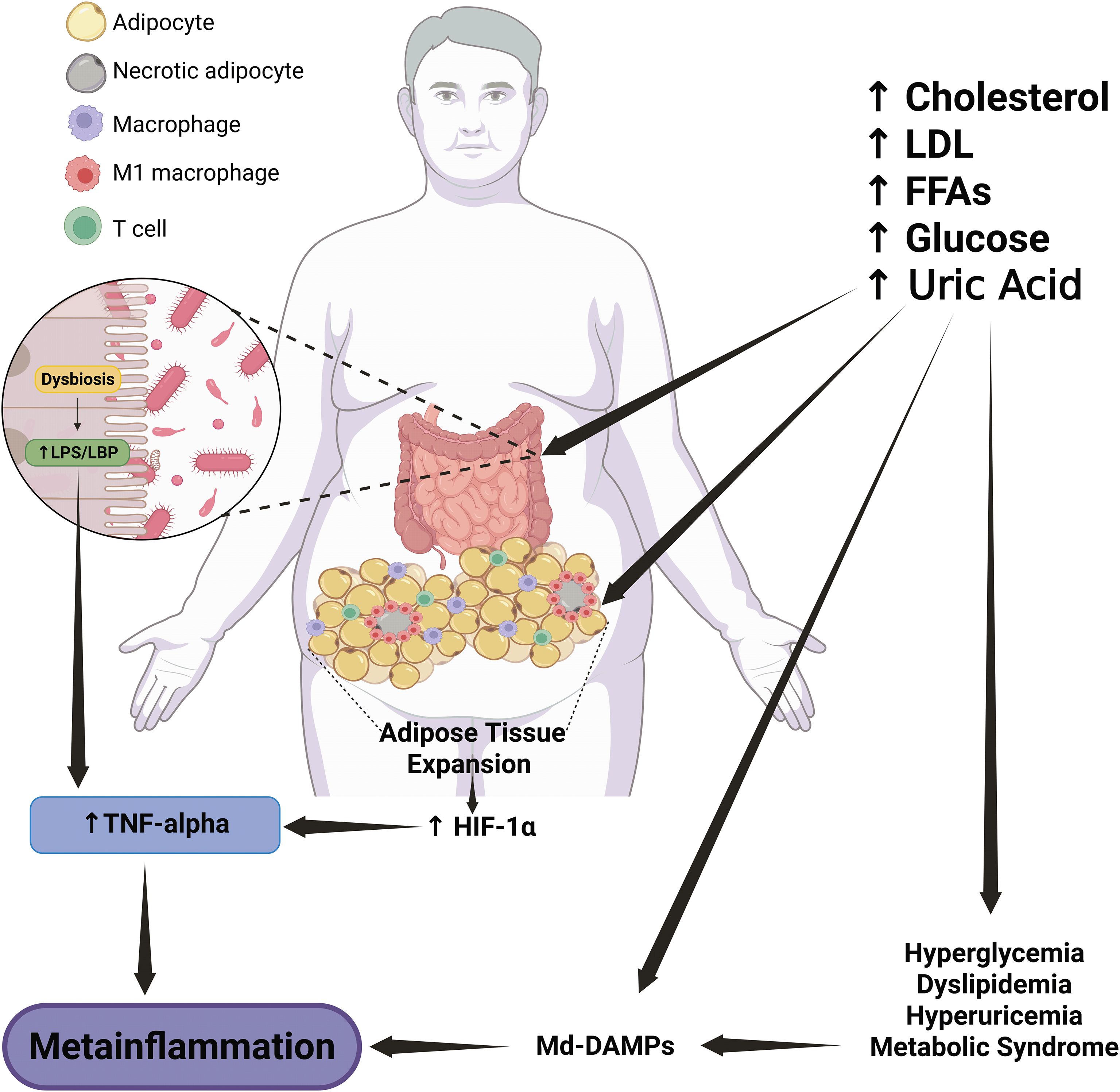
Figure 1. Role of Md-DAMPs in atherosclerotic plaque progression. Pathological entities related to metabolic syndrome, such as hyperglycemia, dyslipidemia, and hyperuricemia, are the primary sources of Md-DAMPs, including LDL, oxLDL, uric acid, and excess glucose. In parallel, metabolic syndrome-related alterations also induce endothelial damage and dysfunction, leading to endothelial cell death, oxidative stress, and immune cell recruitment, particularly circulating monocytes that can infiltrate the tunica intima. In an environment enriched in Md-DAMPs, monocytes tend to adopt a proinflammatory profile similar to that of non-classical monocyte subsets, with increased IL-1 beta production and enhanced vascular patrolling and trafficking. Recruited monocytes differentiate into M1 proinflammatory macrophages, which engulf lipoproteins and produce TNF-alpha, thus turning macrophages into foam cells and starting the process of atherosclerotic plaque formation. Circulating monocytes continue to infiltrate and differentiate into M1 macrophages, which will turn into foam cells and produce proinflammatory cytokines, MCP-1, and EMPs. This milieu, enriched in Md-DAMPs, inflammatory mediators, and profibrotic signals, promotes immune cell recruitment and fibrous cap formation, thus leading to plaque growth and stability in a first stage. In a late phase, TNF-alpha also induces apoptosis of smooth muscle cells and endothelial cells within the vascular endothelium. At the same time, IL-1 beta stimulates the expression of MMPs with the ability to degrade collagen and other MEPs. This scenario results in thinning and weakening of the fibrous core, which promotes plaque instability and rupture and leads to coronary artery disease and myocardial compromise. An upward arrow indicates an increase. A downward arrow indicates a decrease. Connector arrows indicate a causative relationship, with the arrow pointing from the cause to the effect. Abbreviations: Md-DAMPs, metabolite-derived damage-associated molecular patterns; TA, tunica adventitia; TM, tunica media; TI, tunica intima; LDL, low-density lipoprotein; oxLDL, oxidized low-density lipoprotein; UA, uric acid; Glu, glucose; TNF-alpha, tumor necrosis factor alpha; IL, interleukin; MCP-1, monocyte chemoattractant protein-1; EMPs, extracellular matrix proteins; MMPs, matrix metalloproteinases; SMCs, smooth muscle cells; CAD, coronary artery disease.
For decades, we all believed that intestinal flora was just gut bacteria we should keep under control with an enormous variety of antibiotics, often misused without medical supervision and prescription-free (27, 28). Nevertheless, the gut microbiome is a 2-kilogram organ composed of more than 400 bacterial species that are responsible for carbohydrate breakdown, SCFA synthesis, vitamin production, immune system maturation, protection against harmful bacterial colonization, and maintenance of intestinal barrier integrity, among other functions (29–35). Bacteria within the human intestine are in a delicate but complex balance. Intestinal dysbiosis occurs after disruption of intestinal flora equilibrium, altering bacterial composition and function and allowing the proliferation of detrimental microorganism species, such as gram-negative bacteria (36, 37). Gram-negative bacteria include more than 100 species of microorganisms responsible for most in-hospital morbidity and mortality, as well as antibiotic resistance. Bacterial species such as Chlamydia sp., Escherichia coli, Klebsiella sp., Pseudescherichia sp., Salmonella sp., Shigella sp., Neisseria sp., and Helicobacter pylori are perhaps the most well-known gram-negative bacteria responsible for lipopolysaccharide (LPS) production (38, 39). LPS is a complex molecule composed of repeated oligosaccharide units, core sugars, and lipid A, and it is a crucial outer membrane component of bacteria (40). Dysbiosis results in LPS release into the gut lumen, where this endotoxin can bind to lipopolysaccharide-binding protein (LBP), a soluble acute-phase protein primarily produced in the liver and intestine (41). CD14 on monocytes and macrophages can bind the LPS-LBP complex, inducing dimerization of toll-like receptor 4 (TLR4)/myeloid differentiation factor 2 (MD2) and activating myeloid differentiation primary response 88 (MyD88) (42). The TLR4/MD2-MyD88 signaling pathway initiates the canonical cascade of proinflammatory events, leading to activation of IκB kinase (IKK), which, in turn, phosphorylates IκBα, resulting in nuclear factor kappa B (NFκB) dimerization (43). The NFκB dimer can translocate to the nucleus, where it acts as a transcription factor, inducing the expression of cytokines and chemokines such as TNF-alpha, pro-IL-1 beta, IL-6, IL-12, and monocyte chemoattractant protein-1 (MCP-1) (44). This evidence may partially explain how intestinal dysbiosis leads to elevated blood LPS levels, and it also provides a molecular basis for understanding why monocytes and macrophages show an increased ability to produce inflammatory mediators systemically. In this regard, circulating monocytes exhibit a higher capacity to produce TNF-alpha in obesity and type 2 diabetes (T2D), both conditions characterized by elevated LPS and LBP serum levels (45–47). Notably, Sakura et al. reported that men with T2D also showed a significant correlation between serum LBP and aortic pulse wave velocity, a widely used measurement of arterial stiffness that serves as a surrogate marker of subclinical atherosclerosis (47). A study in mice with high-fat diet (HFD)-induced insulin resistance found that increased TNF-alpha levels promote a rise in circulating monocytes, thereby perpetuating and amplifying metainflammation (48). This information concurs with results from mouse models showing that antibiotic-induced intestinal dysbiosis is related to increased IL-1 beta-producing CD68+ macrophages in aortic atherosclerotic plaques, accompanied by higher LPS serum levels (49, 50). Altogether, this evidence supports the role of intestinal dysbiosis as a primary source of systemic LPS, which is an endotoxin that can trigger immune hyperactivation in monocytes and macrophages by promoting proinflammatory cytokine release into the bloodstream.
Adipose tissue expansion refers to the enlargement in size or number of adipose cells within the fat mass, also referred to as hypertrophy and hyperplasia, respectively (51). Adipose tissue expansion is a histologic feature of obesity, primarily occurring in visceral and subcutaneous fat depots around internal organs in the abdominal cavity or the hypodermis in areas such as the abdomen, thighs, and buttocks (51, 52). Adipose tissue hyperplasia is a common feature of fat mass in children and adolescents with weight gain (53). Conversely, adipose tissue hypertrophy is a hallmark of obesity in adulthood, as well as long-term insulin resistance and metabolic disturbances (54). Accumulating evidence in animal models and humans suggests that adipose tissue expansion may also play a crucial role in metainflammation development by starting a local inflammatory cascade that can expand to the rest of the body.
During obesity, fatty acids transported by chylomicrons increase in the bloodstream (55, 56). Then, lipoprotein lipase hydrolyzes chylomicrons in adipose tissue’s vascular stroma, thereby releasing free fatty acids that adipocytes can store as triglyceride droplets via re-esterification (57). Consequently, adipocytes become expanded cells. Adipocyte expansion is a human trait resulting from millions of years of evolution as hunter-gatherers in a new world where the first hominids had to run to catch food (58–60). In this scenario, it is unsurprising that excessive weight gain is associated with the widespread availability of high-sugar, high-fat foods, which have contributed to adipose tissue enlargement, along with other factors such as genetic background and lack of physical activity. Bigger adipose cells require more oxygen, which is available in the proximity of vascular stroma’s capillaries (61, 62). However, adipocytes located far from stromal capillaries may have reduced oxygen bioavailability, leading to hypoxia-inducible factor 1α (HIF-1α) production (63, 64). HIF-1α is a master transcription factor that orchestrates the expression of a wide range of genes involved in hypoxia adaptation, including glycolysis, erythropoiesis, vascular endothelial growth factor (VEGF)-mediated angiogenesis, cell survival, and even apoptosis (65–68). HIF-1α also acts as an immune function regulator by promoting NFκB-dependent TLR4 expression, TNF-alpha and IL-6 synthesis, phagocytosis, and macrophage migration mediated by MCP-1 (69–72). Under prolonged hypoxia, adipocytes produce HIF-1α, which induces downstream expression of MCP-1, a chemokine that attracts circulating monocytes that can infiltrate adipose tissue and differentiate into MDMs (73). The HIF-1α-induced microenvironment, enriched in TNF-alpha and IL-6, instigates infiltrated MDMs and adipose tissue macrophages (ATMs) to adopt a proinflammatory activation state and form crown-like structures characteristic of necrotic adipose tissue (74). In this way, adipose tissue expansion, hypoxia, MDMs, and ATMs work synergistically to unleash an orchestrated inflammatory cascade that initiates metainflammation and propagates a systemic milieu favorable to the development of atherosclerotic disease.
This information indicates that gut dysbiosis and adipose tissue expansion are central igniters of metainflammation through LPS production and HIF-1α activation, thereby simultaneously amplifying a proinflammatory environment favorable for atherosclerosis. Once initiated, metainflammation elicits the release of Md-DAMPs, which are metabolic signals that can directly stimulate immune cells to produce proinflammatory cytokines, chemokines, and reactive oxygen species (ROS) via specific receptors and intracellular pathways.
Role of Md-DAMPs in metainflammation
Prof. Dr. Seung-Yong Seong and Prof. Dr. Polly Matzinger first proposed the term damage-associated molecular patterns (DAMPs) in 2004 to describe a variety of ligands released from injured or dying cells able to trigger innate immune responses, including sterile inflammation (75). Fourteen years later, our research team was among the first to coin the term Md-DAMP to describe endogenous metabolic signals that polarize immune cells toward a proinflammatory profile and contribute to the development of cardiovascular disease (76). Currently, a growing number of research groups have consistently reported various Md-DAMPs, including glutamate, bile acids, lipoproteins, SCFAs, uric acid, and excess glucose, among others (76–78). Compared to prototypical immune ligands such as LPS or TNF-alpha, Md-DAMPs are non-prototypical molecules with immunoregulatory actions in numerous innate immune cells, including monocyte subsets, MDMs, ATMs, eosinophils, neutrophils, and mast cells (78, 79). Unlike classical pathogen-associated molecular patterns (PAMPs) or DAMPs, Md-DAMPs are produced from altered metabolic processes rather than from pathogens or injured cells. In fact, the primary sources of Md-DAMPs are metabolic alterations such as hyperglycemia, insulin resistance, dyslipidemia, and hyperuricemia, among others, which increase the thresholds of molecules as glucose, lipoproteins, glutamate, and uric acid (80, 81). Finally, Md-DAMPs are etiopathogenic factors of metainflammation that lead to the development of critical entities such as T2D, metabolic dysfunction-associated steatotic liver disease (MASLD), chronic kidney disease, hypertension, and atherosclerosis (Table 1). Given the definition of Md-DAMP, we will now examine the molecular mechanisms by which Md-DAMPs influence immune cell activity in the context of metainflammation and cardiovascular disease, with particular emphasis on atherosclerosis.
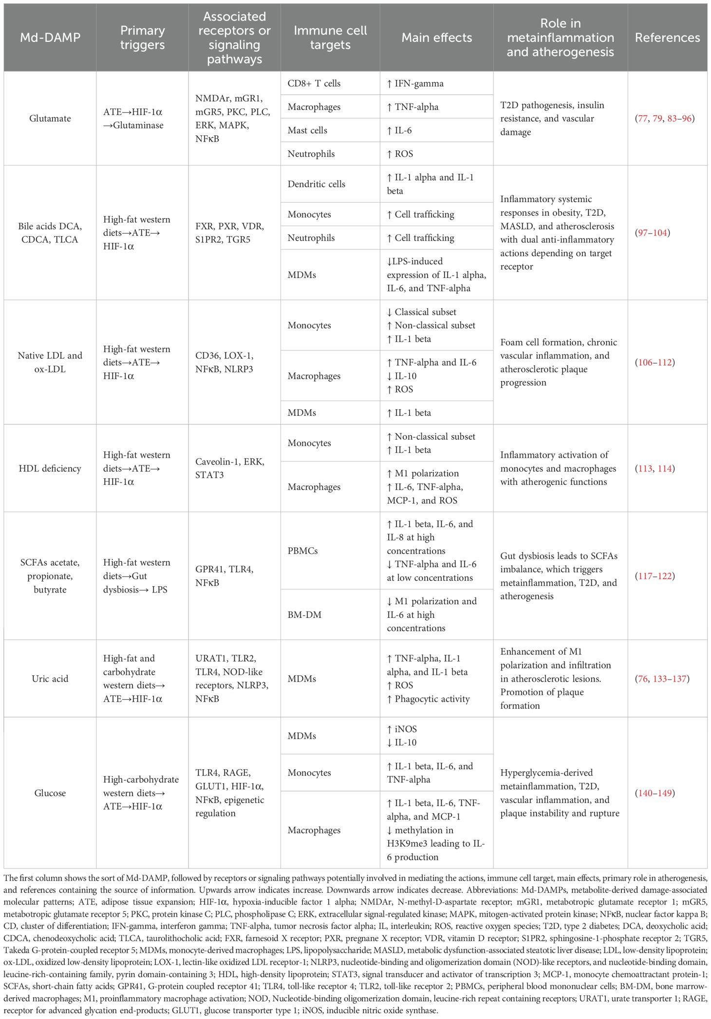
Table 1. Summary of the effects of Md-DAMPs on immune cells involved in metainflammation and atherogenesis.
Glutamate is a neurotransmitter produced by neurons and glial cells that exerts its actions through binding to ionotropic (iGR) or metabotropic (mGR) receptors (82). Notably, hypertrophic white adipose tissue from mice with diet-induced obesity is also a source of glutaminase-mediated glutamate production that concurs with reduced adiponectin secretion and decreased insulin-dependent glucose uptake (83, 84). Besides acting as an excitatory neurotransmitter in the central nervous system, glutamate can behave as an Md-DAMP and increase the production of interferon-gamma (IFN-gamma) in cytotoxic T cells and TNF-alpha in macrophages via mGR1 or mGR5 (79, 85). In CD8+ T cells, glutamate binds to mGR1 and activates phospholipase C (PLC), which in turn cleaves phosphatidylinositol 4,5-bisphosphate (PIP2) into inositol trisphosphate (IP3) and diacylglycerol (DAG) (86). IP3 and DAG are crucial second messengers that promote calcium (Ca2+) release from the endoplasmic reticulum and activate protein kinase C (PKC), respectively (86). The increase in intracellular Ca2+ and phosphorylated PKC activates prominent transcription factors involved in IFN-gamma expression, including nuclear factor of activated T cells (NFAT) and activator protein-1 (AP-1) (87, 88). In parallel, glutamate interaction with mGR5 leads to extracellular-signal-regulated kinase (ERK) phosphorylation and mitogen-activated protein kinase (MAPK) activation, which promote IFN-gamma expression, like that observed with T-cell receptor (TCR) stimulation (89). In macrophages, glutamate interaction with mGR5 activates the PLC-dependent PKC signaling pathway, which in turn promotes NFκB dimerization and translocation to the nucleus, where it upregulates TNF-alpha transcription (90, 91). Simultaneously, the IP3-dependent intracellular Ca2+ increase also triggers TNF-alpha production via calmodulin-dependent kinases (CaMK), thus contributing to glutamate proinflammatory effects on macrophages (92). The role of glutamate in immune cells also extends to polymorphonuclear leukocytes and iGRs, as in murine bone marrow-derived mast cells, which exhibit increased IL-6 synthesis via the N-methyl-D-aspartate receptor (NMDAr) (77). The use of MK-801, an NMDAr antagonist, demonstrated that iGRs mediate glutamate-dependent IL-6 production in mast cells (77). Primary human neutrophils exhibit a progressive, dose-dependent decrease in ROS production when cultured with Co101244, another NMDAr antagonist (93). This body of evidence aligns with the increased glutamate levels observed in patients with T2D, a complex metabolic disease characterized by abnormally high blood glucose levels that significantly increase the risk of atherosclerotic plaque formation (94). With apparent effects on CD8+ T cells, macrophages, mast cells, and neutrophils, glutamate acts as an Md-DAMP via mGRs and iGRs, such as mGR1, mGR5, and NMDAr, expressed in both adaptive and innate immune cells that also play a role in metainflammation (95, 96).
Another type of molecule showing a dual role in metabolism and inflammation is bile acids. Bile acids are produced in hepatocytes and stored in the gallbladder. After dietary fat intake, bile acids are secreted into the duodenum to enable the digestion and absorption of fats. In addition to their roles in lipid metabolism, emerging evidence suggests that bile acids are aberrantly altered in obesity, T2D, and MASLD and act as Md-DAMPs, orchestrating either anti-inflammatory or proinflammatory responses, depending on their chemical structure and target receptors. For instance, deoxycholic acid (DCA) and chenodeoxycholic acid (CDCA) promote the in vitro secretion of IL-1 alpha and IL-1 beta in murine bone marrow-derived dendritic cells, thereby facilitating the recruitment of monocytes and neutrophils to inflamed tissues (97). Both DCA and CDCA are bile acids that share a 24-carbon steroid structure, differing only in the location of the second hydroxyl group, and act primarily through the farnesoid X receptor (FXR), the pregnane X receptor (PXR), the vitamin D receptor (VDR), and sphingosine-1-phosphate receptor 2 (S1PR2) (98, 99). Conversely, a study reported that the secondary bile acid taurolithocholic acid (TLCA) suppresses LPS-induced proinflammatory cytokine expression in primary human MDMs, including IL-1 alpha, IL-6, and TNF-alpha (100). TLCA is a secondary bile acid whose structure substantially differs from DCA and CDCA, showing the amino acid taurine attached to the steroidal backbone through an amide bond (101). Furthermore, TLCA primarily exerts its actions through the Takeda G-protein-coupled receptor 5 (TGR5), a receptor expressed in cytotoxic T cells and macrophages that suppresses antiviral and inflammatory responses (102–104). As outlined, bile acids act as Md-DAMPs by affecting cytokine expression and immune cell activation. However, the effects of bile acids on immune cells are not uniform, as they depend on bile acid structure and target receptors, which may partially explain conflicting evidence regarding their capacity to induce proinflammatory or anti-inflammatory responses. We still need to conduct further research to clarify the role of bile acids in immune activity, including the type of bile acid, the receptor involved, and the concentration thresholds, to understand how these metabolites contribute to metainflammation.
OxLDLs were among the first molecules described as Md-DAMPs due to their role in macrophage differentiation into foam cells, a critical component of atheromatous plaque formation (105). OxLDLs result from the oxidation of native low-density lipoproteins (LDLs), a group of lipophilic proteins synthesized in the liver as very-low-density lipoproteins (VLDLs) initially (106). In the context of dyslipidemia and metabolic dysfunction, monocytes infiltrating the subendothelial space of blood vessels can differentiate into macrophages with proinflammatory activities, including the expression of TNF-alpha and the production of ROS (106, 107). Macrophages also exhibit the ability to uptake oxLDLs via scavenger receptors, such as the cluster of differentiation (CD) 36 and lectin-like oxidized low-density lipoprotein receptor-1 (LOX-1), internalizing oxLDL molecules to form lipid droplets inside the cell (108). The cytosolic accumulation of lipids causes macrophages to transform into foam cells, which not only contribute to the formation of the atheromatous plaque but also enhance inflammatory functions, including the activation of the inflammasome and NFκB pathways, leading to the release of IL-1 beta and TNF-alpha (109).
Nevertheless, accumulating evidence indicates that oxLDLs are not the only lipoproteins that act as Md-DAMPs in metainflammation, metabolic dysfunction, and atherosclerosis. Native LDL also appears to function as an Md-DAMP, affecting a variety of innate immune cells involved in atherogenesis. A previous study from our group demonstrated that native LDLs synergistically act with LPS to decrease CD14++CD16+ classical monocytes and increase CD14+CD16+ non-classical monocytes via CD36, both in vitro and in patients with increased atherogenic risk (110). Another report indicated that native LDL stimulates the differentiation of all monocyte subtypes into IL-6 and TNF-alpha-producing macrophages, which also exhibit a reduced ability to synthesize the anti-inflammatory cytokine IL-10 (111). Consistent with these data, native LDL induces inflammasome activation and IL-1 beta secretion in MDMs from patients with high plasma apolipoprotein B (ApoB), a common form of dyslipidemia (112). Surprisingly, LDL is not the only lipoprotein to exert Md-DAMP effects on human monocytes and macrophages. High-density lipoprotein (HDL) deficiency is associated with decreased circulating classical monocytes and increased non-classical monocytes in patients with metabolic syndrome (113).
Additionally, the in vitro culture of monocytes in the absence of HDL polarizes them towards the non-classical subgroup, characterized by a significant increase in IL-1β production (113). Concurring with this evidence, enrichment of serum medium with HDL abrogates macrophage differentiation towards the M1 proinflammatory profile, inhibiting the production of IL-6, TNF-alpha, MCP-1, and ROS (114). Interestingly, HDL prevents the inflammatory polarization of macrophages by redistributing caveolin-1 in the cell membrane and decreasing the phosphorylation of extracellular signal-regulated kinase (ERK) and signal transducer and activator of transcription 3 (STAT3) (114). As shown, lipoproteins such as oxLDL, native LDL, and HDL play crucial roles as Md-DAMPs, orchestrating the inflammatory activation of monocytes and macrophages and the production of cytokines that contribute to metainflammation and atherogenesis.
Lipids that participate in metainflammation are not limited to complex particles containing cholesterol, triglycerides, and apolipoproteins, but also SCFAs. SCFAs are single lipids produced by microbiota from dietary fiber that comprise a wide variety of fatty acids composed of less than six carbons, including 2-carbon acetic acid, 3-carbon propionic acid, 4-carbon butyric acid, 5-carbon valeric acid, and 6-carbon caproic acid (115). SCFAs serve as an energy source for various cell types, including colonocytes, hepatocytes, adipocytes, neurons, and immune cells. Besides energy metabolism, SCFAs exert Md-DAMP actions via G-protein-coupled receptors (GPRs) and modulation of TLR4-mediated signaling pathways, promoting either proinflammatory or anti-inflammatory responses depending on their concentration and target receptor (116). In this sense, 0.1 mM acetic acid decreases IL-6 and TNF-alpha production in the liver of mice with cecal ligation and puncture-induced sepsis, whereas 20 mM acetic acid stimulates IL-6, IL-8, and IL-1 beta synthesis in peripheral blood mononuclear cells (PBMCs) from healthy donors (117, 118). Propionic acid also exhibits bimodal effects on the inflammatory response. For instance, human intestinal epithelial tissue shows reduced levels of NFκB, MCP-1, and IL-8 when treated with low doses of propionic acid via MAPK activation. However, propionic acid concentrations above 20 mM induce the secretion of IL-6, IL-8, and IL-1 beta in human PBMCs via TLR4 (118, 119). Likewise, different butyric acid concentrations can elicit or suppress inflammatory pathways, as demonstrated in LPS-treated murine bone marrow-derived macrophages, which show decreased IL-6 production at 2 mM but no effect at 0.1 mM (120).
Interestingly, low SCFA thresholds appear to reflect a balance of gut microorganisms linked to immune tolerance. In contrast, an increase in SCFA production may indicate an imbalance in intestinal bacteria, leading to the inflammatory activation of innate immune cells. This idea concurs with prior data showing that butyrate concentrations below 0.5 mM promote the differentiation of naïve CD4+ T cells into IL-10-producing regulatory T cells via GPR41, suggesting that low SCFA thresholds can elicit immune tolerance (121). Conversely, supplementation with acetate, propionate, and butyrate at concentrations above 25 mM stimulates inflammatory activation of RAW264.7 macrophages, serving as a regulatory mechanism that elicits phagocytosis of Klebsiella pneumoniae and prevents dysbiosis (122). Another line of evidence supporting the duality of SCFA in promoting either a proinflammatory or an anti-inflammatory milieu is that SCFA concentration depends on the balance among gut microorganisms, including those belonging to the phyla Bacteroidetes, Actinobacteria, and Firmicutes (123). Therefore, dysbiosis may affect not only the balance of the gut microbiota but also SCFA production, favoring the development of inflammatory conditions or tolerogenic responses, a scenario that may lead to metainflammation and other related diseases such as T2D (124–127). Our understanding of SCFAs as Md-DAMPs is just emerging. However, the clear relationship among gut microbiota, SCFA concentration, and metainflammation reinforces the potential therapeutic use of SCFA supplementation to improve metabolic status, as shown in mice fed an HFD and in individuals with metabolic syndrome (128–130).
Uric acid is an organic heterocyclic compound containing carbon, nitrogen, oxygen, and hydrogen. Uric acid is also the end product of purine metabolism, which is filtered in the kidneys and excreted into the urine (131). A purine-enriched diet, including red meat, seafood, beer, and fructose-sweetened beverages, can increase uric acid production, leading to a condition called hyperuricemia when uric acid levels in the blood exceed 6 mg/dl in women or 7 mg/dl in men (132). Besides being a risk factor for developing gout and urolithiasis, uric acid is an Md-DAMP linking metainflammation with cardiovascular disease and metabolic dysfunction. A previous study from our research group demonstrated that uric acid stimulates the in vitro synthesis of TNF-alpha and TLR4 in human MDMs in a dose-dependent manner while also enhancing the phagocytic activity of these immune cells (76). Notably, the specific blockade of uric acid transport within macrophages abrogated both proinflammatory cytokine production and phagocytic activity, suggesting that uric acid exerts its effects by eliciting a receptor-dependent intracellular signaling cascade (76).
Previous evidence indicates that uric acid interacts with pattern recognition receptors (PRRs), including TLR2, TLR4, nucleotide-binding and oligomerization domain (NOD)-like receptors, and the nucleotide-binding domain, leucine-rich-containing family, pyrin domain-containing 3 (NLRP3) (133). Upon activation, TLR2 and TLR4 recruit MyD88 to interact with the L-1R-associated kinase (IRAK) protein family, leading to IκB phosphorylation and the release of NFκB (134). Along with promoting cytokine expression, NFκB activity aligns with ROS production, which serves as a triggering signal to activate NLRP3 and enhance macrophage inflammatory behavior in the presence of uric acid (135). This evidence supports previous reports that hyperuricemia enhances macrophage proinflammatory capacity and their ability to infiltrate atherosclerotic lesions in the aortas of ApoE-/- mice (136). Interestingly, inhibition of xanthine oxidase, the rate-limiting enzyme in purine degradation that produces uric acid, decreases IL-1 alpha and IL-1 beta gene expression in aortic lesions, also alleviating ROS production and plaque formation (137). Together, this body of experimental findings suggests that uric acid plays a role as an Md-DAMP, conferring inflammatory functions to macrophages through pathways involving urate transporters and NFκB and NLRP3-dependent signaling cascades.
Glucose is a monosaccharide, or simple sugar, that serves as the primary energy source for almost all living organisms (138). Conversely, excess glucose is a leading cause of morbidity and mortality worldwide due to the harmful effects of hyperglycemia on nerves and blood vessels that contribute to developing neuropathy, endothelial dysfunction, and vascular complications (139). Besides altering protein structure and function through glycation, weakening small blood vessels, and promoting ROS production, abnormally high blood glucose levels act as Md-DAMPs, triggering metainflammation and cardiometabolic dysfunction. Almost 10 years ago, our research team reported that 15 mM excess glucose, resembling the blood sugar amount in patients with uncontrolled T2D, promotes the in vitro polarization of human MDMs by increasing inducible nitric oxide synthase (iNOS) and decreasing IL-10 (140). A recent study expanded the body of evidence supporting the proinflammatory effects of glucose on human MDMs, revealing that hyperglycemia increases TLR4 expression, which partially explains the elevation of iNOS in these immune cells (141). High glucose can also increase IL-1 beta, IL-6, and TNF-alpha expression in human monocytes incubated with 24 mM glucose (142).
The molecular mechanisms by which excess glucose exerts its actions as an Md-DAMP remain uncertain but likely involve receptors such as TLR4 and the receptor for advanced glycation end-products (RAGE), glucose transporters such as GLUT1, and epigenetic mechanisms. In this sense, lactic acid and epigenetic changes are pivotal mediators of glucose’s actions. Briefly, accumulation of extracellular glucose leads monocytes and macrophages to increase glucose uptake via GLUT1, upregulating glycolysis that results in lactic acid overproduction (143). Lactate can also inhibit the prolyl hydroxylase domain (PHD), a dioxygenase family member that tags the transcription factor HIF-1α for proteasomal degradation (144). Lactate’s effect results in the accumulation of HIF-1α, promoting increased expression of cytokines and chemokines with inflammatory roles in monocytes and macrophages (145). High glucose prevents methylation of histone H3 (H3K9me3), a key inhibitor of proinflammatory gene expression (146). Sudden accumulation of intracellular glucose induces HIF-1α–dependent IL-6 expression, which activates transcription of the suppressor of variegation 3–9 homolog 1 (SUV39H1) in human and murine macrophages (147). SUV39H1 catalyzes the trimethylation of histone H3 at lysine 9 (H3K9me3), an epigenetic mechanism that acts as a negative feedback regulator of proinflammatory gene expression (148). Conversely, prolonged exposure to high intracellular glucose concentrations suppresses IL-6-mediated feedback, leading to repression of SUV39H1, inhibition of H3K9me3 methylation, and promotion of proinflammatory gene transcription (149). Collectively, this information highlights the profound effect of high glucose in triggering innate immune cell hyperactivity, which contributes to metainflammation that accompanies obesity, T2D, and cardiovascular disease. Understanding the molecular mechanisms triggered by Md-DAMPs, such as excess glucose, is crucial to explaining why monocytes and macrophages exhibit an inflammatory profile in hyperglycemia and how these cells contribute to the development of atherosclerosis.
As outlined, Md-DAMPs resulting from metabolic dysfunction can perpetuate metainflammation, initially driven by gut dysbiosis and adipose tissue expansion. Moreover, Md-DAMPs can stimulate immune cells to produce inflammatory factors and chemotactic agents that play prominent roles in atherosclerotic plaque formation, instability, and rupture (Figure 2).
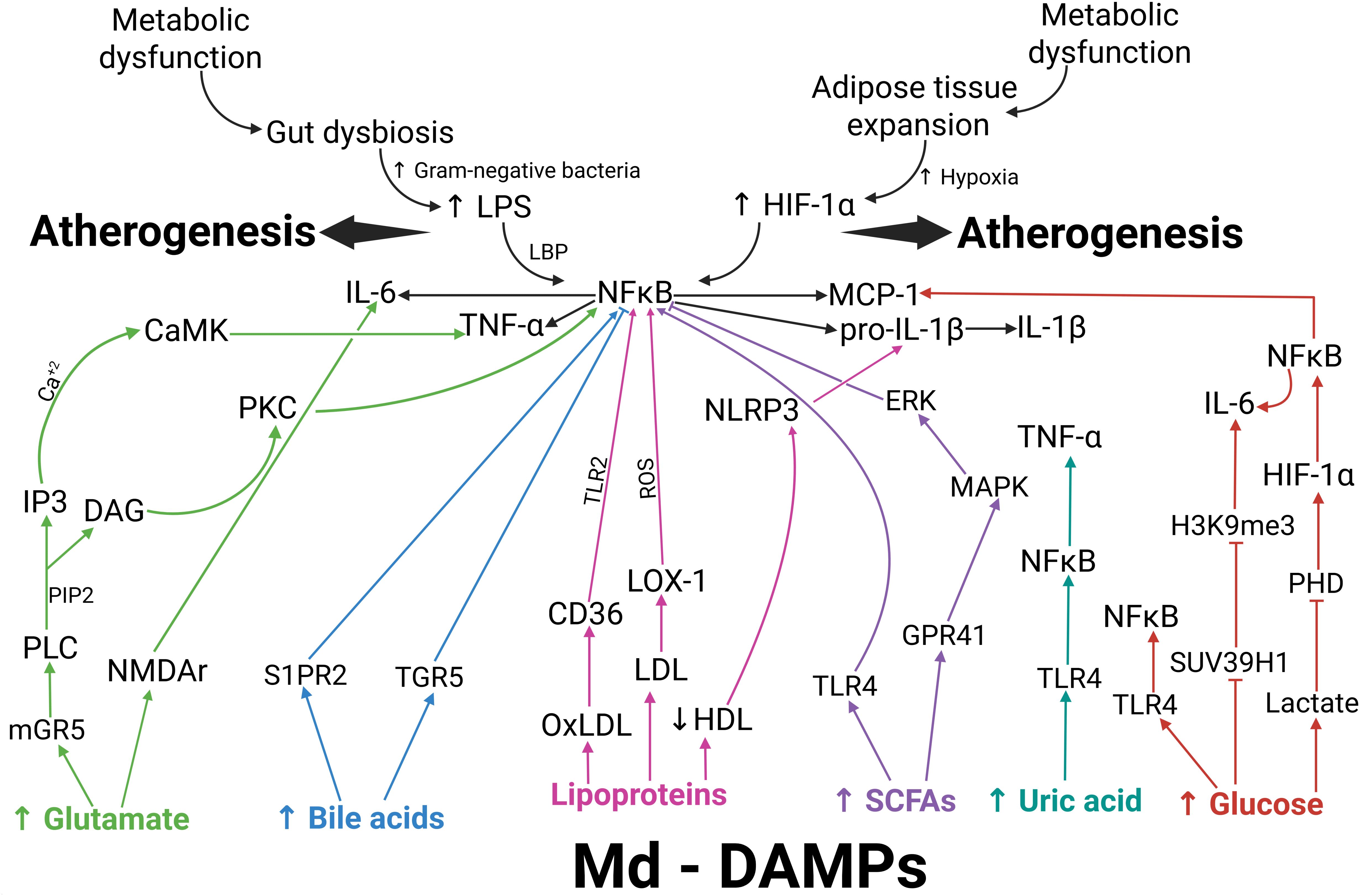
Figure 2. Map of the potential molecular interplay among entries of metainflammation, Md-DAMPs, target receptors, and intracellular mediators in atherogenesis. Metabolic dysfunction favors gut dysbiosis and adipose tissue expansion as primary entries of metainflammation. Gram-negative bacteria proliferate during gut dysbiosis, leading to increased LPS production. Once bound to LBP, LPS activates NFκB via TLR4. Adipose tissue expansion involves adipocyte hypertrophy and hyperplasia. Hypoxia increases as adipocytes expand, thereby activating HIF-1α. HIF-1α can also activate NFκB, thus promoting the expression of IL-6, TNF-alpha, pro-IL-1 beta, and MCP-1. Md-DAMPs are endogenous metabolic signals with the ability to stimulate monocytes, macrophages, and other immune cells to produce proinflammatory cytokines and chemokines via specific receptors and intracellular signaling pathways. After binding to mGR5 (green arrows), glutamate activates PLC, promoting PIP2 cleavage into IP3 and DAG. IP3 increases intracellular calcium, which leads to CaMK activation and TNF-alpha production. DAG activates PKC, which, in turn, leads to NFκB activation and TNF-alpha synthesis. Glutamate can also bind to NMDAr, which elicits IL-6 production. S1PR2 and TGR5 can recognize bile acids (blue arrows), thereby promoting NFκB activation and the expression of proinflammatory cytokines when bile acid levels increase. Ox-LDL acts through CD36 (pink arrows), thus inducing NFκB activation via TLR2. Native LDL exerts its effect via LOX-1 (pink arrows), which also activates NFκB through ROS production. Decreased HDL levels concur with NLRP3 activation, which cleaves pro-IL-1 beta to IL-1 beta (pink arrows). SCFAs exert their actions via TLR4 or GPR41 (purple arrows), promoting NFκB activation directly or via the ERK- and MAPK-dependent signaling pathways, respectively. Uric acid can bind to TLR4 (dark green), thereby activating NFκB and inducing TNF-alpha expression. Increased glucose acts through 3 different pathways (red arrows). After binding to TLR4, uric acid activates NFκB and TNF-alpha expression. Excess glucose inhibits SUV39H1 transcription, thereby suppressing H3K9me3-mediated methylation of the IL-6 gene. Increased intracellular glucose converts to lactic acid, which inhibits PHD activity and prevents proteasomal degradation of HIF-1α, thereby leading to NFκB activation. Metabolic dysfunction results in increased LPS production and HIF-1α activation, thereby triggering metainflammation that creates a milieu favorable to monocytes and macrophages for the production of proinflammatory cytokines and chemokines. In parallel, metabolic dysfunction also allows the release of Md-DAMPs, which directly stimulate monocytes and macrophages to synthesize proinflammatory cytokines and chemokines that can perpetuate and amplify metainflammation. The interplay between metainflammation igniters and Md-DAMPs favors atherosclerotic disease via specific receptors, including mGR5, NMDAr, S1PR2, TGR5, CD36, LOX-1, TLR4, and GPR41, as well as intracellular mediators such as PKC, NFκB, NLRP3, ERK, MAPK, and epigenetic mechanisms. An upward arrow indicates an increase. A downward arrow indicates a decrease. Connector arrows indicate a causative relationship, with the arrow pointing from the cause to the effect. Abbreviations: Md-DAMPs, metabolite-derived damage-associated molecular patterns; LPS, lipopolysaccharide; LBP, lipopolysaccharide-binding protein; HIF-1α, hypoxia-inducible factor 1 alpha; NFκB, nuclear factor kappa B; TNF-α, tumor necrosis factor alpha; IL-6, interleukin 6; pro-IL-1β, pro-interleukin 1 beta; IL-1β, interleukin 1 beta; PLC, phospholipase C; PIP2, phosphatidylinositol 4,5-bisphosphate; IP3, inositol trisphosphate; Ca+2, calcium; DAG, diacylglycerol; CaMK, calmodulin-dependent kinases; PKC, protein kinase C; S1PR2, sphingosine-1-phosphate receptor 2; TGR5, Takeda G-protein-coupled receptor 5; LDL, low-density lipoprotein; oxLDL, oxidized low-density lipoprotein; HDL, high-density lipoprotein; CD36, cluster of differentiations 36; TLR2, Toll-like receptor 2; LOX-1, lectin-like oxidized low-density lipoprotein receptor-1; ROS, reactive oxygen species; NLRP3, nucleotide-binding and oligomerization domain (NOD)-like receptors, and the nucleotide-binding domain, leucine-rich-containing family, pyrin domain-containing 3; SCFAs, short-chain fatty acids; TLR4, Toll-like receptor 4; GPR41, G-protein coupled receptor 41; ERK, extracellular-signal-regulated kinase; MAPK, mitogen-activated protein kinase; SUV39H1, suppressor of variegation 3–9 homolog 1; H3K9me3, trimethylation of histone H3 at lysine 9, PHD, prolyl hydroxylase domain.
Integrating the molecular mechanisms of metainflammation in atherosclerosis
We have reviewed the pathophysiological mechanisms by which gut dysbiosis-derived LPS and adipose tissue expansion trigger metainflammation, with particular emphasis on the activity of monocyte subsets and macrophages. We also elucidated the molecular basis of Md-DAMP function in preserving and amplifying receptor-mediated intracellular signaling pathways that lead to immune cell hyperactivation, proinflammatory cytokine production, and ROS release. Now, we will outline how these actors interact with each other in atherosclerotic plaque formation and rupture, causing acute heart attacks and strokes (Figure 3).
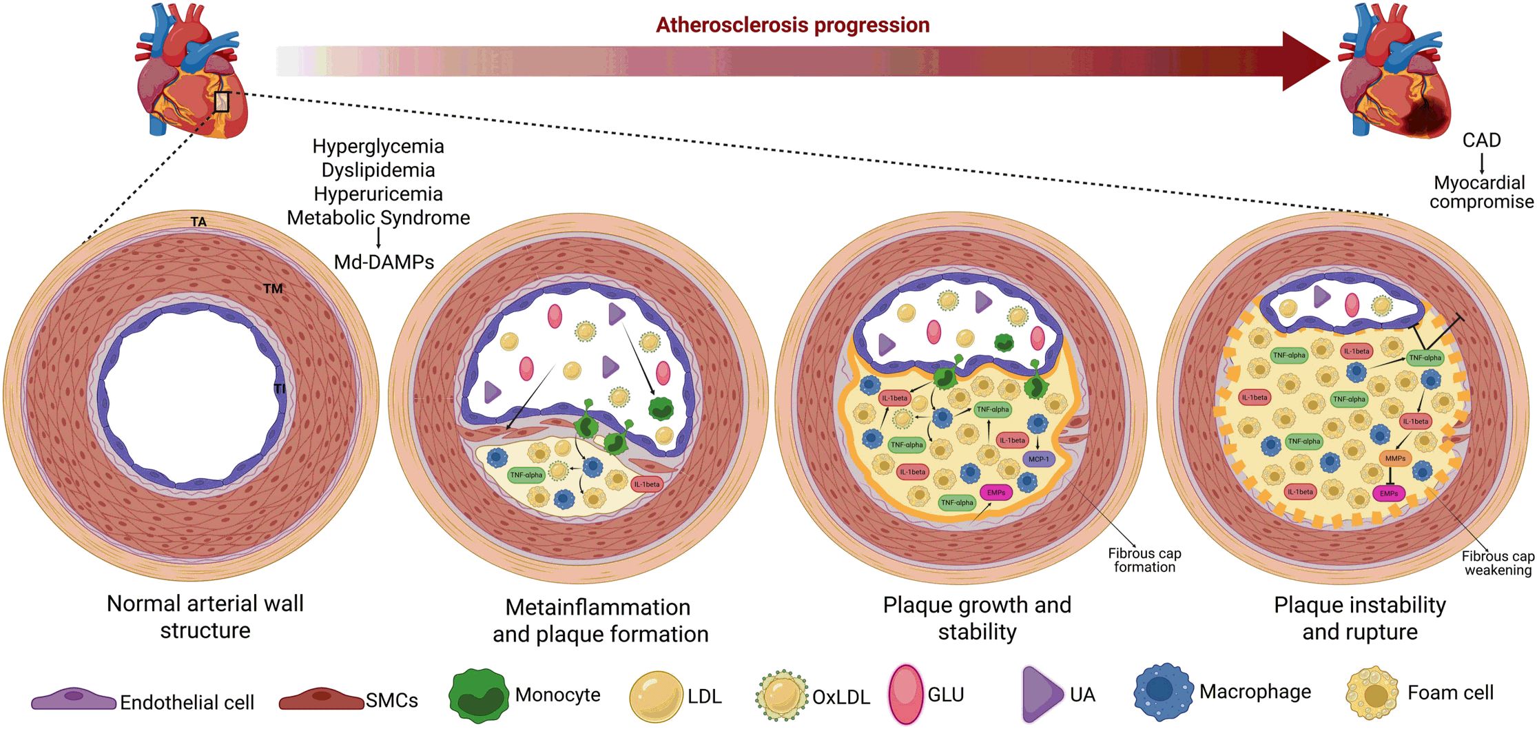
Figure 3. Gut dysbiosis and adipose tissue expansion are crucial triggers of TNF-alpha production and metainflammation. The abnormal increases in cholesterol, FFAs, LDL, glucose, and uric acid, among others, contribute to the development of dyslipidemia, hyperuricemia, hyperglycemia, and metabolic syndrome. These metabolic entities also serve as a source of Md-DAMPs, thereby perpetuating the abnormal increase in these immune triggers in circulation. Md-DAMPs provide micronutrients that alter the balance of microorganism species composing the gut microbiome, resulting in intestinal dysbiosis. Intestinal dysbiosis promotes the proliferation of gram-negative bacteria capable of producing LPS. LPS can cross the intestinal barrier and reach systemic circulation upon binding to LBP, thereby stimulating monocytes and tissue-resident macrophages to produce TNF-alpha and other inflammatory mediators that can trigger metainflammation. In parallel but not necessarily dependent, Md-DAMPs can also directly reach adipocytes within the adipose tissue, resulting in tissue hyperplasia and hypertrophy. The process of adipose tissue expansion induces hypoxia, primarily affecting adipocytes distant from the vascular bed, thereby increasing HIF-1α expression. HIF-1α is a master transcription factor that stimulates adipocytes, monocytes, and macrophages to synthesize TNF-alpha via NFκB, thereby favoring the development of metainflammation. As outlined, gut dysbiosis and adipose tissue expansion converge on TNF-alpha production, a cytokine with potent proinflammatory actions that also triggers metainflammation, immune cell hyperactivity, and metabolic dysfunction. An upward arrow indicates an increase. Bold connector arrows indicate a causative relationship, with the arrow pointing from the cause to the effect. Abbreviations: FFAs, free-fatty acids; LDL, low-density lipoprotein; Md-DAMPs, metabolite-derived damage-associated molecular patterns; LPS, lipopolysaccharide; LBP, lipopolysaccharide-binding protein; TNF-alpha, tumor necrosis factor alpha; HIF-1α, hypoxia-inducible factor 1 alpha; NFκB, nuclear factor kappa B.
Atherosclerotic plaque formation primarily occurs in arteries, a vast component of the circulatory system consisting of muscular and elastic vessels that allow transport of oxygen, nutrients, and hormones from the heart to the rest of the body. Arteries are composed of three superimposed tissue layers, named the tunica intima, the tunica media, and the tunica adventitia. The tunica intima is the innermost layer of the artery, primarily composed of a flattened endothelial cell lamina supported by collagen-enriched elastic fibers. The tunica media plays a critical role in regulating vessel tone due to the presence of smooth muscle cells superimposed around the tunica intima. The tunica adventitia is the artery’s outermost layer, mainly consisting of collagen and elastic fibers, fibroblasts, autonomic nervous fibers, and the vasa vasorum, which together regulate the vessel tone and the attachment of the artery to surrounding tissues.
Atherosclerotic plaque formation occurs in the tunica intima through a complex process in which Md-DAMPs, monocytes, macrophages, and other inflammatory immune cells interact to promote endothelial damage and dysfunction in a first stage. As mentioned, metabolic alterations serve as the primary source of Md-DAMPs, including glutamate, bile acids, SCFAs, uric acid, and glucose, which can promote damage to the inner lining of arteries and lead to endothelial dysfunction. In this regard, prior studies have demonstrated that endothelial cells express both NMDAr and mGR5 (150). In the presence of high extracellular glutamate thresholds, NMDAr can activate Ca2+-permeable channels in endothelial cells, increasing intracellular Ca2+ and potentially inducing mitochondrial Ca2+ overload, ROS generation, vasoconstriction, and endothelial injury (151). In parallel, glutamate interaction with mGR5 on endothelial cells activates the PLC-dependent PKC signaling pathway, leading to NFκB activation and production of TNF-alpha and MCP-1, which mediate immune cell infiltration in the tunica intima (152). Furthermore, bile acid-induced S1PR2 activation plays a critical role in triggering the NFκB-dependent signaling pathway that mediates proinflammatory cytokine release, ROS production, and vascular endothelial damage (153). Similarly, butyrate levels above 5 mM can stimulate the PKC, ERK, and MAPK-dependent signaling pathways via GPR41, resulting in activation of NFκB and AP-1, and production of TNF-alpha and adhesion molecules that cooperatively enhance leukocyte transmigration and vascular inflammation (154). Elevated uric acid levels also induce endothelial dysfunction by stimulating NADPH oxidase activity, leading to superoxide anion production and peroxynitrite formation, which reduces nitric oxide bioavailability and vasodilation (155). Excess glucose is another Md-DAMP that promotes endothelial dysfunction through impairing nitric oxide bioavailability and vascular tone, and increasing oxidative stress, immune cell infiltration, and tissue damage via TLR4, RAGE, and GLUT1 (156). As shown, Md-DAMP effects on the vascular endothelium involve numerous receptors and signaling pathways that result in the release of proinflammatory cytokines, ROS production, immune cell adhesion and transmigration, impaired vascular tone, and endothelial injury.
In turn, endothelial injury produces multiple DAMPs, including heat shock proteins (HSPs), high-mobility group box 1 (HMGB1), and mitochondrial deoxyribonucleic acid (mtDNA) (157, 158). Together with Md-DAMPs, these DAMPs can recruit circulating monocytes and other immune cells to the vascular endothelium. Monocytes infiltrating the tunica intima can differentiate into macrophages and adopt an inflammatory activation profile instigated not only by DAMPs but also by Md-DAMPs, which can extend cytokine production and act as signals to recruit additional innate cells (159). This microenvironment influences macrophages to engulf cholesterol, triglycerides, lipoproteins, and oxLDL accumulated in the injured endothelium, converting macrophages into foam cells (160). These foam cells not only contribute to plaque buildup but also exacerbate metainflammation by producing MCP-1, CXC-chemokine ligand 1 (CXCL1), and CXCL4, which recruit additional leukocytes and platelets and promote monocyte arrest in the atheroma (161, 162). Furthermore, foam cells within the atherosclerotic plaque can synthesize proinflammatory cytokines that perpetuate the inflammatory milieu, including IL-1 beta, IL-6, and TNF-alpha (163). In the absence of changes in habits or proper pharmacotherapy, the atheroma continues to grow due to the accumulation of fats, cell debris, calcium, platelets, monocytes, macrophages, and foam cells, leading to narrowing and hardening of the arteries, also referred to as atherosclerosis. As seen, atheroma formation is the first stage of atherosclerotic disease.
Afterward, the body must stabilize the plaque to prevent it from entering the bloodstream and produce a fibrous cork composed mainly of collagen and extracellular matrix proteins (164). However, the atherosclerotic plaque continues to secrete a variety of cytokines and DAMPs, which affect its stability and can lead to weakening and rupture. In this sense, TNF-alpha can mediate not only inflammatory responses but also increase plaque instability by inducing apoptosis of smooth muscle cells and endothelial cells within the vascular endothelium (165). Previous evidence shows that TNF-alpha stimulates caspase-3 activity in human vascular smooth muscle cells cultured in vitro, which may explain the reduced proliferation and increased apoptosis observed in these cells (166). A recent study reported that human umbilical vascular endothelial cells (HUVECs) exhibit caspase-3-mediated apoptosis when treated with TNF-alpha (167). At the same time, another study found that the apoptosis rate in HUVECs increased with increasing TNF-alpha dose (168). Besides playing a role in inflammasome activation, IL-1 beta is particularly active in atherogenesis due to its ability to promote the expression of matrix metalloproteinases (MMPs), which are key contributors to fibrous cap degradation and plaque instability (169). MMP-1 can degrade type I and III collagen, while MMP-2 and MMP-9 can cleave type IV collagen in both animal models and in vitro cell cultures, supporting their roles in plaque instability and rupture (170, 171).
Md-DAMPs not only participate in atherosclerotic plaque rupture by orchestrating TNF-alpha and IL-1 beta expression but also by inducing apoptosis of endothelial cells, a crucial element of arteries. For instance, 30 mM glucose activates NFκB, upregulating cyclooxygenase-2 (COX-2) and enabling caspase-3 activation, thereby increasing apoptosis in HUVECs (172). Later, another research team replicated these findings in human coronary artery endothelial cells, confirming that excess glucose can weaken atherosclerotic plaque (173). Consistent with increased apoptosis in HUVECs, uric acid also promotes the expression of caspase-3 and B-cell lymphoma 2 (Bcl-2) in a time- and dose-dependent manner (174). Furthermore, recent information revealed that 4 mM of butyric acid provokes DNA damage-related cell apoptosis in HUVECs, thus corroborating emerging evidence linking gut dysbiosis to atherosclerotic disease (175, 176). As outlined, this emerging information consistently shows that proinflammatory cytokines and Md-DAMPs can contribute to weakening atherosclerotic plaque by affecting the viability of smooth muscle cells and endothelial cells, as well as diminishing extracellular matrix protein content. This scenario elicits fibrous cap rupture and thrombus formation, blocking the blood flow within arteries and leading to acute myocardial infarction and cerebrovascular accident (Figure 3).
Conclusion
We have examined the complex interplay between gut dysbiosis-derived LPS and adipose tissue expansion as crucial signals that unleash low-grade systemic inflammation during metabolic dysfunction, also known as metainflammation. This state of metainflammation enables the release of Md-DAMPs as glutamate, bile acids, lipoproteins, butyrate, uric acid, and excess glucose, which can perpetuate proinflammatory polarization of monocytes and macrophages. Md-DAMPs predispose circulating monocytes toward proinflammatory activity by increasing the CD14+CD16+ non-classical monocyte subset, IL-1β synthesis, and monocyte infiltration into inflamed tissues, such as visceral adipose tissue. Priming of monocytes toward an inflammatory profile predisposes macrophages to classical activation once these immune cells have differentiated in peripheral tissues, thereby perpetuating the release of cytokines with proinflammatory actions, such as TNF-alpha. Md-DAMPs exert their inflammatory capacity through specific receptors and intracellular signaling pathways, including mGR1, mGR5, NMDAr, FXR, PXR, VDR, S1PR2, TGR5, CD36, LOX-1, TLR4, GPR41, urate transporter 1 (URAT1), GLUT1, RAGE, PLC, PKC, ERK, and MAPK. This variety of receptors with metabolic functions can also activate molecular mediators such as HIF-1α, CaMK, NFκB, AP-1, NFAT, and NLRP3, which in turn induce the expression of TNF-alpha, IL-1 beta, IL-6, and MCP-1, among others. The role of Md-DAMPs in the humoral and cellular responses associated with metainflammation becomes particularly relevant in atherogenesis, where monocytes, macrophages, and foam cells actively contribute to the formation and rupture of atherosclerotic plaques. Md-DAMPs such as glucose, uric acid, or butyrate continue to stimulate macrophages and foam cells to produce TNF-alpha and IL-1 beta within the atheroma, leading to remodeling of the vascular endothelium. The mechanisms by which Md-DAMPs and proinflammatory cytokines contribute to atherosclerotic plaque instability involve the apoptosis of smooth muscle cells and endothelial cells within the atheroma, as well as the degradation of extracellular matrix proteins. Ultimately, the intricate interplay among Md-DAMPs, monocytes, macrophages, foam cells, and cytokines culminates in atherosclerotic plaque rupture and thrombus formation, both of which are key pathophysiological features of cardiovascular disease. The current knowledge presented in this comprehensive review may help elucidate the molecular basis behind the role of gut dysbiosis-derived LPS and adipose tissue expansion in initiating metainflammation. Moreover, the experimental and clinical evidence discussed here may contribute to a better understanding of the molecular mechanisms through which Md-DAMPs can perpetuate metainflammation by increasing the activity of cytokine-producing immune cells. Collectively, this information may help refresh our conceptual understanding of atherogenesis to identify new molecular targets against immune cell receptors and signaling pathways involved in the development of metainflammation and atherosclerosis.
Author contributions
LG-H: Conceptualization, Data curation, Methodology, Writing – review & editing. KÁ-S: Conceptualization, Data curation, Methodology, Writing – review & editing. LM-G: Data curation, Methodology, Writing – review & editing. AC-V: Data curation, Methodology, Writing – review & editing. ME-V: Data curation, Methodology, Writing – review & editing. NB-H: Data curation, Methodology, Writing – review & editing. MF-S: Data curation, Methodology, Writing – review & editing. GE: Conceptualization, Data curation, Investigation, Supervision, Writing – original draft, Writing – review & editing.
Funding
The author(s) declare that no financial support was received for the research and/or publication of this article.
Acknowledgments
Leslie-Marisol Gonzalez-Hermosillo is a doctoral student from the Programa de Doctorado en Ciencias Biomédicas, Universidad Nacional Autónoma de México (UNAM) and received CONAHCYT fellowship CVU 1272497.
Conflict of interest
The authors declare that the research was conducted in the absence of any commercial or financial relationships that could be construed as a potential conflict of interest.
The author(s) declared that they were an editorial board member of Frontiers, at the time of submission. This had no impact on the peer review process and the final decision.
Generative AI statement
The author(s) declare that no Generative AI was used in the creation of this manuscript.
Any alternative text (alt text) provided alongside figures in this article has been generated by Frontiers with the support of artificial intelligence and reasonable efforts have been made to ensure accuracy, including review by the authors wherever possible. If you identify any issues, please contact us.
Publisher’s note
All claims expressed in this article are solely those of the authors and do not necessarily represent those of their affiliated organizations, or those of the publisher, the editors and the reviewers. Any product that may be evaluated in this article, or claim that may be made by its manufacturer, is not guaranteed or endorsed by the publisher.
References
1. Vozenilek AE, Vetkoetter M, Green JM, Shen X, Traylor JG, Klein RL, et al. Absence of nicotinamide nucleotide transhydrogenase in C57BL/6J mice exacerbates experimental atherosclerosis. J Vasc Res. (2018) 55:98–110. doi: 10.1159/000486337
2. Shi N, Jiang S, Zhao Y, Zhang Y, Duan X, Hong GB, et al. HFD aggravated the arthritis and atherosclerosis by altering the intestinal status and gut microbiota. Mol Med. (2024) 30:270. doi: 10.1186/s10020-024-01014-3
3. Mazhar F, Faucon AL, Fu EL, Szummer KE, Mathisen J, Gerward S, et al. Systemic inflammation and health outcomes in patients receiving treatment for atherosclerotic cardiovascular disease. Eur Heart J. (2024) 45:4719–30. doi: 10.1093/eurheartj/ehae557
4. Pesce G, Gusto G, Johansen P, Khachatryan A, Lopez-Ledesma B, Vukmirica J, et al. Systemic inflammation prevalence in patients with atherosclerotic cardiovascular disease and chronic kidney disease: a population-based study using a nationwide primary care database in Spain. Front Cardiovasc Med. (2025) 12:1538466. doi: 10.3389/fcvm.2025.1538466
5. Dinh KM, Kaspersen KA, Mikkelsen S, Pedersen OB, Petersen MS, Thorner LW, et al. Low-grade inflammation is negatively associated with physical Health-Related Quality of Life in healthy individuals: Results from The Danish Blood Donor Study (DBDS). PloS One. (2019) 14:e0214468. doi: 10.1371/journal.pone.0214468
6. Su Z, Efremov L, and Mikolajczyk R. Differences in the levels of inflammatory markers between metabolically healthy obese and other obesity phenotypes in adults: A systematic review and meta-analysis. Nutr Metab Cardiovasc Dis. (2024) 34:251–69. doi: 10.1016/j.numecd.2023.09.002
7. Perrin EM, O’Shea TM, Skinner AC, Bose C, Allred EN, Fichorova RN, et al. Elevations of inflammatory proteins in neonatal blood are associated with obesity and overweight among 2-year-old children born extremely premature. Pediatr Res. (2018) 83:1110–9. doi: 10.1038/pr.2017.313
8. Gharamti AA, Samara O, Monzon A, Montalbano G, Scherger S, DeSanto K, et al. Proinflammatory cytokines levels in sepsis and healthy volunteers, and tumor necrosis factor-alpha associated sepsis mortality: A systematic review and meta-analysis. Cytokine. (2022) 158:156006. doi: 10.1016/j.cyto.2022.156006
9. Cui G, Florholmen J, and Goll R. Could mucosal TNF transcript as a biomarker candidate help optimize anti-TNF biological therapy in patients with ulcerative colitis? Front Immunol. (2022) 13:881112. doi: 10.3389/fimmu.2022.881112
10. Nasriati F, Hidayat R, Budiman B, and Rinaldi I. Correlation between tumor necrosis factor-alpha levels, free fatty acid levels, and soluble vascular cell adhesion molecule-1 levels in rheumatoid arthritis patients. Open Rheumatol J. (2018) 12:86–93. doi: 10.2174/1874312901812010086
11. Lin TY, Chiu CJ, Kuan CH, Chen FH, Shen YC, Wu CH, et al. IL-29 promoted obesity-induced inflammation and insulin resistance. Cell Mol Immunol. (2020) 17:369–79. doi: 10.1038/s41423-019-0262-9
12. Morita Y, Senokuchi T, Yamada S, Wada T, Furusho T, Matsumura T, et al. Impact of tissue macrophage proliferation on peripheral and systemic insulin resistance in obese mice with diabetes. BMJ Open Diabetes Res Care. (2020) 8. doi: 10.1136/bmjdrc-2020-001578
13. Gallerand A, Stunault MI, Merlin J, Luehmann HP, Sultan DH, Firulyova MM, et al. Brown adipose tissue monocytes support tissue expansion. Nat Commun. (2021) 12:5255. doi: 10.1038/s41467-021-25616-1
14. Marimuthu R, Francis H, Dervish S, Li SCH, Medbury H, and Williams H. Characterization of human monocyte subsets by whole blood flow cytometry analysis. J Vis Exp. (2018) 140. doi: 10.3791/57941
15. Merah-Mourah F, Cohen SO, Charron D, Mooney N, and Haziot A. Identification of novel human monocyte subsets and evidence for phenotypic groups defined by interindividual variations of expression of adhesion molecules. Sci Rep. (2020) 10:4397. doi: 10.1038/s41598-020-61022-1
16. Boyette LB, Macedo C, Hadi K, Elinoff BD, Walters JT, Ramaswami B, et al. Phenotype, function, and differentiation potential of human monocyte subsets. PloS One. (2017) 12:e0176460. doi: 10.1371/journal.pone.0176460
17. Rajasekaran M, Sul OJ, Choi EK, Kim JE, Suh JH, and Choi HS. MCP-1 deficiency enhances browning of adipose tissue via increased M2 polarization. J Endocrinol. (2019) 242:91–101. doi: 10.1530/JOE-19-0190
18. Zhou WBS, Meng J, and Zhang J. Does low grade systemic inflammation have a role in chronic pain? Front Mol Neurosci. (2021) 14:785214. doi: 10.3389/fnmol.2021.785214
19. Perez-Castillo IM, Rueda R, Bouzamondo H, Aparicio-Pascual D, Valino-Marques A, Lopez-Chicharro J, et al. Does lifelong exercise counteract low-grade inflammation associated with aging? A systematic review and meta-analysis. Sports Med. (2025) 55:675–96. doi: 10.1007/s40279-024-02152-8
20. Zhang J, Sjoberg KA, Gong S, Wang T, Li F, Kuo A, et al. Endothelial metabolic control of insulin sensitivity through resident macrophages. Cell Metab. (2024) 36:2383–401 e9. doi: 10.1016/j.cmet.2024.08.008
21. Wang N, Tan HY, Li S, Wang D, Xu Y, Zhang C, et al. SBP2 deficiency in adipose tissue macrophages drives insulin resistance in obesity. Sci Adv. (2019) 5:eaav0198. doi: 10.1126/sciadv.aav0198
22. Neels JG and Olefsky JM. Inflamed fat: what starts the fire? J Clin Invest. (2006) 116:33–5. doi: 10.1172/JCI27280
23. Wang J, Gu X, Yang J, Wei Y, and Zhao Y. Gut microbiota dysbiosis and increased plasma LPS and TMAO levels in patients with preeclampsia. Front Cell Infect Microbiol. (2019) 9:409. doi: 10.3389/fcimb.2019.00409
24. Song B, Zhao K, Zhou S, Xue Y, Lu H, Jia X, et al. Association of the gut microbiome with fecal short-chain fatty acids, lipopolysaccharides, and obesity in young Chinese college students. Front Nutr. (2023) 10:1057759. doi: 10.3389/fnut.2023.1057759
25. Leng S, Zhang X, Zhao R, Jiang N, Liu X, Li X, et al. Mechanical activation of adipose tissue macrophages mediated by Piezo1 protects against diet-induced obesity by regulating sympathetic activity. Metabolism. (2025) 168:156262. doi: 10.1016/j.metabol.2025.156262
26. Lindhorst A, Raulien N, Wieghofer P, Eilers J, Rossi FMV, Bechmann I, et al. Adipocyte death triggers a pro-inflammatory response and induces metabolic activation of resident macrophages. Cell Death Dis. (2021) 12:579. doi: 10.1038/s41419-021-03872-9
27. Defazio J, Fleming ID, Shakhsheer B, Zaborina O, and Alverdy JC. The opposing forces of the intestinal microbiome and the emerging pathobiome. Surg Clin North Am. (2014) 94:1151–61. doi: 10.1016/j.suc.2014.08.002
28. Byrne MK, Miellet S, McGlinn A, Fish J, Meedya S, Reynolds N, et al. The drivers of antibiotic use and misuse: the development and investigation of a theory driven community measure. BMC Public Health. (2019) 19:1425. doi: 10.1186/s12889-019-7796-8
29. Rosenberg E. Diversity of bacteria within the human gut and its contribution to the functional unity of holobionts. NPJ Biofilms Microbiomes. (2024) 10:134. doi: 10.1038/s41522-024-00580-y
30. da Silva Pereira M, Alcantara LM, de Freitas LM, de Oliveira Ferreira AL, and Leal PL. Microbial Rumen proteome analysis suggests Firmicutes and Bacteroidetes as key producers of lignocellulolytic enzymes and carbohydrate-binding modules. Braz J Microbiol. (2025) 56:817–833. doi: 10.1007/s42770-025-01627-8
31. Thananimit S, Pahumunto N, and Teanpaisan R. Characterization of short chain fatty acids produced by selected potential probiotic lactobacillus strains. Biomolecules. (2022) 12. doi: 10.3390/biom12121829
32. Odumosu BT, Bamidele TA, Ofem DW, Agbozo F, and Olasehinde GI. Screening, isolation and biotechnological potentials of foodborne Lactobacillus fermentum strains MT903311 and MT903312. Heliyon. (2023) 9:e14959. doi: 10.1016/j.heliyon.2023.e14959
33. Shen Z, Luo W, Tan B, Nie K, Deng M, Wu S, et al. Roseburia intestinalis stimulates TLR5-dependent intestinal immunity against Crohn’s disease. EBioMedicine. (2022) 85:104285. doi: 10.1016/j.ebiom.2022.104285
34. Segui-Perez C, Huang LZX, Paganelli FL, Lievens E, and Strijbis K. Probiotic Bifidobacterium bifidum strains desialylate MUC13 and increase intestinal epithelial barrier function. Sci Rep. (2025) 15:8778. doi: 10.1038/s41598-025-92125-2
35. Yang L, Chen Y, Bai Q, Chen X, Shao Y, Wang R, et al. Protective Effect of Bifidobacterium lactis JYBR-190 on Intestinal Mucosal Damage in Chicks Infected With Salmonella pullorum. Front Vet Sci. (2022) 9:879805. doi: 10.3389/fvets.2022.879805
36. Taylor SJ, Winter MG, Gillis CC, Silva LAD, Dobbins AL, Muramatsu MK, et al. Colonocyte-derived lactate promotes E. coli fitness in the context of inflammation-associated gut microbiota dysbiosis. Microbiome. (2022) 10:200. doi: 10.1186/s40168-022-01389-7
37. Yang RQ, Chen YH, Wu QY, Tang J, Niu SZ, Zhao Q, et al. Indole produced during dysbiosis mediates host-microorganism chemical communication. Elife. (2023) 12. doi: 10.7554/eLife.85362
38. Krzyzewska-Dudek E, Dulipati V, Kapczynska K, Noszka M, Chen C, Kotimaa J, et al. Lipopolysaccharide with long O-antigen is crucial for Salmonella Enteritidis to evade complement activity and to facilitate bacterial survival in vivo in the Galleria mellonella infection model. Med Microbiol Immunol. (2024) 213:8. doi: 10.1007/s00430-024-00790-3
39. Javed A, Balhuizen MD, Pannekoek A, Bikker FJ, Heesterbeek DAC, Haagsman HP, et al. Effects of escherichia coli LPS structure on antibacterial and anti-endotoxin activities of host defense peptides. Pharm (Basel). (2023) 16. doi: 10.3390/ph16101485
40. O’Donoghue EJ, Sirisaengtaksin N, Browning DF, Bielska E, Hadis M, Fernandez-Trillo F, et al. Lipopolysaccharide structure impacts the entry kinetics of bacterial outer membrane vesicles into host cells. PloS Pathog. (2017) 13:e1006760. doi: 10.1371/journal.ppat.1006760
41. Yesitayi G, Wang Q, Wang M, Ainiwan M, Kadier K, Aizitiaili A, et al. LPS-LBP complex induced endothelial cell pyroptosis in aortic dissection is associated with gut dysbiosis. Microbes Infect. (2025) 27:105406. doi: 10.1016/j.micinf.2024.105406
42. Li X, Li X, Huang P, Zhang F, Du JK, Kong Y, et al. Acetylation of TIR domains in the TLR4-Mal-MyD88 complex regulates immune responses in sepsis. EMBO J. (2024) 43:4954–83. doi: 10.1038/s44318-024-00237-8
43. Li C, Moro S, Shostak K, O’Reilly FJ, Donzeau M, Graziadei A, et al. Molecular mechanism of IKK catalytic dimer docking to NF-kappaB substrates. Nat Commun. (2024) 15:7692. doi: 10.1038/s41467-024-52076-0
44. Wang AG, Son M, Kenna E, Thom N, and Tay S. NF-kappaB memory coordinates transcriptional responses to dynamic inflammatory stimuli. Cell Rep. (2022) 40:111159. doi: 10.1016/j.celrep.2022.111159
45. Morris AM, Noakes M, and Clifton PM. In vitro mononuclear cell production of tumour necrosis factor-alpha and weight loss. Diabetes Res Clin Pract. (2004) 63:179–84. doi: 10.1016/j.diabres.2003.09.012
46. Metz CN, Xue X, Chatterjee PK, Adelson RP, Brines M, Tracey KJ, et al. Increased plasma lipopolysaccharide-binding protein and altered inflammatory mediators in overweight women suggest a state of subclinical endotoxemia. Res Sq. (2023) 27:rs.3.rs-3356683. doi: 10.21203/rs.3.rs-3356683/v1
47. Sakura T, Morioka T, Shioi A, Kakutani Y, Miki Y, Yamazaki Y, et al. Lipopolysaccharide-binding protein is associated with arterial stiffness in patients with type 2 diabetes: a cross-sectional study. Cardiovasc Diabetol. (2017) 16:62. doi: 10.1186/s12933-017-0545-3
48. Breznik JA, Naidoo A, Foley KP, Schulz C, Lau TC, Loukov D, et al. TNF, but not hyperinsulinemia or hyperglycemia, is a key driver of obesity-induced monocytosis revealing that inflammatory monocytes correlate with insulin in obese male mice. Physiol Rep. (2018) 6:e13937. doi: 10.14814/phy2.13937
49. Garshick MS, Nikain C, Tawil M, Pena S, Barrett TJ, Wu BG, et al. Reshaping of the gastrointestinal microbiome alters atherosclerotic plaque inflammation resolution in mice. Sci Rep. (2021) 11:8966. doi: 10.1038/s41598-021-88479-y
50. Jin Y, Wu Y, Zeng Z, Jin C, Wu S, Wang Y, et al. From the cover: exposure to oral antibiotics induces gut microbiota dysbiosis associated with lipid metabolism dysfunction and low-grade inflammation in mice. Toxicol Sci. (2016) 154:140–52. doi: 10.1093/toxsci/kfw150
51. Arner P, Andersson DP, Arner E, Ryden M, and Kerr AG. Subcutaneous adipose tissue expansion mechanisms are similar in early and late onset overweight/obesity. Int J Obes (Lond). (2022) 46:1196–203. doi: 10.1038/s41366-022-01102-6
52. Lu Y, Luo Z, Zhou H, Shi Y, Zhu Y, Guo X, et al. A nanoemulsion targeting adipose hypertrophy and hyperplasia shows anti-obesity efficiency in female mice. Nat Commun. (2024) 15:72. doi: 10.1038/s41467-023-44416-3
53. Murphy J, Dera A, Morais JA, Tsoukas MA, Khor N, Sazonova T, et al. Age of obesity onset affects subcutaneous adipose tissue cellularity differently in the abdominal and femoral region. Obes (Silver Spring). (2024) 32:1508–17. doi: 10.1002/oby.24059
54. Muir LA, Neeley CK, Meyer KA, Baker NA, Brosius AM, Washabaugh AR, et al. Adipose tissue fibrosis, hypertrophy, and hyperplasia: Correlations with diabetes in human obesity. Obes (Silver Spring). (2016) 24:597–605. doi: 10.1002/oby.21377
55. Hasegawa Y, Chen SY, Sheng L, Jena PK, Kalanetra KM, Mills DA, et al. Long-term effects of western diet consumption in male and female mice. Sci Rep. (2020) 10:14686. doi: 10.1038/s41598-020-71592-9
56. Kazmi T, Nagi LF, Iqbal SP, Razzaq S, Hassnain S, Khan S, et al. Relationship between physical inactivity and obesity in the urban slums of lahore. Cureus. (2022) 14:e23719. doi: 10.7759/cureus.23719
57. Dragos SM, Bergeron KF, Desmarais F, Suitor K, Wright DC, Mounier C, et al. Reduced SCD1 activity alters markers of fatty acid reesterification, glyceroneogenesis, and lipolysis in murine white adipose tissue and 3T3-L1 adipocytes. Am J Physiol Cell Physiol. (2017) 313:C295–304. doi: 10.1152/ajpcell.00097.2017
58. Mundi MS, Koutsari C, and Jensen MD. Effects of increased free fatty acid availability on adipose tissue fatty acid storage in men. J Clin Endocrinol Metab. (2014) 99:E2635–42. doi: 10.1210/jc.2014-2690
59. Batta E and Stephens CR. Evolutionary success of the thrifty genotype depends on both behavioral adaptations and temporal variability in the food environment. Sci Rep. (2023) 13:7975. doi: 10.1038/s41598-023-33139-6
60. Geier A, Trost J, Wang K, Schmid C, Krawczyk M, and Schiffels S. PNPLA3 fatty liver allele was fixed in Neanderthals and segregates neutrally in humans. Gut. (2024) 73:1008–14. doi: 10.1136/gutjnl-2023-331594
61. Pasarica M, Sereda OR, Redman LM, Albarado DC, Hymel DT, Roan LE, et al. Reduced adipose tissue oxygenation in human obesity: evidence for rarefaction, macrophage chemotaxis, and inflammation without an angiogenic response. Diabetes. (2009) 58:718–25. doi: 10.2337/db08-1098
62. Chang JC, Durinck S, Chen MZ, Martinez-Martin N, Zhang JA, Lehoux I, et al. Adaptive adipose tissue stromal plasticity in response to cold stress and antibody-based metabolic therapy. Sci Rep. (2019) 9:8833. doi: 10.1038/s41598-019-45354-1
63. Lee YS, Kim JW, Osborne O, Oh DY, Sasik R, Schenk S, et al. Increased adipocyte O2 consumption triggers HIF-1alpha, causing inflammation and insulin resistance in obesity. Cell. (2014) 157:1339–52. doi: 10.1016/j.cell.2014.05.012
64. Seo JB, Riopel M, Cabrales P, Huh JY, Bandyopadhyay GK, Andreyev AY, et al. Knockdown of ant2 reduces adipocyte hypoxia and improves insulin resistance in obesity. Nat Metab. (2019) 1:86–97. doi: 10.1038/s42255-018-0003-x
65. Ye Y, Wang Y, Xu Q, Liu J, Yang Z, Wuren T, et al. In vitro study: HIF-1alpha-dependent glycolysis enhances NETosis in hypoxic conditions. Front Immunol. (2025) 16:1583587. doi: 10.3389/fimmu.2025.1583587
66. Wang S, Xu Q, Liu W, Zhang N, Qi Y, Tang F, et al. Regulation of PHD2 by HIF-1alpha in erythroid cells: insights into erythropoiesis under hypoxia. Int J Mol Sci. (2025) 26. doi: 10.3390/ijms26020762
67. Li X, Li H, Li Z, Wang T, Yu D, Jin H, et al. TRPV3 promotes the angiogenesis through HIF-1alpha-VEGF signaling pathway in A549 cells. Acta Histochem. (2022) 124:151955. doi: 10.1016/j.acthis.2022.151955
68. Liu L, Zhang P, Bai M, He L, Zhang L, Liu T, et al. p53 upregulated by HIF-1alpha promotes hypoxia-induced G2/M arrest and renal fibrosis in vitro and in vivo. J Mol Cell Biol. (2019) 11:371–82. doi: 10.1093/jmcb/mjy042
69. Han S, Xu W, Wang Z, Qi X, Wang Y, Ni Y, et al. Crosstalk between the HIF-1 and Toll-like receptor/nuclear factor-kappaB pathways in the oral squamous cell carcinoma microenvironment. Oncotarget. (2016) 7:37773–89. doi: 10.18632/oncotarget.9329
70. Wang G, Wang J, Li X, Wu Q, Yao R, and Luo X. Hypoxia and TNF-alpha synergistically induce expression of IL-6 and IL-8 in human fibroblast-like synoviocytes via enhancing TAK1/NF-kappaB/HIF-1alpha signaling. Inflammation. (2023) 46:912–24. doi: 10.1007/s10753-022-01779-x
71. Li C, Wang Y, Li Y, Yu Q, Jin X, Wang X, et al. HIF1alpha-dependent glycolysis promotes macrophage functional activities in protecting against bacterial and fungal infection. Sci Rep. (2018) 8:3603. doi: 10.1038/s41598-018-22039-9
72. Safi W, Kraus A, Grampp S, Schodel J, and Buchholz B. Macrophage migration inhibitory factor is regulated by HIF-1alpha and cAMP and promotes renal cyst cell proliferation in a macrophage-independent manner. J Mol Med (Berl). (2020) 98:1547–59. doi: 10.1007/s00109-020-01964-1
73. Conde E, Gimenez-Moyano S, Martin-Gomez L, Rodriguez M, Ramos ME, Aguado-Fraile E, et al. HIF-1alpha induction during reperfusion avoids maladaptive repair after renal ischemia/reperfusion involving miR127-3p. Sci Rep. (2017) 7:41099. doi: 10.1038/srep41099
74. Shang Q, Bai Y, Wang G, Song Q, Guo C, Zhang L, et al. Delivery of adipose-derived stem cells attenuates adipose tissue inflammation and insulin resistance in obese mice through remodeling macrophage phenotypes. Stem Cells Dev. (2015) 24:2052–64. doi: 10.1089/scd.2014.0557
75. Seong SY and Matzinger P. Hydrophobicity: an ancient damage-associated molecular pattern that initiates innate immune responses. Nat Rev Immunol. (2004) 4:469–78. doi: 10.1038/nri1372
76. Martinez-Reyes CP, Manjarrez-Reyna AN, Mendez-Garcia LA, Aguayo-Guerrero JA, Aguirre-Sierra B, Villalobos-Molina R, et al. Uric acid has direct proinflammatory effects on human macrophages by increasing proinflammatory mediators and bacterial phagocytosis probably via URAT1. Biomolecules. (2020) 10. doi: 10.3390/biom10040576
77. Alim MA, Grujic M, Ackerman PW, Kristiansson P, Eliasson P, Peterson M, et al. Glutamate triggers the expression of functional ionotropic and metabotropic glutamate receptors in mast cells. Cell Mol Immunol. (2021) 18:2383–92. doi: 10.1038/s41423-020-0421-z
78. Che Y, Xu W, Ding C, He T, Xu X, Shuai Y, et al. Bile acids target mitofusin 2 to differentially regulate innate immunity in physiological versus cholestatic conditions. Cell Rep. (2023) 42:112011. doi: 10.1016/j.celrep.2023.112011
79. Liang M, Wang X, Yuan Y, Zhou Q, Tong C, and Jiang W. Different effect of glutamine on macrophage tumor necrosis factor-alpha release and heat shock protein 72 expression in vitro and in vivo. Acta Biochim Biophys Sin (Shanghai). (2009) 41:171–7. doi: 10.1093/abbs/gmn020
80. Kang N, Ji Z, Li Y, Gao J, Wu X, Zhang X, et al. Metabolite-derived damage-associated molecular patterns in immunological diseases. FEBS J. (2024) 291:2051–67. doi: 10.1111/febs.16902
81. Wang G, Hua R, Chen X, He X, Dingming Y, Chen H, et al. MX1 and UBE2L6 are potential metaflammation gene targets in both diabetes and atherosclerosis. PeerJ. (2024) 12:e16975. doi: 10.7717/peerj.16975
82. Reiner A and Levitz J. Glutamatergic signaling in the central nervous system: ionotropic and metabotropic receptors in concert. Neuron. (2018) 98:1080–98. doi: 10.1016/j.neuron.2018.05.018
83. Nagao H, Nishizawa H, Bamba T, Nakayama Y, Isozumi N, Nagamori S, et al. Increased dynamics of tricarboxylic acid cycle and glutamate synthesis in obese adipose tissue: IN VIVO METABOLIC TURNOVER ANALYSIS. J Biol Chem. (2017) 292:4469–83. doi: 10.1074/jbc.M116.770172
84. Pan X, Ye L, Guo X, Wang W, Zhang Z, Wang Q, et al. Glutamine production by glul promotes thermogenic adipocyte differentiation through prdm9-mediated H3K4me3 and transcriptional reprogramming. Diabetes. (2023) 72:1574–96. doi: 10.2337/db23-0162
85. Vizcarra EA, Ulu A, Landrith TA, Qiu X, Godzik A, and Wilson EH. Group 1 metabotropic glutamate receptor expression defines a T cell memory population during chronic Toxoplasma infection that enhances IFN-gamma and perforin production in the CNS. Brain Behav Immun. (2023) 114:131–43. doi: 10.1016/j.bbi.2023.08.015
86. de Aquino MTP, Hodo TW, Ochoa SG, Uzhachenko RV, Mohammed MA, Goodwin JS, et al. Glutamate receptor-T cell receptor signaling potentiates full CD8(+) T cell activation and effector function in tumor immunity. iScience. (2025) 28:112772. doi: 10.1016/j.isci.2025.112772
87. Liu H, Zhang M, Zhu X, and Chen G. ITK overexpression enhances T cell cytotoxicity against DLBCL through the TCR-Ca(2+)-Calcineurin-NFAT-IFN-gamma pathway. Sci Rep. (2025) 15:30789. doi: 10.1038/s41598-025-16112-3
88. Tassi I, Cella M, Presti R, Colucci A, Gilfillan S, Littman DR, et al. NK cell-activating receptors require PKC-theta for sustained signaling, transcriptional activation, and IFN-gamma secretion. Blood. (2008) 112:4109–16. doi: 10.1182/blood-2008-02-139527
89. Jong YJ, Kumar V, and O’Malley KL. Intracellular metabotropic glutamate receptor 5 (mGluR5) activates signaling cascades distinct from cell surface counterparts. J Biol Chem. (2009) 284:35827–38. doi: 10.1074/jbc.M109.046276
90. Byrnes KR, Stoica B, Loane DJ, Riccio A, Davis MI, and Faden AI. Metabotropic glutamate receptor 5 activation inhibits microglial associated inflammation and neurotoxicity. Glia. (2009) 57:550–60. doi: 10.1002/glia.20783
91. Wang Y, Peng D, Huang W, Zhou X, Liu J, and Fang Y. Mechanism of altered TNF-alpha expression by macrophage and the modulatory effect of Panax notoginseng saponins in scald mice. Burns. (2006) 32:846–52. doi: 10.1016/j.burns.2006.02.001
92. Ye Y, Huang X, Zhang Y, Lai X, Wu X, Zeng X, et al. Calcium influx blocked by SK&F 96365 modulates the LPS plus IFN-gamma-induced inflammatory response in murine peritoneal macrophages. Int Immunopharmacol. (2012) 12:384–93. doi: 10.1016/j.intimp.2011.12.011
93. Del Arroyo AG, Hadjihambi A, Sanchez J, Turovsky E, Kasymov V, Cain D, et al. NMDA receptor modulation of glutamate release in activated neutrophils. EBioMedicine. (2019) 47:457–69. doi: 10.1016/j.ebiom.2019.08.004
94. Lee HJ, Yeom JW, Yun JH, Jang HB, Yoo MG, Kim HJ, et al. Increased glutamate in type 2 diabetes in the Korean population is associated with increased plasminogen levels. J Diabetes. (2023) 15:777–86. doi: 10.1111/1753-0407.13429
95. Yao X, Wang X, Zhang R, Kong L, Fan C, and Qian Y. Dysregulated mast cell activation induced by diabetic milieu exacerbates the progression of diabetic peripheral neuropathy in mice. Nat Commun. (2025) 16:4170. doi: 10.1038/s41467-025-59562-z
96. Shantaram D, Hoyd R, Blaszczak AM, Antwi L, Jalilvand A, Wright VP, et al. Obesity-associated microbiomes instigate visceral adipose tissue inflammation by recruitment of distinct neutrophils. Nat Commun. (2024) 15:5434. doi: 10.1038/s41467-024-48935-5
97. Oleszycka E, O’Brien EC, Freeley M, Lavelle EC, and Long A. Bile acids induce IL-1alpha and drive NLRP3 inflammasome-independent production of IL-1beta in murine dendritic cells. Front Immunol. (2023) 14:1285357. doi: 10.3389/fimmu.2023.1285357
98. Wilson A, Almousa A, Teft WA, and Kim RB. Attenuation of bile acid-mediated FXR and PXR activation in patients with Crohn’s disease. Sci Rep. (2020) 10:1866. doi: 10.1038/s41598-020-58644-w
99. Kubota H, Ishizawa M, Kodama M, Nagase Y, Kato S, Makishima M, et al. Vitamin D receptor mediates attenuating effect of lithocholic acid on dextran sulfate sodium induced colitis in mice. Int J Mol Sci. (2023) 24. doi: 10.3390/ijms24043517
100. Wammers M, Schupp AK, Bode JG, Ehlting C, Wolf S, Deenen R, et al. Reprogramming of pro-inflammatory human macrophages to an anti-inflammatory phenotype by bile acids. Sci Rep. (2018) 8:255. doi: 10.1038/s41598-017-18305-x
101. Miyazaki T, Ueda H, Ikegami T, and Honda A. Upregulation of taurine biosynthesis and bile acid conjugation with taurine through FXR in a mouse model with human-like bile acid composition. Metabolites. (2023) 13. doi: 10.3390/metabo13070824
102. Sato H, Macchiarulo A, Thomas C, Gioiello A, Une M, Hofmann AF, et al. Novel potent and selective bile acid derivatives as TGR5 agonists: biological screening, structure-activity relationships, and molecular modeling studies. J Med Chem. (2008) 51:1831–41. doi: 10.1021/jm7015864
103. Wan Z, Wang Y, Li C, and Zheng D. The G protein-coupled receptor-related gene signatures for predicting prognosis and immunotherapy response in bladder urothelial carcinoma. Open Life Sci. (2023) 18:20220682. doi: 10.1515/biol-2022-0682
104. Zhao L, Zhang H, Liu X, Xue S, Chen D, Zou J, et al. TGR5 deficiency activates antitumor immunity in non-small cell lung cancer via restraining M2 macrophage polarization. Acta Pharm Sin B. (2022) 12:787–800. doi: 10.1016/j.apsb.2021.07.011
105. Filipek A, Mikolajczyk TP, Guzik TJ, and Naruszewicz M. Oleacein and foam cell formation in human monocyte-derived macrophages: A potential strategy against early and advanced atherosclerotic lesions. Pharm (Basel). (2020) 13. doi: 10.3390/ph13040064
106. Schwarz A, Bonaterra GA, Schwarzbach H, and Kinscherf R. Oxidized LDL-induced JAB1 influences NF-kappaB independent inflammatory signaling in human macrophages during foam cell formation. J BioMed Sci. (2017) 24:12. doi: 10.1186/s12929-017-0320-5
107. Lara-Guzman OJ, Gil-Izquierdo A, Medina S, Osorio E, Alvarez-Quintero R, Zuluaga N, et al. Oxidized LDL triggers changes in oxidative stress and inflammatory biomarkers in human macrophages. Redox Biol. (2018) 15:1–11. doi: 10.1016/j.redox.2017.11.017
108. Rezende L, Couto NFD, Fernandes-Braga W, Epshtein Y, Alvarez-Leite JI, Levitan I, et al. OxLDL induces membrane structure rearrangement leading to biomechanics alteration and migration deficiency in macrophage. Biochim Biophys Acta Biomembr. (2022) 1864:183951. doi: 10.1016/j.bbamem.2022.183951
109. Almansouri M, Patel P, Chamberlain J, and Francis S. OxLDL induces the release of IL-1beta from primed human endothelial and smooth muscle cells via different caspase -1-dependent mechanisms. Vasc Biol. (2022) 4:11–8. doi: 10.1530/VB-22-0009
110. Manjarrez-Reyna AN, Martinez-Reyes CP, Aguayo-Guerrero JA, Mendez-Garcia LA, Esquivel-Velazquez M, Leon-Cabrera S, et al. Native low-density lipoproteins act in synergy with lipopolysaccharide to alter the balance of human monocyte subsets and their ability to produce IL-1 beta, CCR2, and CX3CR1 in vitro and in vivo: implications in atherogenesis. Biomolecules. (2021) 11. doi: 10.3390/biom11081169
111. Al-Sharea A, Lee MK, Moore XL, Fang L, Sviridov D, Chin-Dusting J, et al. Native LDL promotes differentiation of human monocytes to macrophages with an inflammatory phenotype. Thromb Haemost. (2016) 115:762–72. doi: 10.1160/TH15-07-0571
112. Bissonnette S, Lamantia V, Ouimet B, Cyr Y, Devaux M, Rabasa-Lhoret R, et al. Native low-density lipoproteins are priming signals of the NLRP3 inflammasome/interleukin-1beta pathway in human adipose tissue and macrophages. Sci Rep. (2023) 13:18848. doi: 10.1038/s41598-023-45870-1
113. Grun JL, Manjarrez-Reyna AN, Gomez-Arauz AY, Leon-Cabrera S, Ruckert F, Fragoso JM, et al. High-density lipoprotein reduction differentially modulates to classical and nonclassical monocyte subpopulations in metabolic syndrome patients and in LPS-stimulated primary human monocytes in vitro. J Immunol Res. (2018) 2018:2737040. doi: 10.1155/2018/2737040
114. Lee MK, Moore XL, Fu Y, Al-Sharea A, Dragoljevic D, Fernandez-Rojo MA, et al. High-density lipoprotein inhibits human M1 macrophage polarization through redistribution of caveolin-1. Br J Pharmacol. (2016) 173:741–51. doi: 10.1111/bph.13319
115. Heaney LM. Applying mass spectrometry-based assays to explore gut microbial metabolism and associations with disease. Clin Chem Lab Med. (2020) 58:719–32. doi: 10.1515/cclm-2019-0974
116. Coutzac C, Jouniaux JM, Paci A, Schmidt J, Mallardo D, Seck A, et al. Systemic short chain fatty acids limit antitumor effect of CTLA-4 blockade in hosts with cancer. Nat Commun. (2020) 11:2168. doi: 10.1038/s41467-020-16079-x
117. Yang H, Meng L, Ai D, Hou N, Li H, Shuai X, et al. Acetic acid alleviates the inflammatory response and liver injury in septic mice by increasing the expression of TRIM40. Exp Ther Med. (2019) 17:2789–98. doi: 10.3892/etm.2019.7274
118. Mirmonsef P, Zariffard MR, Gilbert D, Makinde H, Landay AL, and Spear GT. Short-chain fatty acids induce pro-inflammatory cytokine production alone and in combination with toll-like receptor ligands. Am J Reprod Immunol. (2012) 67:391–400. doi: 10.1111/j.1600-0897.2011.01089.x
119. Pace F, Rudolph SE, Chen Y, Bao B, Kaplan DL, and Watnick PI. The Short-Chain Fatty Acids Propionate and Butyrate Augment Adherent-Invasive Escherichia coli Virulence but Repress Inflammation in a Human Intestinal Enteroid Model of Infection. Microbiol Spectr. (2021) 9:e0136921. doi: 10.1128/Spectrum.01369-21
120. Chang PV, Hao L, Offermanns S, and Medzhitov R. The microbial metabolite butyrate regulates intestinal macrophage function via histone deacetylase inhibition. Proc Natl Acad Sci U S A. (2014) 111:2247–52. doi: 10.1073/pnas.1322269111
121. Park J, Kim M, Kang SG, Jannasch AH, Cooper B, Patterson J, et al. Short-chain fatty acids induce both effector and regulatory T cells by suppression of histone deacetylases and regulation of the mTOR-S6K pathway. Mucosal Immunol. (2015) 8:80–93. doi: 10.1038/mi.2014.44
122. Wu T, Li H, Su C, Xu F, Yang G, Sun K, et al. Microbiota-derived short-chain fatty acids promote LAMTOR2-mediated immune responses in macrophages. mSystems. (2020) 5. doi: 10.1128/mSystems.00587-20
123. LaBouyer M, Holtrop G, Horgan G, Gratz SW, Belenguer A, Smith N, et al. Higher total faecal short-chain fatty acid concentrations correlate with increasing proportions of butyrate and decreasing proportions of branched-chain fatty acids across multiple human studies. Gut Microbiome (Camb). (2022) 3:e2. doi: 10.1017/gmb.2022.1
124. Cui J, Ramesh G, Wu M, Jensen ET, Crago O, Bertoni AG, et al. Butyrate-producing bacteria and insulin homeostasis: the microbiome and insulin longitudinal evaluation study (MILES). Diabetes. (2022) 71:2438–46. doi: 10.2337/db22-0168
125. Ma H, Yang L, Liu Y, Yan R, Wang R, Zhang P, et al. Butyrate suppresses atherosclerotic inflammation by regulating macrophages and polarization via GPR43/HDAC-miRNAs axis in ApoE-/- mice. PloS One. (2023) 18:e0282685. doi: 10.1371/journal.pone.0282685
126. Liu L, Chen Y, Wu Q, Shu A, and Sun J. Sodium butyrate attenuated diabetes-induced intestinal inflammation by modulating gut microbiota. Evid Based Complement Alternat Med. (2022) 2022:4646245. doi: 10.1155/2022/4646245
127. Umirah F, Neoh CF, Ramasamy K, and Lim SM. Differential gut microbiota composition between type 2 diabetes mellitus patients and healthy controls: A systematic review. Diabetes Res Clin Pract. (2021) 173:108689. doi: 10.1016/j.diabres.2021.108689
128. Gao F, Lv YW, Long J, Chen JM, He JM, Ruan XZ, et al. Butyrate improves the metabolic disorder and gut microbiome dysbiosis in mice induced by a high-fat diet. Front Pharmacol. (2019) 10:1040. doi: 10.3389/fphar.2019.01040
129. Gao Z, Yin J, Zhang J, Ward RE, Martin RJ, Lefevre M, et al. Butyrate improves insulin sensitivity and increases energy expenditure in mice. Diabetes. (2009) 58:1509–17. doi: 10.2337/db08-1637
130. Bouter K, Bakker GJ, Levin E, Hartstra AV, Kootte RS, Udayappan SD, et al. Differential metabolic effects of oral butyrate treatment in lean versus metabolic syndrome subjects. Clin Transl Gastroenterol. (2018) 9:155. doi: 10.1038/s41424-018-0025-4
131. Lu J, Hou X, Yuan X, Cui L, Liu Z, Li X, et al. Knockout of the urate oxidase gene provides a stable mouse model of hyperuricemia associated with metabolic disorders. Kidney Int. (2018) 93:69–80. doi: 10.1016/j.kint.2017.04.031
132. FitzGerald JD, Dalbeth N, Mikuls T, Brignardello-Petersen R, Guyatt G, Abeles AM, et al. 2020 American college of rheumatology guideline for the management of gout. Arthritis Care Res (Hoboken). (2020) 72:744–60. doi: 10.1002/acr.24180
133. Xiao J, Zhang XL, Fu C, Han R, Chen W, Lu Y, et al. Soluble uric acid increases NALP3 inflammasome and interleukin-1beta expression in human primary renal proximal tubule epithelial cells through the Toll-like receptor 4-mediated pathway. Int J Mol Med. (2015) 35:1347–54. doi: 10.3892/ijmm.2015.2148
134. Ouyang X, Li NZ, Guo MX, Zhang MM, Cheng J, Yi LT, et al. Active Flavonoids From Lagotis brachystachya Attenuate Monosodium Urate-Induced Gouty Arthritis via Inhibiting TLR4/MyD88/NF-kappaB Pathway and NLRP3 Expression. Front Pharmacol. (2021) 12:760331. doi: 10.3389/fphar.2021.760331
135. An Y, Zhang H, Wang C, Jiao F, Xu H, Wang X, et al. Activation of ROS/MAPKs/NF-kappaB/NLRP3 and inhibition of efferocytosis in osteoclast-mediated diabetic osteoporosis. FASEB J. (2019) 33:12515–27. doi: 10.1096/fj.201802805RR
136. Kimura Y, Yanagida T, Onda A, Tsukui D, Hosoyamada M, and Kono H. Soluble uric acid promotes atherosclerosis via AMPK (AMP-activated protein kinase)-mediated inflammation. Arterioscler Thromb Vasc Biol. (2020) 40:570–82. doi: 10.1161/ATVBAHA.119.313224
137. Nomura J, Busso N, Ives A, Matsui C, Tsujimoto S, Shirakura T, et al. Xanthine oxidase inhibition by febuxostat attenuates experimental atherosclerosis in mice. Sci Rep. (2014) 4:4554. doi: 10.1038/srep04554
138. Hunt KA, Flynn JM, Naranjo B, Shikhare ID, and Gralnick JA. Substrate-level phosphorylation is the primary source of energy conservation during anaerobic respiration of Shewanella oneidensis strain MR-1. J Bacteriol. (2010) 192:3345–51. doi: 10.1128/JB.00090-10
139. Hossain MJ, Al-Mamun M, and Islam MR. Diabetes mellitus, the fastest growing global public health concern: Early detection should be focused. Health Sci Rep. (2024) 7:e2004. doi: 10.1002/hsr2.2004
140. Torres-Castro I, Arroyo-Camarena UD, Martinez-Reyes CP, Gomez-Arauz AY, Duenas-Andrade Y, Hernandez-Ruiz J, et al. Human monocytes and macrophages undergo M1-type inflammatory polarization in response to high levels of glucose. Immunol Lett. (2016) 176:81–9. doi: 10.1016/j.imlet.2016.06.001
141. Badillo-Garcia LE, Liu Q, Ziebner K, Balduff M, Sevastyanova T, Schmuttermaier C, et al. Hyperglycemia amplifies TLR-mediated inflammatory response of M(IL4) macrophages to dyslipidemic ligands. J Leukoc Biol. (2024) 116:197–204. doi: 10.1093/jleuko/qiae050
142. Hu R, Xia CQ, Butfiloski E, and Clare-Salzler M. Effect of high glucose on cytokine production by human peripheral blood immune cells and type I interferon signaling in monocytes: Implications for the role of hyperglycemia in the diabetes inflammatory process and host defense against infection. Clin Immunol. (2018) 195:139–48. doi: 10.1016/j.clim.2018.06.003
143. Beg M, Zhang W, McCourt AC, and Enerback S. ATGL activity regulates GLUT1-mediated glucose uptake and lactate production via TXNIP stability in adipocytes. J Biol Chem. (2021) 296:100332. doi: 10.1016/j.jbc.2021.100332
144. Singh A, Wilson JW, Schofield CJ, and Chen R. Hypoxia-inducible factor (HIF) prolyl hydroxylase inhibitors induce autophagy and have a protective effect in an in-vitro ischaemia model. Sci Rep. (2020) 10:1597. doi: 10.1038/s41598-020-58482-w
145. Kozlov AM, Lone A, Betts DH, and Cumming RC. Lactate preconditioning promotes a HIF-1alpha-mediated metabolic shift from OXPHOS to glycolysis in normal human diploid fibroblasts. Sci Rep. (2020) 10:8388. doi: 10.1038/s41598-020-65193-9
146. Li MF, Zhang R, Li TT, Chen MY, Li LX, Lu JX, et al. High glucose increases the expression of inflammatory cytokine genes in macrophages through H3K9 methyltransferase mechanism. J Interferon Cytokine Res. (2016) 36:48–61. doi: 10.1089/jir.2014.0172
147. Wang J, Yan W, Peng X, Jiang Y, He L, Peng Y, et al. Functional role of SUV39H1 in human renal tubular epithelial cells under high-glucose ambiance. Inflammation. (2018) 41:1–10. doi: 10.1007/s10753-017-0657-7
148. Liu Y, Hrit JA, Chomiak AA, Stransky S, Hoffman JR, Tiedemann RL, et al. DNA hypomethylation promotes UHRF1-and SUV39H1/H2-dependent crosstalk between H3K18ub and H3K9me3 to reinforce heterochromatin states. Mol Cell. (2025) 85:394–412 e12. doi: 10.1016/j.molcel.2024.11.009
149. Lin SH, Ho WT, Wang YT, Chuang CT, Chuang LY, and Guh JY. Histone methyltransferase Suv39h1 attenuates high glucose-induced fibronectin and p21(WAF1) in mesangial cells. Int J Biochem Cell Biol. (2016) 78:96–105. doi: 10.1016/j.biocel.2016.06.021
150. Beard RS Jr., Reynolds JJ, and Bearden SE. Metabotropic glutamate receptor 5 mediates phosphorylation of vascular endothelial cadherin and nuclear localization of beta-catenin in response to homocysteine. Vascul Pharmacol. (2012) 56:159–67. doi: 10.1016/j.vph.2012.01.004
151. Soda T, Brunetti V, Berra-Romani R, and Moccia F. The emerging role of N-methyl-D-aspartate (NMDA) receptors in the cardiovascular system: physiological implications, pathological consequences, and therapeutic perspectives. Int J Mol Sci. (2023) 24. doi: 10.3390/ijms24043914
152. Zuccolo E, Kheder DA, Lim D, Perna A, Nezza FD, Botta L, et al. Glutamate triggers intracellular Ca(2+) oscillations and nitric oxide release by inducing NAADP- and InsP(3) -dependent Ca(2+) release in mouse brain endothelial cells. J Cell Physiol. (2019) 234:3538–54. doi: 10.1002/jcp.26953
153. Zhang G, Yang L, Kim GS, Ryan K, Lu S, O’Donnell RK, et al. Critical role of sphingosine-1-phosphate receptor 2 (S1PR2) in acute vascular inflammation. Blood. (2013) 122:443–55. doi: 10.1182/blood-2012-11-467191
154. Wang Y, Dou W, Qian X, Chen H, Zhang Y, Yang L, et al. Advancements in the study of short-chain fatty acids and their therapeutic effects on atherosclerosis. Life Sci. (2025) 369:123528. doi: 10.1016/j.lfs.2025.123528
155. Dogru S, Yasar E, and Yesilkaya A. Effects of uric acid on oxidative stress in vascular smooth muscle cells. BioMed Rep. (2024) 21:171. doi: 10.3892/br.2024.1859
156. Wang X, Kang X, Li B, Chen C, Chen L, and Liu D. High Glucose Treatment Induces Nuclei Aggregation of Microvascular Endothelial Cells via the foxo1a-klf2a Pathway. Arterioscler Thromb Vasc Biol. (2025) 45:398–411. doi: 10.1161/ATVBAHA.124.321719
157. Zhou Q, Tu T, Tai S, Tang L, Yang H, and Zhu Z. Endothelial specific deletion of HMGB1 increases blood pressure and retards ischemia recovery through eNOS and ROS pathway in mice. Redox Biol. (2021) 41:101890. doi: 10.1016/j.redox.2021.101890
158. He S, Pan T, Tian R, He Q, Cheng D, Qu H, et al. Fatty acid synthesis promotes mtDNA release via ETS1-mediated oligomerization of VDAC1 facilitating endothelial dysfunction in sepsis-induced lung injury. Cell Death Differ. (2025). doi: 10.1038/s41418-025-01524-5
159. Gao C, Huang Q, Liu C, Kwong CHT, Yue L, Wan JB, et al. Treatment of atherosclerosis by macrophage-biomimetic nanoparticles via targeted pharmacotherapy and sequestration of proinflammatory cytokines. Nat Commun. (2020) 11:2622. doi: 10.1038/s41467-020-16439-7
160. Zhuo S, Song S, Wang C, Wang Z, Zhang M, Lin D, et al. Inflammatory corpuscle AIM2 facilitates macrophage foam cell formation by inhibiting cholesterol efflux protein ABCA1. Sci Rep. (2024) 14:10782. doi: 10.1038/s41598-024-61495-4
161. Bruser L, Teichmann E, and Hinz B. Effect of flavonoids on MCP-1 expression in human coronary artery endothelial cells and impact on MCP-1-dependent migration of human monocytes. Int J Mol Sci. (2023) 24. doi: 10.3390/ijms242216047
162. De Filippo K, Dudeck A, Hasenberg M, Nye E, van Rooijen N, Hartmann K, et al. Mast cell and macrophage chemokines CXCL1/CXCL2 control the early stage of neutrophil recruitment during tissue inflammation. Blood. (2013) 121:4930–7. doi: 10.1182/blood-2013-02-486217
163. Castro CA, Buzinari TC, Lino RLB, Araujo HSS, Anibal FF, Verzola RMM, et al. Profile of IL-6 and TNF in foam cell formation: an improved method using fluorescein isothiocyanate (FITC) probe. Arq Bras Cardiol. (2022) 119:533–41. doi: 10.36660/abc.20210682
164. Schake MA, McCue IS, Curtis ET, Ripperda TJ Jr., Harvey S, Hackfort BT, et al. Restoration of normal blood flow in atherosclerotic arteries promotes plaque stabilization. iScience. (2023) 26:106760. doi: 10.1016/j.isci.2023.106760
165. Oberoi R, Vlacil AK, Schuett J, Schosser F, Schuett H, Tietge UJF, et al. Anti-tumor necrosis factor-alpha therapy increases plaque burden in a mouse model of experimental atherosclerosis. Atherosclerosis. (2018) 277:80–9. doi: 10.1016/j.atherosclerosis.2018.08.030
166. Tang M and Fang J. TNF−alpha regulates apoptosis of human vascular smooth muscle cells through gap junctions. Mol Med Rep. (2017) 15:1407–11. doi: 10.3892/mmr.2017.6106
167. Diaz Sanchez L, Sanchez-Aranguren L, Wang K, Spickett CM, Griffiths HR, and Dias IHK. TNF-alpha-mediated endothelial cell apoptosis is rescued by hydrogen sulfide. Antioxid (Basel). (2023) 12. doi: 10.3390/antiox12030734
168. Jiang C, Fang X, Jiang Y, Shen F, Hu Z, Li X, et al. TNF-alpha induces vascular endothelial cells apoptosis through overexpressing pregnancy induced noncoding RNA in Kawasaki disease model. Int J Biochem Cell Biol. (2016) 72:118–24. doi: 10.1016/j.biocel.2016.01.011
169. Mon NN, Senga T, and Ito S. Interleukin-1beta activates focal adhesion kinase and Src to induce matrix metalloproteinase-9 production and invasion of MCF-7 breast cancer cells. Oncol Lett. (2017) 13:955–60. doi: 10.3892/ol.2016.5521
170. Bertelsen DM, Neergaard JS, Bager CL, Nielsen SH, Secher NH, Svendsen JH, et al. Matrix metalloproteinase mediated type I collagen degradation is an independent predictor of increased risk of acute myocardial infarction in postmenopausal women. Sci Rep. (2018) 8:5371. doi: 10.1038/s41598-018-23458-4
171. Nyante SJ, Wang T, Tan X, Ozdowski EF, and Lawton TJ. Quantitative expression of MMPs 2, 9, 14, and collagen IV in LCIS and paired normal breast tissue. Sci Rep. (2019) 9:13432. doi: 10.1038/s41598-019-48602-6
172. Sheu ML, Ho FM, Yang RS, Chao KF, Lin WW, Lin-Shiau SY, et al. High glucose induces human endothelial cell apoptosis through a phosphoinositide 3-kinase-regulated cyclooxygenase-2 pathway. Arterioscler Thromb Vasc Biol. (2005) 25:539–45. doi: 10.1161/01.ATV.0000155462.24263.e4
173. Kageyama S, Yokoo H, Tomita K, Kageyama-Yahara N, Uchimido R, Matsuda N, et al. High glucose-induced apoptosis in human coronary artery endothelial cells involves up-regulation of death receptors. Cardiovasc Diabetol. (2011) 10:73. doi: 10.1186/1475-2840-10-73
174. Wu G, Liu J, Ma G, Wei Q, and Song X. Hyperuricemia facilitates uric acid-mediated vascular endothelial cell damage by inhibiting mitophagy. Cell Biochem Biophys. (2025) 83:811–21. doi: 10.1007/s12013-024-01512-5
175. Chang MC, Wang TM, Chien HH, Pan YH, Tsai YL, Jeng PY, et al. Effect of butyrate, a bacterial by-product, on the viability and ICAM-1 expression/production of human vascular endothelial cells: Role in infectious pulpal/periapical diseases. Int Endod J. (2022) 55:38–53. doi: 10.1111/iej.13614
Keywords: metainflammation, low-grade systemic inflammation, monocyte-derived macrophages, metabolite-derived damage-associated molecular patterns, low-density lipoproteins, glucose, short-chain fatty acids, gut microbiome
Citation: González-Hermosillo LM, Ávila-Soto KI, Méndez-García LA, Cérbulo-Vázquez A, Esquivel-Velázquez M, Bueno-Hernández N, Fonseca-Sánchez MÁ and Escobedo G (2025) The molecular interplay among gut dysbiosis, adipose tissue, and metabolite-derived damage-associated molecular patterns in metainflammation and atherogenesis. Front. Immunol. 16:1694061. doi: 10.3389/fimmu.2025.1694061
Received: 27 August 2025; Accepted: 13 November 2025; Revised: 12 November 2025;
Published: 26 November 2025.
Edited by:
Philip Calder, University of Southampton, United KingdomReviewed by:
Hector A. Cabrera-Fuentes, Imam Abdulrahman bin Faisal University, Saudi ArabiaHelena Fisk, University of Southampton, United Kingdom
Copyright © 2025 González-Hermosillo, Ávila-Soto, Méndez-García, Cérbulo-Vázquez, Esquivel-Velázquez, Bueno-Hernández, Fonseca-Sánchez and Escobedo. This is an open-access article distributed under the terms of the Creative Commons Attribution License (CC BY). The use, distribution or reproduction in other forums is permitted, provided the original author(s) and the copyright owner(s) are credited and that the original publication in this journal is cited, in accordance with accepted academic practice. No use, distribution or reproduction is permitted which does not comply with these terms.
*Correspondence: Galileo Escobedo, Z2VzY29iZWRvZ0Btc24uY29t
†These authors have contributed equally to this work