- 1Institut Charles Viollette, Polytech-Lille, Université Lille 1 – Sciences et Technologies, Villeneuve d’Ascq, France
- 2Hôpital Victor Provo, Roubaix, France
- 3Génie Biologique et Alimentaire, Institut Charles Violette, ProbioGem Laboratoire, Polytech-Lille, Université Lille 1 – Sciences et Technologies, Villeneuve d’Ascq, France
107 bacterial isolates with Gram positive staining and negative catalase activity, presumably assumed as lactic acid bacteria, were isolated from samples of meconium of 6 donors at Roubaix hospital, in the north of France. All these bacterial isolates were identified by MALDI-TOF mass spectrometry as Enterococcus faecalis. However, only six isolates among which E. faecalis 14, E. faecalis 28, E. faecalis 90, E. faecalis 97, and E. faecalis 101 (obtained from donor 3), and E. faecalis 93 (obtained from donor 5) were active against some Gram-negative bacteria and Gram-positive bacteria , through production of lactic acid, and bacteriocin like inhibitory substances. The identification of these isolates was confirmed by 16rDNA sequencing and their genetic relatedness was established by REP-PCR and pulsed field gel electrophoresis methods. Importantly, the aforementioned antagonistic isolates were sensitive to various classes of antibiotics tested, exhibited high scores of coaggregation and hydrophobicity, and were not hemolytic. Taken together, these properties render these strains as potential candidates for probiotic applications.
Introduction
Human microbiome is undergoing dynamic changes in bacterial content in the gut during pregnancy and development of childhood (Dominguez-Bello et al., 2010). In early childhood, the presence of pathogenic species, or absence of beneficial ones, leads to adverse effects such as initiation of preterm birth (DiGiulio et al., 2008), and development of asthma, allergy, and autism (Hong et al., 2010; Johansson et al., 2011; Wang et al., 2011). Microbes colonizing the gastrointestinal tract of the infant are originated from the mother and surrounding environment during the birth and shortly thereafter (Mshvildadze et al., 2010). The fetus, as well as the intrauterine environment, have been considered sterile, but the presence of microbes was reported in amniotic fluid (Hitti et al., 1987), fetal membranes (Steel et al., 2005), and meconium (Gosalbes et al., 2013). Meconium is the first intestinal discharge of newborns, which consists of a viscous, sticky, dark green substance. It might contain materials ingested during time that the infant has spent in the uterus; these could include epithelial cells, mucus, bile acid, blood, lanugo, and amniotic fluid (Cleary and Wiswell, 1998; Gosalbes et al., 2013). Newborns seem to acquire their first microbiota at birth and maternal vaginal or skin bacteria colonize newborns delivered vaginally or by caesarian section (C-section; Alicea-Serrano et al., 2013). Hu et al. (2013) confirmed that meconium is not sterile and does contain diversified microbiota. Moles et al. (2013) characterized the microbiota from meconium and fecal samples obtained during the first 3 weeks of life from 14 donors. They showed that bacilli and other Firmicutes were important in meconium whilst Proteobacteria dominated in the fecal samples based on molecular method. Staphylococcus predominated in meconium while Enterococcus and Gram-negative bacteria (GNB) were more abundant in fecal samples (Moles et al., 2013). Makino et al. (2013) showed that several Bifidobacterium strains transmitted from the mother are colonizing the infant’s intestine shortly after birth. Thus, the gut of infant might contain different species such as Enterococci, Bifidobacteria, and Lactobacilli that protect mucus of infants from pathogenic species through production of inhibitory substances including hydrogen peroxide, organic acids, and bacteriocins (Vizoso-Pinto et al., 2006; Rodríguez et al., 2012). Enterococci, particularly Enterococcus faecalis and E. faecium are involved in the reduction or prevention of gastro intestinal tract infections (Franz et al., 2011). Enterococci belong to the group of LAB, which is known to produce lactic acid as the end product of sugar fermentation, and antimicrobials, which are active against pathogens including Staphylococcus aureus and Listeria monocytogenes (Campos et al., 2006). Further, many strains of E. faecalis produce bacteriocins named enterocins, a family of safe (Belguesmia et al., 2011), and ribosomally synthesized antimicrobial peptides (AMP; Drider and Rebuffat, 2011). This study aimed at studying and taking advantage of the LAB isolated from meconium sampled at Roubaix hospital in the North of France.
Materials and Methods
Preparation of Samples, Isolation, and Identification of Lactic acid Bacteria
Samples of fresh meconium were collected, in sterile dry plastic containers, from 6 donors (newborn infants), at Roubaix hospital in the North of France. Samples of meconium were stored at 4°C until to be processed. One gram of meconium was resuspended in 9 ml of 0.9% (w/v) of sterile saline solution, and was serially diluted from 10-1 to 10-6. One ml of each dilution was poured onto Petri dishes and melt MRS (de Man-Rogosa-Sharpe) agar medium (Biokar, France; de Man et al., 1960) was poured gently. After agar solidification at room temperature, plates were incubated under 5% CO2 at 37°C for 24–48 h. The grown colonies were Gram-stained and checked for catalase activity. Presumptive LAB strains were selected, maintained at -80°C in MRS with 25% glycerol as stock culture, until use. The presumptive LAB isolates were identified by the VITEK MS v2.0 MALDI-TOF mass spectrometry according to manufacturer’s instructions.
Antimicrobial Activity
The antibacterial activity was assessed against the GNB and Gram-positive bacteria (GPB) listed in Table 1, using the well known agar diffusion test (Naghmouchi et al., 2006). The cell free supernatants (CFS) used for antibacterial activity measurement were obtained by centrifuging (9000 ×g, 10 min, 4°C), overnight cultures of E. faecalis grown at 37°C for 18–24 h, on MRS broth. Wells were performed in solid agar and 50 μl of each CFS or neutralized cell-free supernatant (NCFS; pH6.5) were poured into the wells. The Petri plates were left at room temperature, in sterile conditions, for 1 h before incubation for 18 h at adequate temperature. After this period of incubation, the antibacterial activity was detected by observing the inhibition zones around the well containing the CFS or NCFS. Importantly, the impacts of temperature and pH variations as well as proteinase K, papain, trypsin, and α-chymotrypsin at 1 mg/ml (Sigma-Aldrich Germany) were determined toward Listeria innocua CIP 103982.
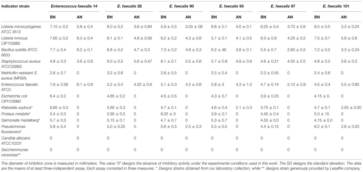
TABLE 1. Inhibitory activity before neutralization of the cell-free supernatant (BN) and after the neutralization of the cell-free supernatant (AN).
Molecular Characterization of the Antagonistic Isolates
Total DNA was extracted from each antagonistic isolates using Wizard® Genomic DNA Purification Kit (Promega, Madison, WI, USA). For 16S rDNA analysis, total DNA was amplified with 16S forward 5′-AGAGTTTGATCMTGGCTCAG-3′ and 16S reverse 5′-GGMTACCTTGTTACGAYTTC-3′ primers (Drago et al., 2011), and the following PCR program: 94°C/3 min, 29 cycles at 94°C/40 s, 55°C/50 s and 72°C/2 min; and finally 72°C/10 min. For REP-PCR analysis, total DNA was amplified with 5′- (GTG)5-3 primer and the following program: 95°C/5 min, 30 cycles at 94°C/1 min, 40°C/1 min, 72°C/8 min, and finally 72°C/16 min. Amplicons were separated on 1% agarose gel. Electrophoresis was performed at 100 V for 2 h using 1X Tris-borate-EDTA. The gels were stained with GEL-RED (Biotium, Canada), and visualized by GelDoc (Bio-Rad, France). Dendrograms were generated automatically using the DIVERSILAB® software (Biomérieux, Craponne, France).
The amplicons for 16S rDNAs were purified with the pJET1Kit (Fermentas, France), and sequenced at Eurofins MWG Operon (Germany). Partial 16S rDNAs sequences were compared to that of E. faecalis ATCC 19433 using ClustalW2 software1.
Challenge Tests: Staphylococcus aureus ATCC 33862 vs. Antagonistic E. faecalis
Enterococcus faecalis was grown in MRS and S. aureus ATCC33862 in Brain Heart Infusion (BHI; 105 CFU/ml) at 37°C for 18 h. After this period, the cultures had reached 108 CFU/ml for E. faecalis, and 105 CFU/ml for S. aureus ATCC33862. Afterward, 0.1 ml of each culture was added to 20 ml of BHI containing leading to 5. 105 CFU/ml (E. faecalis) and 5. 102 CFU/ml (S. aureus ATCC33862). One ml of this coculture was withdrawn at 0, 4, 8, 12, 18, and 24 h, and was serially diluted from 10-1 to 10-6, in 0.9% (w/v) of saline water. Then 1 ml of each dilution was plated on Plates dishes containing MRS agar for E. faecalis, and Chapman agar for S. aureus ATCC33862. The number of colonies was determined after 24 h of incubation at 37°C.
Lactic Acid Quantification
Lactic acid was quantified by HPLC spectra system P1000XR (Thermo, USA). Isolates E. faecalis 28 and E. faecalis 93 were grown in MRS broth at 37°C, samples were withdrawn after 0, 4, 8, 12, 18, and 24 h of incubation, centrifuged (10,000 ×x g, 10 min, 4°C), and sterilized by filtration using Millipore filter (0.2 μm). The CFS were divided into 3 samples. Sample 1 was used to measure pH, sample 2 to assess the antibacterial activity against S. aureus ATCC33862 by the well diffusion method (Naghmouchi et al., 2006), and sample 3 to determine the concentration of lactic acid. All these experiments were performed in triplicate.
Autoaggregation Assay
Autoaggregation assays were performed according to initial protocol of Del Re et al. (2000) and modified by Al Kassaa et al. (2014). Thus, isolates of interest were cultured on MRS for 18 h at 37°C, then cells were centrifuged (5,000 ×g, 4°C, 15 min), washed three times with phosphate saline (PBS) buffer (pH7.0) and were resuspended in sterile PBS to obtain a viable cell count of 108 CFU/ml. The bacterial suspension of 4 ml was mixed by vortexing for 10 s and incubated at room temperature for different time intervals (0, 1, 2, 3, 4, or 5 h). At each interval, the growth was measured at OD600 nm. The percentage of autoaggregation was expressed as follows:
Where OD1 represents the optical density at time (1, 2, 3, 4, or 5 h) and OD2 the data at time 0 h. All experiments were performed in triplicate.
Hydrophobicity
The hydrophobicity was determined using in vitro method to detect the bacterial adhesion to hydrocarbons (Rosenberg et al., 1980). Each antagonistic LAB isolate was grown in MRS broth at 37°C for 18 h. The bacterial cells harvested by centrifugation (5,000 ×g, 4°C, 15 min) were washed twice with PBS (pH7.0), resuspended in the same solution and the OD600 nm was determined. One milliliter of xylene (Fluka, Germany) was added to 3 ml of cell suspension and vortexed for 2 min after 10 min of incubation at room temperature. The aqueous phase was removed after 2 h of incubation at room temperature and the OD600 nm was determined. The percentage of hydrophobicity was calculated using the formula given below. All experiments were performed in triplicate.
Hemolytic Activity
The hemolytic activity was determined by streaking Enterococcal isolates on Columbia blood agar supplemented with 5% (v/v) of human blood or Sheep blood. The plates were incubated at 37°C for 48 h. The presence or absence of zones of clearing around the colonies was interpreted as β-hemolysis (positive hemolytic activity) or χ-hemolysis (negative hemolytic activity), respectively. When observed, greenish zones around the colonies were interpreted as α-hemolysis and taken as negative for the assessment of hemolytic activity (Semedo et al., 2003).
Antimicrobial Susceptibility
Study of antibiotic susceptibility was performed by three independent methods: disk diffusion method, minimal inhibitory concentrations (MICs) using E test (Bio-Mérieux, France), and VITEK 2 system (Bio-Mérieux, France). Related to this, AST-P606 card was used for “Enterococci” encompassing nearly all important antibiotics among which ampicillin, gentamicin, kanamycin, streptomycin, levofloxacin, moxifloxacin, erythromycin, clindamycin, linezolid, teicoplanin, vancomycin, tetracycline, nitrofurantoin chloramphenicoland trimethoprim-sulfamethoxazole. Antibiotic susceptibility and MICs were determined and analyzed according to the French Committee on Antimicrobial Susceptibility Testing (CA-SFM 2013).
Results
Scavenging of Lactic Acid Bacteria and Elucidation of their Antagonism
Hundred and seven LAB isolates were isolated from 6 samples of meconium newborns infants (6 donors) obtained at Roubaix hospital in the North of France. The number of LAB isolates per sample of meconium/donor was as follows: sample 1 (20 LAB isolates), sample 2 (12 LAB isolates), sample 3 (LAB 26 isolates), sample 4 (LAB 12 isolates), sample 5 (LAB 17 isolates), and sample 6 (LAB 20 isolates). In addition to their Gram positive staining, and absence of catalase activity, all these 107 LAB isolates were able to hydrolyze esculine. Importantly, all of them were identified by MALDI TOF mass spectrometry (Biomérieux, France), with high score (>99%), as E. faecalis. Among these E. faecalis species, only six isolates designed as E. faecalis 14, E. faecalis 28, E. faecalis 90, E. faecalis 93, E. faecalis 97, and E. faecalis 101 resulted to be antagonistic. Partial 16S ribosomal DNA sequences of these isolates as well as those of E. faecalis ATCC 19433 were aligned using ClustalW2 software2. This alignment shows a percentage of similarity higher than 99.7% to E. faecalis, confirming phylogenetic identification of our isolates. When the 16S rDNA sequence of E. coli ATCC 11229 was compared to that of enterococci, the percentage of similarity was estimated to 76.33%, which clearly is very low. These antagonistic isolates were able to produce lactic acid and bacteriocin like inhibitory substances (BLIS) and consequently to inhibit growth of GNB and GPB (Table 1). This antagonism was restricted to GPB when the pH of CFS was adjusted to 6.5 (Table 1). The BLIS produced by the aforementioned antagonistic strains resulted to be insensitive to pH and temperature variations but sensitive to proteases. Remarkably, only E. faecalis 90 and E. faecalis 101 were active against Pseudomonas fluorescens (Table 1).
Genetic Patterns of the Antagonistic Strains
Genetic patters gathered by REP-PCR permitted to create two main groups. Indeed, group 1 contains isolates E. faecalis 14, E. faecalis 28, E. faecalis 90, E. faecalis 97, and E. faecalis 101, whereas group 2 contains only isolate E. faecalis 93 (Figure 1). Based on their genetic patterns and analysis of the dendrogram and pulsed field gel electrophoresis (PFGE; data not shown here), we assume that E. faecalis 14 and E. faecalis 28, then E. faecalis 90 and E. faecalis 93, E. faecalis 97 and E. faecalis 101 have similar DNA patterns.
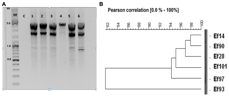
FIGURE 1. Genetic relatedness of antagonistic strains. (A) REP-PCR products were separated on 1% agarose gel. Lane M corresponds to the DNA markers (Thermo, USA), lane C corresponds to the PCR control (PCR carried without DNA template), lanes 1, 2, 3, 4, 5, 6 correspond to genetic patterns of Enterococcus faecalis 14 (Ef14), E. faecalis 28 (Ef28), E. faecalis 90 (Ef90), E. faecalis 93 (Ef93), E. faecalis 97 (Ef97), and E. faecalis 101 (Ef101). (B) Dendrogram outlining Pearson correlation of antagonistic E. faecalis 14 (Ef14), E. faecalis 28 (Ef28), E. faecalis 90 (Ef90), E. faecalis 93 (Ef93), E. faecalis 97 (Ef97) and E. faecalis 101 (Ef101).
Highlights on Anti-S. aureus ATCC33862 Activity
The data gathered from the challenge tests indicate that inhibition of S. aureus ATCC 33862 by E. faecalis 28 and E. faecalis 93 was due to production of lactic acid and bacteriocin. The number of S. aureus ATCC33862 cells has decreased of about 5.5 Log, 5 Log, and 2 log in presence of E. faecalis 28, E. faecalis 93, and E. faecalis ATCC29212 (Figures 2A–C).
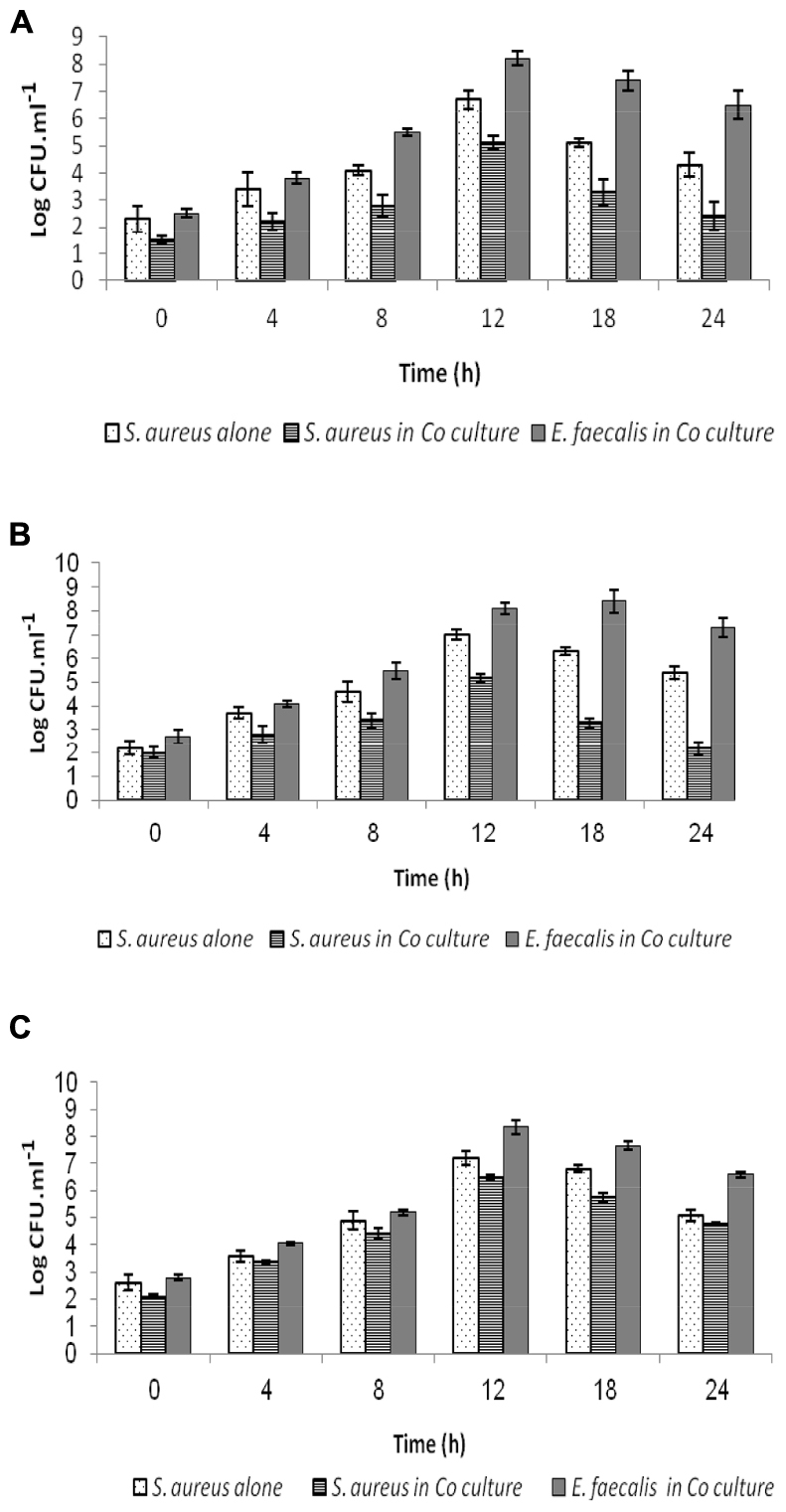
FIGURE 2. Interplay between Staphylococcus aureus ATC C33862 and enterococci during coculture. Samples were withdrawn at regular intervals of time and plated onto agar Brain Heart for counting of S. aureus and on agar MRS for counting of enterococci E. faecalis 28 (A), E. faecalis 93 (B), and E. faecalis ATCC 29212 (C). The values expressed in Log CFUT ml-1 (±SD) are the average of at least three independent experiments.
Production of Lactic Acid and Bacteriocins like Inhibitory Substances
Enterococcus faecalis 28 and E. faecalis 93 isolates produced up to 7.06 g/l of lactic acid, after 24 h of culture. As shown on Figures 3A,B, the drop of pH observed during coculture, was correlated to increase of lactic acid production. The anti-staphylococcal activity was ascribed to both lactic acid and bacteriocins, and this activity appeared to increase until 18 h of co-culture.
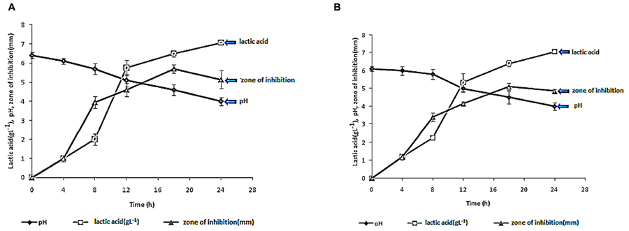
FIGURE 3. Measurement of pHs, amount of lactic acid (g L-1) and inhibitory activity of the cell free supernatants (CFS) prepared from E. faecalis 28 (A) and E. faecalis 93 (B) after different times of growth. The data are the average of at least three independent experiments.
Cell Surface Properties of the Antagonistic Isolates
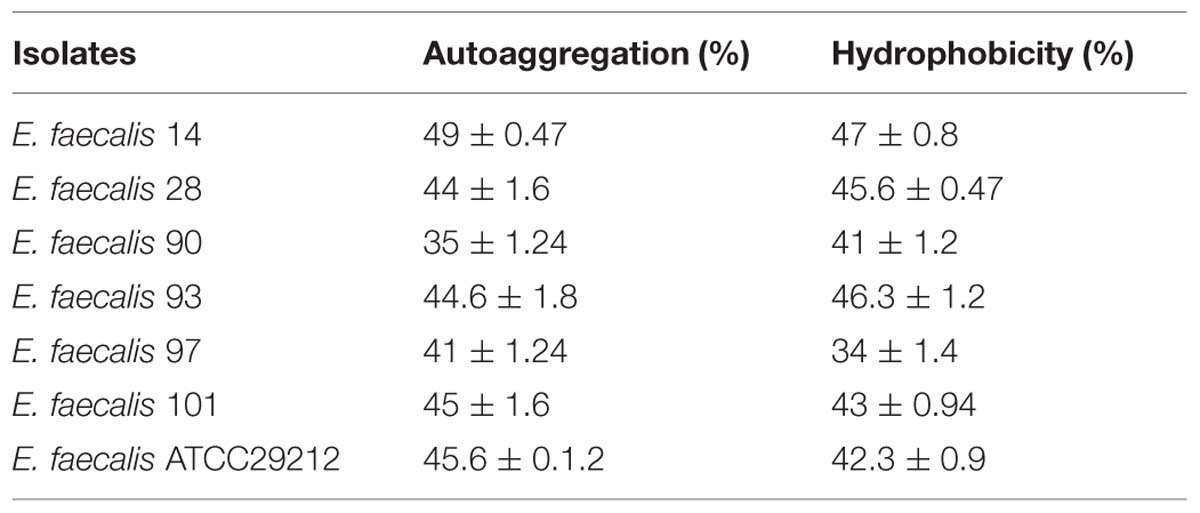
Table 2 shows the scores of hydrophobicity and autoaggregation of antagonistic isolates. The highest scores were registered for E. faecalis 14; that was isolated from donor 3.
Antagonistic Strains are Sensitive to Antibiotics and are Not Hemolytic
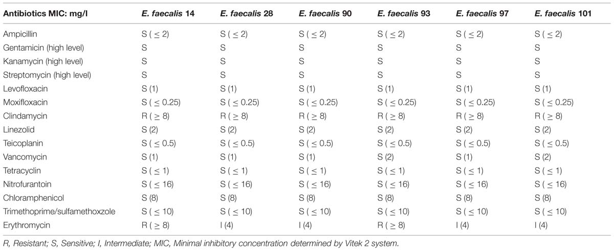
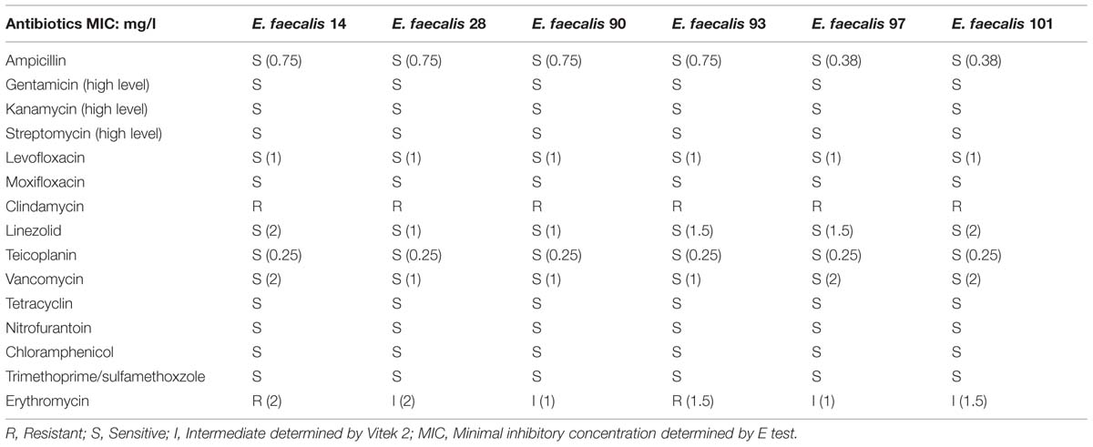
As indicated in Tables 3A,B, antagonistic strains are sensitive to antibiotics tested. To be noted that E. faecalis 14 and E. faecalis 93 are resistant to erythromycin, whilst isolates E. faecalis 28, E. faecalis 90, E. faecalis 97, and E. faecalis 101 exhibit intermediate resistance to this drug. Further, all antagonistic isolates are sensitive to ampicillin, gentamicin, kanamycin, streptomycin, levofloxacin, moxifloxacin, linezolid, teicoplanin, vancomycin, tetracycline, nitrofurantoin, and chloramphenicol but are resistant to clindamycin and trimethoprim-sulfamethoxazole. The data analysis was performed according to “Antibiogram committee of French Microbiology of Society” based on the European Committee on Antimicrobial Susceptibility Testing recommendations. Remarkably, these strains were not hemolytic (data not shown).
Accession Numbers
The accession numbers were KP057871 (E. faecalis 14), KP057872 (E. faecalis 28), KP057873 (E. faecalis 90), KP057874 (E. faecalis 93), KP057875 (E. faecalis 97), KP057876 (E. faecalis 101), NR_115765.1 (E. faecalis ATCC 19433), and GQ340751.1 Escherichia coli ATCC 11229.
Discussion
This study aimed at strictly isolating LAB and particularly bacteriocinogenic LAB from meconium of newborns infants. Thus, 107 bacterial isolates were obtained from meconium of 6 donors at Roubaix Hospital in the north of France. All the bacterial isolates were identified as E. faecalis. Interestingly, Borderon et al. (1996), Adlerberth et al. (2006) showed that E. faecalis colonizes the gut of vaginally and cesarean-delivered term and preterm neonates Clinical isolates of enterococci are weakly diverse comparatively to those obtained from the environment, but E. faecalis remains the dominant species (Kuhn et al., 2003). According to Fisher and Phillips (2009), this weak diversity could be linked to the virulence factors present in this species.
Isolates E. faecalis 14, E. faecalis 28, E. faecalis 90, E. faecalis 93, E. faecalis 97, and E. faecalis 101 produce inhibitory compounds that comprise lactic acid and BLIS. The concomitant production of these antimicrobials inhibited a set of GNB and GPB, including the robust methicillin-resistant S. aureus (MRSA), which is frequently encountered in hospital environment. Remarkably, this antagonism was abolished when the CFS was neutralized, highlighting thereof the role of BLIS. Vancomycin is the standard antibiotic used for the treatment of MRSA infections. Recently, Liang et al. (2014), suggested daptomycin as a reasonable alternative for treating MRSA mainly for osteoarticular infections (OAIs). The activity of these antibiotics could be potentialized with bacteriocins, which are overall safe (Belguesmia et al., 2011), ribosomally synthesized AMP (Drider and Rebuffat, 2011). Genotyping of these isolates performed by REP-PCR and PFGE (data not shown) concluded to three different strains. Percent Identity Matrix based on 16S ribosomal DNA confirms a high score of similarity of enterococci sequences and a low score of similarity with E. coli ATCC 11229 used as different bacterial taxon.
In this study, E. faecalis 28 and E. faecalis 93, obtained from two different sources, were the most actives isolates. In their presence, the counts of S. aureus ATCC33862 has drastically decreased. Further, the antagonistic isolates displayed high scores of autoaggregation and hydrophobicity. These two characteristics might be important for the interplay of these isolates with S. aureus ATCC33862. Clewell et al. (2002) reported that production of aggregation substance (Agg) on the donor cell-surface facilitates contact with recipient cell by binding to enterococcal binding substance.
Importantly, these antagonistic strains were not hemolytic and resulted to be sensitive to antibiotic usually used to treat enterococci infections. The resistance to clindamycin and Trimethoprime/sulfamethoxzole is thought to be a species characteristic. According to the opinion of the FEEDAP panel on the updating of criteria used in the assessment of bacteria for resistance to antibiotics of human and veterinary importance, strains carrying an acquired resistance to antimicrobial(s) should not be used as feed additives, unless it can be demonstrated that it is a result of chromosomal mutation(s) (European Food Safety Authority [EFSA], 2005).
This study unveiled the presence of only E. faecalis species in samples of meconium obtained from six donors at Roubaix hospital in the north of France. Off 107 E. faecalis isolates, only E. faecalis 14, E. faecalis 28, E. faecalis 90, E. faecalis 93, E. faecalis 97, and E. faecalis 101 resulted to be bacteriocinogenic. In the best of our knowledge, this is the first report dealing with bacteriocinogenic LAB from meconium. The number of antagonistic isolates is relatively low (5.60%) compared to the frequency usually reported in the literature (Nes et al., 2014). The high hydrophobicity and aggregation scores registered for the aforementioned antagonistic isolates, as well as absence of hemolytic activity and sensibility to various antibiotic render these strains as potential candidates for probiotic applications.
Conflict of Interest Statement
The authors declare that the research was conducted in the absence of any commercial or financial relationships that could be construed as a potential conflict of interest.
Acknowledgments
AA was a recipient of Ph.D. scholarship from Iraqi-French governments. The authors are indebted to Prof. Mike Chikindas (Rutgers University, USA), and Abdellah Benachour (Université de Caen, France) for their critical reading of the manuscript, and to Dr. Sylvie Rousseau (Roubaix Hospital) for providing us with meconium samples used in this study.
Footnotes
References
Adlerberth, I., Lindberg, E., Aberg, N., Hesselmar, B., Saalman, R., and Strannegard, I. L. (2006). Reduced enterobacterial and increased staphylococcal colonization of the infantile bowel: an effect of hygienic lifestyle. Pediatr. Res. 59, 96–101. doi: 10.1203/01.pdr.0000191137.12774.b2
PubMed Abstract | Full Text | CrossRef Full Text | Google Scholar
Al Kassaa, I., Hamze, M., Hober, D., Chihib, N. E., and Drider, D. (2014). Identification of vaginal Lactobacilli with potential probiotic properties isolated from women in North Lebanon. Microb. Ecol. 67, 722–734. doi: 10.1007/s00248-014-0384-7
PubMed Abstract | Full Text | CrossRef Full Text | Google Scholar
Alicea-Serrano, A. M., Contreras, M., Magris, M., Hidalgo, G., and Dominguez-Bello, M. G. (2013). Tetracycline resistance genes acquired at birth. Arch. Microbiol. 195, 447–451. doi: 10.1007/s00203-012-0864-4
PubMed Abstract | Full Text | CrossRef Full Text | Google Scholar
Belguesmia, Y., Madi, A., Sperandio, D., Merieau, A., Feuilloley, M., Prévost, H., et al. (2011). Growing insights into the safety of bacteriocins: the case of enterocin S37. Res. Microbiol. 162, 159–163. doi: 10.1016/j.resmic.2010.09.019
PubMed Abstract | Full Text | CrossRef Full Text | Google Scholar
Borderon, J. C., Lionnet, C., Rondeau, C., Suc, A. I., Laugier, J., and Gold, F. (1996). Current aspects of fecal flora of the newborn without antibiotherapy during the first 7 days of life: enterobacteriaceae, enterococci, staphylococci. Pathol. Biol. 44, 416–422.
Campos, C. A., Rodriguez, O., Calo-Mata, P., Prado, M., and Barros-Velazquez, J. (2006). Preliminary characterization of bacteriocins from Lactococcus lactis, Enterococcus faecium and Enterococcus mundtii strains solated from turbot (Psetta maxima). Food Res. Int. 39, 356–364. doi: 10.1016/j.foodres.2005.08.008
Cleary, G. M., and Wiswell, T. E. (1998). Meconium-stained amniotic fluid and the meconium aspiration syndrome. an update. Pediatr. Clin. North Am. 45, 511–529. doi: 10.1016/S0031-3955(05)70025-0
Clewell, D. B., Francia, M. V., Flannagan, S. E., and An, F. Y. (2002). Enterococcal plasmid transfer: sex pheromones, transfer origins, relaxases, and the Staphylococcus aureus issue. Plasmid 48, 193–201. doi: 10.1016/S0147-619X(02)00113-0
PubMed Abstract | Full Text | CrossRef Full Text | Google Scholar
de Man, J. C., Rogosa, M., and Sharpe, M. E. (1960). A medium for the cultivation of lactobacilli. J. Appl. Bacteriol. 23, 130–135. doi: 10.1111/j.1365-2672.1960.tb00188.x
Del Re, B., Sgorbati, B., Miglioli, M., and Palenzona, D. (2000). Adhesion, autoaggregation and hydrophobicity of 13 strains of Bifidobacterium longum. Lett. Appl. Microbiol. 31, 438–442. doi: 10.1046/j.1365-2672.2000.00845.x
PubMed Abstract | Full Text | CrossRef Full Text | Google Scholar
DiGiulio, D. B., Romero, R., Amogan, H. P., Kusanovic, J. P., Bik, E. M., Gotsch, F., et al. (2008). Microbial prevalence, diversity and abundance in amniotic fluid during preterm labor: a molecular and culture-based investigation. PLoS ONE 3:e3056. doi: 10.1371/journal.pone.0003056
PubMed Abstract | Full Text | CrossRef Full Text | Google Scholar
Dominguez-Bello, M. G., Costello, E. K., Contreras, M., Magris, M., Hidalgo, G., Fierer, N., et al. (2010). Delivery mode shapes the acquisition and structure of the initial microbiota across multiple body habitats in newborns. Proc. Natl. Acad. Sci. U.S.A. 107, 11971–11975. doi: 10.1073/pnas.1002601107
PubMed Abstract | Full Text | CrossRef Full Text | Google Scholar
Drago, L., Mattina, R., Nicola, L., Rodighiero, V., and De Vechi, E. (2011). Macrolide resistance and in vitro selection of resistance to antibiotics in Lactobacillus isolates. J. Microbiol. 49, 651–656. doi: 10.1007/s12275-011-0470-1
PubMed Abstract | Full Text | CrossRef Full Text | Google Scholar
Drider, D., and Rebuffat, S. (2011). Prokaryotic Antimicrobial Peptides: From Genes to Applications. New York, NY: Springer. doi: 10.1007/978-1-4419-7692-5
European Food Safety Authority [EFSA]. (2005). Opinion of the scientific panel on. (additives) and products or substances used in animal feed on the updating of the criteria used in the assessment of bacteria for resistance to antibiotics of human or veterinary importance (question no. EFSA-Q-2004-079). EFSA J. 223, 1–12.
Fisher, K., and Phillips, C. (2009). The ecology, epidemiology and virulence of Enterococcus. Microbiology 155, 1749–1757. doi: 10.1099/mic.0.026385-0
PubMed Abstract | Full Text | CrossRef Full Text | Google Scholar
Franz, C. M., Huch, M., Abriouel, H., Holzapfel, W., and Glávez, A. (2011). Enterococci as probiotics and their implication in food safety. Int. J. Food. Microbiol. 151, 125–140. doi: 10.1016/j.ijfoodmicro.2011.08.014
PubMed Abstract | Full Text | CrossRef Full Text | Google Scholar
Gosalbes, M. J., Llop, S., Vallès, Y., Moya, A., Ballester, F., and Francino, M. P. (2013). Meconium microbiota types dominated by lactic acid or enteric bacteria are differentially associated with maternal eczema and respiratory problems in infants. Clin. Exp. Allergy 43, 198–211. doi: 10.1111/cea.12063
PubMed Abstract | Full Text | CrossRef Full Text | Google Scholar
Hitti, J., Riley, D. E., Krohn, M. A., Hillier, S. L., Agnew, K. J., Krieger, J. N., et al. (1987). Broad-spectrum bacterial rDNA polymerase chain reaction assay for detecting amniotic fluid infection among women in premature labor. Clin. Infect. Dis. 24, 1228–1232. doi: 10.1086/513669
PubMed Abstract | Full Text | CrossRef Full Text | Google Scholar
Hong, P. Y., Lee, B. W., Aw, M., Shek, L. P., Yap, G. C., Chua, K. Y., et al. (2010). Comparative analysis of fecal microbiota in infants with and without eczema. PLoS ONE 5:e9964. doi: 10.1371/journal.pone.0009964
PubMed Abstract | Full Text | CrossRef Full Text | Google Scholar
Hu, J., Nomura, Y., Bashir, A., Fernandez-Hernandez, H., Itzkowitz, S., Pei, Z., et al. (2013). Diversified microbiota of meconium is affected by maternal diabetes status. PLoS ONE 8:e78257. doi: 10.1371/journal.pone.0078257
PubMed Abstract | Full Text | CrossRef Full Text | Google Scholar
Johansson, M. A., Sjögren, Y. M., Persson, J. O., Nilsson, C., and Sverremark-Ekström, E. (2011). Early colonization with a group of Lactobacilli decreases the risk for allergy at five years of age despite allergic heredity. PLoS ONE 6:e23031. doi: 10.1371/journal.pone.0023031
PubMed Abstract | Full Text | CrossRef Full Text | Google Scholar
Kuhn, I., Iversen, A., Burman, L. G., Olsson-Liljequist, B., Franklin, A., Finn, M., et al. (2003). Comparison of enterococcal populations in animals, humans, and the environment– a European study. Int. J. Food Microbiol. 88, 133–145. doi: 10.1016/S0168-1605(03)00176-4
Liang, S. Y., Khair, H. N., McDonald, J. R., Babcock, H. M., and Marschall, J. (2014). Daptomycin versus vancomycin for osteoarticular infections due to methicillin-resistant Staphylococcus aureus (MRSA): a nested case-control study. Eur. J. Clin. Microbiol. Infect. Dis. 33, 659–664. doi: 10.1007/s10096-013-2001-y
PubMed Abstract | Full Text | CrossRef Full Text | Google Scholar
Makino, H., Kushiro, A., Ishikawa, E., Kubota, H., Gawad, A., Sakai, T., et al. (2013). Mother-to-infant transmission of intestinal bifidobacterial strains has an impact on the early development of vaginally delivered infant’s microbiota. PLoS ONE 8:e78331. doi: 10.1371/journal.pone.0078331
PubMed Abstract | Full Text | CrossRef Full Text | Google Scholar
Moles, L., Gómez, M., Heilig, H., Bustos, G., Fuentes, S., de Vos, W., et al. (2013). Bacterial diversity in meconium of preterm neonates and evolution of their fecal microbiota during the first month of life. PLoS ONE 28:e66986. doi: 10.1371/journal.pone.0066986
PubMed Abstract | Full Text | CrossRef Full Text | Google Scholar
Mshvildadze, M., Neu, J., Shuster, J., Theriaque, D., Li, N., and Mai, V. (2010). Intestinal microbial ecology in premature infants assessed using with non –culture- based technique. J. Pediatr. 156, 20–25. doi: 10.1016/j.jpeds.2009.06.063
PubMed Abstract | Full Text | CrossRef Full Text | Google Scholar
Naghmouchi, K., Drider, D., Kheadr, E., Lacroix, C., Prévost, H., and Fliss, I. (2006). Multiple characterizations of Listeria monocytogenes sensitive and insensitive variants to divergicin M35, a new pediocin-like bacteriocin. J. Appl. Microbiol. 100, 29–39. doi: 10.1111/j.1365-2672.2005.02771.x
PubMed Abstract | Full Text | CrossRef Full Text | Google Scholar
Nes, I. F., Diep, D. B., and Ike, Y. (2014). “Enterococcal bacteriocins and antimicrobial proteins that contribute to niche control,” in Enterococci: From Commensals to Leading Causes of Drug Resistant Infection [Internet], eds M. S. Gilmore, D. B. Clewell, Y. Ike, and N. Shankar (Boston, MA: Massachusetts Eye and Ear Infirmary).
Rodríguez, E., Arqués, J. L., Rodríguez, R., Peirotén,Á., Landete, J. M., and Medina, M. (2012). Antimicrobial properties of probiotic strains isolated from brest-fed infants. J. Funct. Foods. 4, 542–551. doi: 10.1016/j.jff.2012.02.015
Rosenberg, M., Gutnick, D., and Rosenberg, E. (1980). Adherence of bacteria to hydrocarbons: a simple method for measuring cell-surface hydrophobicity. FEMS Microbiol. Lett. 9, 29–33. doi: 10.1111/j.1574-6968.1980.tb05599.x
PubMed Abstract | Full Text | CrossRef Full Text | Google Scholar
Semedo, T., Santos, M. A., Lopes, M. F. S., Marques, J. J. F., Crespo, M. T. B., and Tenreiro, R. (2003). Virulence factors in food, clinical and reference enterococci: a common trait in the genus? Syst. Appl. Microbiol. 26, 13–22. doi: 10.1078/072320203322337263
PubMed Abstract | Full Text | CrossRef Full Text | Google Scholar
Steel, J. H., Malatos, S., Kennea, N., Edwards, A. D., Miles, L., Duggan, P., et al. (2005). Bacteria and inflammatory cells in fetal membranes do not always cause preterm labor. Pediatr. Res. 57, 404–411. doi: 10.1203/01.PDR.0000153869.96337.90
PubMed Abstract | Full Text | CrossRef Full Text | Google Scholar
Vizoso-Pinto, M. G., Franz, C., Schillinger, U., and Holzapfel, W. H. (2006). Lactobacillus spp with in vitro probiotic properties from human faeces and traditional fermented products. Int. J. Food Microbiol. 109, 205–214. doi: 10.1016/j.ijfoodmicro.2006.01.029
PubMed Abstract | Full Text | CrossRef Full Text | Google Scholar
Wang, L., Christophersen, C. T., Sorich, M. J., Gerber, J. P., Angley, M. T., and Conlon, M. A. (2011). Low relative abundances of the mucolytic bacterium Akkermansia muciniphila and Bifidobacterium spp. in feces of children with autism. Appl. Environ. Microbiol. 77, 6718–6721. doi: 10.1128/AEM.05212-11
PubMed Abstract | Full Text | CrossRef Full Text | Google Scholar
Keywords: meconium, Enterococcus faecalis, antagonism, anti-Staphylococcus activity, enterocins
Citation: Al Atya AK, Drider-Hadiouche K, Ravallec R, Silvain A, Vachee A and Drider D (2015) Probiotic potential of Enterococcus faecalis strains isolated from meconium. Front. Microbiol. 6:227. doi: 10.3389/fmicb.2015.00227
Received: 26 November 2014; Accepted: 07 March 2015;
Published online: 02 April 2015
Edited by:
Tzi Bun Ng, The Chinese University of Hong Kong, ChinaReviewed by:
Atte von Wright, University of Eastern Finland, FinlandPeter Pristas, Institue of Animal Physiology – Slovak Academy of Sciences, Slovakia
Copyright © 2015 Al Atya, Drider-Hadiouche, Ravallec, Silvain, Vachee and Drider. This is an open-access article distributed under the terms of the Creative Commons Attribution License (CC BY). The use, distribution or reproduction in other forums is permitted, provided the original author(s) or licensor are credited and that the original publication in this journal is cited, in accordance with accepted academic practice. No use, distribution or reproduction is permitted which does not comply with these terms.
*Correspondence: Djamel Drider, Institut Charles Viollette, Polytech-Lille, Université Lille 1 – Sciences et Technologies, Office C315, Avenue Paul Langevin, Cité Scientifique, 59655 Villeneuve d’Ascq, FranceZGphbWVsLmRyaWRlckB1bml2LWxpbGxlMS5mcg==
 Ahmed K. Al Atya1
 Djamel Drider