 Quentin Menetrey
 Chloé Dupont
 Raphaël Chiron1,3
 Hélène Marchandin
- 1HydroSciences Montpellier, Univ Montpellier, CNRS, IRD, Montpellier, France
- 2HydroSciences Montpellier, Univ Montpellier, CNRS, IRD, Laboratoire d’Ecologie Microbienne Hospitalière, CHU Montpellier, Montpellier, France
- 3HydroSciences Montpellier, Univ Montpellier, CNRS, IRD, Centre de Ressources et de Compétences de la Mucoviscidose, CHU Montpellier, Montpellier, France
- 4HydroSciences Montpellier, Univ Montpellier, CNRS, IRD, Département de Microbiologie, CHU Nîmes, Nîmes, France
Cystic Fibrosis (CF) airways favor abnormal microbial development. Infections are considered as polymicrobial and competition can be observed between microorganisms. The current literature on bacterial competition in CF mostly consists of studies with limited numbers of strains, mainly focused on the major pathogens Pseudomonas aeruginosa (Pa) and Staphylococcus aureus (Sa) and does not give a comprehensive overview of the overall importance of bacterial interactions or the behavior of less often encountered emerging bacteria such as Achromobacter. In this context, we screened a panel of 39 strains from six CF patients, of either clinical or domestic environmental origin, distinguished according to genotype and belonging to four opportunistic pathogens, Pa (n = 15), Sa (n = 3), Stenotrophomonas maltophilia (Sm, n = 10) and Achromobacter xylosoxidans (Ax, n = 11). We investigated their capacity to compete in terms of growth, motility, and pigment production on agar media through 203 crossing experiments. Eleven strains selected via the initial screening results were further studied for competitive growth in liquid medium and biofilm formation. Competition was noted for 33% (67/203) of the pairs of strains with 85 modifications observed between monocultures and co-cultures, impacting growth (23.6%), motility (13.8%), and/or pigment production (6.1%). Under all conditions of the study (clinical, environmental strains; intra-, inter-patients; intra-, inter-species levels), competition was significantly more frequent among pairs of strains with at least one clinical strain. While Pa mainly outcompeted other species, in one patient with chronic colonization by Ax and sporadic colonization by Pa, we showed that some Ax inhibited the growth and pigmentation of Pa whereas biofilm formation was drastically reduced. Enlarging the panel of strains tested in competition assays gave new perspectives on the complex interactions taking place among the CF airway community. Indeed, the frequent occurrence of varied, strain-dependent interactions is revealed here. We report the first results of competition assays for Ax with the ability of certain strains to outcompete Pa. Our results are linked to the patient’s colonization history and question the importance of bacterial competitiveness in the colonization pattern of CF airways.
Introduction
Cystic fibrosis (CF) is a genetic disease caused by a mutation in the Cystic Fibrosis Transmembrane conductance Regulator gene coding for a transmembrane channel allowing the transport of chloride ions. Different organs are affected, including the respiratory tract, where thickening mucus, mucociliary clearance defects, and a decrease in anti-microbial defenses are observed. These alterations allow a variety of microorganisms, of endogenous and environmental origin, to multiply in the cystic fibrosis respiratory tract (CFRT; Burns et al., 1998; Lyczak et al., 2002; Ciofu et al., 2013). The main opportunistic pathogens identified in young children with CF are Staphylococcus aureus and Haemophilus influenzae whereas Pseudomonas aeruginosa predominates in older patients (Registre français de la mucoviscidose – Bilan des données, 2017). In addition, certain other opportunistic bacterial pathogens of environmental origin are encountered in CF patients and, despite being less frequently identified, some of them, such as Achromobacter xylosoxidans and Stenotrophomonas maltophilia are considered as emerging (Lyczak et al., 2002; Hansen et al., 2009; Lambiase et al., 2011; Parkins and Floto, 2015; Esposito et al., 2017; Registre français de la mucoviscidose – Bilan des données, 2017). Cultivation-independent studies have led to increasing knowledge about the diversity of the bacterial community in CF lungs and its dynamics throughout the evolution of this disease. They have revealed a higher phylogenetic diversity than expected from cultivation-based studies, a diversity that progressively decreases despite the overall abundance of microbiota remaining high, and the existence of co-occurring bacterial networks suggestive of functional associations within the CF polymicrobial consortium (Filkins et al., 2015; Quinn et al., 2016). Pulmonary infections in CF patients have been considered as polymicrobial for more than a decade now (Sibley et al., 2006; Peters et al., 2012; O’Brien and Welch, 2019) and pulmonary exacerbations have been shown to be associated with modifications in the CF community and a disruption/fragmentation of bacterial networks observed during stable clinical status (Quinn et al., 2016). These microbiota-disease associations must also be interpreted in an ecological perspective as they take place in the pulmonary environment, i.e., a restricted space subjected to diverse selective pressures (immune system, acidic environment, antibiotic cures, etc.; Rogers et al., 2013).
Due to both the diversity of bacterial species and the abundance of the community present in CF airways, competition between species for space and nutrients is established through physical and chemical interactions (Høiby et al., 2010; Armbruster et al., 2019). These interactions can be mutualistic or synergistic as they can improve the coexistence of microorganisms on epithelial surfaces and also enable them to use metabolic by-products more efficiently (Whiley et al., 2014, 2015). Conversely, other microorganisms can develop antagonistic interactions, for example by secreting effectors that inhibit the growth of co-colonizing species (Bragonzi et al., 2012; Michelsen et al., 2014; Tognon et al., 2019). These interactions are facilitated within multi-species biofilms and involve intercellular communication via quorum sensing (QS) molecules able to modulate the expression of certain virulence factors, bacterial growth, and/or regulate the host’s immune system (Duan et al., 2003; Hogan et al., 2004; Williams and Cámara, 2009; Tashiro et al., 2013; Pollitt et al., 2014; Beaume et al., 2015; Whiley et al., 2015; O’Brien and Fothergill, 2017). However, despite an increasing interest in characterizing interactions between species, their clinical relevance and potential implications in the progression of polymicrobial pathologies are still poorly understood (He and Shi, 2009; Limoli and Hoffman, 2019).
In CF, it is likely that pathogens develop specific competitive strategies as they develop in a narrow, hostile environment subjected to many selection pressures (Schwab et al., 2014). Competition between microorganisms should thus to be considered as a dynamic phenomenon, highly dependent on the strains’ biotic and abiotic environment and the colonization history of the patient’s airways (Palmer et al., 2005; Andersen et al., 2015). A better knowledge and characterization of these bacterial interactions could help us to understand the dynamics of bacterial colonization of the respiratory tract in CF patients and therefore studies on clinically documented CF strains warrant more consideration. Indeed, most studies on the bacterial competition that may occur in the CF lung have focused on the two major CF pathogens, P. aeruginosa and S. aureus (Baldan et al., 2014; Filkins et al., 2015; Hotterbeekx et al., 2017; Limoli et al., 2017; Tognon et al., 2017) and available data have mostly been obtained from a limited number of strains, including reference strains that may not reflect the true behavior of clinical strains. In this context, the main objective of our study was to investigate the existence of interactions between clinically documented representatives of opportunistic pathogenic species colonizing the respiratory tract of CF patients and the domestic environment of CF patients, a potential source for airway colonization (Heirali et al., 2016), and to specify the importance and nature of these interactions. We studied important features that may be modified through bacterial competition to confer an advantage to one of the partners, i.e., growth, motility, biofilm production, and production of pigments as an indirect marker for P. aeruginosa virulence. A focus was then made on the competitive ability of A. xylosoxidans, which had never been studied before.
Materials and Methods
Biological Materials, Bacterial Strains, and Patients
The strains under study were isolated during routinely performed analyses of sputum samples from six CF patients (Patients A–F among whom Patients A, C, and F were three patients chronically colonized by A. xylosoxidans included from previous studies (Dupont et al., 2015, 2016, 2018) in which they were known as Patients 12, 2, and 5, respectively) attending the CF center at Montpellier University Hospital, France (n = 24) or from the domestic environment of three of these patients (n = 15; Table 1). Strains were stored frozen at −80°C. The 39 bacterial strains belonged to four species: 11 A. xylosoxidans (Ax) including one environmental strain; 15 non-mucoid P. aeruginosa (Pa) including seven environmental strains; 10 S. maltophilia (Sm) including seven environmental strains and three methicillin-susceptible S. aureus (Sa; Table 1). Bacterial strains were genotyped by multi locus sequence typing (MLST) as previously described (Enright et al., 2000; Curran et al., 2004; Kaiser et al., 2009; Spilker et al., 2012). Microbiological data resulting from patient follow-up were retrospectively recorded to specify the patient’s colonization profile (co-isolated species, colonization type). Chronic and intermittent colonization’s were defined according to the modified Leeds criteria by isolating bacterium from > 50% or ≤ 50% of respiratory samples collected over the previous 12 months for patients with at least four samples analyzed during that period, respectively (Lee et al., 2003). Sporadic colonization refers to a general definition of bacterial identification occurring at irregular scattered or isolated intervals.
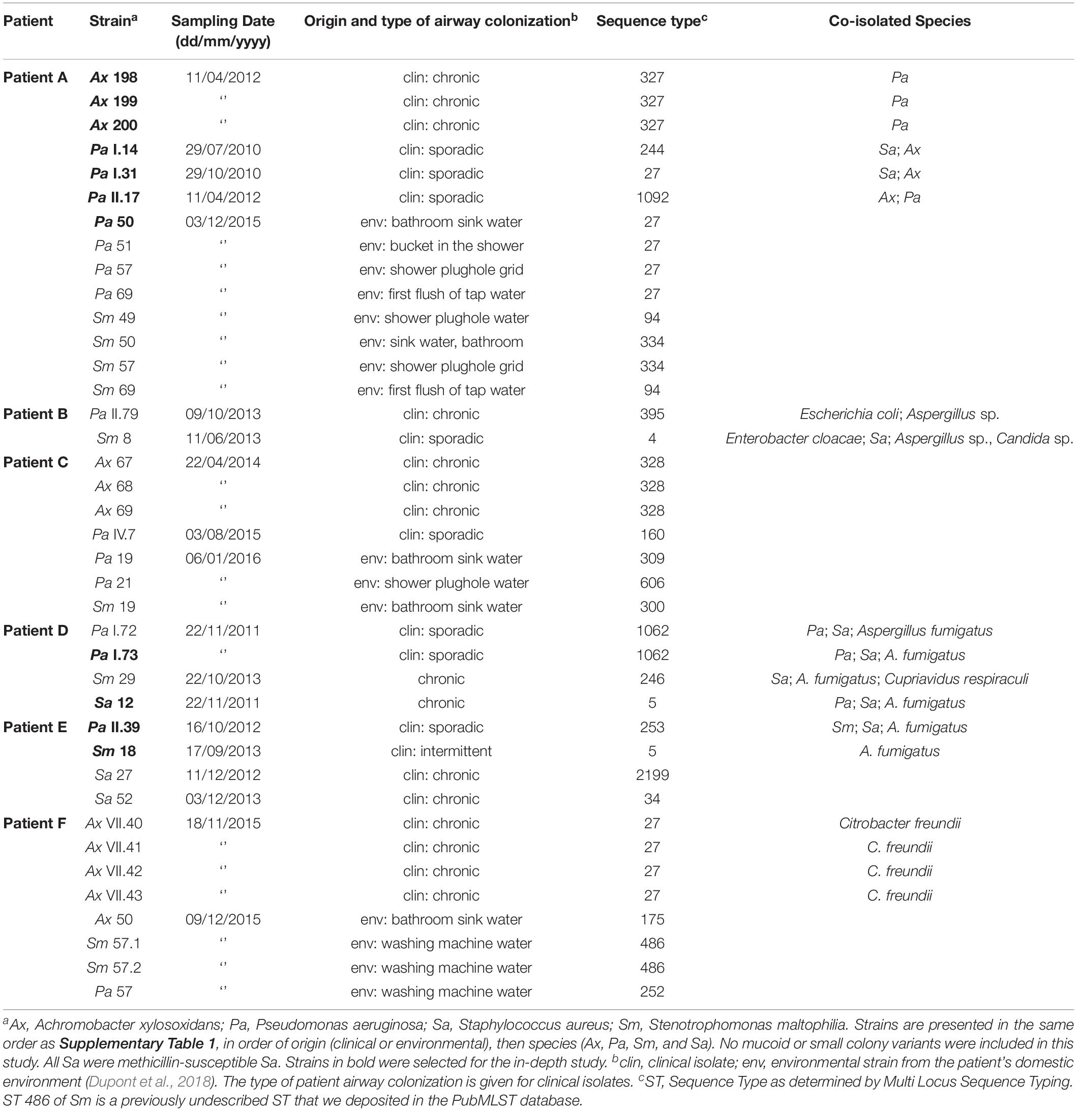
Table 1. Isolation and genotypic characteristics of the 39 strains under study.
Study Design
Interactions between the 39 strains in our study were investigated through 203 crossing experiments to evaluate competition in terms of growth, motility, and global pigment production on agar media (Figure 1 and Supplementary Table 1). During this first part of the study (see “screening study” hereafter), all pairs of strains were tested by the three approaches detailed below: proximity assay, test on bacterial layer, and swimming motility evaluation. For assays including at least one Pa strain, these three assays simultaneously allowed us to visually detect any modification in pigment production by Pa. Among the 203 pairs of strains tested, 80.8% (164/203) included at least an emerging pathogen (Ax or Sm); 33 pairs included strains from the same species and 170 strains from different species, 121 came from the same patient and 82 from different patients, and 122 included strains of the same origin (clinical or environmental) while 81 included strains of different origins (clinical and environmental). All results obtained from the co-culture assays were compared with those obtained from monoculture assays on the two strains of the tested pair. Each experiment was performed at least twice. Based on the results obtained from this screening stage of our study, an in-depth study was then conducted on 11 selected strains originating from three patients to assess competition in terms of bacterial growth by establishing curves for growth and/or the ability to form biofilm, with each assay repeated 4 times (Figure 1 and Supplementary Table 1).
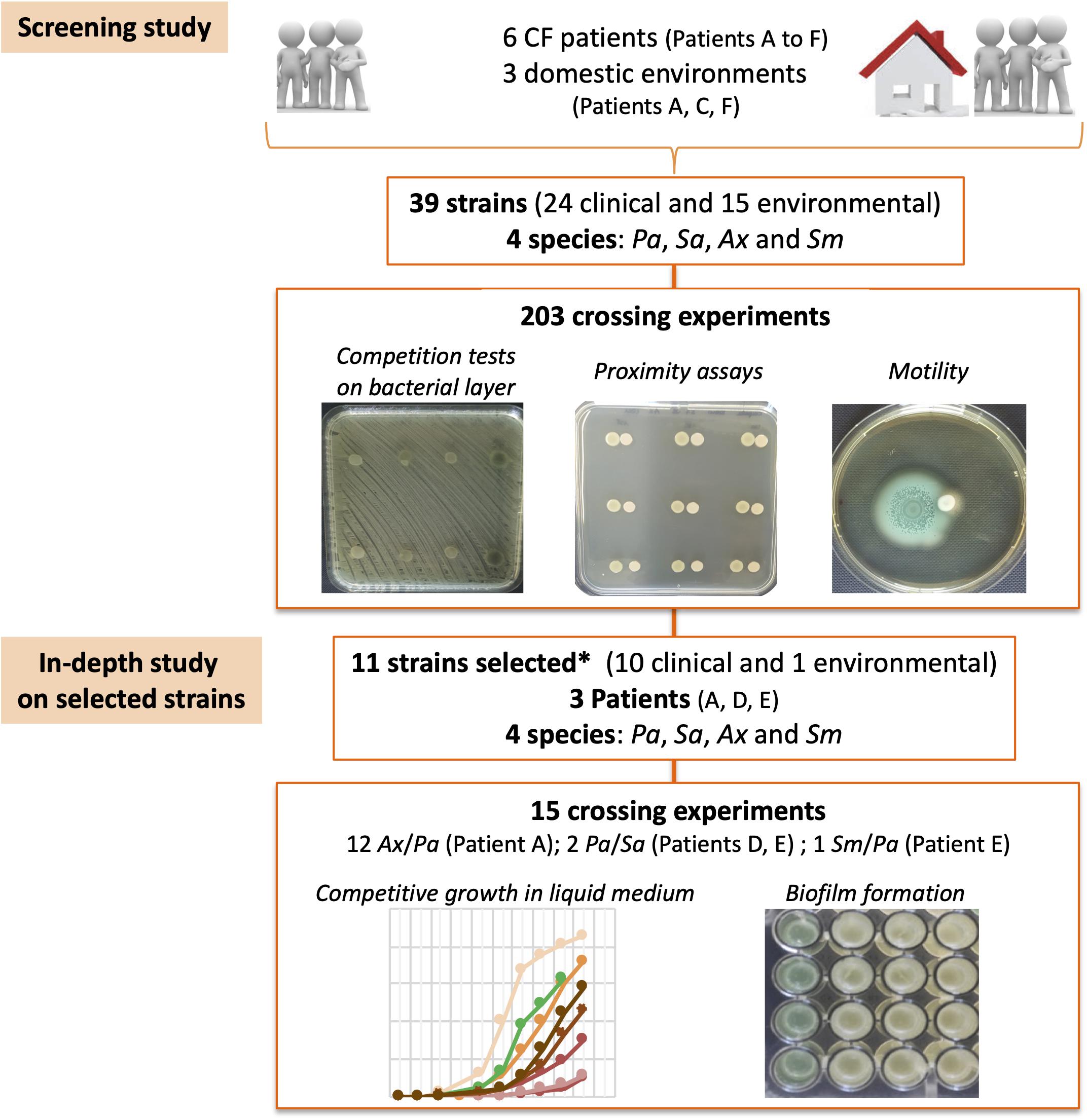
Figure 1. Study design flowchart. CF, Cystic Fibrosis; Pa, Pseudomonas aeruginosa; Sa, Staphylococcus aureus; Ax, Achromobacter xylosoxidans; Sm, Stenotrophomonas maltophilia. ∗ Selected strains were those for which the most important growth inhibition of one partner was observed during the screening experiments (two co-isolated strains were also included to investigate intra-sample diversity of Ax/Pa competition in Patient A).
Screening Study: Culture Conditions and Competition Assays
Culture Conditions
Bacteria were cultured on trypticase soy (TS) agar for 24 h at 37°C. Liquid cultures were then performed in TS broth incubated overnight at 37°C and shaken (175 rpm) under aerobic conditions (slightly open tubes). Before any experiment, the OD600nm of each bacterial suspension was adjusted to 0.5, equivalent to 2–6.108 colony forming units (CFU)/mL. Growth curves were established for the four species included in the study (n = 18 strains; 7 Pa, 1 Sa, 5 Ax, and 5 Sm). All the five Ax tested displayed a longer time to enter the exponential growth phase compared to other species (7 Pa, 1 Sa, and 5 Sm; Supplementary Figure 1), supporting the application of specific co-culture conditions for Pa–Ax pair testing, as in previous studies among species displaying distinct growth rates (Duan et al., 2003; Pompilio et al., 2015). Consequently, a time offset of 4 h was retained for Ax deposited 4 h before Pa in the various experiments, and the OD600nm of Pa was adjusted to 0.1 to reduce the nutrient competition with Ax. Incubation was carried out at 37°C for 48 h for all assays. Sm strains showed heterogeneous growth characteristics and specific conditions were not applied to co-cultures including an Sm strain in this study (Supplementary Figure 1).
Proximity Assays
Tests were performed according to Whiley et al. (2015) with modifications: 10 μL-spots of two strains were deposited opposite each other, at three different distances, 0.4, 0.6, and 0.8 cm, respectively. Growth zones were visually observed to search for a decrease or potentiation of growth of the bacteria under study. Controls consisted of depositing each of the two strains in spots opposite each other.
Competition Tests on Bacterial Layer
Tests were performed according to Beaume et al. (2015) with modifications. Briefly, TS agar was inoculated by swabbing a bacterial suspension. Spots (10 μL) of a “competitor” bacterium in suspension were then deposited on the surface of the seeded agar. The diameter of the growth inhibition zones of the bacterial layer around the competitor bacterium’s spot was measured (in mm) and any modification to the spot’s aspect (growth, pigmentation) was recorded.
Motility
Swimming motility was studied according to Kirov et al. (2004) with modifications. Briefly, 10 μL of bacterial suspension was deposited on the appropriate semi-solid agar and the same volume of competitor strain in suspension was deposited 1.5 cm away from the first spot. Aspect and size of the competing bacteria’s motility zones were compared to those observed for the corresponding strain deposited alone on the agar surface and opposite itself.
In-Depth Study on Selected Strains
This part of the study included crossing experiments conducted on 11 strains selected from three patients: nine strains belong to pairs of strains for which the most important growth inhibition of one partner was observed during the screening experiments; the two remaining strains, Ax 198 and Ax 199, were co-isolated with a selected strain and were included to complete the panel of strains in Patient A and to investigate intra-sample diversity of Ax/Pa competition (Table 1 and Supplementary Tables 1, 2). Strains were further studied for competitive growth in liquid medium and their ability to form biofilm. Pairwise comparisons were conducted for (i) clinical strains Pa II.39 and Sm 18 isolated about 1-year apart from Patient E, (ii) clinical strains Pa I.73 and Sa 12 isolated from the same sputum sample in Patient D, (iii) clinical strains Pa II.39 (Patient E) and Sa 12 (Patient D), and (iv) seven strains related to Patient A, i.e., three Ax clinical strains (Ax 198, Ax 199, Ax 200 corresponding to adaptive variants of identical MLST genotype but distinct colony morphotypes co-cultured from a sputum specimen in a chronically colonized patient), and 4 Pa (three clinical strains Pa I.14, Pa I.31, and Pa II.17, sporadically colonizing the patient’s airways, and one environmental strain, Pa 50). Ax 198, 199, and 200 were co-cultured from a sputum specimen sampled in April 2012 together with Pa strain II.17. Clinical strains Pa I.14 and Pa I.31 were cultured from sputum samples 2 years before, in July and October 2010, respectively. Pa 50 was recovered from water collected from the washbasin in the bathroom at Patient 12’s house in December 2015 (Supplementary Figure 2).
Competitive Growth in Liquid Medium
Overnight cultures were made to obtain strains at the beginning of the stationary phase as described in 2.4.1. OD600nm of bacterial suspensions were adjusted to 0.5 (± 0.02) and each suspension was put into a 50 mL tube containing TS broth (1% of the total volume, 300 μL in 30 mL) with the following exception: for Ax/Pa co-culture experiments (and comparative Pa monoculture), the OD600nm of Pa suspensions were adjusted to 0.1 (± 0.01) and the Ax bacterial suspension was inoculated in TS broth 4 h before the introduction of an equal volume of Pa suspension. The tubes were incubated at 37°C under aerobic conditions (slightly open tubes) and spun at 175 rpm for the duration of the experiment. Bacterial cells were enumerated (CFU/mL) after 24 and 48 h on TS agar (total count) and selective media (co-cultures) using EasySpiral Pro according to the manufacturer’s instructions (Interscience®). Based on the respective antimicrobial susceptibility profiles of the 11 selected strains, TS agar plates with either 5 mg/L of gentamicin or 8 mg/L of colistin were used as the selective media for Ax/Sm and Sa, respectively, after checking that similar growth was obtained on selective and non-selective media. Pa cells were enumerated on non-selective TS agar plates. To test the effect of culture supernatants, overnight cultures were centrifuged (10 min at 4000 rpm) then filtered using a 0.22 μm sterile supernatant filter. Fifteen mL of the supernatant were mixed with 15 ml of double strength TS broth before introducing the bacterial suspension to be tested. The conditions of further analysis were similar to those described above.
Biofilm Formation
Briefly, an overnight culture was grown aerobically in TS broth under agitation (175 rpm) at 37°C, then adjusted with sterile broth medium to an OD600nm = 0.5 or to an OD600nm = 0.1 for Pa when tested in Pa/Ax co-culture (and comparative Pa monoculture assay). Suspensions were diluted 100-fold. For dual-species cultures, these suspensions were mixed in a 1:1 ratio. Each well of a polystyrene 96-well microtiter plate (Nunc®) was seeded with 100 μL of standardized bacterial inoculum (single or dual cultures; Pompilio et al., 2015; Magalhães et al., 2017) and incubated for 48 h at 37°C, under aerobic conditions in a standard incubator. Biofilm quantification was performed according to Harvey et al. (2007) with modifications. Absorbance at 570 nm was determined prior to discarding the liquid content of the microtiter plate. The wells were then washed three times with tap water before adding crystal violet (CV) solution (1%). After washing, the CV was solubilized with 200 μL of ethanol 95° and 125 μL were transferred to a new microtiter plate for a new absorbance measure at 570 nm. Strains were categorized as non-adherent, weakly, moderately, or strongly adherent according to Stepanovic et al. (2000).
Statistical Analysis
A Pearson’s chi-squared test was carried out for the comparison of results of the screening assays depending on the species and the condition, i.e., intra-patient versus inter-patient, intra-species versus inter-species, and origin of the strains (co-culture of clinical strains, co-culture of environmental strains, co-culture of clinical versus environmental strains). To compare bacterial concentrations observed in broth medium during co-cultures versus monocultures and values obtained during the evaluation of biofilm formation, the Friedman test (non-parametric ANOVA with paired samples) followed by a Dunn’s post-test was performed when at least three groups were compared. Otherwise, when both data groups followed a Gaussian distribution, a paired t-test was used as a parametric test. A Wilcoxon signed-rank test was used as a non-parametric test when at least one of the two groups did not follow a normal distribution. Data were analyzed using GraphPad Prism (GraphPad Software, La Jolla, CA). A two-tailed P-value < 0.05 was considered statistically significant (∗: P-value < 0.05, ∗∗: P-value < 0.01, ∗∗∗: P-value < 0.001).
Results
Bacterial Strains and Patients’ Bacterial Colonization Profiles
The 39 strains studied included four species isolated from 21 samples (13 clinical and eight environmental samples) belonging to 25 genotypes (two to five according to the species with multiple strains; Table 1). The panel of strains included various species co-isolated from one sample and up to three strains belonging to one species and originating from the same sample, either of different genotypes or of identical genotype, i.e., cultural variants. The six patients were chronically colonized by either one pathogen (patients B, C, E, and F) or two (patients A and D) and five of them had sporadic or intermittent colonization by other species (Table 1).
Screening Study
Overall Results
A total of 85 modifications in growth, motility or pigment production were observed among the monoculture and co-culture tests for 67 of the 203 pairs of strains studied (33%). One modification was observed for 51 of the 67 pairs of strains (76.1%), two modifications for 14 of the 67 pairs (20.9%), and three modifications for two of the 57 pairs which had at least one Pa (3.5%).
Modifications in bacterial growth revealed either by the competition tests on bacterial layers or by the proximity assays were mostly observed (23.6% of the 203 crossing experiments) before changes in swimming motility (13.8%; Table 2). Induction or inhibition of overall pigment production by Pa strains was observed for 6.1% of pairs of strains that had at least one Pa (9/148), a notable decrease in pigmentation or even a complete absence of pigmentation being observed in seven of these cases. These modifications in Pa pigmentation were systematically associated with at least one other modification.
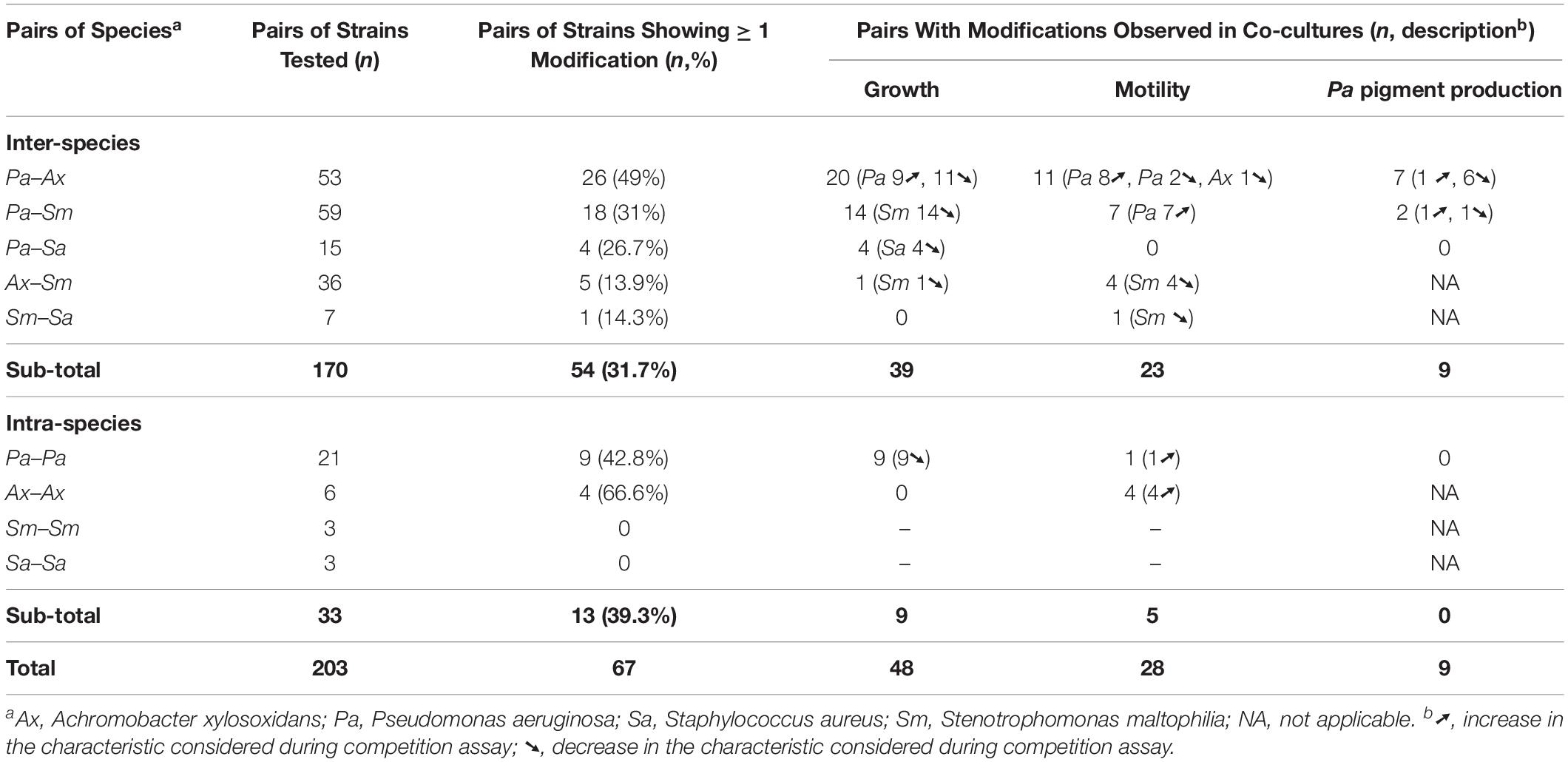
Table 2. Summary of the prevalence and type of interactions observed according to the crossing experiments.
Interactions observed were not significantly different in pairs of strains originating from the same patient or from different patients, either from the patient’s airways or domestic environment with similar distributions in the interaction type (Figure 2). However, interactions affecting growth and motility were significantly more frequent for pairs of clinical strains and those comprising at least one clinical strain compared with pairs of environmental strains (Figure 2).
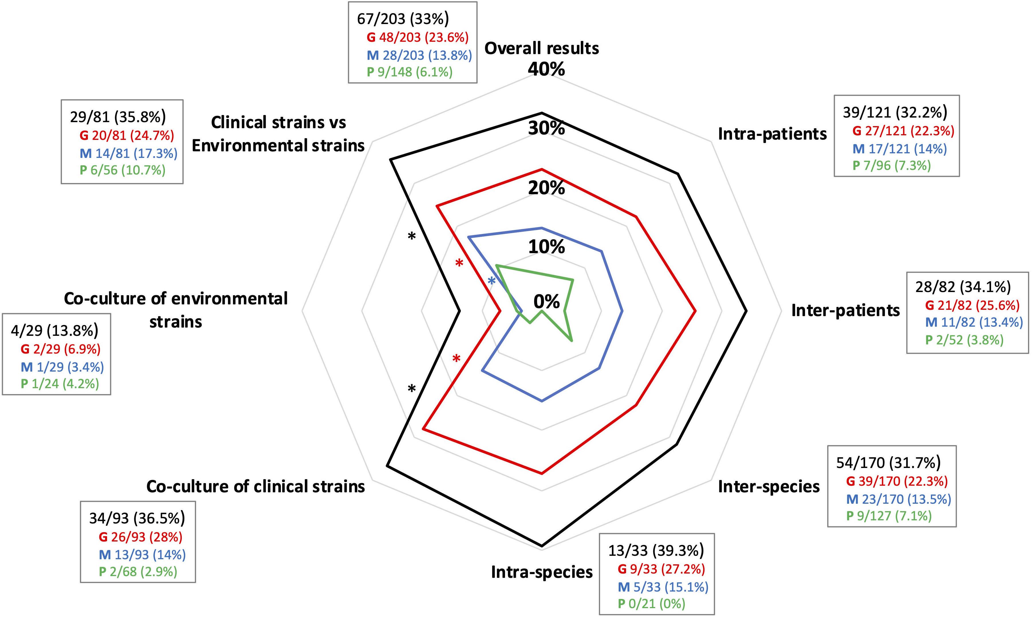
Figure 2. Rates of crossing experiments (%) showing competitive effects on growth, motility and Pseudomonas aeruginosa overall pigment production according to seven crossing conditions. Black, rate of pairs of strains showing at least one type of interaction; red, rate of pairs of strains showing interactions affecting growth; blue, rate of pairs of strains showing interactions affecting motility; green, rate of pairs of strains showing interactions affecting P. aeruginosa pigment production. Detailed results (number of pairs of strains showing an interaction out of the total crossing experiments performed in the category, %) are given next to the corresponding crossing condition with the same color code than above (G, growth; M, motility; P, Pa global pigment production). ∗, significant P-value < 0.05.
Under the study conditions, competition was significantly more frequent for pairs that had a Pa and/or an Ax strain, 38.5% (57/148) and 36.8% (35/95), respectively, compared with those comprising an Sm and/or an Sa strain, 22.9% (24/105) and 20% (5/25), respectively (P-value < 0.05; Table 2).
Interspecies interactions evaluated through 170 co-cultures of which 155 (91.2%) included an emerging pathogen, Ax or Sm, are presented in Table 2. They were observed in both intrapatient (30 pairs of strains out of 101 tested) and interpatient (24/69) conditions. Pa outcompeted its bacterial partner in the majority of the cases, except in 16 crossing experiments where growth, motility and/or pigmentation of 10 Pa strains were affected in co-culture with Ax (seven strains) or Sm (one strain; Table 2 and Supplementary Table 2), nine pairs of which were later included in the in-depth study. Interestingly, the three variants of Ax (Ax 198, Ax 199, and Ax 200) of identical MLST but distinct colony morphotypes co-cultured from a sputum specimen showed different competitive ability toward Pa strains (Supplementary Table 1). The first data on competition assays between Ax and Sm showed that Ax outcompeted Sm in all cases, either by decreasing Sm growth (n = 1, Ax/Sm pair of isolates from the environment of patients C and A, respectively) or motility (n = 4). Finally, out of eight pairwise comparisons between Sa and Sm, one interaction affecting Sm motility was observed (12.5%).
Intraspecific competitions were observed for Pa and Ax only in both intrapatient (nine pairs of strains out of 20 tested) and interpatient (4/13) conditions. For Pa, competition mostly affected bacterial growth (Table 2) and was mainly observed for pairs of clinical/environmental strains (n = 6 out of nine pairs) and of different genotypes (n = 5; Supplementary Table 1). In these cases, the Pa environmental strain decreased the growth of the clinical isolate. Unlike the observations made for Pa, all four modifications observed in the intra-Ax assays concerned clinical strains and bacterial motility (Table 2 and Supplementary Figure 3).
In-Depth Study on 9 Selected Strains From Three Patients
Intermittent Sm (ST 5) Versus Sporadic Pa (ST 253) Colonizing Patient E
The Sm 18 growth inhibition observed during the screening assay (mean inhibition zone diameter of 21.6 mm; Figure 3A) was confirmed by growth curve analysis showing a significant decrease in Sm growth in the presence of Pa II.39 (by a factor of 23.2 after 24 h, P-value < 0.001 and of 31.3 after 48 h, P-value < 0.01) while the growth of Pa remained unchanged in the presence of Sm (Figure 3B). Pa supernatant had no inhibitory effect on Sm suggesting that the competition between both species required a direct contact between the two bacteria (Figure 3B). Regarding biofilm formation, Sm 18 and Pa II.39 were moderate and strong biofilm producers, respectively. During co-culture, a significant decrease in dual-species biofilm production was observed (P-value < 0.001) in comparison with Pa biofilm (Figure 3C) despite the unchanged growth of Pa in planktonic culture.
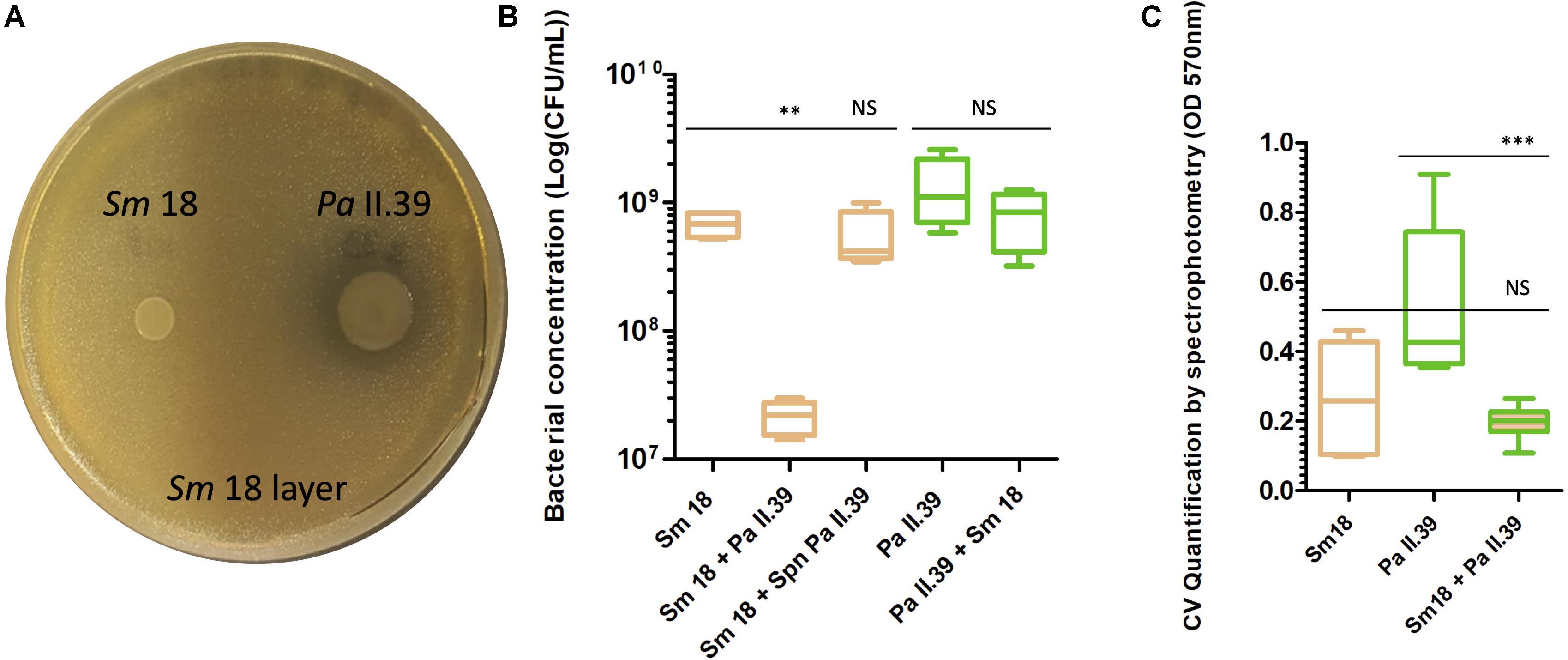
Figure 3. Interactions between intermittent Stenotrophomonas maltophilia (Sm 18, ST 5) and sporadic Pseudomonas aeruginosa (Pa II.39, ST 253) isolated about 1-year apart in Patient E. (A) Inhibition of Sm 18 growth by Pa II.39 during competition tests on bacterial layer after 48 h of co-culture. (B) Bacterial concentration after 48 h of culture in liquid medium of Sm 18 and Pa II.39 in monoculture, co-culture or Sm culture in presence of Pa supernatant. CFU, colony forming unit; Spn, supernatant. (C) Quantification of biofilm formed after 48 h of monoculture or co-culture. CV, crystal violet. The color indicates the species whose cells are numbered in (B) orange for Sm and green for Pa. The same color code is used in (C) to indicate the species whose biofilm formation has been measured; hatched box-plots with mixed colors indicate global bacterial quantification in biofilm experiments. NS, not significant; ∗∗, significant P-value < 0.01, ∗∗∗, significant P-value < 0.001.
Chronic Sa (ST 5) of Patient D Versus Sporadic Pa Originating From the Same Patient (ST 1062) and a Sporadic Pa From Another Patient (ST 253)
In screening experiments, Sa 12 growth was inhibited by both Pa strains; however, the strain originating from the same patient (Pa I.73) had limited inhibition (inhibition zone diameter of 23.2 mm after 48 h) compared to Pa II.39 from another patient (31 mm; Figure 4A). This was confirmed by drawing a growth curve showing that, after both 24 and 48 h of liquid co-culture, the growth curves of Pa/Sa pairs of strains showed a significant decrease in Sa growth (by a factor of 3.3 and 4.7 after 24 h and by a factor of 3.4 and 5.7 after 48 h for both Pa/Sa pairs, respectively, P-value < 0.001) and no modification in Pa growth (Figure 4B). However, any Pa supernatants affected the growth of Sa, suggesting that direct contact mediated the outcompetition of Sa by Pa (Figure 4B). A growth inhibition of an adapted Sa strain, chronically colonizing the patient airways, was observed in both cases but was different according to origin, i.e., from the same patient or from a different patient, and/or genotype of the Pa strain although not significantly (P-value = 0.06). Finally, a significant decrease in dual-species biofilm formation was observed for strains originating from different patients alone compared with Pa II.39 biofilm formation, although growth of the Pa strain remained unchanged in planktonic culture (P-value < 0.01; Figure 4C).
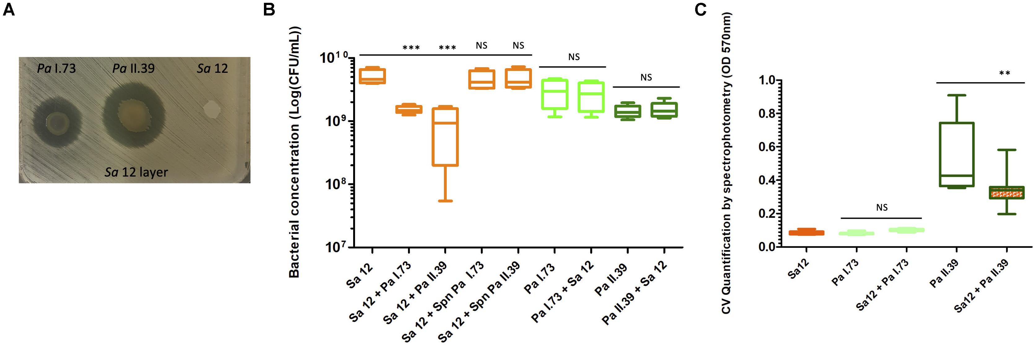
Figure 4. Interactions between chronic Staphylococcus aureus (Sa 12, ST 5) versus sporadic Pseudomonas aeruginosa originating from Patient D (Pa I.73, ST 1062, same sample than Sa 12) and a sporadic P. aeruginosa from Patient E (Pa II.39, ST 253). (A) Inhibition of Sa 18 growth by Pa I.73 and Pa II.39 during competition tests on bacterial layer after 48 h of co-culture. (B) Bacterial concentration after 48 h of culture in liquid medium of Sa 12, Pa I.73, and Pa II.39 in monoculture, Sa/Pa co-cultures or Sa culture in presence of Pa supernatant. CFU, colony forming unit; Spn, supernatant. (C) Quantification of biofilm formed after 48 h of monoculture or co-culture. CV, crystal violet. The color indicates the species and strains whose cells are numbered in (B) orange for Sa, light green for Pa I.73, and dark green for Pa II.39. The same color code is used in (C) to indicate the species and strains whose biofilm formation has been measured; hatched box-plots with mixed colors indicate global bacterial quantification in biofilm experiments. NS, not significant; ∗∗, significant P-value < 0.01, ***, significant P-value < 0.001.
Chronic Ax (ST 327) Versus Three Sporadic Pa of Different Genotypes (ST 27, ST 244 and ST 1092) and an Environmental Pa (ST 27) in Patient A
A timeline representation of selected strains in Patient A is shown in Supplementary Figure 2. Strains were selected based on the results of screening assays, particularly on a marked inhibition of Pa growth, pigmentation, and motility in presence Ax 200 (Figures 5A,B). After co-culturing clinical strain Ax 200 (the most competitive Ax strain in screening assays) with each of the four selected Pa strains in liquid medium for 24 h, the growth of all Pa was significantly reduced (by a factor of 3.9–10.8, P-value < 0.01; Figure 5C) whereas no modification in Ax growth was observed. The Ax supernatant decreased Pa growth by a factor of 1.1–1.6 but this was not significant (Figure 5C). After 48h of co-culture, the growth of Ax 200 was not decreased by any of the four Pa strains whereas Ax 200 retained the ability to significantly inhibit the growth of Pa I.14 and of the co-isolated Pa strain (Pa II.17; P-value < 0.05). The effect of the Ax 200 supernatant remained unchanged (with no significant inhibitory effect on Pa growth decreased by a factor of 0.9–1.4; Figure 5C). We also explored growth of Ax and Pa on intra-clinical samples by co-culturing each of the three Ax variants, Ax 198, Ax 199, and Ax 200, with Pa II.17. Pa growth was slightly, but not significantly, reduced by Ax 198 (factor 2, P-value = 0.07) whereas it was significantly reduced by Ax 199 and Ax 200 (factors 3.9 and 5, respectively, P-value < 0.05; Supplementary Figure 4A). After 48 h of co-culturing, similar results were observed (data not shown). A distinct ability to form biofilm was noted for the seven strains studied in Patient A, the three Ax variants being either moderate biofilm producers (Ax 198) or strong biofilm producers (Ax 199 and Ax 200) and the four Pa strains being either low biofilm producers (Pa I.14), moderate biofilm producers (Pa I.31 and the environmental Pa 50) or strong biofilm producers (Pa II.17). Under all conditions of co-culture, a significant reduction in biofilm production was observed for Pa and/or Ax compared with each of the monocultures (P-value < 0.05; Figure 5D and Supplementary Figure 4B) unrelated to growth modifications observed for planktonic cultures except for Pa II.17 in co-culture with Ax 200 or Ax 199.
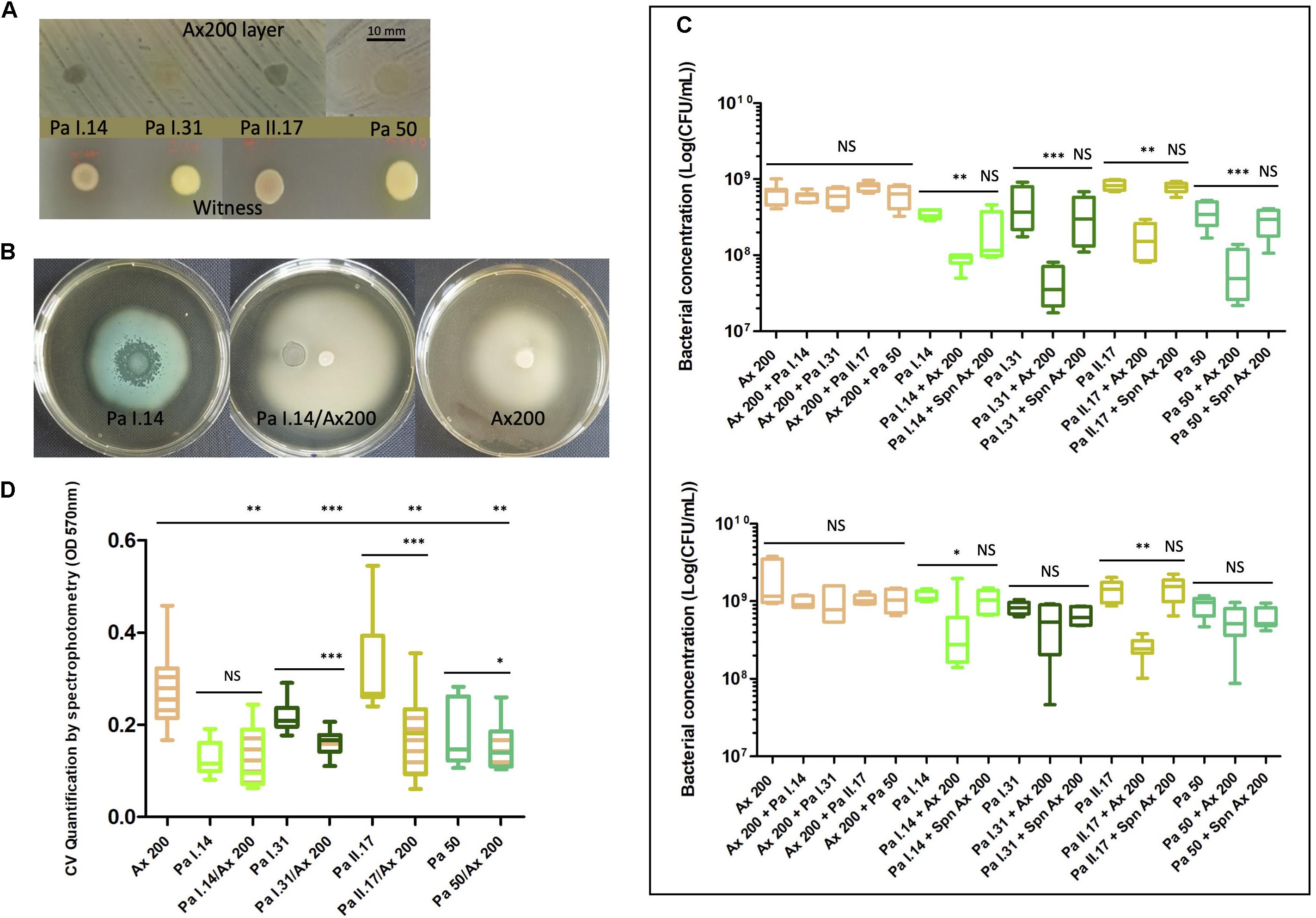
Figure 5. Interactions between chronic Achromobacter xylosoxidans (Ax; ST 327) versus three sporadic Pseudomonas aeruginosa (Pa) of different genotypes (ST 27, ST 244 and ST 1092) and an environmental Pa (ST 27) in Patient A. Strain isolation characteristics are presented in Supplementary Figure 2. (A) Inhibition of Pa I.14, Pa I.31, Pa II.17, and Pa 50 development on Ax 200 layer after 48 h of co-culture compared to corresponding monocultures. (B) Inhibition of Pa I.14 pigmentation and swimming motility during the co-culture with Ax 200 compared to monocultures. (C) Bacterial concentration after 24 h (top) and 48 h (bottom) of culture in liquid medium of Ax 200, Pa I.14, Pa I.31, Pa II.17, and Pa 50 in monocultures and Ax/Pa co-cultures or Pa culture in presence of Ax supernatant. CFU, colony forming unit; Spn, supernatant. (D) Quantification of biofilm formed after 48 h of monoculture or co-culture. CV: crystal violet. The color indicates the species and strains whose cells are numbered in (C) and (D) orange for Ax, light green for Pa I.14, and dark green for Pa I.31, light brown for Pa II.17, and turquoise for the environmental Pa 50. The same color code was used in C to indicate the species and strains whose biofilm formation has been measured; hatched box-plots with mixed colors indicate global bacterial quantification in biofilm experiments. NS, not significant; ∗, significant P-value < 0.05; ∗∗, significant P-value < 0.01; ∗∗∗, significant P-value < 0.001.
Discussion
Competitive Abilities of Emerging CF Pathogens
Concerning bacterial competition that may occur in the CF lung, most studies have always focused on Pa and deciphering the mechanisms of bacterial interactions with other opportunistic pathogens by studying the communication signals of its QS and secretion systems. As Pa–Sa co-isolation is common in CF, a majority of studies have focused on these opportunistic pathogens (Baldan et al., 2014; Filkins et al., 2015; Hotterbeekx et al., 2017; Limoli et al., 2017; Tognon et al., 2017) whereas fewer studies have addressed the question of interactions between Pa and other bacteria such as members of the Burkholderia cepacia complex (Al-Bakri et al., 2004; Schwab et al., 2014; Bragonzi et al., 2012; Smalley et al., 2015) and various members of the commensal microbiota of the CFRT (Shinzato and Saito, 1994; Sibley et al., 2008; Whiley et al., 2014; Gao et al., 2018). Pa has generally proved to outcompete other species, but more complex and reciprocal interactions between species have been further demonstrated with protection from killing by antimicrobial agents conferred by Pa to other species or modulation or potentiation of Pa metabolism and virulence by commensal members of the host microbiota like streptococci, Gemella or anaerobes (Duan et al., 2003; Hoffman et al., 2006; Michelsen et al., 2014; Whiley et al., 2015; Gao et al., 2018).
Ax and Sm are considered as emerging in CF (Lyczak et al., 2002; Lambiase et al., 2011; Parkins and Floto, 2015; Esposito et al., 2017). Although these bacteria may be co-isolated with Pa and/or Sa and have the ability to persistently colonize patients’ airways, so far no study has evaluated the competitive ability of members of the genus Achromobacter in CF and Sm competitive ability has rarely been studied in the context of CF (Pompilio et al., 2015; Magalhães et al., 2017; McDaniel et al., 2020).
Interactions Between Sm and Pa
In our study of 10 Sm and 15 Pa strains, we showed that Pa/Sm interactions affecting bacterial growth were the most frequent and that, in all cases, Sm growth was markedly inhibited by Pa, as previously observed for a couple of strains co-isolated from the lung of a chronically infected CF patient (Pompilio et al., 2015). However, several interactive traits were specifically observed in our study: (i) when motility modifications were observed, Pa motility increased systematically, unlike the observation made by Pompilio and colleagues (Pompilio et al., 2015); (ii) Pa virulence modulation by Sm previously reported is indirectly confirmed by global modifications to Pa pigment production. A couple of strains for which Pa pigment production decreased in our study suggested a potential decrease in Pa virulence caused by certain Sm strains which has never previously been described (Pompilio et al., 2015), and (iii) a significant decrease in Pa biofilm formation unlike previous studies showing either a significant increase in biofilm biomass between a CF Sm strain and a Pa reference strain (Magalhães et al., 2017) or no difference in total biofilm biomass after 6 days of mixed cultures of CF clinical strains (Pompilio et al., 2015). Interactions not previously reported require further investigations to specify whether they could be strain-dependent, according to strain’s origin, genotype or context of isolation, and/or mediated by specific mechanisms.
Ax Competitive Ability
This study showed interspecies interactions mainly in Ax/Pa tests and, to a lesser extent, in the Ax/Sm assays. We reported the first data on competition between Ax and Sm showing that Ax outcompeted Sm in all cases. A more complex interplay was noted between Ax and Pa, with Pa’s growth, motility and/or pigment production being either increased or decreased. In the current context wherein Pa is recognized as a highly competitive microorganism and, in the absence of any data on Ax/Pa competition, this prompted us to investigate pairs of strains in which Ax outcompetes Pa more thoroughly. This revealed additional competition in terms of biofilm formation with the biomass of Ax/Pa biofilms being significantly reduced compared with monocultures, as previously observed for Pa/Sa pairs (Magalhães et al., 2017; this study), and for Pa and the unusual CF pathogen Inquilinus limosus (Magalhães et al., 2017). The ability of Ax to form biofilm has been previously demonstrated (Trancassini et al., 2014; Filipic et al., 2017) but interactive modifications have never been described before. In our study, we showed that biofilm formation was disturbed during the interaction between Ax and Pa and, unlike the observations made earlier for Sm (Pompilio et al., 2015; Magalhães et al., 2017) and Burkholderia cenocepacia (Bragonzi et al., 2012), neither partner cooperated and both reduced their biofilm productions. This observation, associated with the growth modifications in Ax/Pa co-cultures, is highly suggestive of reciprocal interactions between Ax and Pa as previously described between Pa and other species like Sa, Sm, and B. cepacia complex (Bragonzi et al., 2012; Pompilio et al., 2015; Limoli et al., 2017; Magalhães et al., 2017; Tognon et al., 2017).
We therefore underline the need for additional studies on emerging CF pathogens to better delineate their importance in the complex interactions taking place in CF airways.
Multiple Interactions Suggestive of QS-Regulated Competition
Multiple types of interactions were observed for a quarter of the pairs of strains for which competition was noted in our study. The induction of Pa pigment production, in particular, was systematically associated with another effect on growth or bacterial motility. This emphasizes that various competition strategies could be established simultaneously between microorganisms (Köhlerx et al., 2009) and suggests a regulation by a common QS mechanism (Smalley et al., 2015). Competition systems have been largely described for Pa (Tashiro et al., 2013; Schwab et al., 2014; Filkins et al., 2015; Smalley et al., 2015; Bernier et al., 2016). For Sa, the major QS system, the accessory gene regulator (agr) system, has also been widely studied (Le and Otto, 2015). By contrast, mechanisms supporting Pa/Sm interactions have yet to be more deeply deciphered. At present, studies have shown that the Sm antimicrobial activity exhibited by Pa is not mediated by components of the Pa supernatant but established in a contact-dependent manner (Pompilio et al., 2015; this study). Others underline a role for signal molecules of the diffusible signal factor (DSF) family, cis-2-unsaturated fatty acids, produced by Sm on Pa virulence, persistence, biofilm formation, and stress tolerance (Ryan et al., 2008; Twomey et al., 2012), as well as a pro-killing effect of Sm on Pa involving the type IVA secretion system recently described in Sm (Nas et al., 2019). In our study, Pa culture supernatants had no effect on Sa growth and this result must be further investigated as it contrasts with previous studies showing that Pa exoproducts determine antimicrobial activity against Sa (Radlinski et al., 2017). Finally, the mechanisms of Achromobacter QS are not yet elucidated even though cyclic-di-GMP has been suspected of acting in QS signaling associated with biofilm formation as previously demonstrated for Pa (Valentini and Filloux, 2016; Nielsen et al., 2019). This also warrants further investigation.
Complex Interactions Revealed Through the Study of a Large Panel of Documented Isolates
Present studies, apart from three evaluating intraspecific Pa interactions (Ghoul et al., 2015; Chatterjee et al., 2017; Bara et al., 2018) and one multispecies study (Bernier et al., 2016), including more than 50 strains, mostly include limited numbers of strains. Similarly, most published studies include reference strains. Taking Pa as an example, strains PAO1 and PA14 are regularly included in studies on CF bacterial interactions as the sole Pa strains (Beaume et al., 2015; Smalley et al., 2015; Bernier et al., 2016; Magalhães et al., 2017; Tognon et al., 2017; Anand et al., 2018). However, these two strains, originating from a human wound and a burns patient, respectively (Klockgether et al., 2010; Mathee, 2018), may not reflect the true behavior of CF clinical strains. When clinical CF strains are studied, with a few exceptions, there is usually no information given on the colonization history or profile of patients and these are major factors in the competitive traits of bacterial isolates. Indeed, isolates from early and later stages of infection have previously been shown to have distinct competitiveness (Michelsen et al., 2014; Ghoul et al., 2015; Morgan et al., 2020) or differences in gene expression that may influence the strains’ competitive ability (Nielsen et al., 2017). Finally, little information on the genotypes of clinical strains is usually available in these studies.
Enlarging the panel of clinically documented strains tested in competition assays in our study showed divergent competitive ability between species depending on the pairs of strains under consideration – from indifference to competition – affecting the three characteristics under evaluation and among strains of a same species. These results completed scarce available observations as follows: (i) intraspecific interactions previously demonstrated for Pa alone were observed in our study for the two species Pa and Ax (Chatterjee et al., 2017; Bara et al., 2018; Oluyombo et al., 2019); (ii) considering the strain’s clinical or environmental origin, a significant higher rate of interactions was observed among clinical strains and between clinical and environmental strains, compared with pairs of environmental strains with a free lifestyle. Only two previous studies included Pa clinical CF and free-living environmental strains (Chatterjee et al., 2017; Bara et al., 2018), one of which included strains from CF homes (Bara et al., 2018). These studies showed that CF and environmental isolates did not differ significantly in their competitive ability (Bara et al., 2018) and that similar rates of antagonistic interactions were observed between environmental and clinical strains, and among environmental strains, although some environmental strains inhibited clinical strains (Chatterjee et al., 2017); and finally (iii), we found divergent competitive ability depending on the context of isolation (from the same patient, the same sample or from different patients) and among distinct but clonally related morphotypes isolated from one sample, resulting from diversification within adapted bacterial populations (Dupont et al., 2015).
Ax Competitiveness and Colonization Pattern of CF Airways
Major bacterial pathophysiological traits represented by growth, motility, biofilm formation, and Pa pigment production were shown to be affected by bacterial competition for a third of the pairs of strains in our study. Interpreting the consequences of these interactions remains a challenge (Hibbing et al., 2010) as species may be co-localized or not in the CF lung and as, for most interactions, dual interpretations, either beneficial or deleterious for a bacterium, can be drawn. The example of flagellar motility modifications is iconic. Flagellar motility is indeed a key parameter of bacterial adhesion and biofilm formation and its inhibition was shown important for stabilizing cell aggregates in several species including Pa (Guttenplan and Kearns, 2013). Recently, the downregulation of flagellar motility has also been confirmed as decisive for biofilm formation in Ax (Nielsen et al., 2019) although a previous study on 69 clinical isolates did not find any correlation between swimming phenotype and biofilm formation (Filipic et al., 2017). However, the interspecific, intraspecific, and intraclonal increase in swimming motility observed in our study represents another opportunity to persist within the CF airways, in the same way as the “exploratory motility” recently studied in Pa (Limoli et al., 2019).
The involvement of such highly complex interactions in the pulmonary colonization of CF patients and its dynamics remains to be elucidated. Some studies highlight the importance of the colonization sequence in driving cooperation and competition between CF Pa strains and oral commensal streptococci (Whiley et al., 2015) and also among Pa strains (Qin et al., 2012). Intraspecific interactions may explain certain observations that implanting a second strain of Pa in a patient who is already colonized by Pa has little chance of success (Qin et al., 2012). Although no specific studies have been made on Ax, we previously demonstrated that patients chronically colonized by Ax harbored highly phenotypically and genotypically diversified Ax populations but that all the variants observed were clonally related (Dupont et al., 2015; Dupont et al., 2016). As previously observed, no unrelated Ax strains were able to become implanted in these patients (Ridderberg et al., 2011; Amoureux et al., 2013). In our study, for patient A chronically colonized by Ax and sporadically by Pa and for whom the highest number of strains were studied, we found that complex, reciprocal competition between these two species occurred. However, our major observation was the ability for clinical strains of Ax to outcompete clinical and environmental Pa. These observations, highly atypical in the overall literature dealing with bacterial competition in CF, questioned the importance of bacterial competitiveness in the colonization pattern of CF airways when linked to the patient’s colonization history. Finally, in Patient A, we also observed distinct competitive ability between Ax strains of identical MLST genotypes but distinct cultural morphotypes generated through adaptation during persistent colonization. Intraspecies interactions between these adaptive Ax variants found in chronic colonization may confer a selective advantage to some variants among the Ax community that may contribute to the species persistence in the patient’s airways (Limoli et al., 2019).
Conclusion and Outlook
Despite the limitations of not having explored further mechanisms of the observed interactions and the fact that this was an in-depth study conducted only on selected strains, the results of our study on 39 CF-related strains with clinical and genotypic documentation provide new perspectives on the complex interactions that may take place among the CF airway community. This is the first study describing the competition capacity of Ax strains, adding new findings to the scarce data on Sm strains and highlighting the importance of bacterial competitiveness in the colonization pattern of CF airways. A broader exploration of the bacterial interactions between Ax and Pa strains, by diversifying sources of isolates (clinical non-CF, hospital environment, natural environments) is required to search for specific competition that might be established in the lungs of CF patients. It would also be interesting to test Achromobacter species other than Ax which are prevalent in CF patients (Spilker et al., 2013). We recommend screening as a valuable first-stage strategy to detect the most competitive strains before selecting strains for further testing. Besides additional in vitro investigations on pigment production quantification, the decrease in Pa virulence by some Ax strains must be specified using in vivo models such as Caenorhabditis elegans and/or zebrafish embryos as previously used for CF pathogens (Clatworthy et al., 2009; Dupont et al., 2017). Finally, the role of QS systems also remains to be explored to understand the mechanisms underlying Pa inhibition by Ax.
Data Availability Statement
The datasets presented in this study (the previously undescribed Stenotrophomonas maltophilia ST 486) can be found in online repositories. The names of the repository/repositories and accession number(s) can be found at: https://pubmlst.org/bigsdb?page=info&db=pubmlst_smaltophilia_isolates&id=654.
Ethics Statement
The studies involving human participants were reviewed and approved by the Institutional Review Board at Nîmes University Hospital (IRB no. 19.02.01 for the study of clinical isolates and collection of patients’ microbiological results and IRB no. 15/07.05 for the study of isolates from the domestic environment and the sampling at the patients’ homes). Written informed consent from the participants’ legal guardian/next of kin was not required to participate in this study in accordance with the national legislation and the institutional requirements.
Author Contributions
CD and HM conceived and designed the study. HM designed the method and collected the clinical isolates. CD, RC, and EJ-B designed the method, collected the environmental isolates, and critically revised the manuscript. RC collected the clinical data. QM, CD, and HM collected the microbiological data and analyzed and interpreted the data. QM and CD performed the microbial analyses. QM and HM drafted the manuscript. All authors read and approved the final manuscript.
Funding
This work was presented in part at the 42nd European Cystic Fibrosis Conference, 5–8 June 2019, Liverpool, United Kingdom.
Conflict of Interest
The authors declare that the research was conducted in the absence of any commercial or financial relationships that could be construed as a potential conflict of interest.
Acknowledgments
We gratefully acknowledge Missita Fofana, Sabrina Khedairia, and Marion Normandin for their assistance. We warmly thank Teresa Sawyers, English medical writer at our institution, for editing this manuscript and Mylène Toubiana for her help with the statistical analyses.
Supplementary Material
The Supplementary Material for this article can be found online at: https://www.frontiersin.org/articles/10.3389/fmicb.2020.558160/full#supplementary-material
References
Al-Bakri, A. G., Gilbert, P., and Allison, D. G. (2004). Immigration and emigration of Burkholderia cepacia and Pseudomonas aeruginosa between and within mixed biofilm communities. J. Appl. Microbiol. 96, 455–463. doi: 10.1111/j.1365-2672.2004.02201.x
Amoureux, L., Bador, J., Siebor, E., Taillefumier, N., Fanton, A., and Neuwirth, C. (2013). Epidemiology and resistance of Achromobacter xylosoxidans from cystic fibrosis patients in Dijon, Burgundy: first French data. J. Cyst. Fibros. 12, 170–176. doi: 10.1016/j.jcf.2012.08.005
Anand, R., Moss, R. B., Sass, G., Banaei, N., Clemons, K. V., Martinez, M., et al. (2018). Small colony variants of Pseudomonas aeruginosa display heterogeneity in inhibiting Aspergillus fumigatus biofilm. Mycopathologia 183, 263–272.
Andersen, S. B., Marvig, R. L., Molin, S., Krogh Johansen, H., and Griffin, A. S. (2015). Long-term social dynamics drive loss of function in pathogenic bacteria. Proc. Natl. Acad. Sci. U.S.A. 112, 10756–10761. doi: 10.1073/pnas.1508324112
Armbruster, C. R., Coenye, T., Touqui, L., and Bomberger, J. M. (2019). Interplay between host-microbe and microbe-microbe interactions in cystic fibrosis. J. Cyst. Fibros. 19, S47–S53. doi: 10.1016/j.jcf.2019.10.015
Baldan, R., Cigana, C., Testa, F., Bianconi, I., De Simone, M., Pellin, D., et al. (2014). Adaptation of Pseudomonas aeruginosa in Cystic Fibrosis airways influences virulence of Staphylococcus aureus in vitro and murine models of co-infection. PLoS One 9:e89614. doi: 10.1371/journal.pone.0089614
Bara, J. J., Matson, Z., and Remold, S. K. (2018). Life in the cystic fibrosis upper respiratory tract influences competitive ability of the opportunistic pathogen Pseudomonas aeruginosa. R. Soc. Open Sci. 5:180623. doi: 10.1098/rsos.180623
Beaume, M., Köhler, T., Fontana, T., Tognon, M., Renzoni, A., and van Delden, C. (2015). Metabolic pathways of Pseudomonas aeruginosa involved in competition with respiratory bacterial pathogens. Front. Microbiol. 6:321. doi: 10.3389/fmicb.2015.00321
Bernier, S. P., Workentine, M. L., Li, X., Magarvey, N. A., O’Toole, G. A., and Surette, M. G. (2016). Cyanide toxicity to Burkholderia cenocepacia is modulated by polymicrobial communities and environmental factors. Front. Microbiol. 7:725. doi: 10.3389/fmicb.2016.00725
Bragonzi, A., Farulla, I., Paroni, M., Twomey, K. B., Pirone, L., Lorè, N. I., et al. (2012). Modelling co-infection of the cystic fibrosis lung by Pseudomonas aeruginosa and Burkholderia cenocepacia reveals influences on biofilm formation and host response. PLoS One 7:e52330. doi: 10.1371/journal.pone.0052330
Burns, J. L., Emerson, J., Stapp, J. R., Yim, D. L., Krzewinski, J., Louden, L., et al. (1998). Microbiology of sputum from patients at cystic fibrosis centers in the United States. Clin. Infect. Dis. 27, 158–163. doi: 10.1086/514631
Chatterjee, P., Davis, E., Yu, F., James, S., Wildschutte, J. H., Wiegmann, D. D., et al. (2017). Environmental pseudomonads inhibit cystic fibrosis patient-derived Pseudomonas aeruginosa. Appl. Environ. Microbiol. 83:e02701-16.
Ciofu, O., Hansen, C. R., and Høiby, N. (2013). Respiratory bacterial infections in cystic fibrosis. Curr. Opin. Pulm. Med. 19, 251–258. doi: 10.1097/MCP.0b013e32835f1afc
Clatworthy, A. E., Lee, J. S. W., Leibman, M., Kostun, Z., Davidson, A. J., and Hung, D. T. (2009). Pseudomonas aeruginosa infection of zebrafish involves both host and pathogen determinants. Infect. Immun. 77, 1293–1303.
Curran, B., Jonas, D., Grundmann, H., Pitt, T., and Dowson, C. G. (2004). Development of a multilocus sequence typing scheme for the opportunistic pathogen Pseudomonas aeruginosa. J. Clin. Microbiol. 42, 5644–5649. doi: 10.1128/JCM.42.12.5644-5649.2004
Duan, K., Dammel, C., Stein, J., Rabin, H., and Surette, M. G. (2003). Modulation of Pseudomonas aeruginosa gene expression by host microflora through interspecies communication. Mol. Microbiol. 50, 1477–1491. doi: 10.1046/j.1365-2958.2003.03803.x
Dupont, C., Aujoulat, F., Chiron, R., Condom, P., Jumas-Bilak, E., and Marchandin, H. (2017). Highly diversified Pandoraea pulmonicola population during chronic colonization in cystic fibrosis. Front. Microbiol. 8:1892. doi: 10.3389/fmicb.2017.01892
Dupont, C., Jumas-Bilak, E., Doisy, C., Aujoulat, F., Chiron, R., and Marchandin, H. (2018). Chronic airway colonization by Achromobacter xylosoxidans in cystic fibrosis patients is not sustained by their domestic environment. Appl. Environ. Microbiol. 84:e01739-18.
Dupont, C., Jumas-Bilak, E., Michon, A. L., Chiron, R., and Marchandin, H. (2016). Impact of high diversity of Achromobacter populations within Cystic Fibrosis sputum samples on antimicrobial susceptibility testing. J. Clin. Microbiol. 55, 206–215.
Dupont, C., Michon, A. L., Jumas-Bilak, E., Nørskov-Lauritsen, N., Chiron, R., and Marchandin, H. (2015). Intrapatient diversity of Achromobacter spp. involved in chronic colonization of Cystic Fibrosis airways. Infect. Genet. Evol. 32, 214–223. doi: 10.1016/j.meegid.2015.03.012
Enright, M. C., Day, N. P., Davies, C. E., Peacock, S. J., and Spratt, B. G. (2000). Multilocus sequence typing for characterization of methicillin-resistant and methicillin-susceptible clones of Staphylococcus aureus. J. Clin. Microbiol. 38, 1008–1015.
Esposito, A., Pompilio, A., Bettua, C., Crocetta, V., Giacobazzi, E., Fiscarelli, E., et al. (2017). Evolution of Stenotrophomonas maltophilia in cystic fibrosis lung over chronic infection: a genomic and phenotypic population study. Front. Microbiol. 8:1590. doi: 10.3389/fmicb.2017.01590
Filipic, B., Malesevic, M., Vasiljevic, Z., Lukic, J., Novovic, K., Kojic, M., et al. (2017). Uncovering differences in virulence markers associated with Achromobacter species of CF and non-CF origin. Front. Cell. Infect. Microbiol. 7:224. doi: 10.3389/fcimb.2017.00224
Filkins, L. M., Graber, J. A., Olson, D. G., Dolben, E. L., Lynd, L. R., Bhuju, S., et al. (2015). Coculture of Staphylococcus aureus with Pseudomonas aeruginosa drives S. aureus towards fermentative metabolism and reduced viability in a cystic fibrosis model. J. Bacteriol. 197, 2252–2264.
Gao, B., Gallagher, T., Zhang, Y., Elbadawi-Sidhu, M., Lai, Z., Fiehn, O., et al. (2018). Tracking polymicrobial metabolism in cystic fibrosis airways: Pseudomonas aeruginosa metabolism and physiology are influenced by Rothia mucilaginosa-derived metabolites. mSphere 3:e00151-18.
Ghoul, M., West, S. A., Johansen, H. K., Molin, S., Harrison, O. B., Maiden, M. C., et al. (2015). Bacteriocin-mediated competition in cystic fibrosis lung infections. Proc. Biol. Sci. 282:20150972. doi: 10.1098/rspb.2015.0972
Guttenplan, S. B., and Kearns, D. B. (2013). Regulation of flagellar motility during biofilm formation. FEMS Microbiol. Rev. 37, 849–871. doi: 10.1111/1574-6976.12018
Hansen, C. R., Pressler, T., Nielsen, K. G., Jensen, P. Ø., Bjarnsholt, T., and Høiby, N. (2009). Inflammation in Achromobacter xylosoxidans infected cystic fibrosis patients. J. Cyst. Fibros. 9, 51–58. doi: 10.1016/j.jcf.2009.10.005
Harvey, J., Keenan, K. P., and Gilmour, A. (2007). Assessing biofilm formation by Listeria monocytogenes strains. Food Microbiol. 24, 380–392. doi: 10.1016/j.fm.2006.06.006
He, X. S., and Shi, W. Y. (2009). Oral microbiology: past, present and future. Int. J. Oral Sci. 1, 47–58. doi: 10.4248/ijos.09029
Heirali, A., McKeon, S., Purighalla, S., Storey, D. G., Rossi, L., Costilhes, G., et al. (2016). Assessment of the microbial constituents of the home environment of individuals with Cystic Fibrosis (CF) and their association with lower airways infections. PLoS One 11:e0148534. doi: 10.1371/journal.pone.0148534
Hibbing, M. E., Fuqua, C., Parsek, M. R., and Peterson, S. B. (2010). Bacterial competition: surviving and thriving in the microbial jungle. Nat. Rev. Microbiol. 8, 15–25. doi: 10.1038/nrmicro2259
Hoffman, L. R., Déziel, E., D’Argenio, D. A., Lépine, F., Emerson, J., McNamara, S., et al. (2006). Selection for Staphylococcus aureus small-colony variants due to growth in the presence of Pseudomonas aeruginosa. Proc. Natl. Acad. Sci. U.S.A. 103, 19890–19895. doi: 10.1073/pnas.0606756104
Hogan, D. A., Vik, A., and Kolter, R. (2004). A Pseudomonas aeruginosa quorum-sensing molecule influences Candida albicans morphology. Mol. Microbiol. 54, 1212–1223. doi: 10.1111/j.1365-2958.2004.04349.x
Høiby, N., Bjarnsholt, T., Givskov, M., Molin, S., and Ciofu, O. (2010). Antibiotic resistance of bacterial biofilms. Int. J. Antimicrob. Agents 35, 322–332. doi: 10.1016/j.ijantimicag.2009.12.011
Hotterbeekx, A., Kumar-Singh, S., Goossens, H., and Malhotra-Kumar, S. (2017). In vivo and in vitro interactions between Pseudomonas aeruginosa and Staphylococcus spp. Front. Cell. Infect. Microbiol. 7:106. doi: 10.3389/fcimb.2017.00106
Kaiser, S., Biehler, K., and Jonas, D. (2009). A Stenotrophomonas maltophilia multilocus sequence typing scheme for inferring population structure. J. Bacteriol. 191, 2934–2943.
Kirov, S. M., Castrisios, M., and Shaw, J. G. (2004). Aeromonas flagella (polar and lateral) are enterocyte adhesins that contribute to biofilm formation on surfaces. Infect. Immun. 72, 1939–1945. doi: 10.1128/iai.72.4.1939-1945.2004
Klockgether, J., Munder, A., Neugebauer, J., Davenport, C. F., Stanke, F., Larbig, K. D., et al. (2010). Genome diversity of Pseudomonas aeruginosa PAO1 laboratory strains. J. Bacteriol. 192, 1113–1121.
Köhler, T., Buckling, A., and van Delden, C. (2009). Cooperation and virulence of clinical Pseudomonas aeruginosa populations. Proc. Natl. Acad. Sci. U.S.A. 106, 6339–6344. doi: 10.1073/pnas.0811741106
Lambiase, A., Catania, M. R., Del Pezzo, M., Rossano, F., Terlizzi, V., Sepe, A., et al. (2011). Achromobacter xylosoxidans respiratory tract infection in cystic fibrosis patients. Eur. J. Clin. Microbiol. Infect. Dis. 30, 973–980.
Le, K. Y., and Otto, M. (2015). Quorum-sensing regulation in staphylococci – an overview. Front. Microbiol. 6:1174. doi: 10.3389/fmicb.2015.01174
Lee, T. W., Brownlee, K. G., Conway, S. P., Denton, M., and Littlewood, J. M. (2003). Evaluation of a new definition for chronic Pseudomonas aeruginosa infection in cystic fibrosis patients. J. Cyst. Fibros. 2, 29–34. doi: 10.1016/S1569-1993(02)00141.8
Limoli, D. H., and Hoffman, L. R. (2019). Help, hinder, hide and harm: what can we learn from the interactions between Pseudomonas aeruginosa and Staphylococcus aureus during respiratory infections? Thorax 74, 684–692.
Limoli, D. H., Warren, E. A., Yarrington, K. D., Donegan, N. P., Cheung, A. L., and O’Toole, G. A. (2019). Interspecies interactions induce exploratory motility in Pseudomonas aeruginosa. eLife 8:e47365. doi: 10.7554/eLife.47365
Limoli, D. H., Whitfield, G. B., Kitao, T., Ivey, M. L., Davis, M. R. Jr., Grahl, N., et al. (2017). Pseudomonas aeruginosa alginate overproduction promotes coexistence with Staphylococcus aureus in a model of Cystic Fibrosis respiratory infection. mBio 8:e00186-17.
Lyczak, J. B., Cannon, C. L., and Pier, G. B. (2002). Lung infections associated with cystic fibrosis. Clin. Microbiol. Rev. 15, 194–222. doi: 10.1128/cmr.15.2.194-222.2002
Magalhães, A. P., Lopes, S. P., and Pereira, M. O. (2017). Insights into cystic fibrosis polymicrobial consortia: the role of species interactions in biofilm development, phenotype, and response to in-use antibiotics. Front. Microbiol. 7:2146. doi: 10.3389/fmicb.2016.02146
Mathee, K. (2018). Forensic investigation into the origin of Pseudomonas aeruginosa PA14 - old but not lost. J. Med. Microbiol. 67, 1019–1021. doi: 10.1099/jmm.0.000778
McDaniel, M. S., Schoeb, T., and Swords, W. E. (2020). Cooperativity between Stenotrophomonas maltophilia and Pseudomonas aeruginosa during polymicrobial airway infections. Infect. Immun. 13:e00855-19.
Michelsen, C. F., Christensen, A. M., Bojer, M. S., Høiby, N., Ingmer, H., and Jelsbak, L. (2014). Staphylococcus aureus alters growth activity, autolysis, and antibiotic tolerance in a human host-adapted Pseudomonas aeruginosa lineage. J. Bacteriol. 196, 3903–3911.
Morgan, B. G., Warren, P., Mewis, R. E., and Rivett, D. W. (2020). Bacterial dominance is due to effective utilisation of secondary metabolites produced by competitors. Sci. Rep. 10:2316.
Nas, M. Y., White, R. C., DuMont, A. L., Lopez, A. E., and Cianciotto, N. P. (2019). Stenotrophomonas maltophilia encodes a VirB/VirD4 type IV secretion system that modulates apoptosis in human cells and promotes competition against heterologous bacteria, including Pseudomonas aeruginosa. Infect. Immun. 87:e00457-19.
Nielsen, S. M., Meyer, R. L., and Nørskov-Lauritsen, N. (2017). Differences in gene expression profiles between early and late isolates in monospecies Achromobacter biofilm. Pathogens 6:E20. doi: 10.3390/pathogens6020020
Nielsen, S. M., Penstoft, L. N., and Nørskov-Lauritsen, N. (2019). Motility, biofilm formation and antimicrobial efflux of sessile and planktonic cells of Achromobacter xylosoxidans. Pathogens 8:E14. doi: 10.3390/pathogens8010014
O’Brien, S., and Fothergill, J. L. (2017). The role of multispecies social interactions in shaping Pseudomonas aeruginosa pathogenicity in the cystic fibrosis lung. FEMS Microbiol. Lett. 364:fnx128. doi: 10.1093/femsle/fnx128
O’Brien, T. J., and Welch, M. (2019). Recapitulation of polymicrobial communities associated with cystic fibrosis airway infections: a perspective. Future Microbiol. 14, 1437–1450.
Oluyombo, O., Penfold, C. N., and Diggle, S. P. (2019). Competition in biofilms between cystic fibrosis isolates of Pseudomonas aeruginosa is shaped by R-pyocins. mBio 10:e01828-18.
Palmer, K. L., Mashburn, L. M., Singh, P. K., and Whiteley, M. (2005). Cystic fibrosis sputum supports growth and cues key aspects of Pseudomonas aeruginosa physiology. J. Bacteriol. 187, 5267–5277. doi: 10.1128/JB.187.15.5267-5277.2005
Parkins, M. D., and Floto, R. A. (2015). Emerging bacterial pathogens and changing concepts of bacterial pathogenesis in cystic fibrosis. J. Cyst. Fibros. 14, 293–304. doi: 10.1016/j.jcf.2015.03.012
Peters, B. M., Jabra-Rizk, M. A., O’May, G. A., Costerton, J. W., and Shirtliff, M. E. (2012). Polymicrobial interactions: impact on pathogenesis and human disease. Clin. Microbiol. Rev. 25, 193–213.
Pollitt, E. J., West, S. A., Crusz, S. A., Burton-Chellew, M. N., and Diggle, S. P. (2014). Cooperation, quorum sensing, and evolution of virulence in Staphylococcus aureus. Infect. Immun. 82, 1045–1051.
Pompilio, A., Crocetta, V., De Nicola, S., Verginelli, F., Fiscarelli, E., and Di Bonaventura, G. (2015). Cooperative pathogenicity in cystic fibrosis: Stenotrophomonas maltophilia modulates Pseudomonas aeruginosa virulence in mixed biofilm. Front. Microbiol. 6:951. doi: 10.3389/fmicb.2015.00951
Qin, X., Zerr, D. M., McNutt, M. A., Berry, J. E., Burns, J. L., and Kapur, R. P. (2012). Pseudomonas aeruginosa syntrophy in chronically colonized airways of cystic fibrosis patients. Antimicrob. Agents Chemother. 56, 5971–5981.
Quinn, R. A., Whiteson, K., Lim, Y. W., Zhao, J., Conrad, D., LiPuma, J. J., et al. (2016). Ecological networking of cystic fibrosis lung infections. NPJ Biofilms Microbiomes 2:4.
Radlinski, L., Rowe, S. E., Kartchner, L. B., Maile, R., Cairns, B. A., Vitko, N. P., et al. (2017). Pseudomonas aeruginosa exoproducts determine antibiotic efficacy against Staphylococcus aureus. PLoS Biol. 15:e2003981. doi: 10.1371/journal.pbio.2003981
Registre français de la mucoviscidose – Bilan des données (2017). Vaincre la Mucoviscidose et Institut National D’études Démographiques. Paris: INED.
Ridderberg, W., Bendstrup, K. E., Olesen, H. V., Jensen-Fangel, S., and Nørskov-Lauritsen, N. (2011). Marked increase in incidence of Achromobacter xylosoxidans infections caused by sporadic acquisition from the environment. J. Cyst. Fibrosis. 10, 466–499. doi: 10.1016/j.jcf.2011.07.004
Rogers, G. B., Hoffman, L. R., Carroll, M. P., and Bruce, K. D. (2013). Interpreting infective microbiota: the importance of an ecological perspective. Trends Microbiol. 21, 271–276. doi: 10.1016/j.tim.2013.03.004
Ryan, R. P., Fouhy, Y., Garcia, B. F., Watt, S. A., Niehaus, K., Yang, L., et al. (2008). Interspecies signaling via the Stenotrophomonas maltophilia diffusible signal factor influences biofilm formation and polymyxin tolerance in Pseudomonas aeruginosa. Mol. Microbiol. 68, 75–86. doi: 10.1111/j.1365-2958.2008.06132.x
Schwab, U., Abdullah, L. H., Perlmutt, O. S., Albert, D., Davis, C. W., Arnold, R. R., et al. (2014). Localization of Burkholderia cepacia complex bacteria in cystic fibrosis lungs and interactions with Pseudomonas aeruginosa in hypoxic mucus. Infect. Immun. 82, 4729–4745.
Shinzato, T., and Saito, A. (1994). A mechanism of pathogenicity of “Streptococcus milleri group” in pulmonary infection: synergy with an anaerobe. J. Med. Microbiol. 40, 118–123. doi: 10.1099/00222615-40-2-118
Sibley, C. D., Parkins, M. D., Rabin, H. R., Duan, K., Norgaard, J. C., and Surette, M. G. (2008). A polymicrobial perspective of pulmonary infections exposes an enigmatic pathogen in cystic fibrosis patients. Proc. Natl. Acad. Sci. U.S.A. 105, 15070–15075. doi: 10.1073/pnas.0804326105
Sibley, C. D., Rabin, H., and Surette, M. G. (2006). Cystic fibrosis: a polymicrobial infectious disease. Future Microbiol. 1, 53–61. doi: 10.2217/17460913.1.1.53
Smalley, N. E., An, D., Parsek, M. R., Chandler, J. R., and Dandekar, A. A. (2015). Quorum sensing protects Pseudomonas aeruginosa against cheating by other species in a laboratory coculture model. J. Bacteriol. 197, 3154–3159.
Spilker, T., Vandamme, P., and Lipuma, J. J. (2012). A multilocus sequence typing scheme implies population structure and reveals several putative novel Achromobacter species. J. Clin. Microbiol. 50, 3010–3015.
Spilker, T., Vandamme, P., and Lipuma, J. J. (2013). Identification and distribution of Achromobacter species in cystic fibrosis. J. Cyst. Fibros. 12, 298–301. doi: 10.1016/j.jcf.2012.10.002
Stepanovic, S., Vukovic, D., Dakic, I., Savic, B., and Svabic-Vlahovic, M. (2000). A modified microtiter-plate test for quantification of staphylococcal biofilm formation. J. Microbiol. Methods 40, 175–179.
Tashiro, Y., Yawata, Y., Toyofuku, M., Uchiyama, H., and Nomura, N. (2013). Interspecies interaction between Pseudomonas aeruginosa and other microorganisms. Microbes Environ. 28, 13–24. doi: 10.1264/jsme2.me12167
Tognon, M., Köhler, T., Gdaniec, B. G., Hao, Y., Lam, J. S., Beaume, M., et al. (2017). Co-evolution with Staphylococcus aureus leads to lipopolysaccharide alterations in Pseudomonas aeruginosa. ISME J. 11, 2233–2243. doi: 10.1038/ismej.2017.83
Tognon, M., Köhler, T., Luscher, A., and van Delden, C. (2019). Transcriptional profiling of Pseudomonas aeruginosa and Staphylococcus aureus during in vitro co-culture. BMC Genomics 20:30. doi: 10.1186/s12864-018-5398-y
Trancassini, M., Iebba, V., Citerà, N., Tuccio, V., Magni, A., Varesi, P., et al. (2014). Outbreak of Achromobacter xylosoxidans in an Italian Cystic fibrosis center: genome variability, biofilm production, antibiotic resistance, and motility in isolated strains. Front. Microbiol. 5:138. doi: 10.3389/fmicb.2014.00138
Twomey, K. B., O’Connell, O. J., McCarthy, Y., Dow, J. M., O’Toole, G. A., Plant, B. J., et al. (2012). Bacterial cis-2-unsaturated fatty acids found in the cystic fibrosis airway modulate virulence and persistence of Pseudomonas aeruginosa. ISME J. 6, 939–950. doi: 10.1038/ismej.2011.167
Valentini, M., and Filloux, A. (2016). Biofilms and cyclic di-GMP (c-di-GMP) signaling: lessons from Pseudomonas aeruginosa and other bacteria. J. Biol. Chem. 291, 12547–12555. doi: 10.1074/jbc.R115.711507
Whiley, R. A., Fleming, E. V., Makhija, R., and Waite, R. D. (2015). Environment and colonisation sequence are key parameters driving cooperation and competition between Pseudomonas aeruginosa cystic fibrosis strains and oral commensal streptococci. PLoS One 10:e0115513. doi: 10.1371/journal.pone.0115513
Whiley, R. A., Sheikh, N. P., Mushtaq, N., Hagi-Pavli, E., Personne, Y., Javaid, D., et al. (2014). Differential potentiation of the virulence of the Pseudomonas aeruginosa cystic fibrosis Liverpool epidemic strain by oral commensal streptococci. J. Infect. Dis. 209, 769–780. doi: 10.1093/infdis/jit568
Keywords: cystic fibrosis, opportunistic pathogens, Achromobacter, Stenotrophomonas, competition, growth, motility, biofilm
Citation: Menetrey Q, Dupont C, Chiron R, Jumas-Bilak E and Marchandin H (2020) High Occurrence of Bacterial Competition Among Clinically Documented Opportunistic Pathogens Including Achromobacter xylosoxidans in Cystic Fibrosis. Front. Microbiol. 11:558160. doi: 10.3389/fmicb.2020.558160
Received: 01 May 2020; Accepted: 14 August 2020;
Published: 10 September 2020.
Edited by:
Giovanni Di Bonaventura, University of Studies G. d’Annunzio Chieti and Pescara, ItalyReviewed by:
Oana Ciofu, University of Copenhagen, DenmarkCristina Cigana, IRCCS San Raffaele Scientific Institute, Italy
Robert Quinn, Michigan State University, United States
Copyright © 2020 Menetrey, Dupont, Chiron, Jumas-Bilak and Marchandin. This is an open-access article distributed under the terms of the Creative Commons Attribution License (CC BY). The use, distribution or reproduction in other forums is permitted, provided the original author(s) and the copyright owner(s) are credited and that the original publication in this journal is cited, in accordance with accepted academic practice. No use, distribution or reproduction is permitted which does not comply with these terms.
*Correspondence: Hélène Marchandin, aGVsZW5lLm1hcmNoYW5kaW5AdW1vbnRwZWxsaWVyLmZy