 Guanlin Li
 Florian Weidner
 Jinghui Hu
 Hans Gellersen1,3*
- 1School of Computing and Communications, Lancaster University, Lancaster, United Kingdom
- 2School of Computing Science, University of Glasgow, Glasgow, United Kingdom
- 3Department of Computer Science, Aarhus University, Aarhus, Denmark
Introduction: Head-mounted displays (HMDs) are central to virtual and augmented reality (VR/AR) experiences, yet their reliance on head movement for interaction imposes physical demands on the neck. While neck pain is already a prevalent musculoskeletal issue, neck muscle activity during HMD usage remains poorly understood.
Methods: In this paper, we present the first empirical study of neck muscle activity during head fixation in a seated VR pointing task. Using surface electromyography, we measured muscle activity across 67 head poses in 15 participants.
Results: Our findings describe how neck muscle workload increases with rotation angle, is asymmetric across directions, and exhibits distinct temporal dynamics depending on head rotation angle. We contribute an open dataset and a detailed characterisation of neck muscle activity during prolonged head fixation, providing a physiological baseline for future research on the ergonomics of immersive technologies.
Discussion: Our findings highlight how specific head orientations and fixation durations differentially load the neck musculature, offering the basis for a physiological explanation for discomfort during prolonged VR use and establishing a baseline for future ergonomic assessments.
1 Introduction
Head-mounted displays (HMDs) enable interaction within 3D environments primarily through head movement, which controls the user’s view and often serves as an input modality. These interactions require the neck muscles to move, rotate, and stabilise the head — effects which are exacerbated by the additional weight of the HMD (Chen and Wu, 2023; Souchet et al., 2022; Kazemi and Lee, 2023). Despite the fact that virtual and augmented reality (VR/AR) have seen a rapid uptake (Fares et al., 2024), the effect of head-based interaction in VR on the neck has not yet been quantified. We provide the first empirical characterisation of neck muscle activity via surface electromyography over time while using a VR-HMD in a seated fixation task, considering head and torso rotation. Having a deep understanding of how an HMD impacts the neck musculature is essential for the ergonomic design of AR/VR user interfaces. Once developers and designers understand how head fixation and rotation affect neck muscle activity, they can build user interfaces that mitigate negative effects and, by that, ensure productive and enjoyable usage.
Even without considering HMDs, neck pain is one of the most common musculoskeletal disorders (27 per 1,000 in 2019, Kazeminasab et al. (2022)). Modern headsets with a weight of around 600–650 g (Apple Vision Pro1) introduce additional mass and shift the wearer’s centre of gravity forward (Chen and Wu, 2023; Souchet et al., 2022; Kazemi and Lee, 2023), exacerbating the problem. Besides, over-extended sessions are common in VR/MR applications, such as immersive gaming, reading, or 3D modelling, forcing users to continuously engage their neck to control the HMD. Specifically, these repetitive or sustained loads on the neck muscles can cause fatigue, localised pain, and, in some cases, longer-term postural issues (Gosselin et al., 2004).
Within those neck muscle loads, sustained head fixation appears to be both important and potentially discomfort-inducing. This happens because humans tend to hold their heads very still while performing tasks that require focused visual attention, such as reading detailed text, inspecting fine graphical elements, or searching for specific items in a complex virtual scene (Rayner, 1998; Fang et al., 2015; Sidenmark and Gellersen, 2019). This stillness, combined with the need for neck muscles to maintain a constant posture and perform continuous contractions, can quickly lead to muscle strain and discomfort (Sadamoto et al., 1983; McNeil et al., 2015). Additionally, prior work in ergonomics and human factors has shown that sustained muscle contractions, particularly in low-load static postures, can be more discomforting over time than movements of comparable effort (Mousavi-Khatir et al., 2018).
To avoid these negative consequences and optimise user interfaces for AR/VR, fundamental knowledge about neck muscle activity characteristics during HMD usage is essential. However, few investigations have directly measured neck muscle activity during HMD usage (e.g., Kim and Shin, 2018), and none have measured it during sustained off-centre fixations (e.g., during reading on a second virtual screen). Notably, previous work by Zhang et al. examined neck muscle activity during head pointing at targets in the visible range of an HMD (Zhang et al., 2023). They integrate their data into an overview of neck muscle activity and a model to predict it. However, the study did not encompass the full range of head movement and focused primarily on short fixations (as they occur during brief glances away from the neutral head position). With that in mind, we conducted a study (N = 15) in which participants were tasked with turning their heads from a neutral position to a target angle. There, they were shown a video clip that required their focus for 30 s. After that, they returned to the centre position, rested for 5 s, and moved on to the next target. Neck muscle activity was measured using surface electromyography (sEMG), with electrodes positioned on the sternocleidomastoid muscle (SCM), splenius capitis muscle (SPL), and extensor muscle group (EXT, cf. Figure 3). We collected data for 67 head poses for head rotations in the range of
In this study, we set out to characterise neck muscle activity during head fixation. We explore how neck muscle activity develops during sustained head fixation across a large neck rotation range and the torso’s role during head rotation. Among others, we outline that activity increases significantly for larger head rotation angles. We also describe the asymmetry in neck muscle activity and a stronger increase for upward movements than for downward movements. In addition, we characterise temporal dynamics: Neck muscle activity increases slowly and steadily over time during head fixation on targets above the horizon, but decreases sharply within the first 5 s during fixation on targets presented to the left or right on the horizon. The core contributions of our work are:
• An open-source data set of neck muscle activity.
• A characterisation of neck muscle activity, split by muscles, during head fixation over 30 s.
• Implications for user interface design based on our descriptive results on neck muscle activity.
Our findings provide foundational knowledge that can inform the development of more ergonomic and healthier systems.
2 Related work
The neck plays a vital part during human-computer interaction (HCI) as it controls head movements and, in addition to eye movements, enables us to look around and interact in VR-HMD (Sidenmark and Gellersen, 2019). This makes neck ergonomics especially important. Thus, it has been investigated in various contexts such as desktop settings (Lee et al., 2018), tablet interaction (Chiu et al., 2015), smartphone scenarios (Ning et al., 2015; Yoon et al., 2021; Choi et al., 2016), mid-air interaction (Kim et al., 2020), and, notably, extended reality. Before this work outlines the related work on (neck) ergonomics in XR and ergonomics assessment methods, we provide a primer in neck physiology.
2.1 Physiology of the neck
The human head weighs approximately 4.5–5 kg, and its position relative to the trunk significantly influences cervical spine loading (Kroemer, 2007; Winters and Crago, 2000; Debnath, 2023). Even small deviations from neutral posture increase the torque on cervical joints, requiring sustained activation of neck muscles to maintain equilibrium. Forward head posture or prolonged rotation amplifies these demands, as extensor and flexor muscle groups must generate counterbalancing forces against gravity and inertial loads. Over time, such static or asymmetric loading patterns can lead to elevated muscle activity, fatigue, and discomfort, particularly in tasks involving head fixation or constrained movement.
Head yaw rotation is normally 80°for one side, while neck flexion and extension are about normally 80°and 50°, respectively (Ahmad Sukari et al., 2021). To achieve these head movements, neck muscles like the semispinalis capitis (part of the extensor muscle group, EXT), sternocleidomastoid muscle (SCM), and splenius capitis muscle (SPL) are deeply involved (cf. Figure 3). According to Vasavada et al. (1998), most of the extension moment-generating capacity comes from EXT (37%) and the SPL (30%). In addition, levator scapulae, trapezius, erector spinae, and suboccipital muscles also contribute 5%–10% each (Vasavada et al., 1998). Flexion is dominated by the SCM (69%), with small contributions from longus capitis and colli (17% total) and scalenus anterior (14%) (Vasavada et al., 1998). Lateral rotation comes from trapezius (32%), followed by 10%–20% from SPL, SCM, semispinalis, and suboccipital muscles (Vasavada et al., 1998). Generally, these muscles work together to move and stabilise the head (Moore et al., 2013).
In this work, we used head flexion/extension to describe the user looking down/up, left/right lateral rotation for looking left/right, and left/right lateral rotation with extension/flexion for the four corners, such as upper right and lower left (cf. Table 1). Note that left and right always describe directions from an egocentric perspective (participant view).
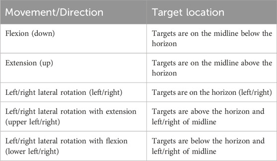
Table 1. Description of movements and direction terms used throughout the paper.
2.2 Neck muscle activity in HMD
Previous studies have investigated neck muscle activity when users use phones, laptops, and desktop computers (Lee et al., 2017; Ning et al., 2015; Yoon et al., 2021; Yadegaripour et al., 2021). However, the relationship between neck muscle activity and HMD has received limited attention to date. Astrologo et al. (2024) report that HMD leads to increased head movements during slow and fast rotations (due to the decreased field of view of HMDs) and higher load on the muscles (due to the additional weight of HMDs (Chihara and Seo, 2018)). This has been confirmed for short interactions by Zhang et al. (2023) and for prolonged usage (60 min) in a setup where the content was placed in front of the user (
Notably, Zhang et al. (2023) recently conducted a study measuring and modelling VR users’ neck muscle activity levels while pointing at targets for 2 s in VR. To do this, they relied on surface electromyography data (sEMG, cf. Section 2.3), a technique used to measure the electrical activity of muscles. They placed electrodes on two neck muscles, the SCM and SPL, to measure EMG while participants fixated on targets for 2 s (63 different positions) within
Therefore, this study aims to further investigate and contribute to a deeper understanding of how head rotation affects neck muscle activity over long fixation durations (30 s) and across the full range of head rotation. We also included the neck extensor group (EXT, in addition to SCM and SPL) as a primary contributor to head movement, and investigated how torso rotation compensates for neck muscle activity. By that, we aim to provide insights for scenarios where content is placed at angles that require sustained, extended head fixation. That could be, for example, multi-screen setups in VR/MR for working (e.g., reading, 3d modeling, or coding (Rayner, 1998)) or 360°-videos where viewers follow aeroplanes high up in the sky (looking up) or observe content that happens below them (e.g., while standing on a cliff in VR (Lo et al., 2017)).
2.3 Surface electromyography (sEMG)
Experiments with sEMG involve placing electrodes on the skin’s surface above the muscles of interest and recording the electrical signals generated by muscle contractions (Merletti and Farina, 2016). By analysing sEMG data, researchers and practitioners can evaluate muscle activation and estimate discomfort and fatigue (Merletti and Farina, 2016). Generally, sEMG is a valuable tool in human-computer interaction, supporting ergonomics assessment (Yoon et al., 2021; Choi et al., 2016), interaction design (Li et al., 2014), and acting as a direct input method to control devices (Subba and Chingtham, 2019). However, sEMG signals can be affected by noise from other electrical devices and movement artefacts and thus need adequate preprocessing (Merletti and Farina, 2016; Halaki et al., 2012). Besides, accurate placement of electrodes, often done via manual palpation, is critical for reliable and comparable measurements (Merletti and Farina, 2016). By that, sEMG provides a direct, real-time, and non-invasive measurement of muscle activity during tasks or activities (Merletti and Farina, 2016). It can capture muscle activity from specific muscles or muscle groups with high temporal resolution, valuable information for our study (Merletti and Farina, 2016). In VR (Figas et al., 2023), highlighted asymmetries of various muscles (trapezius, SCM) during short-term motions while wearing a VR-HMD, speculating about an effect of handedness. Zhang et al. (2023) characterised neck muscle activity for the visible Field of View (FOV) and small fixation durations.
As ergonomic issues in HMDs are common, they have been studied extensively (Chen and Wu, 2023; Souchet et al., 2022; Kazemi and Lee, 2023; Chen et al., 2021). Yet, comprehensive knowledge of neck muscle activity during prolonged head fixation remains incomplete, which complicates the development of solutions for work-associated ergonomic musculoskeletal issues. A thorough and precise understanding of neck muscle dynamics during HMD use, particularly during prolonged periods of head fixation, is crucial. This understanding enables researchers, developers, and designers to take specific measures to refine device form factor, interaction methods, and user interface design, aiming to enhance comfort by minimising neck muscle strain. Our objective is to offer insights into neck muscle activity using sEMG.
3 Study methods and materials
This work aims to investigate how head fixations affect neck muscle activity, simulating users’ daily tasks in HMDs, such as reading and searching. In our study, participants were instructed to watch a video while wearing a VR HMD. Every 30 s, the video would stop and change position. The video paused if the participants’ head-forward vector (measured via the HMD) was not pointing at the centre of the video frame. This means participants had to align their heads to the target to watch the video. The video continued playing once they realigned their head. We measured neck muscle activity (via sEMG), head orientation, and torso rotation.
3.1 Task description
A trial was started with the participants looking straight ahead at a target at (0°, 0°). After that, a blue target (diameter = 2.68°visual angle) appeared (Figure 1a). If the target is farther away than 20°, a blue line guides participants’ movement. Participants were instructed to rotate their heads towards the target. Once reached, the video frame appeared (size = 33.4°
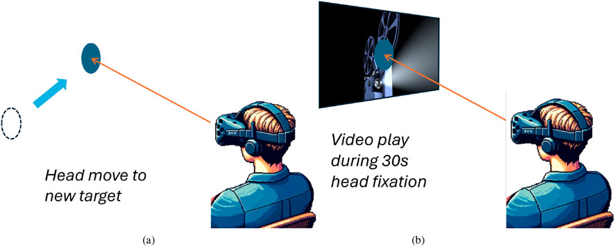
Figure 1. Task description. Figure 1a: A new head fixation target appeared. Participants were instructed to rotate their heads towards the target. A blue arrow appears to guide participants’ head movement. Figure 1b: Once reached, a video frame appeared and started playing as long as the participants’ head forward vector remained within the region of the target. (a) Head moves from neutral to a new target. (b) Head holds the fixation for 30 s before returning to neutral.
3.2 Procedure
Participants arrived at the lab, signed an informed consent form, filled in a demographics questionnaire, and were fitted with the sEMG device, HMD, and tracking equipment. Our goal was to sample neck muscle activity across a wide field of view (FOV). We initially defined 105 measurement points spanning from
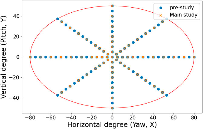
Figure 2. Fixation target distribution. The blue dots are the 105 pre-study head fixation targets, and the orange targets are the 67 of the main study. The red ellipse outlines the average person’s head movement range (Ahmad Sukari et al., 2021).
Pilot tests showed that blocks longer than 15 min lead to muscle discomfort and fatigue. We stayed below that and opted to design for 10–12 min per block, depending on how fast participants moved between targets. Thus, each block contained 16 to 17 trials. Participants were given 5-s breaks between trials and 5-min breaks between blocks to minimise fatigue (Yates et al., 1987; Lind, 1959). The sequence of fixation positions was randomised for each participant to avoid any order effects. In addition, participants had the opportunity to practice the task until they felt confident.
During the study, participants were seated on a non-revolving chair. They were allowed to make natural torso movements but needed to keep their lower limbs stationary on the chair and recenter their torso after each trial. The study was approved by the university’s ethics committee, and the average total duration per participant was approximately 120 min. Participants received £10 compensation for their time. EMG was recorded only within the head fixation 30-s periods.
3.3 Measures
3.3.1 EMG
3.3.1.1 Measurement setup
sEMG was employed in both pre-study and main study to measure neck muscle activity (8-Channel EMG Biosignalsplux Kit3). We bilaterally measured 6 EMG channels (1,000 Hz sampling rate; 16-bit resolution). We cleaned the skin with 70% alcohol wipes4 and Nuprep gel5 before attaching electrodes. Medical tape6 was used to strengthen the electrode adhesion and ensure good signal acquisition because the neck skin is unstable during head movement. We used gelled self-adhesive disposable Ag/AgCl electrodes (round; diameter: 24 mm)7.
3.3.1.2 Electrode placement
We measure signals at the SCM, SPL, and neck extensor muscle group (EXT) locations (cf. Figure 3). The electrodes at the EXT primarily measure the activity of the semispinalis capitis, the SPL and the upper trapezius because these three muscles cover each other. Therefore, more than 72% of extension moment-generating capacity, 69% of flexion moment-generating capacity, and 70% of lateral rotation moment-generating capacity muscles are included in the measurement (cf. Section 2.1). Final placements were determined through manual palpation of each subject, following the instructions of Sommerich et al. (2000).
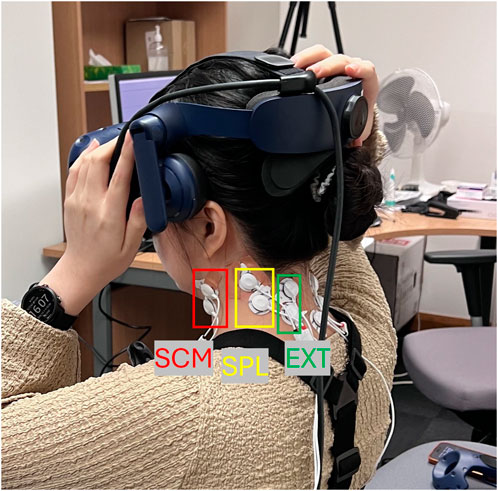
Figure 3. sEMG electrode placement on SCM, SPL, and EXT. Another three electrodes were placed on the participant’s right side.
3.3.1.3 Data filtering and artefact removal
The raw EMG signal was filtered by a bandpass filter with a 15 Hz–450 Hz frequency to remove noise and highlight relevant frequencies (Luca, 1997). After performing full wave rectification, we applied the Savitzky-Golay filter to smooth the EMG data for reduction of high-frequency noise and highlight the signal envelope (Kline and De Luca, 2014; Staudenmann et al., 2006). While the moving average filter is a simple and efficient option, it may not preserve signal features as well as the Savitzky-Golay filter, which utilises polynomial fitting for a more sophisticated smoothing process (Kawala-Sterniuk et al., 2020). Then, the processed EMG would be divided by the corresponding Maximum voluntary contraction (MVC) value measured before, and the output would be normalised EMG.
3.3.1.4 Data normalisation
Given that numerous internal and external factors influence EMG amplitudes, such as muscle size and skin conduction, the recorded amplitudes cannot be directly compared without normalisation (Halaki et al., 2012). MVC EMG amplitudes are commonly used to normalise the EMG signal (Criswell, 2010; Yang and Winter, 1984; Netto and Burnett, 2006). MVC is each channel’s absolute maximum EMG amplitude (not related to the specific head movement). We measured MVC by manual resistance for each desired head direction, including head flexion, extension, and left and right rotation (Figure 4). During the manual resistance, users were expected to gradually increase the force and manual resistance to the maximum and maintain it for 10 s. To avoid muscle fatigue accumulation, there was a 10-s rest period between each manual resistance test (Yates et al., 1987; Lind, 1959). We used the maximum amplitude as the muscle MVC. Before the MVC measurement, we performed a baseline measurement in the resting position for 20 s. We treated the average as the minimum value for each electrode pair. The minimum EMG generally contains noise, cross-talk, or electrode offset. With minimum and maximum value, we normalised data with
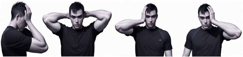
Figure 4. Manual resistance for normalising EMG data involves moving the head against resistance in four directions: flexion, extension, left rotation, and right rotation (Attwood et al., 2022).
3.3.2 Torso rotation
We measured torso movement with an HTC Vive tracker 3.08 attached to the participant’s chest. The tracker recorded the torso’s orientation when the user’s head was at the centre before the head movement for a fixation trial and the mean torso orientation during head fixation trials. The final torso movement is the difference between the above two recorded positions. Note that torso rotation was only measured during the main study.
3.4 Apparatus
We developed the task scenarios with Unity 2021.3.14f1. We used an HTC VIVE Pro Eye VR headset for the study, with 110°diagonal FOV, 2,880
3.5 Participants
We recruited 19 participants. 4 were excluded due to neck muscle pain and excessive hair growth on the neck, preventing electrode placement. In the end, we included data from N = 15 participants (5 identified as male, 9 identified as female, and 1 identified as non-binary). All were students from the local university. On average, participants were M = 25.1 years old (SD = 3.4, Min = 21, Max = 33). 12 self-reported being right-handed, and 3 being left-handed. All participants reported no neck or shoulder pain or related disease, and no dermatological conditions.
4 Results
We present a description of neck muscle activity over time and target angle. We further characterise the influence on torso rotation during head rotation in VR. The study’s results are presented in three parts, each corresponding to a research aim: viewing angle Section 4.1, development over time in Section 4.2, and torso rotation in Section 4.3. If not explicitly mentioned, all angles are the angles of the HMD, consisting of neck and torso rotation. If not explicitly split up, neck muscle activity is the sum of all 6 normalised EMG measurements. Raw data and scripts for data processing are available in the Supplementary Material.
4.1 Influence of viewing angle
4.1.1 Average neck muscle activity
Figure 5 illustrates the average neck muscle activity during head fixations of all participants over 30 s. For each measurement point, we averaged the data of the 30-s interval. We linearly interpolated the data from the measurement points to generate the heatmap.
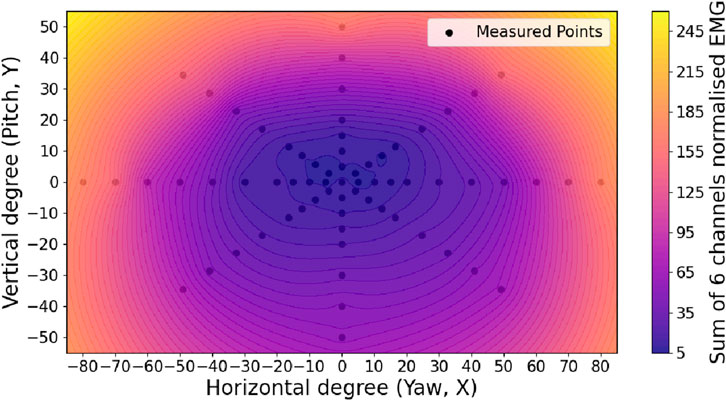
Figure 5. Mean neck muscle activity by head pose over 30 s.
Average neck muscle activity in the centre (vertical: 10°–15°; horizontal:
4.1.2 Individual muscle contribution
Figure 6a illustrates the individual muscles %MVC along the vertical axis (extended and flexed neck). The figure shows an asymmetry of neck muscle activity in the upper and lower areas. The upper area requires more than twice the total neck muscle activity (analogous to Figure 5 in Section 4.1.2). Most neck muscle activity in extended positions is caused by the left and right SCM (up to 60 %MVC), while the left and right EXT account for a significant amount of neck muscle activity in flexed positions (keeping it from “falling” further down; about 20 %MVC). In upper visual field targets, the left and right SCM together account for up to 70% of total muscle activity, while for horizontal rotations, each SCM contributes approximately 50%.
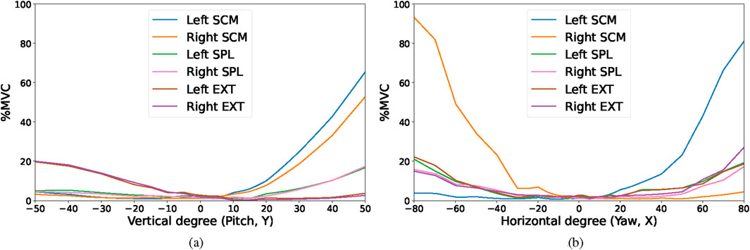
Figure 6. Individual EMG channels neck muscle activity, averaged over 30 s, along horizontal and vertical direction with %MVC (individual normalised EMG). Figure 6a shows the mean neck muscle activity in the vertical axis (x = 0) and Figure 6b in the horizontal axis (y = 0). (a) Neck muscle activity in vertical axis (x = 0). Major changes happen at −10° and 15° (vertical). SCM pair is the major contributor to extension and EXT pair to flexion. (b) Neck muscle activity in horizontal axis (y = 0). Major changes start happening at −30° and 20° (horizontal). SCM pairs are major contributors during the rotational movements.
Figure 6b illustrates the individual neck muscles along the horizontal axis. While there is a slight asymmetry in individual muscle contribution (e.g., left and right SCM), we could not find a systematic asymmetry upon closer inspection of other points and data. Left and right SCM %MVC reach around 80 in the outermost head position, indicating a higher risk of neck fatigue in these positions. SPL and EXT increase when head fixation is toward the periphery, but generally below 20 %MVC. All neck muscle activity remains low between horizontal −30°and 20°.
4.2 Temporal dynamics of muscle activity
4.2.1 Breakdown by individual directions
Figure 7 shows neck muscle activity along the eight measured directions for the 30-s interval. Each sub-figure corresponds to a direction. Each line illustrates the temporal behaviour of one target. Neck muscle activity increases with more eccentric angles for all directions and positions. In addition, for very eccentric lateral rotations and rotations with extended necks, data show a pronounced decrease directly after participants reached the target point. These results may imply that transitioning toward the position involves more neck muscle engagement than simply sustaining or stabilising it (as in Section 2.1). For positions with a very extended neck without rotations, neck muscle activity initially increases. This suggests that reaching the position may be relatively easy, whereas maintaining and stabilising the head there may require greater neck muscle activity. For all positions with a flexed neck (with and without rotation), neck muscle activity is low, especially compared to positions with an extended neck in the outermost positions. There are also no particularly outstanding spikes, increases, or decreases throughout 30 s.
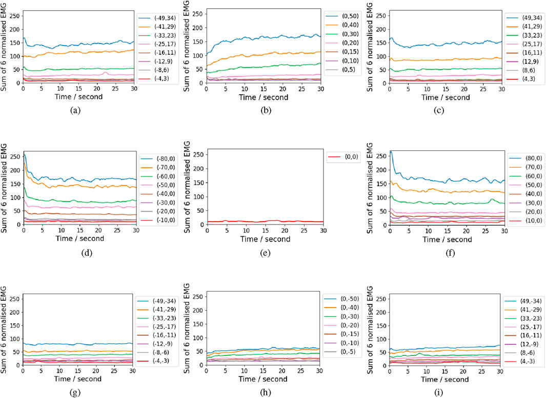
Figure 7. Neck muscle activity over the 30 s along the eight measured directions and the neutral position. Each sub-figure corresponds to one direction. Each line corresponds to the normalised neck muscle activity at one head position. The legends indicate the positioning (yaw, pitch) coordinates. (a,c,d,f) show spikes in the beginning, followed by steady activity. (b,h) show initial increases for eccentric angles. (g,i) are stable. Generally, activity increases at more eccentric angles.
4.2.2 Individual neck muscles over time
Figure 8 shows neck muscle activity of the eight outermost target positions as a line chart, illustrating the behaviour of the different muscles and muscle groups for selected data points (each graph shows neck muscle activity at measurement point). The individual normalised EMG increase beyond 100 in Figure (a) (c) (d) (f). This may be explained by MVC and baseline measurements, which may be subject to variability, influenced by individual or contextual factors.
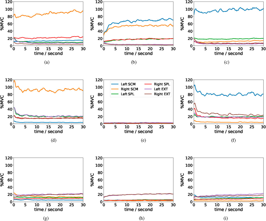
Figure 8. Examples of neck muscle activity in 30 s with 6 EMG channels shown in different lines at the outermost positions with %MVC (individual normalised EMG). (a–f) show that the SCM dominates neck muscle activity for horizontal and lateral extended rotations with relatively stable activity over time after an initial drop. (g–i) are stable.
SCM activity in the upper direction (b) increases sharply within the first 3 seconds by about 100%, whereas in lateral rotations, it decreases by roughly 30% over the same period in subfigures (d, f). EXT activity remains low. SCM remain high about 100 %MVC and slightly increases during the periods in (a, c). The SCM dominate neck muscle activity for lateral rotations with and without extension. The initial peaks (d, f) are primarily because of high SCM and EXT (ca. 20% and 40% above average); their activity decreases after a short time. The SPL pair stays relatively stable. Generally, the extensor muscle group (EXT) is responsible for moving the head in extended positions, but relaxes shortly after the target orientation is reached.
For the lower area, SCM decreases for rotations with a flexed neck (or starts low), but EXT increases over time, suggesting an increased activity for stabilisation (keeping the head in position). The other channels remain low during flexion.
4.3 Torso rotation
Figure 9 illustrates torso rotation by head rotation. The torso’s rotation is roughly symmetric along the horizontal axis, increasing towards more eccentric lateral rotations. Asymmetry is also found in the vertical axis, where looking up led to less torso compensation. Torso rotation in the horizontal direction is higher than in the vertical direction, where horizontal torso rotation can be up to 14°while around 7°in the vertical direction. Besides, we observe a relatively large no-torso rotation area in the centre,
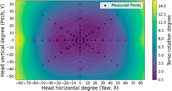
Figure 9. Interpolated heat map of mean torso rotation during 30-s fixations with measured points. The X and Y-axes are the head fixation positions, and the colour indicates torso rotation.
We calculated the correlation between average neck muscle activity and torso rotation for lateral rotation (left/right) and centrally extended/flexed positions (up/down): The Pearson Correlation Coefficient (PCC) between horizontal torso movement and mean neck muscle activity over 30 s is
5 General discussion
In this paper, we characterised neck muscle activity during head fixation while wearing an HMD, quantifying real-time EMG data. Specifically, we investigated the effect of target locations, fixation time, and torso rotation on neck muscle activity. In the following, we contextualise our findings to deepen the understanding of neck muscle activity.
5.1 Neck muscle activity in time and space
We found neck muscle activity to be asymmetric along the vertical axis. This aligns with the natural range of motion of the head, which is larger for extension than for flexion (Swartz et al., 2005) and agrees with prior findings in studies without HMD Forsberg et al. (1985). The difference in range of motion, muscle sizes, and strengths between SCM and EXT is a major source of this asymmetry (Vasavada et al., 1998; Vasavada et al., 2001). Comparing our data and asymmetry with results from Zhang et al. (2023), while comparable for neck extension, results differ for targets that require flexion. Their results indicate a decrease in neck muscle activity between −10° and −25°before activity rises again between −25° and −30°. In our data, neck muscle activity increased steadily for more eccentric targets without any decrease. We assume that the reason for this is the extensor group (EXT), which adds additional neck muscle activity to the signal (see Figure 6a) (Moore et al., 2013). Notably, our vertical asymmetry and horizontal symmetry correspond with the natural head movement range, which is also asymmetric along the vertical direction but symmetric along the horizontal axis (Ahmad Sukari et al., 2021). These findings emphasise the importance of considering muscle-specific contributions when interpreting EMG data in HMD contexts (rather than total EMG), especially for postures involving vertical head movement.
Neck muscle activity exhibits distinct temporal dynamics within the first 5 s of head fixation (Figures 7, 8). For neutral and extended rotations, EMG signals begin at a high level and then decline. In contrast, for pure extension movements without rotation, muscle activity increases over time. These patterns may be driven by a combination of factors: the need for deceleration and stabilisation (Pope-Ford, 2013; Latash, 2018), but also gradual postural adjustments. Consequently, our results suggest that certain head angles are not inherently unsuitable for interaction, but rather that their viability depends on the specific context and duration of use.
Our results show that the sternocleidomastoid (SCM) is a primary contributor to elevated neck muscle activity overall and its temporal dynamics during head fixation. Given that the SCM is relatively small and generates less force compared to other neck muscles (Vasavada et al., 1998), it still is the primary contributor and likely operates at higher activation levels to drive head stabilisation and movement. This suggests that reducing SCM workload is key to improving neck ergonomics in HMD-based interactions. To that end, future VR research should explore strategies to alleviate SCM strain through both hardware (e.g., dynamic load balancing on the HMD, advanced neck exoskeletons) and user interface design (e.g., adaptive interfaces that balance SCM load by repositioning, interaction pacing, and multimodal input).
Muscle fatigue is likely to occur when a muscle operates at a high percentage of its %MVC (Gandevia et al., 1995). As illustrated in Figures 8a–d,f, 6b, the %MVC (normalised EMG) for the left and right SCM consistently range between 80 and 100, suggesting an increased risk of muscle fatigue under these conditions.
Torso rotation was highly correlated with horizontal head rotation (PCC = 0.984). This was most likely because participants began rotating their torsos as they approached the mechanical limits of neck rotation, a strategy known to reduce neck strain (Guo et al., 2021). In this context, torso rotation might serve as a practical proxy for neck strain in the horizontal plane, as it is easier to integrate than EMG. The central region with minimal torso movement (Figure 9) may represent a “neck comfort zone,” where head poses can be maintained without compensatory body movement and low neck muscle activity.
Together, these findings highlight the complex interplay between muscle-specific activation, temporal dynamics, and compensatory strategies like torso rotation in shaping neck load during VR use. Understanding these patterns is essential for designing more ergonomic VR systems that align with the physical capabilities and limitations of the human body.
5.2 UI design implications
Our data provides a detailed characterisation of neck muscle activity across both spatial and temporal dimensions. This characterisation offers insights into how different head poses and fixation durations affect neck muscle activity in the context of HMD-based interactions. In addition, the temporal dynamics of muscle activation, especially the early-phase changes, can inform user interface design strategies. Building on these findings, we outline several recommendations for reducing neck muscle load:
1. Fixations in lower area require little muscle activity: Both spatial and temporal data show that the head fixates in positions below the horizon (flexion) lead to relatively low neck muscle activity, even for rather eccentric target positions. Thus, this area can be considered an appropriate (fallback) solution for user interface designers if content cannot be placed at the centre.
2. Use the area above but close to the horizon: For head shifts to targets above the horizon, relatively close to the centre, neck muscle activity is comparable to targets below the horizon (0°–20°vertical and −30°–+30°. Here, targets can be placed without leading to considerable neck strain within 30 s.
3. Avoid UIs that require repeated fixations in the upper left and right: Looking up is generally expensive, and neck extension with lateral rotation towards eccentric targets leads to spikes in neck muscle activity. Thus, repeated short head shifts (
4. Fixations with strongly extended neck are tolerable — if short: Head shifts requiring large neck extensions without rotations are forgiving within the first few seconds, but activity increases quickly (
5. Avoid fixations that require large lateral rotations: While low for targets close to the centre, lateral rotations along the horizon lead to spikes in neck muscle activity for more eccentric target locations (
6. Torso rotation as proxy for “neck comfort zone”: We observed that as cervical rotation approached participants’ comfortable limits, trunk rotation increased non-linearly, suggesting that trunk involvement co-occurs with high cervical loading and may serve as an indirect marker of neck loading strategy: frequently used interface elements can be put in the no-torso-rotation area (0°–2.5°). For this, torso rotation could be tracked with inverse kinematics e.g., FinalIK for Unity9.
5.3 Limitations and future work
Generalisation is limited, as our sample consisted of only 15 participants, predominantly university students around 25 years old and mostly right-handed, resulting in a very young and healthy user group. This study was conducted exclusively using the HTC VIVE Pro Eye HMD, which limits generalisability to other headsets with different weights and centres of gravity. However, given that most contemporary HMDs fall within a similar weight range and design, we expect that the overall patterns of muscle activity and temporal dynamics would remain consistent across devices, though absolute values may vary. Future work should validate these findings across a broader range of HMDs to confirm the robustness of our findings.
As we set out to characterise muscle load, we only provide pure EMG data and no other subjective measures about fatigue, perceived exertion, or similar. Thus, our data does not allow precise statements about ergonomics, although high neck muscle activity suggests poor ergonomics Cheung et al. (2003). Future work should correlate target locations with subjective measures to provide insights into ergonomic limits.
The nature of EMG and common measurement biases, such as the precision of electrode placement and skin conditions during signal acquisition, introduce additional measurement errors. We ensured proper placement through manual palpation and prepared the skin before electrode application to avoid these biases, but we can not fully prevent them. In addition, the manual measurement of MVC relies on participants’ subjective feelings, introducing potential bias that affects normalisation. It can lead to values of %MVC above 100. In our case, this mostly happens in the beginning of the measurements. One possible reason is that we measure MVC with isometric contractions. However, the values above 100% MVC happen at the end of a head movement during dynamic contractions.
We cover a large angular area, but at the cost of sampling density. Distributing our fixation point along three axes provides us with high-quality data along this axis. However, data is interpolated between the axes and thus is less accurate. This needs to be considered when interpreting the data, especially the heat maps.
Our design implications are grounded in sEMG-derived muscle activation during fixations and therefore do not capture vertebral mechanics, ligamentous or capsular loading, or subjective discomfort. Low muscle activation does not imply that a posture is biomechanically safe. Prolonged head fixation, particularly at eccentric poses, may still impose adverse loads on cervical joints and soft tissues. We present the implications as guidance only and recommend validating placement policies with measures of spinal mechanics and user-reported comfort in future work.
Finally, our data showed peculiar neck EMG dynamics in the onset of head fixation. Future studies should record and analyse neck EMG during head movement to further explain those dynamics.
6 Conclusion
This work presents the first empirical characterisation of neck muscle activity during prolonged head fixation in VR, offering a detailed view of how different head poses and durations affect muscle load. Our findings reveal spatial asymmetries, distinct temporal dynamics, and the dominant role of the sternocleidomastoid (SCM) in supporting head stabilisation, particularly during upward and lateral gaze. These insights are central for understanding the physiological demands of HMD-based interaction and provide a foundation for ergonomic design in AR/VR. By releasing an open dataset and highlighting key muscle-specific and temporal patterns, we enable future research to build on this work. As immersive technologies continue to expand into everyday use, ensuring their long-term usability and safety becomes not only a design challenge but a public health concern. Our study contributes to this effort by offering actionable insights and a physiological baseline for designing healthier, more sustainable VR experiences.
Data availability statement
The datasets presented in this study can be found in online repositories. The names of the repository/repositories and accession number(s) can be found below: https://zenodo.org/records/16785914.
Ethics statement
The studies involving humans were approved by Lancaster University Faculty of Science and Technology Research Ethics Committee. The studies were conducted in accordance with the local legislation and institutional requirements. The participants provided their written informed consent to participate in this study.
Author contributions
GL: Writing – review and editing, Writing – original draft. FW: Writing – review and editing, Writing – original draft. JH: Writing – review and editing. HG: Writing – review and editing.
Funding
The authors declare that financial support was received for the research and/or publication of this article. This work was supported by the European Research Council (ERC) under the European Union’s Horizon 2020 research and innovation program (Grant No. 101021229).
Conflict of interest
The authors declare that the research was conducted in the absence of any commercial or financial relationships that could be construed as a potential conflict of interest.
Generative AI statement
The authors declare that Generative AI was used in the creation of this manuscript. Generative AI was used for text revision and for generating Figure 4.
Any alternative text (alt text) provided alongside figures in this article has been generated by Frontiers with the support of artificial intelligence and reasonable efforts have been made to ensure accuracy, including review by the authors wherever possible. If you identify any issues, please contact us.
Publisher’s note
All claims expressed in this article are solely those of the authors and do not necessarily represent those of their affiliated organizations, or those of the publisher, the editors and the reviewers. Any product that may be evaluated in this article, or claim that may be made by its manufacturer, is not guaranteed or endorsed by the publisher.
Supplementary material
The Supplementary Material for this article can be found online at: https://www.frontiersin.org/articles/10.3389/frvir.2025.1682866/full#supplementary-material
References
Ahmad Sukari, A. A., Singh, S., Bohari, M. H., Idris, Z., Ghani, A. R. I., and Abdullah, J. M. (2021). Examining the range of motion of the cervical spine: utilising different bedside instruments. Malays. J. Med. Sci. 28, 100–105. doi:10.21315/mjms2021.28.2.9
Apti, A., Çolak, T. K., Akçay, B., Apti, A., Çolak, T. K., and Akçay, B. (2023). Normative values for cervical and lumbar range of motion in healthy young adults. J. Turkish Spinal Surg. 34, 113–117. doi:10.4274/jtss.galenos.2023.33042
Astrologo, A. N., Nano, S., Klemm, E. M., Shefelbine, S. J., and Dennerlein, J. T. (2024). Determining the effects of AR/VR HMD design parameters (mass and inertia) on cervical spine joint torques. Appl. Ergon. 116, 104183. doi:10.1016/j.apergo.2023.104183
Attwood, M. J., Hudd, L.-J. W., Roberts, S. P., Irwin, G., and Stokes, K. A. (2022). Eight weeks of self-resisted neck strength training improves neck strength in age-grade rugby union players: a pilot randomized controlled trial. Sports Health 14, 500–507. doi:10.1177/19417381211044736
Chen, Y., and Wu, Z. (2023). A review on ergonomics evaluations of virtual reality. Work 74, 831–841. doi:10.3233/WOR-205232
Chen, Y., Wang, X., and Xu, H. (2021). Human factors/ergonomics evaluation for virtual reality headsets: a review. CCF Trans. Pervasive Comput. Interact. 3, 99–111. doi:10.1007/s42486-021-00062-6
Cheung, K., Hume, P. A., and Maxwell, L. (2003). Delayed onset muscle soreness. Sports Med. 33, 145–164. doi:10.2165/00007256-200333020-00005
Chihara, T., and Seo, A. (2018). Evaluation of physical workload affected by mass and center of mass of head-mounted display. Appl. Ergon. 68, 204–212. doi:10.1016/j.apergo.2017.11.016
Chiu, H.-P., Tu, C.-N., Wu, S.-K., and Chien-Hsiou, L. (2015). Muscle activity and comfort perception on neck, shoulder, and forearm while using a tablet computer at various tilt angles. Int. J. Human–Computer Interact. 31, 769–776. doi:10.1080/10447318.2015.1064639
Choi, J.-H., Jung, M.-H., and Yoo, K.-T. (2016). An analysis of the activity and muscle fatigue of the muscles around the neck under the three most frequent postures while using a smartphone. J. Phys. Ther. Sci. 28, 1660–1664. doi:10.1589/jpts.28.1660
Criswell, E. (2010). Cram’s introduction to surface electromyography. Burlington: Jones and Bartlett Publishers.
Debnath, U. K. (2023). “Biomechanics of the cervical spine,” in Handbook of orthopaedic trauma implantology (Singapore: Springer), 1831–1852. doi:10.1007/978-981-19-7540-0_113
Fang, Y., Nakashima, R., Matsumiya, K., Kuriki, I., and Shioiri, S. (2015). Eye-head coordination for visual cognitive processing. PloS One 10, e0121035. doi:10.1371/journal.pone.0121035
Fares, O. H., Aversa, J., Lee, S. H., and Jacobson, J. (2024). Virtual reality: a review and a new framework for integrated adoption. Int. J. Consumer Stud. 48, e13040. doi:10.1111/ijcs.13040
Figas, G., Hadamus, A., Błażkiewicz, M., and Kujawa, J. (2023). Symmetry of the neck muscles’ activity in the electromyography signal during basic motion patterns. Sensors 23, 4170. doi:10.3390/s23084170
Forsberg, C.-M., Hellsing, E., Linder-Aronson, S., and Sheikholeslam, A. (1985). EMG activity in neck and masticatory muscles in relation to extension and flexion of the head. Eur. J. Orthod. 7, 177–184. doi:10.1093/ejo/7.3.177
Gallagher, H., Caldwell, E., and Albery, C. (2008). Neck muscle fatigue resulting from prolonged wear of weighted helmets.
S. C. Gandevia, R. M. Enoka, A. J. McComas, D. G. Stuart, C. K. Thomas, and P. A. Pierce (1995). Fatigue: neural and muscular mechanisms, vol. 384 of advances in experimental medicine and biology. (Boston, MA: Springer US). doi:10.1007/978-1-4899-1016-5
Gosselin, G., Rassoulian, H., and Brown, I. (2004). Effects of neck extensor muscles fatigue on balance. Clin. Biomech. 19, 473–479. doi:10.1016/j.clinbiomech.2004.02.001
Guo, R., Zhou, C., Wang, C., Tsai, T.-Y., Yu, Y., Wang, W., et al. (2021). In vivo primary and coupled segmental motions of the healthy female head-neck complex during dynamic head axial rotation. J. biomechanics 123, 110513. doi:10.1016/j.jbiomech.2021.110513
Halaki, M., Ginn, K., Halaki, M., and Ginn, K. (2012). “Normalization of EMG signals: to normalize or not to normalize and what to normalize to?,” in Computational intelligence in electromyography analysis - a perspective on current applications and future challenges (London: IntechOpen). doi:10.5772/49957
Kawala-Sterniuk, A., Podpora, M., Pelc, M., Blaszczyszyn, M., Gorzelanczyk, E. J., Martinek, R., et al. (2020). Comparison of smoothing filters in analysis of EEG data for the medical diagnostics purposes. Sensors 20, 807. doi:10.3390/s20030807
Kazemi, R., and Lee, S. C. (2023). Human factors/ergonomics (HFE) evaluation in the virtual reality environment: a systematic review. Int. J. Human–Computer Interact. 0, 4533–4549. doi:10.1080/10447318.2023.2227835
Kazeminasab, S., Nejadghaderi, S. A., Amiri, P., Pourfathi, H., Araj-Khodaei, M., Sullman, M. J. M., et al. (2022). Neck pain: global epidemiology, trends and risk factors. BMC Musculoskelet. Disord. 23, 26. doi:10.1186/s12891-021-04957-4
Kim, E., and Shin, G. (2018). Head rotation and muscle activity when conducting document editing tasks with a head-mounted display. Proc. Hum. Factors Ergonomics Soc. Annu. Meet. 62, 952–955. doi:10.1177/1541931218621219
Kim, J. H., Ari, H., Madasu, C., and Hwang, J. (2020). Evaluation of the biomechanical stress in the neck and shoulders during augmented reality interactions. Appl. Ergon. 88, 103175. doi:10.1016/j.apergo.2020.103175
Kline, J. C., and De Luca, C. J. (2014). Error reduction in EMG signal decomposition. J. Neurophysiology 112, 2718–2728. doi:10.1152/jn.00724.2013
Kroemer, K. H. E. (2007). Anthropometry and biomechanics: anthromechanics. In Biomechanics in ergonomics (CRC Press). 2 ednPages: 48
Latash, M. L. (2018). Muscle coactivation: definitions, mechanisms, and functions. J. Neurophysiology 120, 88–104. doi:10.1152/jn.00084.2018
Lee, S., Lee, Y., and Chung, Y. (2017). Effect of changes in head postures during use of laptops on muscle activity of the neck and trunk. Phys. Ther. Rehabilitation Sci. 6, 33–38. doi:10.14474/ptrs.2017.6.1.33
Lee, B., Shin, J., Bae, H., and Saakes, D. (2018). “Interactive and situated guidelines to help users design a personal desk that fits their bodies,” in Proceedings of the 2018 Designing Interactive Systems Conference (New York, NY, USA: Association for Computing Machinery), 637–650. doi:10.1145/3196709.3196725
Li, H., Chen, X., and Li, P. (2014). “Human-computer interaction system design based on surface EMG signals,” in Proceedings of 2014 International Conference on Modelling, Identification and Control, 94–98. doi:10.1109/ICMIC.2014.7020734
Lind, A. R. (1959). Muscle fatigue and recovery from fatigue induced by sustained contractions. J. Physiology 147, 162–171. doi:10.1113/jphysiol.1959.sp006231
Lo, W.-C., Fan, C.-L., Lee, J., Huang, C.-Y., Chen, K.-T., and Hsu, C.-H. (2017). “360°Video viewing dataset in head-mounted virtual reality,” in Proceedings of the 8th ACM on Multimedia Systems Conference (New York, NY, USA: Association for Computing Machinery), MMSys’17), 211–216. doi:10.1145/3083187.3083219
Luca, C. J. D. (1997). The use of surface electromyography in biomechanics. J. Appl. Biomechanics 13, 135–163. doi:10.1123/jab.13.2.135
McNeil, C. J., Allen, M. D., Olympico, E., Shoemaker, J. K., and Rice, C. L. (2015). Blood flow and muscle oxygenation during low, moderate, and maximal sustained isometric contractions. Am. J. Physiology. Regul. Integr. Comp. Physiology 309, R475–R481. doi:10.1152/ajpregu.00387.2014
Merletti, R., and Farina, D. (2016). Surface electromyography: physiology, engineering, and applications. John Wiley and Sons.
Moore, K. L., Dalley, A. F., and Agur, A. M. R. (2013). Clinically oriented anatomy. Lippincott Williams and Wilkins.
Mousavi-Khatir, R., Talebian, S., Toosizadeh, N., Olyaei, G. R., and Maroufi, N. (2018). The effect of static neck flexion on mechanical and neuromuscular behaviors of the cervical spine. J. Biomechanics 72, 152–158. doi:10.1016/j.jbiomech.2018.03.004
Netto, K. J., and Burnett, A. F. (2006). Reliability of normalisation methods for EMG analysis of neck muscles. Work 26, 123–130. doi:10.3233/wor-2006-00500
Ning, X., Huang, Y., Hu, B., and Nimbarte, A. D. (2015). Neck kinematics and muscle activity during Mobile device operations. Int. J. Industrial Ergonomics 48, 10–15. doi:10.1016/j.ergon.2015.03.003
Pope-Ford, R. D. (2013). Assessment of neck and shoulder muscle coactivations and the effect on the musculoskeletal system.
Rayner, K. (1998). Eye movements in reading and information processing: 20 years of research. Psychol. Bull. 124, 372–422. doi:10.1037/0033-2909.124.3.372
Sadamoto, T., Bonde-Petersen, F., and Suzuki, Y. (1983). Skeletal muscle tension, flow, pressure, and EMG during sustained isometric contractions in humans. Eur. J. Appl. Physiology Occup. Physiology 51, 395–408. doi:10.1007/BF00429076
Sidenmark, L., and Gellersen, H. (2019). “Eye&Head: synergetic eye and head movement for gaze pointing and selection,” in Proceedings of the 32nd Annual ACM Symposium on User Interface Software and Technology, New Orleans LA USA (Florida: ACM), 1161–1174. doi:10.1145/3332165.3347921
Sommerich, C. M., Joines, S. M. B., Hermans, V., and Moon, S. D. (2000). Use of surface electromyography to estimate neck muscle activity. J. Electromyogr. Kinesiol. 10, 377–398. doi:10.1016/S1050-6411(00)00033-X
Souchet, A. D., Lourdeaux, D., Pagani, A., and Rebenitsch, L. (2022). A narrative review of immersive virtual reality’s ergonomics and risks at the workplace: cybersickness, visual fatigue, muscular fatigue, acute stress, and mental overload. Virtual Real. 27, 19–50. doi:10.1007/s10055-022-00672-0
Staudenmann, D., Kingma, I., Daffertshofer, A., Stegeman, D., and van Dieen, J. (2006). Improving EMG-based muscle force estimation by using a high-density EMG grid and principal component analysis. IEEE Trans. Biomed. Eng. 53, 712–719. doi:10.1109/TBME.2006.870246
Subba, T., and Chingtham, T. S. (2019). “A survey: EMG signal-based controller for human–computer interaction,” in Advances in communication, cloud, and big data. Editors H. K. D. Sarma, S. Borah, and N. Dutta (Singapore: Springer), 117–125. doi:10.1007/978-981-10-8911-4_13
Swartz, E. E., Floyd, R. T., and Cendoma, M. (2005). Cervical spine functional anatomy and the biomechanics of injury due to compressive loading. J. Athl. Train. 40, 155–161. Available online at: https://pmc.ncbi.nlm.nih.gov/articles/PMC1250253/.
Thuresson, M., Äng, B., Linder, J., and Harms-Ringdahl, K. (2005). Mechanical load and EMG activity in the neck induced by different head-worn equipment and neck postures. Int. J. Industrial Ergonomics 35, 13–18. doi:10.1016/j.ergon.2004.06.008
Vasavada, A. N., Li, S., and Delp, S. L. (1998). Influence of muscle morphometry and moment arms on the moment-generating capacity of human neck muscles. Spine 23, 412–422. doi:10.1097/00007632-199802150-00002
Vasavada, A. N., Li, S., and Delp, S. L. (2001). Three-dimensional isometric strength of neck muscles in humans. Spine 26, 1904–1909. doi:10.1097/00007632-200109010-00018
J. M. Winters, and P. E. Crago (2000). Biomechanics and neural control of posture and movement. (New York, NY: Springer). doi:10.1007/978-1-4612-2104-3
Yadegaripour, M., Hadadnezhad, M., Abbasi, A., Eftekhari, F., and Samani, A. (2021). The effect of adjusting screen height and keyboard placement on neck and back discomfort, posture, and muscle activities during laptop work. Int. J. Human–Computer Interact. 37, 459–469. doi:10.1080/10447318.2020.1825204
Yang, J. F., and Winter, D. A. (1984). Electromyographic amplitude normalization methods: improving their sensitivity as diagnostic tools in gait analysis. Archives Phys. Med. rehabilitation 65, 517–521. Available online at: https://pubmed.ncbi.nlm.nih.gov/6477083/.
Yates, J. W., Kearney, J. T., Noland, M. P., and Felts, W. M. (1987). Recovery of dynamic muscular endurance. Eur. J. Appl. Physiology Occup. Physiology 56, 662–667. doi:10.1007/BF00424807
Yoon, W., Choi, S., Han, H., and Shin, G. (2021). Neck muscular load when using a smartphone while sitting, standing, and walking. Hum. Factors 63, 868–879. doi:10.1177/0018720820904237
Zhang, Y., Chen, K., and Sun, Q. (2023). “Toward optimized VR/AR ergonomics: modeling and predicting user neck muscle contraction,” in Special Interest Group on Computer Graphics and Interactive Techniques Conference Conference Proceedings, Los Angeles CA USA (New York: ACM), 1–12. doi:10.1145/3588432.3591495
Keywords: virtual reality, head-mounted display, 3D user interface, head movement, ergonomics, EMG, surface electromyography
Citation: Li G, Weidner F, Hu J and Gellersen H (2025) Quantifying neck muscle activity during head fixation in VR. Front. Virtual Real. 6:1682866. doi: 10.3389/frvir.2025.1682866
Received: 09 August 2025; Accepted: 07 November 2025;
Published: 28 November 2025.
Edited by:
Stephen Palmisano, University of Wollongong, AustraliaReviewed by:
Juno Kim, University of New South Wales, AustraliaJonathan Shemmell, University of Wollongong, Australia
Copyright © 2025 Li, Weidner, Hu and Gellersen. This is an open-access article distributed under the terms of the Creative Commons Attribution License (CC BY). The use, distribution or reproduction in other forums is permitted, provided the original author(s) and the copyright owner(s) are credited and that the original publication in this journal is cited, in accordance with accepted academic practice. No use, distribution or reproduction is permitted which does not comply with these terms.
*Correspondence: Hans Gellersen, aC5nZWxsZXJzZW5AbGFuY2FzdGVyLmFjLnVr