 Maira Rezende Formenton1*
 Lidiane Gonçalves de Lima2
 Flávia Gardilin Vassalo3
 Jean Guilherme Fernandes Joaquim3
 Laryssa Petrocini Rosseto4
 Denise Tabacchi Fantoni1
- 1Department of Surgery, School of Veterinary Medicine and Animal Science, University of São Paulo, São Paulo, Brazil
- 2Veterinary Practitioner, Ribeirao Preto, Brazil
- 3Physical Therapy Department, Instituto Bioethicus, Botucatu, Brazil
- 4FisioPet, Ribeirao Preto, Brazil
Goniometry is a low-cost, user-friendly and widely available technique used by different veterinary medicine professionals to estimate joint range of motion (ROM). Studies providing breed-specific reference ranges for goniometric measurements are scarce and there is a lack of information regarding joint angles in French Bulldogs. This prospective study set out to determine normal ROM for the carpus, elbow, shoulder, tarsus, stifle and hip joints in healthy, adult French Bulldogs using goniometry. We hypothesized ROM would be similar in this and other dog breeds. Twenty dogs met the inclusion criteria. Sample size was calculated using power analysis based on previous studies. Goniometric measurements were made by a single examiner. Limbs were measured in random order and three measurements made per joint. Dogs were not sedated. Joint angles measured in French Bulldogs in this study were similar to those reported in Labrador Retrievers (shoulder, carpal, and tarsal flexion), Rottweilers (shoulder, carpus, and hip flexion), and Dachshunds (hip, stifle, and tarsal flexion). Similar flexion angles and ROM were detected in right and left limb joints. Findings of this study suggest similar ROM in French Bulldogs and other dog breeds. Lack of radiographic assessment and the fact that goniometric measurements were made by a single examiner were the major limitations of this study.
Introduction
Goniometric measurement of joint angles is widely used by orthopedic surgeons and physical therapists to estimate joint range of motion (ROM). Goniometry is a static, low-cost, user-friendly method, and an extremely efficient and reliable ROM assessment tool (1–3). It is also thought to be a useful technique for routine monitoring of patient progression and response to physical rehabilitation, given the close relationship between decreased joint angles and joint stiffness in osteoarthritic patients (1, 4). There is a great need to determine breed-specific ROM in dogs (5, 6), as related literature is limited to a few studies in Labrador retrievers (2), German shepherds (7), and Rottweilers (8).
French Bulldogs have recently enjoyed increasing popularity among brachycephalic breeds (9). Kyphosis and several vertebral malformations have been reported in dogs of this breed, with significant clinical and body conformation implications (10). Still, popular as French Bulldogs may be, joint angles have not been quantified in this breed.
Joint angles can be measured with animals in the lateral recumbent or standing position, via manipulation of thoracic and pelvic limb joints (i.e., passive joint flexion, extension, abduction and adduction, and measurement of joint angles achieved during these movements) (2, 7). Goniometric assessment includes measurements of shoulder flexion and extension, as well as elbow, carpus, hip, stifle and tarsus flexion and extension (11, 12). This study set out to determine normal ROM of the shoulder, elbow, carpus, hip, stifle and tarsus joints in healthy, non-sedated French Bulldogs using goniometry.
Methods
This study was approved by the Ethics Committee for Animal Use of the School of Veterinary Medicine and Animal Science, University of São Paulo, protocol No. 1571120219. Patients were recruited from five different cities located in the state of São Paulo, Brazil. Patient selection was based on clinical history and evaluation, including inspection, joint palpation and ancillary tests. Inclusion criteria were as follows: male or female adult dogs aged 16–48 months, with body condition score ranging from 4 to 6 on a 1-to-9 scale (13). Owners were interrogated as to history of orthopedic conditions or trauma. Animals were inspected in the standard anatomical position for signs of musculoskeletal changes, pain or asymmetries. Subjective muscle asymmetry findings were assessed using perimetric measurements taken with a measuring tape. Clinical and gait assessment at the walk and trot were carried out to search for lameness and joint abnormalities; these were followed by joint palpation in full ROM for signs of crepitus, effusion or instability. Patients were also submitted to specific orthopedic tests, such as the tibial compression (cranial tibial thrust), the patellar luxation and the Ortolani test (14).
Seven exclusion criteria (EC) were defined to ensure the selection of dogs with no apparent musculoskeletal dysfunction that might interfere with goniometric assessment, as follows:
• EC1: Signs of lameness, joint noises and/or limb rotation at the walk and/or trot.
• EC2: Presence of atrophy, asymmetries, or musculoskeletal changes confirmed by perimetric measurement.
• EC3: Joint crepitation, edema, or instability on palpation.
• EC4: Pain manifestations during clinical evaluation.
• EC5: Pregnancy.
• EC6: Positive tibial compression (cranial tibial thrust) test.
• EC7: Positive Ortolani test.
Goniometric assessment was performed with dogs lying on the examination table. Dogs were manually restrained with their owner's assistance; no sedation was required. A plastic 35 cm, 0° to 180° system universal goniometer with two-degree increments (Carci®, São Paulo-SP, Brazil) was used. Dogs were placed in left or right lateral recumbency and the goniometer positioned according to instructions given previous authors (2, 15). Measurements were made by a single examiner (animal rehabilitation specialist with 3 years of professional experience). Range of motion was measured in flexion and extension; three measurements were made per joint for increased accuracy. Limb order was randomly selected using Random Number Generator App.
Shoulder, elbow, carpus, hip, stifle, and tarsus ROM were measured with joints in maximum flexion and extension. Data were analyzed using Google Sheets software. Arithmetic means, standard deviations (SD) and coefficients of variation (CV) were calculated for statistical analysis. Minimum sample size (6 animals) was determined by power analysis (p < 0.05; 95%) based on carpal and hip extension measurements derived from similar trials (2) and a pilot study. Correct placement of the goniometer is shown in Figure 1. Anatomical landmarks used as reference points for goniometer positioning are described in Table 1.
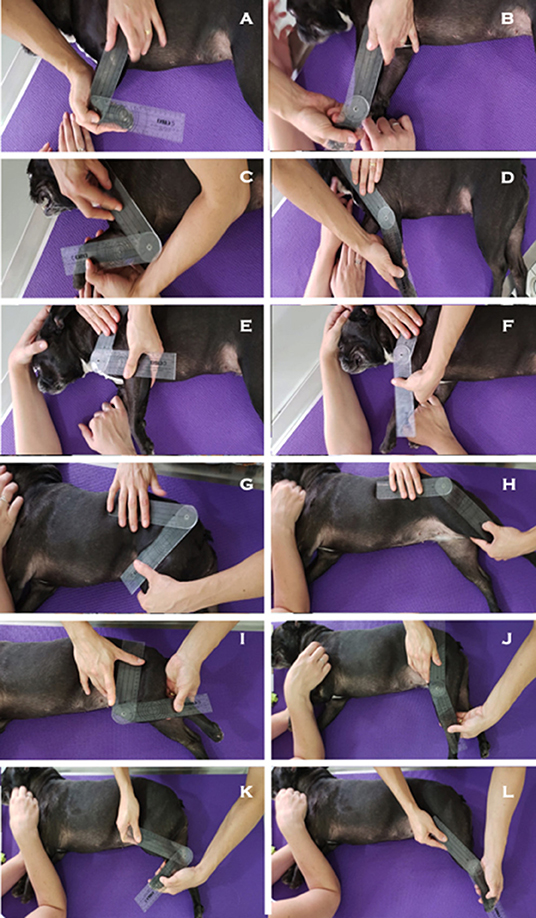
Figure 1. Correct goniometer positioning (A) carpal flexion; (B) carpal extension; (C) elbow flexion; (D) elbow extension; (E) shoulder flexion; (F) shoulder extension; (G) hip flexion; (H) hip extension; (I) stifle flexion; (J) stifle extension; (K) tarsal flexion; and (L) tarsal extension.
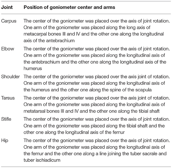
Table 1. Anatomical landmarks used as reference points for goniometer positioning prior to joint flexion and extension angle measurements, according to instructions given elsewhere (2, 15).
Results
Thirty-seven dogs were evaluated; of these, 17 (46%) met one or more EC; the final sample comprised 20 dogs (54%; 5 males and 15 females). Joint angle and ROM data of dogs in this sample are shown in Table 2.
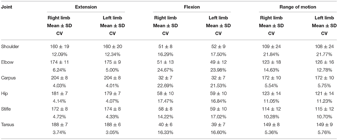
Table 2. Mean maximum flexion and extension angles and range of motion of right and left thoracic and pelvic limb joints of French Bulldogs, and respective standard deviations (SD) and coefficients of variation (CV).
Coefficients of variation express standard deviation as a percentage of the average and may be low, medium or high (>10%, 10–20%, and 20–30%, respectively). Coefficients of variation above 30% are thought to be too high to ensure data quality. In this study, CV values fell within the low, medium or high ranges and did not exceed 30%. Therefore, this data set was deemed homogeneous. Also, comparative analysis of right and left side measurements revealed similar angles, suggesting symmetrical muscle thickness, and ROM overall.
Discussion
This study described normal ROM of the shoulder, elbow, carpus, hip, stifle and tarsus joints in healthy, non-sedated French Bulldogs based on goniometric measurements. Shoulder extension angles in this study were similar to those reported in Labrador Retrievers (2) and cats (16), whereas shoulder, carpal, and tarsal flexion angles reflected those reported in Labrador Retrievers (2). Shoulder, carpus, and hip flexion angles were also comparable to ranges reported in Rottweilers (8). Range of motion was similar in left and right limbs. Full shoulder extension movements are thought to be uncommon in dogs (2). This may explain conflicting findings in this study and the reluctance of some animals to extend their shoulders. Dogs in this sample did not show signs of pain; however, arthritis or other joint/bone abnormalities cannot be ruled out, as radiographic assessment was not performed.
According to Freund et al. (3), radiographic and goniometric measurements of the canine stifle differ. However, radiographic assessment of joint angles is uncommon and goniometric measurement in lateral recumbency is the method of choice in routine practice of physical therapy (3, 4).
Studies comparing goniometric and radiographic measurements in non-sedated and sedated dogs failed to reveal significant impacts of sedation on radiographic measurements of joint angle (2). French Bulldogs in this study did not require sedation given their docile temperament.
Findings of this study are in keeping with data reported in a goniometric study evaluating hip, stifle and tarsal flexion angles in Dachshunds (17). In that study (17) pelvic limb muscle mass was thought to make landmarks for goniometer placement particularly difficult to palpate compared to long-legged dogs. Precise location of anatomical landmarks mitigates problems associated with placement of a flat goniometer on chunky, curvy limbs.
In this study, standard deviations, and coefficients of variation were calculated for improved accuracy of angle estimates (3, 7, 18). Coefficients of variation express standard deviation as a percentage of the average and may be low, medium or high (>10%, 10–20%, and 20–30%, respectively). Coefficients of variation above 30% are thought to be too high to ensure data quality. In this study, CV values fell within the low, medium or high ranges and did not exceed 30%. Therefore, this data set was deemed homogeneous. Also, comparative analysis of right and left side measurements revealed similar angles, suggesting symmetrical muscle thickness and ROM overall.
Comparison of findings reported by different investigators is thought to add reliability to research data (2, 17). Similar goniometric studies in dogs of the French Bulldog breed are lacking, therefore no comparisons could be made. Goniometric data collection by multiple examiners is also recommended. However, goniometric measurements made by different experienced examiners are not thought to be significantly different (2) and manipulation by a single examiner may minimize stress levels in canine patients. Also, the single examiner in this study does have the advantage of consistency in technique.
Major limitations of this study include: lack of radiographic confirmation of joint/limb soundness, lack of comparative radiographic measurements of ROM and technical difficulties associated with goniometric measurements in dogs with well-developed pelvic and thoracic limb muscles such as French Bulldogs.
Conclusion
This study revealed symmetrical ROM in left and right pelvic and thoracic limb joints in French Bulldogs. Similar ROM in French Bulldogs and other dog breeds support the reliability of data collected in this study. However, assessment by more than one examiner and the inclusion of radiographic measurements might have further increased data accuracy. Findings of this study will benefit future studies and practitioners who are rehabilitating French Bulldogs.
Data Availability Statement
All datasets generated for this study are included in the article/supplementary material.
Ethics Statement
The animal study was reviewed and approved by University of São Paulo. Written informed consent was obtained from the owners for the participation of their animals in this study.
Author Contributions
MF contributed with study design, writing, and review. LL contributed to the goniometric assessment of the dogs. FV and LR contributed with study design, writing, and pictures. JJ and DF contributed to review.
Conflict of Interest
FV and JJ currently work for Bioethicus Institute; LR works for FisioPet.
The remaining authors declare that the research was conducted in the absence of any commercial or financial relationships that could be construed as a potential conflict of interest.
References
1. Norkin CC, White DJ. Measurement of Joint Motion : A Guide to Goniometry. Philadelphia, PA: F.A. Davis Company (2011).
2. Jaegger G, Marcellin-little DJ, Levine D. Reliability of goniometry in Labrador Retrievers. AJVR. (2002) 63:979–86. doi: 10.2460/ajvr.2002.63.979
3. Freund KA, Kieves NR, Hart JL, Foster SA, Jeffery U, Duerr FM. Assessment of novel digital and smartphone goniometers for measurement of canine stifle joint angles. Am J Vet Res. (2016) 77:749–55. doi: 10.2460/ajvr.77.7.749
4. Corfield GS, Read RA, Eastley KA, Richardson JL, Robertson ID, Day R. Assessment of the hip reduction angle for predicting osteoarthritis of the hip in the Labrador Retriever. Aust Vet J. (2007) 85:212–6. doi: 10.1111/j.1751-0813.2007.00143.x
5. Steiss JE, . Canine rehabilitation. In: Braund KG, editor. Clinical Neurology in Small Animals - Localization, Diagnosis and Treatment. Ithaca, NY: International Veterinary Information Service (2003). p. 131–46. Available online at: http://www.ivis.org/special_books/Braund/steiss2/IVIS.pdf. (accessed January 25, 2019).
6. Lamoreaux Hesbach A. Techniques for objective outcome assessment. Clin Tech Small Anim Pract. (2007) 22:146–54. doi: 10.1053/j.ctsap.2007.09.002
7. Thomas TM, Marcellin-Little DJ, Roe SC, Lascelles BDX, Brosey BP. Comparison of measurements obtained by use of an electrogoniometer and a universal plastic goniometer for the assessment of joint motion in dogs Todd. AJVR. (2006) 67:1974–9. doi: 10.2460/ajvr.67.12.1974
9. Waters A. Brachycephalic tipping point: time to push the button? Vet Rec. (2017) 180:288. doi: 10.1136/vr.j1479
10. Wyatt SE, Lafuente P, Ter Haar G, Packer RMA, Smith H, De Decker S. Gait analysis in French bulldogs with and without vertebral kyphosis. Vet J. (2019) 244:45–50. doi: 10.1016/j.tvjl.2018.12.008
11. Alievi MM, Schossler JE, Teixeira MW. Tarsocrural joint goniometry after temporary immobilization with external skeletal fixation in dogs. Cienc. Rural. (2004) 34:425–8. doi: 10.1590/S0103-84782004000200014
12. Edge-Hughes L. Hip and sacroiliac disease: selected disorders and their management with physical therapy. Clin Tech Small Anim Pract. (2007) 22:183–94. doi: 10.1053/j.ctsap.2007.09.007
13. Mawby DI, Bartges JW, D'Avignon A, Laflamme DP, Moyers TD, Cottrell T. Comparison of various methods for estimating body fat in dogs. J Am Anim Hosp Assoc. (2004) 40:109–14. doi: 10.5326/0400109
14. Piermattei DL, Flo GL, DeCamp CE. Chapter 1: Orthopedic examination and diagnostic tools. In: Brinker, Piermattei, and Flo's Handbook of Small Animal Orthopedics and Fracture Repair. St. Louis, MO: Saunders/Elsevier (2006). p. 3–24. doi: 10.1016/B978-0-7216-9214-2.50004-6
15. Zink MC, Van Dyke JB. Canine Sports Medicine and Rehabilitation. Hoboken, NJ: Wiley-Blackwell; (2013). p. 465. doi: 10.1002/9781118783443
16. Jaeger GH, Marcellin-little DJ, Depuy V, Lascelles BDX. Validity of goniometric joint measurements in cats. AJVR. (2007) 68:822–6. doi: 10.2460/ajvr.68.8.822
17. Thomovsky SA, Chen AV, Kiszonas AM, Lutskas LA. Goniometry and limb girth in miniature Dachshunds. J Vet Med. (2016) 2016:1–5. doi: 10.1155/2016/5846052
Keywords: canine, goniometry, small animals, physical therapy, range of motion, dogs
Citation: Formenton MR, de Lima LG, Vassalo FG, Joaquim JGF, Rosseto LP and Fantoni DT (2019) Goniometric Assessment in French Bulldogs. Front. Vet. Sci. 6:424. doi: 10.3389/fvets.2019.00424
Received: 01 July 2019; Accepted: 08 November 2019;
Published: 13 December 2019.
Edited by:
David Levine, University of Tennessee at Chattanooga, United StatesReviewed by:
Stephanie Shaver, Midwestern University, United StatesMaria Fahie, Western University of Health Sciences, United States
Tisha Harper, University of Illinois at Urbana-Champaign, United States
Copyright © 2019 Formenton, de Lima, Vassalo, Joaquim, Rosseto and Fantoni. This is an open-access article distributed under the terms of the Creative Commons Attribution License (CC BY). The use, distribution or reproduction in other forums is permitted, provided the original author(s) and the copyright owner(s) are credited and that the original publication in this journal is cited, in accordance with accepted academic practice. No use, distribution or reproduction is permitted which does not comply with these terms.
*Correspondence: Maira Rezende Formenton, bWFpcmFmb3JtZW50b25AZ21haWwuY29t