 Samar Afif Jarrah1
 Louise Bach Kmetiuk2
 Fabrizia Valleriani3
 Barbara Bonfini3
 Alessio Lorusso3
 Violetta Vasinioti4
 Nicola Decaro4
 Marco Tulio dos Santos1
 Kledir Anderson Hofstaetter Spohr1
 Annamaria Pratelli4
 Anna Serroni3
 Sara Capista3
 Valéria Regia Franco Sousa1
 Alexander Welker Biondo2
 Luciano Nakazato1
 Valéria Dutra1*
- 1Laboratory of Molecular Biology, Federal University of Mato Grosso, Cuiabá, MT, Brazil
- 2Department of Veterinary Medicine, Federal University of Paraná, Curitiba, PR, Brazil
- 3Department of Virology, Istituto Zooprofilattico Sperimentale dell'Abruzzo e del Molise “G. Caporale”, Teramo, TE, Italy
- 4Department of Veterinary Medicine, University of Bari, Valenzano, BA, Italy
SARS-CoV-2 was a worldwide threat during the COVID-19 pandemic, and the state of Mato Grosso had the second highest mortality rate in Brazil, with 427. 4 deaths/100,000 inhabitants. However, no large-scale study among dogs and cats in such highly infected areas of Brazil has so far been conducted. Accordingly, the present study reports on a serosurvey among dogs and cats in Cuiabá, capital of Mato Grosso from November 2020 to July 2021, where the human mortality rate was 605/100,000 at that time. Overall, 33/762 dogs (4.3%) and 4/182 cats (2.2%) were found to be seropositive for SARS-CoV-2 through ELISA, and 3/762 dogs (0.4%) and 3/182 cats (1.6%) were seropositive through the serum neutralization test. Cats presented higher seroprevalence with higher titers of neutralizing antibodies. Although N-protein based ELISA may be a good screening test, cross-reactivity with other canine coronaviruses may impair its diagnostic use among dogs.
1. Introduction
Severe acute respiratory syndrome coronavirus 2 (SARS-CoV-2) was identified in Wuhan, China, at the end of 2019, as the cause of the coronavirus disease 2019 (COVID-19), which led to a pandemic scenario in early 2020 (1). Pneumonia and acute respiratory distress syndrome were the main clinical human signs of COVID-19 (2).
As the pandemic spread, reports of dogs and cats that were naturally infected by SARS-CoV-2 soon emerged worldwide. These animals were generally asymptomatic and their cases were associated with infected owners (3). Investigations on animal susceptibility to SARS-CoV-2 showed that cats intranasally inoculated with high doses of SARS-CoV-2 were able to infect other cats by means of airborne aerosols. Infected cats presented neutralizing antibodies and lesions in their respiratory tract but no clinical signs. They were more susceptible to SARS-CoV-2 infection than dogs (4). These findings have highlighted the reverse zoonotic potential of the disease cycle between owners and their pets, considering that outdoor access may have exposed dogs and cats to a contaminated environment. Several reports on experimental and natural infection with SARS-CoV-2 among animals have been published, with confirmation of these animals' susceptibility, regarding dogs (4–6), cats (4, 5, 7–12), ferrets (4, 6, 9), hamsters (13), non-human primates (14–17), and bats (18).
Cuiaba, the capital of the state of Mato Grosso, in central Brazil, has been severely affected by the COVID-19 pandemic. There were 48,152 confirmed cases in 2020, 67,548 in 2021 and 132,667 up to May 2022, out of a total population of 612,547 inhabitants. This state presented the second highest nationwide COVID-19 mortality rate, with 427.4 deaths/100,000 inhabitants. In the state capital, the rate was 605 deaths/100,000 habitants (19, 20). The first report from Brazil of a pet infected with SARS-CoV-2 also came from Cuiabá (12). Furthermore, also in Mato Grosso, a free-ranging black-tailed marmoset (Mico melanurus) that had been hit by a car was also found to be infected with this virus (21). These reports thus showed the impact of this highly contaminated environment on natural infection among domestic and wild animal species.
The first large-scale study on companion animals anywhere in the world was conducted in Italy and showed that 15/451 dogs (3.3%) and 11/191 cats (5.8%) presented SARS-CoV-2 neutralizing antibodies, but that there were no RT-PCR positive samples (22). Another study conducted in Texas, USA, showed that 3/17 cats (17.6%) and 1/59 dogs (1.7%) were positive for SARS-CoV-2 through RT-PCR and that 7/16 cats (43.8%) and 7/59 dogs (11.9%) presented neutralizing antibodies (23). Recently, 9/29 cats (31.0%) and 4/10 dogs (40.0%) in Rio de Janeiro were found to be positive through RT-PCR or serum neutralization (24). Higher prevalence was observed among pets whose owners were positive for COVID-19, which suggested that human contact may be a determining factor for infections among dogs and cats (23–26).
Despite the worldwide threat posed by SARS-CoV-2 during the pandemic, in which the state of Mato Grosso had the second highest mortality rate in Brazil, no large-scale study on dogs and cats in highly infected areas of Brazil has so far been conducted. Accordingly, the present study reports on a serosurvey among dogs and cats in Cuiabá, the state capital of Mato Grosso, from November 2020 to July 2021, where the human COVID-19 mortality rate was very high at that time.
2. Method
2.1. Sampling
This study was based on blood sampling collected according to convenience at the Veterinary Teaching Hospital (VTH) of the Federal University of Mato Grosso, in central Brazil, between November 2020 and July 2021. The animals brought to the VTH for attendance were mostly from the cities of Cuiaba and Varzea Grande. No restrictions were imposed on the sampling, with regard to medical history, age, breed, vaccination status, owner medical history or reason for attendance. After serological results, owners of seropositive pets were contacted about their SARS-CoV-2 infection status at the time of sampling.
In total, serum samples from 762 dogs (296 in November–December 2020 and 466 in January–July 2021) and 182 cats (68 in November–December 2020 and 114 in January–March 2021) were collected. These were placed in cryovial tubes and were immediately sent in dry ice to the Department of Veterinary Medicine, University of Bari, Valenzano, Italy, and to the Istituto Zooprofilattico Sperimentale dell'Abruzzo e del Molise “G. Caporale”, Teramo, Italy, for detection of SARS-CoV-2 antibodies. Serum samples were analyzed by means of ELISA to detect antibodies against the SARS-CoV-2 nucleocapsid (N) protein, and through the serum neutralization (SN) assay to assess presence of SARS-CoV-2 neutralizing antibodies. One positive and one negative control serum sample were kindly provided by the Istituto Nazionale Malattie Infettive “Lazzaro Spallanzani” (INMI, Rome, Italy) and were included in the assays. The B.1 SARS-CoV-2 isolate was used as the reference strain, and the viral titer of the stock was determined by means of the TCID50 assay, as previously described (27). The B.1 lineage strain (virus name: hCoV-19/Italy/ABR-IZSGC-TE46419/2020, accession ID: EPI_ISL_529023) has been a human isolate adapted on VERO E6 cells (fourth passage), first identified by qRT-PCR on human nasopharyngeal swab (28), and then by high-throughput sequencing to obtain the whole genome sequence (29). Viral isolation was performed on VERO E6 cells under biosafety level 3 (BSL-3) conditions. The B.1 strain was propagated into VERO E6 cells using MEM supplemented with 10% FBS. Cells were seeded in 175 cm2 flasks at 106 cells/mL and after 24 h were infected with 5 mL of a viral suspension at 0.01 multiplicity of infection. The flasks were incubated at 37°C in a humidified atmosphere of 5% CO2 and observed daily under an inverted optical microscope. When cytopathic effect (CPE) affected 80–90% of the cell monolayer, the supernatant was collected and centrifuged at 4°C 2,000 rpm for 10 min to remove the cellular pellet. Then, the supernatant was aliquoted and stored at −80°C. Before use, the virus was titrated in serial 1 log dilutions (from 1 log to 8 log) in 96-well culture plates of Vero E6 cells to determine the 50% tissue culture infective dose (TCID50). Plates were incubated at 37°C and checked every day to identify CPE using an inverted optical microscope. The endpoint titers were calculated according to the Reed and Muench method based on 10 replicates for titration (30).
2.2. Cell culture
The grivet monkey (Cercopithecus aethiops) kidney epithelial cell line Vero E6 (C1008) was kindly provided by the INMI. The cells were maintained in minimal essential medium (MEM, Sigma Aldrich, Merck Life Science S.r.l., Milan, Italy), supplemented with 10% fetal bovine serum (FBS, Sigma Aldrich, Merck Life Science S.r.l., Milan, Italy), 106 IU/L of penicillin, 10 g/L of streptomycin, 5 × 10⁶ IU/L of nystatin and 125 mg/L of gentamicin (IZSAM). The cell line was regularly checked for mycoplasma contamination, and its absence was verified through PCR (Mycoplasma Detection Testing, Thermo Fisher, Waltham, MA, USA).
2.3. Enzyme-linked immunosorbent assay
A double-antigen ELISA kit was used for detection of specific antibodies against the SARS-CoV-2 nucleocapsid protein in animal serum samples. Specific IgG antibodies binding to the SARS-CoV-2 N protein were determined using ERADIKIT COVID19-IgG (cat: 26867-02; In3diagnostic, Turin, Italy). The results were defined based on the calculated ratio described in the following formula and were expressed as percentages: PR (%) = (OD test sample–OD negative control)/(OD positive control–OD negative control). Values ≥ 40% were considered positive for the presence of antibodies against SARS-CoV-2.
2.4. Serum neutralization assay
A previously described SN assay (27) was applied to assess the presence of neutralizing antibodies against SARS-CoV-2 in dog and cat serum samples. Before testing, the serum samples were inactivated by heating at 56°C for 30 min. Twofold serial dilutions (from 1:10 to 1:1280) of the tested samples and the positive and negative control samples were prepared in 96-well plates using MEM supplemented with 2% FBS. Negative control serum samples included in the analysis were of human and canine/feline origin, one for each species and collected before SARS-CoV-2 emergence. Positive serum samples were of human and canine/feline origin as previously described (31, 32). Serum samples from dogs and cats tested negative also for alpha coronaviruses by neutralization assays. Subsequently, an equal volume of 100 TCID 50/mL of viral isolate was added to the diluted serum samples, and the plates were incubated for 30 min at 37°C in 5% CO2. After incubation, the serum-virus solutions were transferred to 96-well plates containing confluent Vero E6 cells that had been seeded on the previous day. These plates were incubated for 72 h at 37°C in 5% CO2 and were observed using an inverted microscope for detection of any virus-specific cytopathic effect (CPE). The neutralization titer was defined as the reciprocal of the highest dilution without any CPE in the wells, and the positive threshold was set at 1:10.
2.5. Ethics statement
This project was approved by the Ethics Committee on Animal Use at the Federal University of Mato Grosso (protocol number 23108.043344/2020-62).
3. Results
Overall, the ELISA test revealed that samples from 33/762 dogs (4.3%) dogs and 4/182 cats (2.2%) were seropositive, while the SN assay showed that samples from 3/762 dogs (0.39%) (SN titers ranging from 1:20 to 1:40) and 3/182 cats (1.64%) (SN titers ranging from 1:40 to 1:320) were seropositive. Information on all the SN-positive pets was gathered and is presented in Table 1. The cat owners did not always provide information about when their own SARS-CoV-2 infection started. Only one dog (ID 418) presented a history of respiratory disorder. All the seropositive pets were from households in which the owner was positive for SARS-CoV-2 at the time of sampling. The dog owner with the highest neutralizing antibody titer reported having contracted SARS-CoV-2 infection 1 month before the dog was sampled.
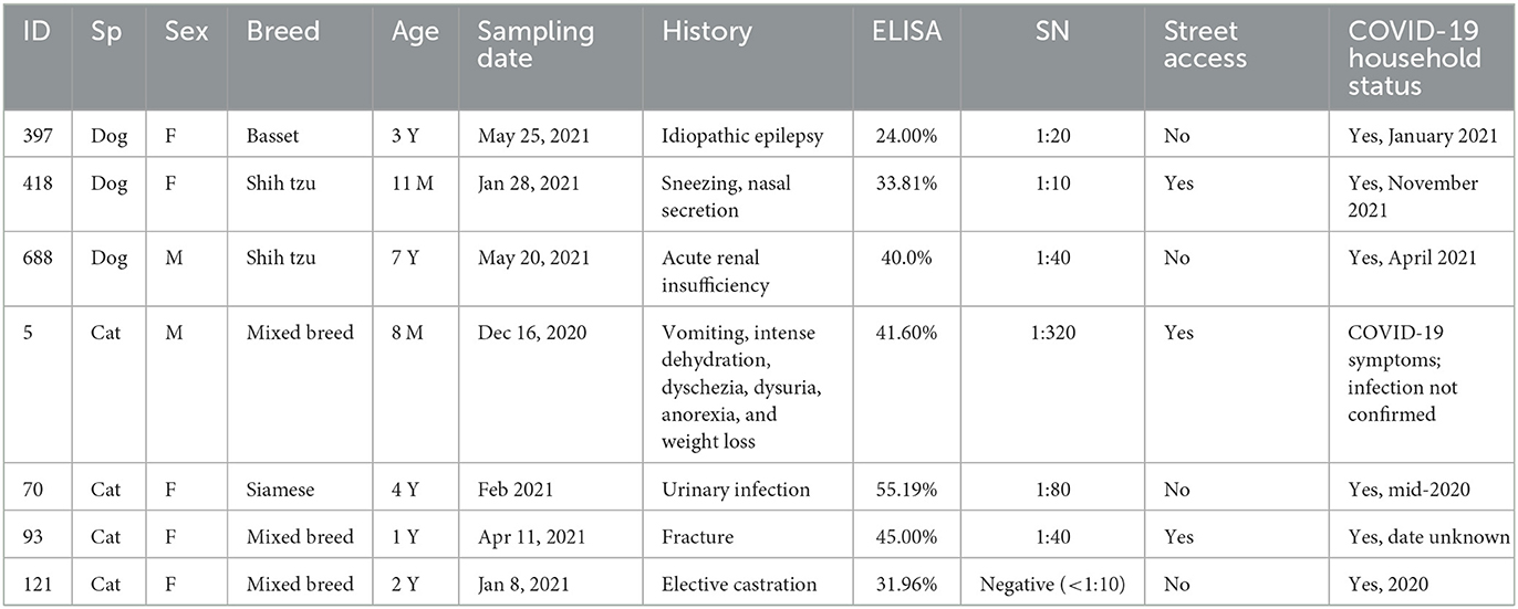
Table 1. Epidemiological data on dogs and cats that were seropositive for SARS-CoV-2 through serum neutralization (SN) assays.
4. Discussion
This study showed that SARS-CoV-2 antibodies were present in serum samples from dogs and cats attended at the Veterinary Teaching Hospital, in the same way as reported previously in several other studies worldwide (4, 6, 11, 22–24, 26, 33–35). In brief, these studies were conducted during the pandemic and reported occurrences of infection among cats and dogs through positive PCR test results, sometimes in the presence of clinical signs, along with presence of SARS-CoV-2 neutralizing antibodies. These previous results were obtained both from owned pets that had been kept indoors and from stray animals.
Since most previous studies also used ELISA as the screening test in serological surveys (25, 31, 35, 36), the discrepancy found in the present study between ELISA and SN findings from dogs may have been due to cross-reactions with other endemic coronaviruses (25, 35). Only 3 out of 33 ELISA-positive canine serum samples were also positive through SN. Not surprisingly, the SARS-CoV-2 N protein has been associated with cross-reactions with other animal coronaviruses such as the feline coronavirus (FCoV) and the canine coronavirus (CCoV). Moreover, the N protein has been considered to be a conserved viral protein, sharing common antigenic epitopes related to SARS-CoV-2 (35). In addition, previous studies have suggested that SARS-CoV-2 infection was not very efficient to elicit N protein specific antibodies (37). Lastly, the high proportion of SN-negative dogs that tested positive through ELISA may be explained by a milder course of infection, compared with cats (6, 38). As the study herein was limited to only an ELISA assay detecting N-protein, future studies should include at least one major surface protein, such as receptor binding site, spike protein, or trimeric spike proteins. Accordingly, higher human levels of neutralizing antibodies have been correlated with severe illness (39). In contrast to what was observed in the dogs of the present study, feline serum samples displayed higher agreement between ELISA and SN results, since out of four cat samples that were positive through ELISA, three of them also tested positive through SN. These results corroborated the results from previous studies on cats, in which higher seropositivity rates were observed and higher neutralizing antibody titers were developed than among dogs (6, 26, 31, 40).
A SARS-CoV-2 serosurvey study in France showed that only 5/449 dogs (1.1%) tested positive through ELISA before the emergence of the virus, while 25/443 dogs (5.5%) displayed antibodies during the COVID-19 pandemic. Seroconversion was observed in the cases of at least 8/218 dogs (3.7%) that were sampled twice during the survey period (25). Also in that study, dogs showed low susceptibility to SARS-CoV-2, such that viral transmission from and between dogs was weak or absent. However, comparative high seropositivity to SARS-CoV-2 was reported in 73/388 (18.9%) cats and 39/243 (16.0%) dogs from three veterinary clinics of Poland (33). This seropositivity was probably biased due to sampling dogs and cats with owner concerns and veterinary care and high infection rate in human population during the fourth SARS-CoV-2 wave in Poland (33). In the present study, samples were collected over a long period of time with different SARS-CoV-2 strains and epidemiological situations, which may have influenced both owner and pet seropositivity.
Lastly, pets sharing the household with a SARS-CoV-2-infected owner were found to present higher risk of infection in a previous study (26). All the pets presenting neutralizing antibodies in the present study were living in COVID-19-positive households. This confirms that pet owners constitute an associated risk factor for infection among their pets. As previously suggested, omicron transmission may occur by back-and-forth, and thus human re-infection with other novel viral strains detected in animals should be consider (41, 42). In such scenario, SARS-CoV-2 infection in humans and animals should always include sequencing to identify novel strains for passive and active immunotherapy development (41–43).
The present study showed that dogs and cats in areas of widespread human COVID-19 presented low rates of occurrence of SARS-CoV-2 antibodies. Positive pets mostly displayed non-specific mild clinical signs. Considering that positive pets were associated with COVID-19-positive households, infected owners should take the same precautions regarding isolation from their pets as they should do in relation to other people. The present study was the first large-scale serological survey on SARS-CoV-2 among pets carried out in central Brazil and was conducted in the area with the second highest COVID-19 human mortality rate nationwide.
Data availability statement
The original contributions presented in the study are included in the article/supplementary material, further inquiries can be directed to the corresponding author.
Ethics statement
This project was approved by the Ethics Committee on Animal Use at the Federal University of Mato Grosso (Protocol No. 23108.043344/2020-62). Written informed consent was obtained from the owners for the participation of their animals in this study.
Author contributions
LN and VD contributed to the conception and design of the study. SJ, MdS, KS, AP, VS, AB, LN, and VD performed field work and sample processing. FV, AS, SC, BB, AL, VV, and ND performed serological analyses. SJ, LK, AB, LN, and VD wrote the first draft of the manuscript. SJ, LK, FV, BB, AL, VV, ND, MdS, KS, AS, SC, AP, VS, AB, LN, and VD wrote sections of the manuscript. All authors contributed to the manuscript revision and read and approved the submitted version.
Funding
This research was partially funded by ERAnet ICRAD—project Musecov: Multi-scale Eco-evolution of Coronaviruses: from surveillance toward emergence prediction (ND); Ricerca Corrente IZSPB 01/20, project Valutazione della circolazione di SARS-CoV-2 e di altri coronavirus nel cane e nel gatto (CoronaPets) (ND); Ricerca Corrente 2020, project PanCO Epidemiologia e Patogenesi dei coronavirus umani ed animali (AL); Ricerca Strategica 2020 project, Suscettibilità dei mammiferi a SARS-CoV-2: rischi di zoonosi inversa e possibilità in medicina traslazionale (AL); European Union's Horizon 2020 Research and Innovation programme (One Health European Joint Programme under grant agreement No. 773830, AL). AB and VD was funded through the Brazilian National Council for Scientific and Technological Development (CNPq) (2020-1/402341).
Acknowledgments
The authors kindly thank all Veterinary Teaching Hospital professionals who helped in the attendance of dogs and cats during the survey period. They also thank all contributors involved in research institutions in Brazil and Italy for their full support, which made it possible to conduct this research. The authors are grateful to Eleonora Lorusso and Costantina Desario, University of Bari, Italy.
Conflict of interest
The authors declare that the research was conducted in the absence of any commercial or financial relationships that could be construed as a potential conflict of interest.
Publisher's note
All claims expressed in this article are solely those of the authors and do not necessarily represent those of their affiliated organizations, or those of the publisher, the editors and the reviewers. Any product that may be evaluated in this article, or claim that may be made by its manufacturer, is not guaranteed or endorsed by the publisher.
References
1. Zhou P, Yang X-L, Wang X-G, Hu B, Zhang L, Zhang W, et al. A pneumonia outbreak associated with a new coronavirus of probable bat origin. Nature. (2020) 579:270–3. doi: 10.1038/s41586-020-2012-7
2. Anka AU, Tahir MI, Abubakar SD, Alsabbagh M, Zian Z, Hamedifar H, et al. Coronavirus disease 2019 (COVID-19): An overview of the immunopathology, serological diagnosis and management. Scand J Immunol. (2021) 93:e12998–e12998. doi: 10.1111/sji.12998
3. de Morais HA, dos Santos AP, do Nascimento NC, Kmetiuk LB, Barbosa DS, Brandão PE, et al. Natural infection by SARS-CoV-2 in companion animals: a review of case reports and current evidence of their role in the epidemiology of COVID-19. Front Veter Sci. (2020) 7:823. doi: 10.3389/fvets.2020.591216
4. Shi J, Wen Z, Zhong G, Yang H, Wang C, Huang B, et al. Susceptibility of ferrets, cats, dogs, and other domesticated animals to SARS–coronavirus 2. Science. (2020) 80:eabb7015. doi: 10.1101/2020.03.30.015347
5. de Souza Barbosa AB, Kmetiuk LB, de Carvalho OV, Brandão APD, Doline FR, Lopes SRRS, et al. Infection of SARS-CoV-2 in domestic dogs associated with owner viral load. Res Vet Sci. (2022) 153:61–5. doi: 10.1016/j.rvsc.2022.10.006
6. Bosco-Lauth AM, Hartwig AE, Porter SM, Gordy PW, Nehring M, Byas AD, et al. Experimental infection of domestic dogs and cats with SARS-CoV-2: Pathogenesis, transmission, and response to reexposure in cats. Proc Natl Acad Sci. (2020) 117:26382–8. doi: 10.1073/pnas.2013102117
7. Agopian RG, da Luz SCG, Zebral AGB, de Sousa GF, de Oliveira IA V, Lima LS, et al. First reported cases of SARS-CoV-2 infection in pets in São Paulo, Brazil. Vet World. (2022) 15:2593–6. doi: 10.14202/vetworld.2022.2593-2596
8. Epifanio I da S, Rodrigues DDS, de Lima LB, Nogueira MA de A, Felix LR, et al. First report of severe acute respiratory syndrome coronavirus 2 detection in two asymptomatic cats in the state of Pernambuco, Northeastern Brazil. Vet world. (2021) 14:2839–42. doi: 10.14202/vetworld.2021.2839-2842
9. Van Den Brand JMA, L.haagmans B, Leijten L, Van Riel D, E.martina BE, Osterhaus ADME, et al. Pathology of experimental SARS coronavirus infection in cats and ferrets. Vet Pathol. (2008) 45:551–62. doi: 10.1354/vp.45-4-551
10. Gaudreault NN, Trujillo JD, Carossino M, Meekins DA, Morozov I, Madden DW, et al. SARS-CoV-2 infection, disease and transmission in domestic cats. Emerg Microbes Infect. (2020) 9:2322–32. doi: 10.1080/22221751.2020.1833687
11. Chiba S, Halfmann PJ, Hatta M, Maemura T, Fan S, Armbrust T, et al. Protective Immunity and Persistent Lung Sequelae in Domestic Cats after SARS-CoV-2 Infection. Emerg Infect Dis. (2021) 27:660. doi: 10.3201/eid2702.203884
12. Dutra V, Jarrah SA, Kmetiuk LB, de Carvalho OV, Ito de., Sousa ATH, Franco Souza VR, et al. Persistent SARS-CoV-2 antigen presence in multiple organs of a naturally infected cat from Brazil. J Venom Anim Toxins Incl Trop Dis. (2022) 7:28. doi: 10.1590/1678-9199-jvatitd-2021-0074
13. Sia SF, Yah L-M, Chin AW, Fund K, Poon LL, Nicholls JM, et al. Pathogenesis and transmission of SARS-CoV-2 virus in golden syrian hamsters. Prepr (Version 1) available Res Sq. (2020). doi: 10.21203/rs.3.rs-20774/v1
14. Rockx B, Kuiken T, Herfst S, Bestebroer T, Lamers MM, Oude Munnink BB, et al. Comparative pathogenesis of COVID-19, MERS, and SARS in a nonhuman primate model. Science. (2020) 368:1012–5. doi: 10.1126/science.abb7314
15. Woolsey C, Borisevich V, Prasad AN, Agans KN, Deer DJ, Dobias NS, et al. Establishment of an African green monkey model for COVID-19 and protection against re-infection. Nat Immunol. (2021) 22:86–98. doi: 10.1038/s41590-020-00835-8
16. Munster VJ, Feldmann F, Williamson BN, van Doremalen N, Pérez-Pérez L, Schulz J, et al. Respiratory disease in rhesus macaques inoculated with SARS-CoV-2. Nature. (2020) 585:268–72. doi: 10.1038/s41586-020-2324-7
17. Singh DK, Singh B, Ganatra SR, Gazi M, Cole J, Thippeshappa R, et al. Responses to acute infection with SARS-CoV-2 in the lungs of rhesus macaques, baboons and marmosets. Nat Microbiol. (2021) 6:73–86. doi: 10.1038/s41564-020-00841-4
18. Schlottau K, Rissmann M, Graaf A, Schön J, Sehl J, Wylezich C, et al. SARS-CoV-2 in fruit bats, ferrets, pigs, and chickens: an experimental transmission study. The Lancet Microbe. (2020) 1:e218–25. doi: 10.1016/S2666-5247(20)30089-6
19. COVID-19 - Painel COVID-19 - Estado de Mato Grosso. Available online at: http://www.saude.mt.gov.br/painelcovidmt2/ (accessed November 29, 2022).
20. Coronavírus Brasil. Available online at: https://covid.saude.gov.br/ (accessed November 29, 2022).
21. Pereira AH, Vasconcelos AL, Silva VL, Nogueira BS, Silva AC, Pacheco RC, et al. Natural SARS-CoV-2 Infection in a Free-Ranging Black-Tailed Marmoset (Mico melanurus) from an Urban Area in Mid-West Brazil. J Comp Pathol. (2022) 194:22–7. doi: 10.1016/j.jcpa.2022.03.005
22. Patterson EI, Elia G, Grassi A, Giordano A, Desario C, Medardo M, et al. Evidence of exposure to SARS-CoV-2 in cats and dogs from households in Italy. Nat Commun. (2020) 11:6231. doi: 10.1038/s41467-020-20097-0
23. Hamer SA, Pauvolid-Corrêa A, Zecca IB, Davila E, Auckland LD, Roundy CM, et al. SARS-CoV-2 Infections and Viral Isolations among Serially Tested Cats and Dogs in Households with Infected Owners in Texas, USA. Viruses. (2021) 13:938. doi: 10.3390/v13050938
24. Calvet GA, Pereira SA, Ogrzewalska M, Pauvolid-Corrêa A, Resende PC, Tassinari W de S, et al. Investigation of SARS-CoV-2 infection in dogs and cats of humans diagnosed with COVID-19 in Rio de Janeiro, Brazil. PLoS ONE. (2021) 16:e0250853. doi: 10.1371/journal.pone.0250853
25. Laidoudi Y, Sereme Y, Medkour H, Watier-Grillot S, Scandola P, Ginesta J, et al. SARS-CoV-2 antibodies seroprevalence in dogs from France using ELISA and an automated western blotting assay. One Heal. (2021) 13:100293. doi: 10.1016/j.onehlt.2021.100293
26. Fritz M, Rosolen B, Krafft E, Becquart P, Elguero E, Vratskikh O, et al. High prevalence of SARS-CoV-2 antibodies in pets from COVID-19+ households. One Heal. (2021) 11:100192. doi: 10.1016/j.onehlt.2020.100192
27. Valleriani F, Mancuso E, Vincifori G, Teodori L, Di Marcantonio L, Spedicato M, et al. Neutralization of SARS-CoV-2 variants by serum from BNT162b2 vaccine recipients. Viruses. (2021) 13:2011. doi: 10.3390/v13102011
28. Danzetta ML, Amato L, Cito F, Di Giuseppe A, Morelli D, Savini G, et al. SARS-CoV-2 RNA persistence in naso-pharyngeal swabs. Microorganisms. (2020) 8:1124. doi: 10.3390/microorganisms8081124
29. Lorusso A, Calistri P, Savini G, Morelli D, Ambrosij L, Migliorati G, et al. Novel SARS-CoV-2 variants in Italy: The role of veterinary public health institutes. Viruses. (2021) 13:549. doi: 10.3390/v13040549
30. Ramakrishnan MA. Determination of 50% endpoint titer using a simple formula. (2016). Available online at: http://dx.doi.org/10.5501/wjv.v5.i2.85 (accessed February 5, 2023). doi: 10.5501/wjv.v5.i2.85
31. Dileepan M, Di D, Huang Q, Ahmed S, Heinrich D, Ly H, et al. Seroprevalence of SARS-CoV-2 (COVID-19) exposure in pet cats and dogs in Minnesota, USA. Virulence. (2021). 12:1597–609. doi: 10.1080/21505594.2021.1936433
32. Decaro N, Grassi A, Lorusso E, Patterson EI, Lorusso A, Desario C, et al. Long-term persistence of neutralizing SARS-CoV-2 antibodies in pets. Transbound Emerg Dis. (2022) 69:3073–6. doi: 10.1111/tbed.14308
33. Kaczorek-Łukowska E, Wernike K, Beer M, Wróbel M, Małaczewska J, Mikulska-Skupień E, et al. High Seroprevalence against SARS-CoV-2 among Dogs and Cats, Poland, 2021/2022. Animals. (2022) 12:2016. doi: 10.3390/ani12162016
34. Bessière P, Vergne T, Battini M, Brun J, Averso J, Joly E, et al. SARS-CoV-2 Infection in Companion Animals: Prospective Serological Survey and Risk Factor Analysis in France. Viruses. (2022) 14:1178. doi: 10.3390/v14061178
35. Barua S, Hoque M, Adekanmbi F, Kelly P, Jenkins-Moore M, Torchetti MK, et al. Antibodies to SARS-CoV-2 in dogs and cats, USA. Emerg Microbes Infect. (2021) 10:1669–74. doi: 10.1080/22221751.2021.1967101
36. Stevanovic V, Vilibic-Cavlek T, Tabain I, Benvin I, Kovac S, Hruskar Z, et al. Seroprevalence of SARS-CoV-2 infection among pet animals in Croatia and potential public health impact. Transbound Emerg Dis. (2021) 68:1767–73. doi: 10.1111/tbed.13924
37. Diezma-Díaz C, Álvarez-García G, Regidor-Cerrillo J, Miró G, Villanueva-Saz S, Dolores Pérez M, et al. A comparative study of eight serological methods shows that spike protein-based ELISAs are the most accurate tests for serodiagnosing SARS-CoV-2 infections in cats and dogs. Front Vet Sci. (2023) 10:1121935. doi: 10.3389/fvets.2023.1121935
38. Lam TTY, Jia N, Zhang YW, Shum MHH, Jiang JF, Zhu HC, et al. Identifying SARS-CoV-2-related coronaviruses in Malayan pangolins. Nature. (2022) 583:282–5. doi: 10.1038/s41586-020-2169-0
39. Zhang B, Zhou X, Zhu C, Song Y, Feng F, Qiu Y, et al. Immune phenotyping based on the neutrophil-to-lymphocyte ratio and IgG level predicts disease severity and outcome for patients With COVID-19. Front Mol Biosci. (2020) 7:157. doi: 10.3389/fmolb.2020.00157
40. Barroso R, Vieira-Pires A, Antunes A, Fidalgo-Carvalho I. Susceptibility of pets to SARS-CoV-2 Infection: lessons from a seroepidemiologic survey of cats and dogs in Portugal. Microorganisms. (2022) 10:345. doi: 10.3390/microorganisms10020345
41. Saied AA, Metwally AA. SARS-CoV-2 variants of concerns in animals: An unmonitored rising health threat. Virusdisease. (2022) 33:466–76. doi: 10.1007/s13337-022-00794-8
42. Saied AA, Metwally AA, Mohamed HMA, Haridy MAM. The contribution of bovines to human health against viral infections. Environ Sci Pollut Res Int. (2021) 28:46999–7023. doi: 10.1007/s11356-021-14941-z
Keywords: Brazil, companion animals, pets, serosurvey, coronavirus, SARS-CoV-2
Citation: Jarrah SA, Kmetiuk LB, Valleriani F, Bonfini B, Lorusso A, Vasinioti V, Decaro N, dos Santos MT, Spohr KAH, Pratelli A, Serroni A, Capista S, Sousa VRF, Biondo AW, Nakazato L and Dutra V (2023) SARS-CoV-2 antibodies in dogs and cats in a highly infected area of Brazil during the pandemic. Front. Vet. Sci. 10:1111728. doi: 10.3389/fvets.2023.1111728
Received: 30 November 2022; Accepted: 06 February 2023;
Published: 23 February 2023.
Edited by:
Tomomi Takano, Kitasato University, JapanReviewed by:
Asmaa A. Metwally, Faculty of Veterinary Med, EgyptGianvito Lanave, University of Bari Aldo Moro, Italy
Copyright © 2023 Jarrah, Kmetiuk, Valleriani, Bonfini, Lorusso, Vasinioti, Decaro, dos Santos, Spohr, Pratelli, Serroni, Capista, Sousa, Biondo, Nakazato and Dutra. This is an open-access article distributed under the terms of the Creative Commons Attribution License (CC BY). The use, distribution or reproduction in other forums is permitted, provided the original author(s) and the copyright owner(s) are credited and that the original publication in this journal is cited, in accordance with accepted academic practice. No use, distribution or reproduction is permitted which does not comply with these terms.
*Correspondence: Valéria Dutra,  dmFsZXJpYWR1dHJhLmR1dHJhQGdtYWlsLmNvbQ==