 Shin-ichi Hayama
 Setsuko Nakanishi1
 Aki Tanaka
 Yoshi Kawamoto
- 1School of Veterinary Medicine, Nippon Veterinary and Life Science University, Musashino, Tokyo, Japan
- 2Tohoku Wildlife Management Center, Sendai, Miyagi, Japan
- 3School of Veterinary Nursing and Technology, Nippon Veterinary and Life Science University, Musashino, Tokyo, Japan
Wild Japanese monkeys (Macaca fuscata) were exposed to radiation after the Fukushima Daiichi nuclear accident in 2011. To clarify the biological effects of radiation exposure on their fetal growth, pregnant monkeys and their fetuses were analyzed. These animals were collected between 2008 and 2020 (before and after the accident in 2011) living in Fukushima City, approximately 70 km from the nuclear power plant. Multiple regression analyses were conducted with fetal body weight (FBW) and fetal head circumference (FHS) as objective variables, and maternal and fetal factors as explanatory variables. The maternal factors were relative exposure dose rate (REDR), age, body weight, body length, fat index, and parity. The fetal factors were crown ramp length (CRL) and sex. Multiple regression analyses showed that FBR and FHS growth were positively associated with CRL, maternal body length, and negatively associated with REDR. Since the relative growth of FBR and FHS to CRL decreased with increasing REDR, radiation exposure due to the nuclear accident may have contributed to the delayed fetal growth observed in Japanese monkeys.
1. Introduction
The accident at the Fukushima Daiichi Nuclear Power Plant (FDNPP) in March 2011 exposed many people and wildlife to radioactive materials. In order to mitigate the damage to crops, the population of Japanese monkeys in Fukushima City has been systematically managed since 2008 in accordance with the laws and regulations established by Fukushima Prefecture. Several papers have been published on the health effects of radiation exposure on Japanese monkeys in Fukushima City (1, 2), located approximately 70 km from the FDNPP (Figure 1). As a result of this accident, this population of Japanese monkeys (Macaca fuscata) became the first wild primate in the world to be exposed to radiation (2).
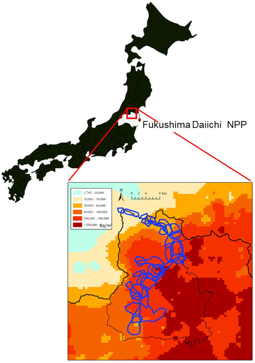
Figure 1. Soil contamination levels by radiocesium concentrations (Bq/m2) and the distribution of monkey troops (irregular enclosed blue outlines) in Fukushima City. This map was generated according to the soil contamination map created by the Ministry of Education, Culture, Sports, Science and Technology (converted to values for July 2, 2011).
The effects of long-term exposure to low doses of radiation on the fetus are among the many health concerns. Low birth weight and high rates of microcephaly have been reported in children born to mothers exposed to the atomic bombs in Hiroshima and Nagasaki (3). Another paper (4) has shown that these children have intellectual disability due to abnormal brain development. In wild animals, Møller et al. (5) reported that the brain weights of birds captured near the Chernobyl NPP were lower than those of birds captured elsewhere. Our study also found that the body weight and head size relative to the crown–rump length (CRL) were significantly lower in fetuses conceived after the FDNPP accident (2011–2016) than in fetuses conceived prior to the FDNPP accident (2008–2010) (2). No other studies from Chernobyl or Fukushima have tracked fetal development over time in the same wildlife populations or compared fetal development before and after long-term radiation exposure (2). However, this previous study did not account for the fetal radiation exposure dose-rate.
In cows, the accumulation of radioactive materials is higher in the fetus than in the mother (6). However, in Japanese monkeys, the accumulation of radioactive materials in the fetus is unknown, it is difficult to estimate the exact exposure dose-rate. In this study, the internal dose rate in the mother was adopted as an indicator of the relative dose rate to the fetus.
In some primates, including humans, fetus growth is known to be affected by maternal age, parity, body size and nutritional status, and the sex of the fetus (7–10). The purpose of this study is to determine the factors that influence the growth of fetal body weight (FBW) and fetal head size (FHS) relative to CRL. In this study, the maternal factors (body weight, body length, parity, fat index, age and the relative exposure dose-rate) and the fetal factors (CRL and sex) were evaluated by multiple regression analysis. Because monkeys are taxonomically close to humans, this study may be useful for research on the health effects of radiation exposure on humans.
2. Materials and methods
2.1. Animals and ethics
This study was approved by the Institutional Animal Care and Use Committee of Nippon Veterinary and Life Science University (No. 2022S-1). All experiments were performed in accordance with relevant guidelines and regulations. Carcasses of Japanese monkeys were provided by Fukushima City. Monkeys were culled to prevent crop damage with the permission of the governor of Fukushima Prefecture, according to the Fukushima Japanese Monkey Management Plan, which was established based on the Wildlife Protection and Hunting Management Law. Monkeys were captured using box traps and euthanized by a gun by licensed hunters at the request of Fukushima City. The methods for capture and euthanasia were in accordance with the guidelines of the management plan and do not present an ethical concern. This euthanasia method was also in accordance with guidelines published by the Wildlife Research Center of Kyoto University (11). The Japanese monkeys inhabiting the study area were not listed as an endangered species on the Japanese Red List, as revised by the Ministry of the Environment in 2012 (12). The sample size of monkey mothers and fetuses was 32 before and 54 after the accident.
2.2. Mothers and muscle samples
Pregnant Japanese monkey carcasses were collected from 2008 to 2020, transported under refrigerated conditions to our laboratory, and subjected to necropsies. The body weight (BW) of each monkey was measured in grams. Body length (BL) was measured in millimeters as the straight-line distance from the top of head to the rump, at the dorsal surface of the sciatic protuberances while the monkey was in a recumbent position. During the necropsy, fat indices (FI) to evaluate the nutritional status were calculated that the ratio of the mesenteric fat weight to body weight was proportional to the percentage of body fat in Japanese monkeys (2). The FI was defined as mesenteric fat weight (g) divided by body weight (g) and multiplied by 1,000. Age was classified as sub-adult or adult by tooth eruption; in adults (13), all permanent teeth are erupted, corresponding to an age of at least 7 years old. Parity was assessed as primiparous or multiparous. Nipples of primiparous animals were not developed because they were not sucked by the young at the time of capture (14). Multiparous individuals had elongated teats on one or both sides.
During the necropsy after the FDNPP accident, 500–1,000 g of muscle tissue from the hind limb of each mother was collected; this tissue type was used because organs weighing 500 g or more were required to measure the radiocesium concentration. The muscle tissue was stored at −30°C until it was used for radioactivity measurements.
2.3. Fetuses
After the fetuses were removed from the uterus during the necropsy, FBW was measured in grams, and CRL (i.e., the length of the fetus from the top of its head to bottom of torso) was measured in millimeters. CRL is a common somatometric measure for age assessment in physical and neurological examinations (15).
Fetuses were preserved in 10% neutral buffered formalin. FHS was defined as the product of the biparietal diameter and occipital frontal diameter. The biparietal diameter is a basic biometric parameter used to assess fetal size and is the maximum width of the head. The occipital frontal diameter was measured as the maximum length between the forehead and occipital region.
All specimens were measured by the same person in millimeters, using a caliper. Specimens with a CRL greater than 80 mm (fetal age of about 3 months or greater) were included in the analysis. This is because in fetuses of this size, the cranium is ossified and there is less error in the external measurements.
The fetuses were divided into those from pregnancies between 2008 and 2010 (pre-accident) and those from pregnancies in 2011 or later (post-accident). The average conception date of Japanese monkeys in Fukushima City is November 19 (SD = 29.2 days) (16), and the average gestation period is 180 days (17). Therefore, the fetuses conceived during the mating season in 2010, the year before the accident, were likely exposed to radiation from the accident in March 2011 for fewer than about 60 days in the second trimester of pregnancy. Because the sensitivity to radiation exposure in the fetal period is highest in the first trimester of pregnancy (4, 18, 19), fetuses conceived in 2010 were included in the pre-accident group in this study.
2.4. Radioactivity measurements
The muscle radiocesium concentration was measured in mothers that were pregnant after the accident. The concentration of radioactive cesium in the muscle tissue before the accident was treated as 0 Bq/kg (muscle samples were not collected for this group).
The radioactivity of radiocesium in the muscle samples was analyzed using a germanium semiconductor spectrometer (GC2020-7500SL-2002 CSL; Canberra, Meriden, CT) and a NaI (T1) scintillation detector (AT1320A; Atometex, Minsk, Belarus). Data were corrected to the background radiation dose in the measurement environment as-needed. 134Cs was detected using 604.70 and 795.85 keV gamma-rays, whereas 137Cs was detected using 661.6 keV gamma-rays. The radioactivity of radiocesium was adjusted to the value on the day of capture based on its physical half-life. The limit of detection was 10 Bq/kg. The muscle radiocesium concentration was calculated as the combined concentration of 134Cs and 137Cs per kilogram of fresh muscle.
2.5. Relative exposure dose-rate
Urushihara et al. (20) estimated the radiocesium dose-rate using the ERICA tool [version 1.2; (21)], with some modifications, and determined dose conversion coefficients (DCCs) using the equation provided by the ERICA tool for Japanese monkeys [Supplementary Table S4 in Urushihara et al. (20)]. The total dose-rate in the mother should include external and internal dose-rate, the total dose-rate and internal dose-rate are highly correlated in Japanese monkeys in Fukushima [r = 0.98, p < 0.0001; Urushihara et al. (20)]. Then, the estimated internal exposure dose-rate in the mother was used as an indicator of the relative exposure dose-rate (REDR) for the fetus by estimating the muscle radiocesium concentration in the mother using the DCCs obtained by Urushihara et al. (20).
2.6. Statistics
The Shapiro–Wilk test was used to test the normality of continuous variables. Wilcoxon single rank test was performed to compare medians for mother monkey body weight, body length, and fat index, and fetal CRL, body weight and head size by pre- and post-accident. Multiple non-parametric linear regressions were performed with dependent variables (FBW and FHS) and explanatory variables, including maternal factors (BW, BL, Parity, FI, and Age, and REDR) and fetal factors (CRL and Sex). Stata/IC 16 (StataCorp, College Station, TX) was used for all analyses. Two-sided tests were used with a 5% significance level.
3. Results
Descriptive statistics for Japanese monkey mothers and fetuses in Fukushima are shown in Table 1 with median (range) for mother monkey body weight (g), body length (mm), and fat index, and fetal CRL (mm), body weight (g) and head size (mm2) by pre- and post-accident with value of ps, respectively.
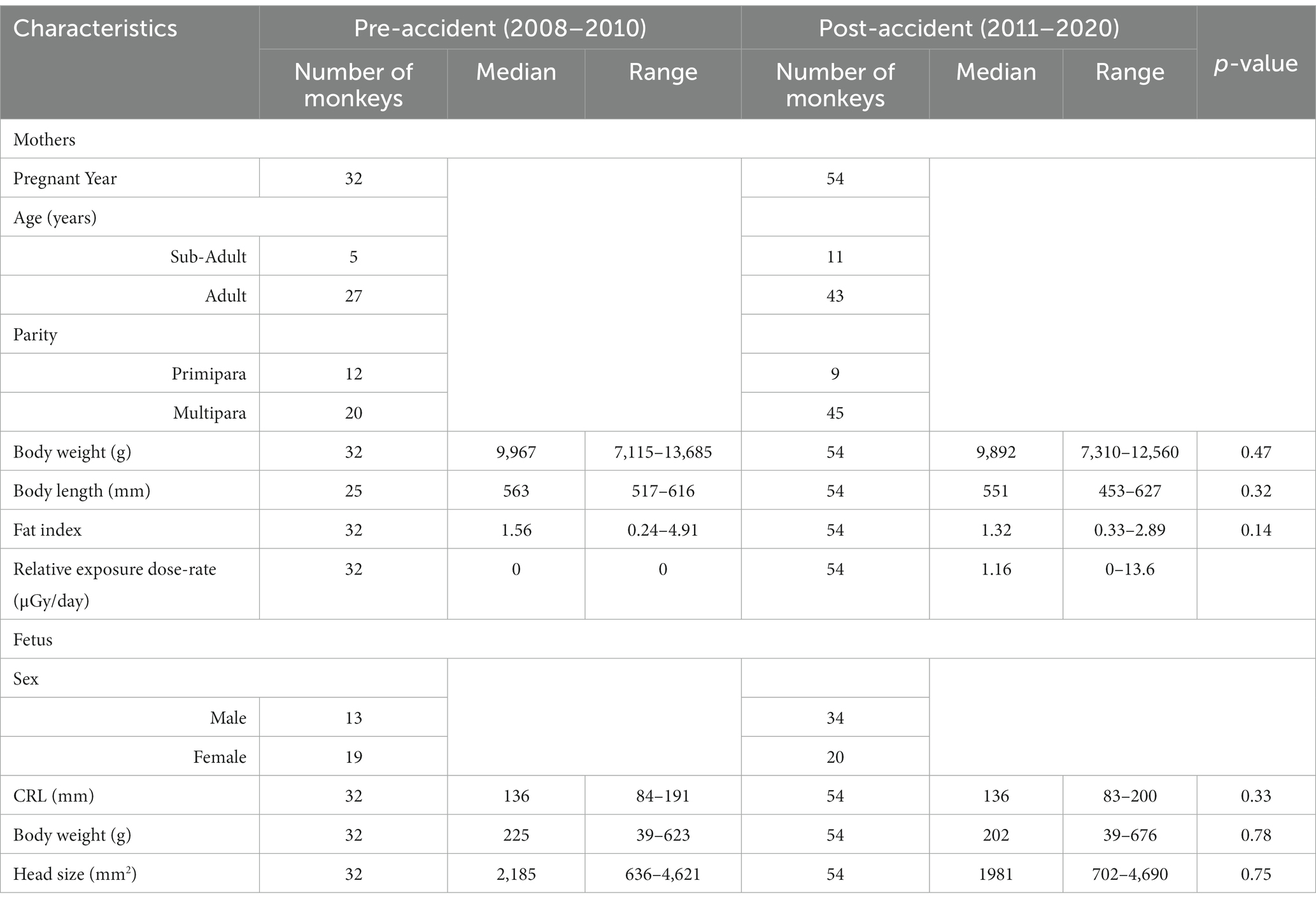
Table 1. Characteristics of mothers (N = 86) and fetuses (N = 86) based on Japanese monkey carcasses collected in Fukushima, Japan from 2008 and 2020.
The growth of FBW and FHS relative to CRL tended to be delayed after the accident. Associations between FBW and CRL in pre- and post-accident groups are described in Figure 2, and associations between FHS and CRL in pre- and post-accident groups are described in Figure 3.
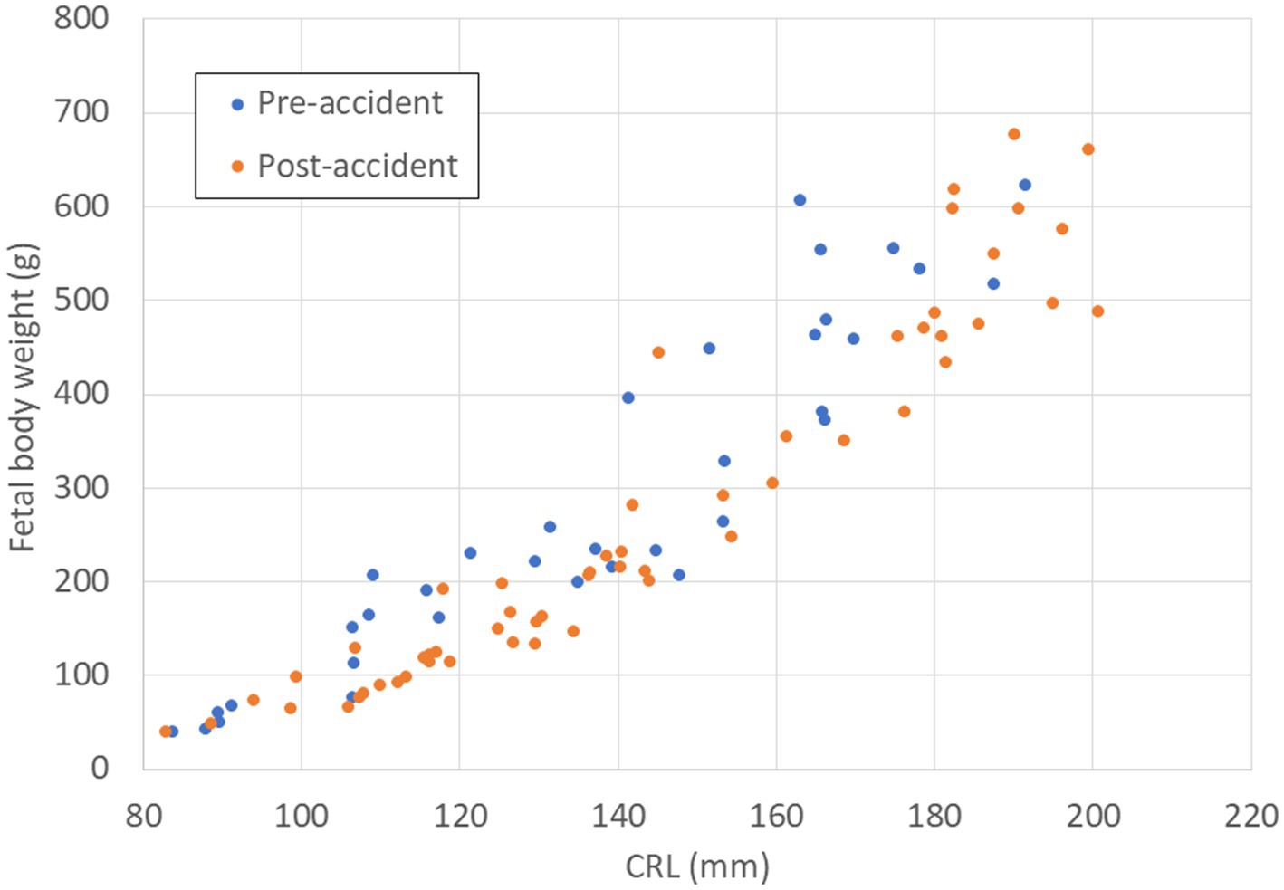
Figure 2. Relationship between CRL (mm) and FBW (g) in fetal Japanese monkeys before and after the Fukushima Daiichi nuclear power plant accident. Blue circles indicate fetal monkeys before the accident; red circles indicate fetal monkeys after the accident. CRL, crown–rump length; FBW, fetal body weight.
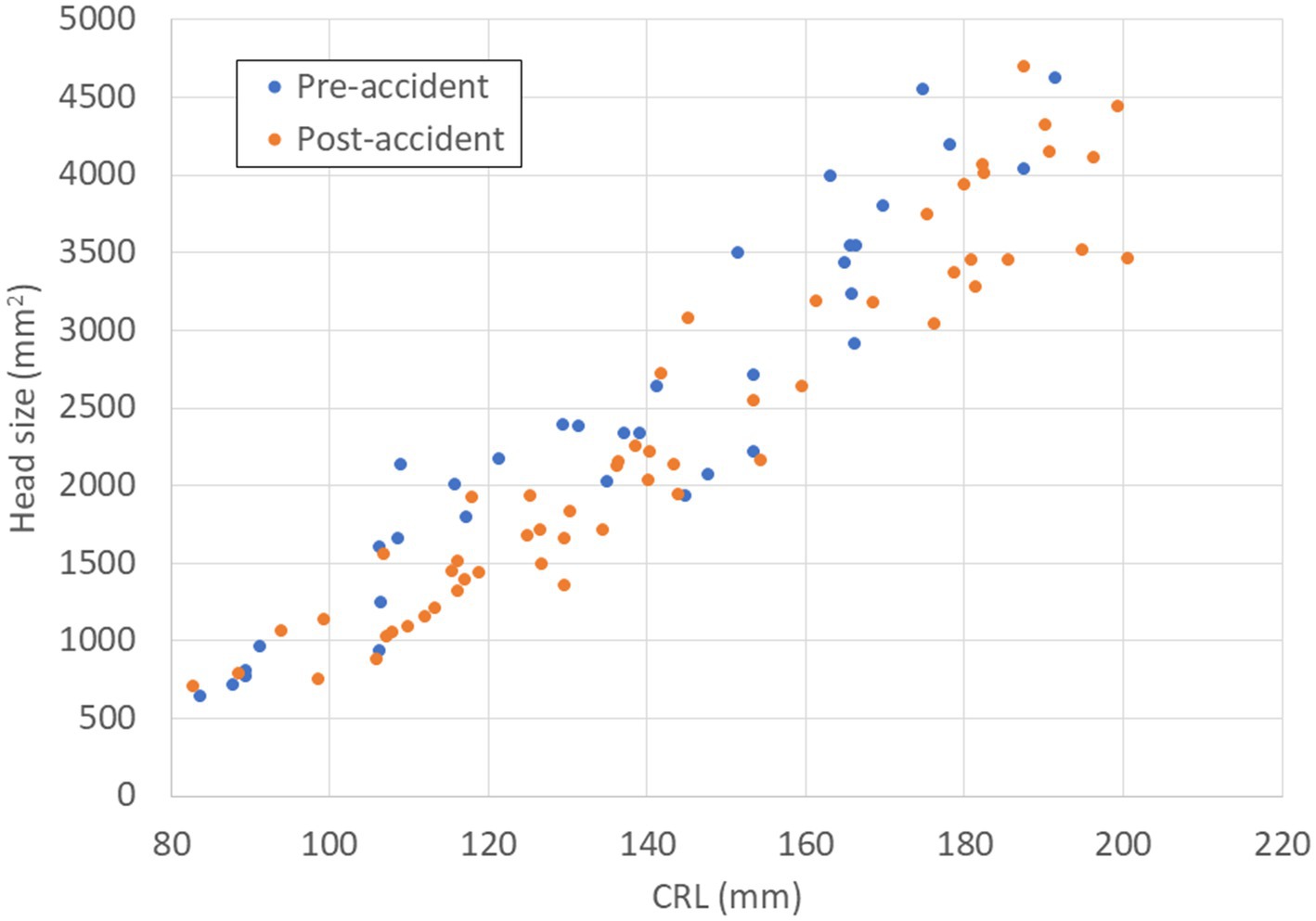
Figure 3. Relationship between CRL (mm) and FHS (mm2) in fetal Japanese monkeys before and after the Fukushima Daiichi nuclear power plant accident. Blue circles indicate fetal monkeys before the accident; red circles indicate fetal monkeys after the accident. CRL, crown–rump length; FHS, fetal head size.
Table 2 shows the results of multiple non-parametric linear regression analyses of fetal body weight and head size of Japanese monkeys collected in Fukushima from 2008 to 2020 as dependent variables and associated factors as independent variables including age, delivery, fat index, relative exposure dose-rate (μGy/day), body weight, body length, fetal CRL, and sex. Both fetal body weight and fetal head size were positively associated with CRL (p < 0.001) and mother body length (FBW: p < 0.02; FHS: p < 0.05), and negatively associated with relative exposure dose-rate (FBW: p < 0.01; FHS: p < 0.05).
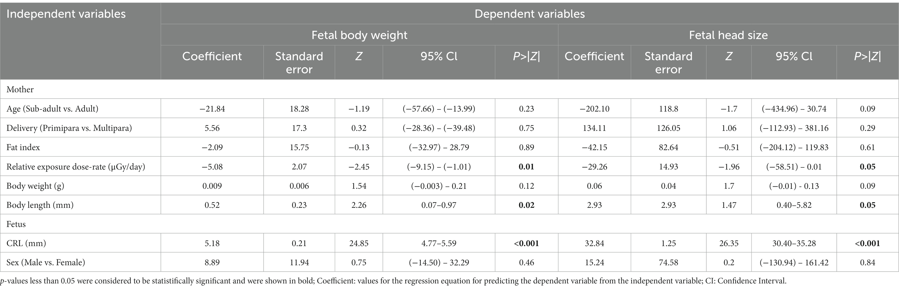
Table 2. Multiple non-parametric linear regression analyses of dependent variables as fetal body weight and head size of Japanese monkeys collected in Fukushima from 2008 to 2020 and associated factors (n = 86).
No significant relationships were found with fetal growth parameters and other factors.
4. Discussion
In this study, the relationship between fetal growth (FBW and FHS) and various factors was analyzed in wild Japanese monkeys exposed to radiation during the FDNPP accident. The only factor that was negatively associated with relative growth parameters (both FBW and FHS) was REDR.
This is the first study to clarify the influence of radiation to delayed fetal growth in wild animals. Based on our results, the relative growth of fetuses is expected to return to the pre-exposure state if radiation exposure is reduced in the future. However, the radioactive cesium concentration in the muscle tissues in this population decreased year by year after the accident until 2015, with no additional decrease after 2016 (22). Therefore, it is not possible to predict when the growth rate will return to pre-accident levels.
Scherb and Hayashi (23) analyzed prefecture-specific spatiotemporal trends in the frequency of low birth weight in Japan using annual counts for 26.158 million live births from 1995 to 2018 provided by the Japanese Ministry of Health, Labor and Welfare. A logistic regression of low birth weight proportions against the additional dose-rate after accidents adjusted for prefecture-specific spatiotemporal base-line trends yielded an odds ratio per μSv/h of 1.098 (95% confidence interval, 1.058–1.139, p < 0.0001). These results are not directly comparable to ours, but suggest similar conclusions.
CRL and BL were significantly associated with relative growth in FBW and FHS. Because CRL increases with fetal growth, a positive association is an inevitable consequence. BL also positively affects fetal growth as in human case studies (10). There are also reports that maternal height is associated with increased birth weight (9, 24). However, REDR, used the internal dose-rate in the mother,increases in proportion to the BL (20). The contribution of the muscle radiocesium concentration to the estimation of REDR may offset the effect of BL.
Maternal age, parity, and fetal sex were not significantly related to fetal growth. On the other hand, it has been reported that these factors are associated with increased birth weight in humans and captive macaques (7, 9). These factors have also been associated with fetal growth retardation (25). Hopper et al. (8) reported that maternal birth weight of rhesus monkeys did not correlate with infant birth weight, and increased parity of the mother was associated with higher birth weight of the infant.
In this study, it is possible that the exact age of the mothers and their history of parity were unknown, and thus a clear causal relationship with fetal growth could not be determined. Although there have been several reports of maternal pre-pregnancy weight and weight gain during pregnancy affecting birth weight (24, 26), it was not possible to obtain these data in this study, so a similar test could not be performed.
Jhonsenn et al. (10) used ultrasound to estimate fetal body weight from fetus measurements and examined the association between intrauterine growth of the fetus and maternal factors in humans. They found that male fetuses were heavier than female fetuses at the same gestational week and that maternal height and age were associated with increased fetal weight.
On the other hand, in this study (10), maternal weight and body mass index had no effect on fetal weight growth. In our study, FI and BW were not significantly related to fetal growth. This may be due to the large individual differences in maternal weight and nutritional status during pregnancy in both humans and monkeys. Japanese monkeys are conceived from the autumn to early winter and deliver in the spring, and body fat and weight fluctuate substantially during these periods. Monkeys eat large amounts of acorns and other foods in the fall and gain 20–30% body weight through early winter, followed by weight loss until minimum in the spring (27). This seasonal variation may weaken the relationship between FI or BW and fetal growth in Japanese monkeys.
5. Conclusion
This study evaluated maternal factors (weight, length, parity, fat index, age, and relative dose rate) and fetal factors (CRL and sex) by multiple regression analysis to identify factors associated with fetal body weight (FBW) and fetal head size (FHS) growth relative to CRL. The results revealed that only REDR had a significant negative association with the relative growth of FBW and FHS. However, REDR is a relative measure of convenience in this study. In addition, the actual exposure dose should be higher than the REDR because it is probably the minimum exposure for the fetus. The exact fetal exposure dose-rate needs to be clarified in future studies.
Data availability statement
The raw data supporting the conclusions of this article will be made available by the authors, without undue reservation.
Ethics statement
Carcasses of animals were provided by Fukushima City with the permission of the governor of Fukushima Prefecture under the Wildlife Protection and Hunting Management Law. The methods for capture and euthanasia were in accordance with the guidelines of the management plan by the Environmental Agency of Japanese government. This research was approved by the Animal Research Committee in Nippon Veterinary and Life Science University (#2022S-1).
Author contributions
SN and FK collected specimens. S-iH, YK, and TO designed the study. AT analyzed the data. S-iH wrote the paper. All authors contributed to the article and approved the submitted version.
Funding
This research was funded by the Cooperative Research Program of The Primate Research Institute (Kyoto University), Act Beyond Trust, The Promotion and Mutual Aid Corporation for Private Schools of Japan, The Sumitomo Foundation, and the Japan Society for the Promotion of Science (KAKENHI Grant nos. 25517008, 16K08087, and 20K06400).
Acknowledgments
This study was made possible by the cooperation of Fukushima City. We would like to thank everyone involved in this study. We would also like to express our gratitude to members of Nippon Veterinary and Life Science University for their assistance.
Conflict of interest
The authors declare that the research was conducted in the absence of any commercial or financial relationships that could be construed as a potential conflict of interest.
Publisher’s note
All claims expressed in this article are solely those of the authors and do not necessarily represent those of their affiliated organizations, or those of the publisher, the editors and the reviewers. Any product that may be evaluated in this article, or claim that may be made by its manufacturer, is not guaranteed or endorsed by the publisher.
References
1. Ochiai, K, Hayama, S, Nakiri, S, Nakanishi, S, Ishii, N, Uno, T, et al. Low blood cell counts in wild Japanese monkeys after the Fukushima Daiichi nuclear disaster. Sci Rep. (2014) 4:5793. doi: 10.1038/srep05793
2. Hayama, S, Tsuchiya, M, Ochiai, K, Nakiri, S, Nakanishi, S, Ishii, N, et al. Small head size and delayed body weight growth in wild Japanese monkey fetuses after the Fukushima Daiichi nuclear disaster. Sci Rep. (2017) 7:3528. doi: 10.1038/s41598-017-03866-8
3. Miller, RW, and Blot, WJ. Small head size after in-utero exposure to atomic radiation. Lancet. (1972) 2:784–7.
4. Otake, M, and Schull, WJ. Radiation-related brain damage and growth retardation among the prenatally exposed atomic bomb survivors. Int J Radiat Biol. (1998) 74:159–71. doi: 10.1080/095530098141555
5. Møller, AP, Bonissoil-Alquati, A, Rudolfsen, G, and Mousseau, TA. Chernobyl birds have smaller brains. PLoS One. (2011) 6:e16862. doi: 10.1371/journal.pone.0016862
6. Fukuda, T, Kino, Y, Abe, Y, Yamashiro, H, Kuwahara, Y, Nihei, H, et al. Distribution of artificial radionuclides in abandoned cattle in the evacuation zone of the Fukushima Daiichi Nuclear Power Plant. PLoS One. (2013) 8:e54312. doi: 10.1371/journal.pone.0054312
7. Naiken, S, Griffiths, M-A, Edouard, L, and Padayatchy, N. Factors influencing reproduction in captive-bred Cynomolgus monkeys (Macaca fascicularis) from Mauritius. Amer J Primatol. (2015) 77:1290–8. doi: 10.1002/ajp.22482
8. Hopper, K, Capozzi, D, and Newsome, JT. Effects of maternal and infant characteristics on birth weight and gestation length in a colony of Rhesus Macaques (Macaca mulatta). Comp Med. (2008) 58:597–603.
9. Terada, M, Matsuda, Y, Ogawa, M, Matsui, H, and Satoh, S. Effects of maternal factors on birth weight in Japan. J Pregnancy. (2013) 2013:1–5. doi: 10.1155/2013/172395
10. Johnsen, SL, Rasmussen, S, Wilsgaad, T, Sollien, R, and Kiserud, T. Longitudinal reference ranges for estimated fetal weight. Acta Obste Gynecol. (2006) 85:286–97. doi: 10.1080/00016340600569133
11. Wildlife Research Center of Kyoto University. Guidelines for wildlife research issued by the wildlife research Center of Kyoto University. Available at: https://www.wrc.kyoto-u.ac.jp/en/guidelines/wild.html.
12. Japanese Ministry of Environment. State of Japan's environment at a glance: Extinct and endangered species listed in the red data book. Available at: http://www.env.go.jp/en/nature/biodiv/reddata.html.
13. Iwamoto, M, Watanabe, T, and Hamada, Y. Eruption of permanent teeth in Japanese monkeys (Macaca fuscata). Primate research. (1987) 3:18–28. (in Japanese with English summary). doi: 10.2354/psj.3.18
14. Hayama, S, Kamiya, S, and Nigi, H. Morphological changes of female reproductive organs of Japanese monkeys with reproductive conditions. Primates. (1997) 38:359–67. doi: 10.1007/BF02381877
15. Newell-Morris, LL. Age determination in monkey fetuses and neonates In: GC Ruppenthal and DJ Reese, editors. Nursery care of nonhuman primates. New York: Plenum Press (1979). 93–115.
16. Hayama, S, Nakiri, S, and Konno, F. Pregnancy rate and conception date in a wild population of Japanese monkeys. J Vet Med Sci. (2011) 73:809–12. doi: 10.1292/jvms.10-0420
17. Nigi, H. Some aspects related to conception of the Japanese monkey (Macaca fuscata). Primates. (1976) 17:81–7. doi: 10.1007/BF02381568
18. De Santis, M, Straface, G, Cavaliere, AF, Caruso, A, Cichocki, F, Venga, L, et al. First trimester maternal thyroid X-ray exposure and neonatal birth weight. Reprod Toxicol. (2005) 20:3–4. doi: 10.1016/j.reprotox.2004.12.002
19. Kusama, T, and Ota, K. Radiological protection for diagnostic examination of pregnant women. Congenit Anom. (2002) 42:10–4. doi: 10.1111/j.1741-4520.2002.tb00848.x
20. Urushihara, Y, Suzuki, T, Shimizu, Y, Ohtaki, M, Kuwahara, Y, Suzuki, M, et al. Haematological analysis of Japanese macaques (Macaca fuscata) in the area affected by the Fukushima Daiichi Nuclear Power Plant accident. Sci Rep. (2018) 8:16748. doi: 10.1038/s41598-018-35104-0
21. Brown, JE, Alfonso, B, Avila, R, Beresford, NA, Copplestone, D, and Hosseini, A. A new version of the ERICA tool to facilitate impact assessments of radioactivity on wild plants and animals. J Environ Radioact. (2016) 153:141–8. doi: 10.1016/j.jenvrad.2015.12.011
22. Hayama, S, Tanaka, A, Nakanishi, S, Konno, F, Kawamoto, Y, Ochiai, K, et al. Time dependence of 137Cs contamination in wild Japanese monkeys after the Fukushima Daiichi nuclear accident. Environ Sci Pollut Res. (2022) 29:88359–68. doi: 10.1007/s11356-022-23707-0
23. Scherb, H, and Hayashi, K. Spatiotemporal association of low birth weight with Cs-137 deposition at the prefecture level in Japan after the Fukushima nuclear power plant accidents: an analytical-ecologic epidemiological study. Environ Health. (2020) 19:82. doi: 10.1186/s12940-020-00630-w
24. Romo, A, Carceller, R, and Tobajas, J. Intrauterine growth retardation (IUGR): epidemiology and etiology. Pediatr Endocrinol Rev. (2009) 6:332–6.
25. Sharma, D, Shastri, S, and Sharma, P. Intrauterine growth restriction: antenatal and postnatal aspects. Clin Med Insights Pediatr. (2016) 10:67–83. doi: 10.4137/CMPed.S40070
26. Liu, L, Hong, Z, and Zhang, L. Associations of pre-pregnancy body mass index and gestational weight gain with pregnancy outcomes in nulliparous women delivering single live babies. Sci Rep. (2015) 5:12863. doi: 10.1038/srep12863
Keywords: gestational development, fetal body weight, fetal head size, nuclear accident, radiation exposure, Japanese monkeys, teratology, birth defects
Citation: Hayama S-i, Nakanishi S, Tanaka A, Konno F, Kawamoto Y and Omi T (2023) Influence of radiation exposure to delayed fetal growth in wild Japanese monkeys after the Fukushima accident. Front. Vet. Sci. 10:1151361. doi: 10.3389/fvets.2023.1151361
Edited by:
Paula Alexandra Oliveira, University of Trás-os-Montes and Alto Douro, PortugalReviewed by:
Ana Calado, University of Trás-os-Montes and Alto Douro, PortugalMaria Lurdes Pinto, University of Trás-os-Montes and Alto Douro, Portugal
Copyright © 2023 Hayama, Nakanishi, Tanaka, Konno, Kawamoto and Omi. This is an open-access article distributed under the terms of the Creative Commons Attribution License (CC BY). The use, distribution or reproduction in other forums is permitted, provided the original author(s) and the copyright owner(s) are credited and that the original publication in this journal is cited, in accordance with accepted academic practice. No use, distribution or reproduction is permitted which does not comply with these terms.
*Correspondence: Shin-ichi Hayama, aGF5YW1hQG52bHUuYWMuanA=