In the published article, there was an error in Figure 2C as published. The CD206 flow data in the DSF group has been corrected. The corrected Figure 2C and its caption appear below.
FIGURE 2
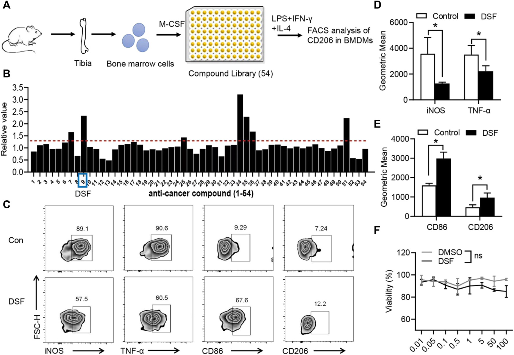
DSF significantly promotes pro-inflammatory macrophages into the M2-type phenotype. (A) The diagram of screening assay. Bone marrow cells were isolated from the tibia of wild-type mice. BMDMs were induced by M-CSF (50 ng/ml). Then the cells were treated with LPS (100 ng/ml), IFN-γ (20 ng/ml), and IL-4 (20 ng/ml) for 6 h and underwent FACS analysis of CD206 expression. (B) Quantitative analysis of the candidates in screening assay. (C) BMDMs were pretreated with DSF (5 μM) or DMSO (0.01%). Then the cells were treated with LPS (100 ng/ml), IFN-γ (20 ng/ml), and IL-4 (20 ng/ml) for 6 h. FACS analysis of iNOS, TNF-α, CD86, and CD206. (D,E) Quantitative analysis of geometric mean of iNOS (D), TNF-α (D), CD86 (E), and CD206 (E). (F) CCK-8 analysis of BMDMs after treatment with DSF at different dosages. Data are presented as the mean ± SEM (n = 3) (D-F). *p < 0.05.
Additionally, in the published article, there was an textual error. A correction has been made to Results, “DSF decreased macrophage pro-inflammatory phenotype and can promote the transition of macrophage from M1 to M2 phenotype”, paragraph 1.
This sentence previously stated:
“while the expression levels of CD206 and Arg1”
The corrected sentence appears below:
“while the expression levels of CD86 and CD206”
The authors apologize for these errors and state that they do not change the scientific conclusions of the article in any way. The original article has been updated.
Statements
Funding
This work was supported by grants from the National Natural Science Foundation of China (81702209).
Publisher’s note
All claims expressed in this article are solely those of the authors and do not necessarily represent those of their affiliated organizations, or those of the publisher, the editors and the reviewers. Any product that may be evaluated in this article, or claim that may be made by its manufacturer, is not guaranteed or endorsed by the publisher.
Summary
Keywords
macrophages, tendon-bone injury, disulfiram, gasdermin D, fibrosis
Citation
Zhou Q, Wang W, Yang F, Wang H, Zhao X, Zhou Y, Fu P and Xu Y (2022) Corrigendum: Disulfiram suppressed peritendinous fibrosis through inhibiting macrophage accumulation and its pro-inflammatory properties in tendon bone healing. Front. Bioeng. Biotechnol. 10:1054283. doi: 10.3389/fbioe.2022.1054283
Received
05 October 2022
Accepted
12 October 2022
Published
10 November 2022
Volume
10 - 2022
Edited and reviewed by
Dmitriy Sheyn, Cedars Sinai Medical Center, United States
Updates
Copyright
© 2022 Zhou, Wang, Yang, Wang, Zhao, Zhou, Fu and Xu.
This is an open-access article distributed under the terms of the Creative Commons Attribution License (CC BY). The use, distribution or reproduction in other forums is permitted, provided the original author(s) and the copyright owner(s) are credited and that the original publication in this journal is cited, in accordance with accepted academic practice. No use, distribution or reproduction is permitted which does not comply with these terms.
*Correspondence: Yiqin Zhou, drzhouyiqin@163.com; Peiliang Fu, fupeiliang@163.com Yaozeng Xu, xuyaozeng@163.com
This article was submitted to Biomaterials, a section of the journal Frontiers in Bioengineering and Biotechnology
Disclaimer
All claims expressed in this article are solely those of the authors and do not necessarily represent those of their affiliated organizations, or those of the publisher, the editors and the reviewers. Any product that may be evaluated in this article or claim that may be made by its manufacturer is not guaranteed or endorsed by the publisher.